LITERATURE REVIEW Colposcopy Findings In High-Grade ...
9
Vol. 4, No. 2, Jul-Dec 2020 eISSN : 2579-8324 pISSN : 2579-8323 ANDALAS OBSTETRICS AND GYNECOLOGY JOURNAL Alamat Korespondensi: Ruang Redaksi Andalas Obstetrics and Gynecology Journal, Lantai 3 PPDS Obstetri dan Ginekologi Universitas Andalas, RSUP DR. M. Djamil Padang, Jl. Perintis Kemerdekaan Padang, Sumatera Barat 25127 Website: http://jurnalobgin.fk.unand.ac.id/index.php/JOE 194 Received : June 28 th , 2020 Accepted : June 30 th , 2020 Correspondence: Andi Friadi, email: [email protected] LITERATURE REVIEW Colposcopy Findings In High-Grade Cervical Precancer Lesion Andi Friadi Affiliations: Sub Division of Gynecological Oncology, Obstetrics and Gynecology Department, Faculty of Medicine, Andalas University, Dr. M. Djamil Central General Hospital Padang Correspondence: Andi Friadi, email: [email protected], Hp: 085263076356 Abstract Cervical precancer lesion can generally be seen in the transformation zone. Colposcopy allows us to see an image of enlarge precancer lesion in the transformation zone. The colpocopist should consider some important things to determine the appearance of low-grade lesion or high-grade lesion. Two important things are the description of abnormal epithelium and the description of abnormal blood vessels. The description of the abnormal epithelial seen after administration of acetic acid 3-5%, acetowhite looks faster and disappears slower. The “white” lesion is more concentrated like the color of shells, with clear border and surface contour. To find the abnormal blood vessels more clearly, we can use the green filter. High-grade lesion shows rough mosaic and rough punctation or both. In addition, finding the cervical blood vessels can help us to determine high-grade lesion. By understanding the description of the epithelial cervix and abnormal blood vessels , we will easily distinguish high-grade lesions from low grade lesion. Keywords: cervical precancer, colposcopy findings INTRODUCTION Colposcopy accompanied by directional biopsy is the gold standard to establish a diagnosis of cervical precancer lesions. Colposcopy is a microscope that can help us to visualize the cervix with a magnification of 6 to 40 times. To be able to correctly interpret the description of the colposcopy, we need a knowledge of histopathological change occurs in the epithelial and cervical stroma. Epithelial is a layer that is not colored, while stroma has a red color due to blood vessels contained in there. The red color is reflected back to the examiner. Thickness, structure and density of epithelium can affect reflected light. In the epithelial of the precancer lesions, there is an increase in thickness and changes in the structure that is reflected to the examiner instead of the red color of the cervical stroma, but we will see white color (especially after the administration of acetic acid). 1 The white color with the strict boundary seen in the transformation zone is a pathognomonic finding in colposcopy to diagnose precancer lesion. 2 When acetic acid applied in the area of precancer lesion, the nucleoproteins that are precipitate in the precancer cells will obstruct the light, so that the light will be reflected and exhibit an acetowhite image. In low grade lesions, acetic acid will enter until the middle of the cervical epithelial, so that the white color will be slowly visible. While in high-grade lesion,
Transcript of LITERATURE REVIEW Colposcopy Findings In High-Grade ...
Vol. 4, No. 2, Jul-Dec 2020
eISSN : 2579-8324 pISSN : 2579-8323
ANDALAS OBSTETRICS AND GYNECOLOGY JOURNAL Alamat Korespondensi: Ruang Redaksi Andalas Obstetrics and Gynecology Journal, Lantai 3 PPDS Obstetri dan Ginekologi Universitas Andalas, RSUP DR. M. Djamil Padang, Jl. Perintis Kemerdekaan Padang, Sumatera Barat 25127 Website: http://jurnalobgin.fk.unand.ac.id/index.php/JOE
194 Received : June 28th, 2020 Accepted : June 30th, 2020 Correspondence: Andi Friadi, email: [email protected]
LITERATURE REVIEW
Colposcopy Findings In High-Grade Cervical Precancer Lesion Andi Friadi Affiliations: Sub Division of Gynecological Oncology, Obstetrics and Gynecology Department, Faculty of Medicine, Andalas University, Dr. M. Djamil Central General Hospital Padang Correspondence: Andi Friadi, email: [email protected], Hp: 085263076356 Abstract Cervical precancer lesion can generally be seen in the transformation zone. Colposcopy allows us to see an image of enlarge precancer lesion in the transformation zone. The colpocopist should consider some important things to determine the appearance of low-grade lesion or high-grade lesion. Two important things are the description of abnormal epithelium and the description of abnormal blood vessels. The description of the abnormal epithelial seen after administration of acetic acid 3-5%, acetowhite looks faster and disappears slower. The “white” lesion is more concentrated like the color of shells, with clear border and surface contour. To find the abnormal blood vessels more clearly, we can use the green filter. High-grade lesion shows rough mosaic and rough punctation or both. In addition, finding the cervical blood vessels can help us to determine high-grade lesion. By understanding the description of the epithelial cervix and abnormal blood vessels , we will easily distinguish high-grade lesions from low grade lesion. Keywords: cervical precancer, colposcopy findings INTRODUCTION Colposcopy accompanied by directional biopsy is the gold standard to establish a diagnosis of cervical precancer lesions. Colposcopy is a microscope that can help us to visualize the cervix with a magnification of 6 to 40 times. To be able to correctly interpret the description of the colposcopy, we need a knowledge of histopathological change occurs in the epithelial and cervical stroma. Epithelial is a layer that is not colored, while stroma has a red color due to blood vessels contained in there. The red color is reflected back to the examiner. Thickness, structure and density of epithelium can affect reflected light. In the epithelial of the precancer lesions, there is an increase in thickness and changes in the structure that is reflected to the examiner instead of the red color of the cervical stroma, but we will see white color (especially after the administration of acetic acid).1 The white color with the strict boundary seen in the transformation zone is a pathognomonic finding in colposcopy to diagnose precancer lesion.2
When acetic acid applied in the area of precancer lesion, the nucleoproteins that are precipitate in the precancer cells will obstruct the light, so that the light will be reflected and exhibit an acetowhite image. In low grade lesions, acetic acid will enter until the middle of the cervical epithelial, so that the white color will be slowly visible. While in high-grade lesion,
Vol. 4, No. 2, Jul-Dec 2020
eISSN : 2579-8324 pISSN : 2579-8323
ANDALAS OBSTETRICS AND GYNECOLOGY JOURNAL Alamat Korespondensi: Ruang Redaksi Andalas Obstetrics and Gynecology Journal, Lantai 3 PPDS Obstetri dan Ginekologi Universitas Andalas, RSUP DR. M. Djamil Padang, Jl. Perintis Kemerdekaan Padang, Sumatera Barat 25127 Website: http://jurnalobgin.fk.unand.ac.id/index.php/JOE
195 Received : June 28th, 2020 Accepted : June 30th, 2020 Correspondence: Andi Friadi, email: [email protected]
acetic acid administration will immediately show a clearer white color.1 The clearer white lesion, then the longer white color is seen, this gives a picture of a high-grade of lesions.3
There are several agreements on the criteria that distinguish the extent of the high grade compare to low grade, such as acetowhite appearance after applied of acetic acid, firmly the border between the acetowhite area with the surrounding area, the blood vessel, and the description of atypical blood vessels.4 To diagnose precancer lesion, the three main modalities which is cytology, colposcopy and histopathology should be interconnected. The abnormal cytology (pap smear) results will help the clinician to see and look for precancer lesions in the cervix.1 Around 70% to 75% patients with the pap smear result of high-grade Intraepithelial lesion (HSIL) is a high-grade of precancer lesion (cervical intraepithelial neoplasia (CIN) 2-3), and about 1% to 4% is a cervical cancer.5
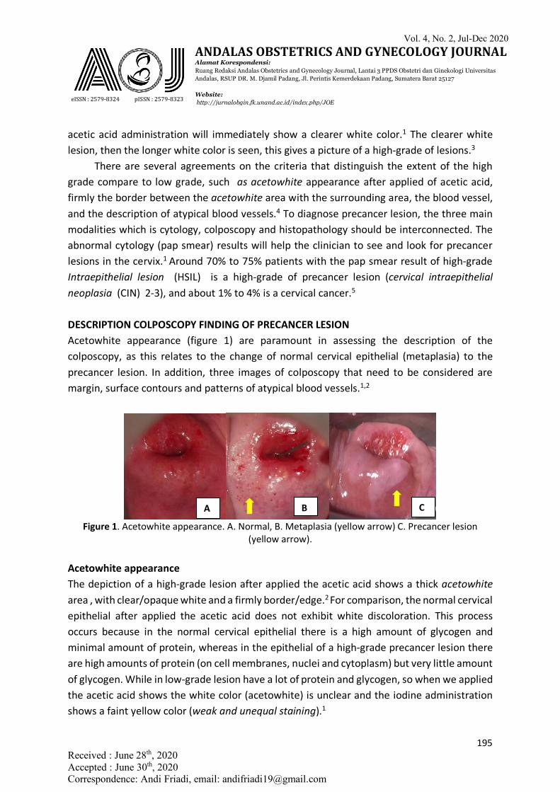
DESCRIPTION COLPOSCOPY FINDING OF PRECANCER LESION Acetowhite appearance (figure 1) are paramount in assessing the description of the colposcopy, as this relates to the change of normal cervical epithelial (metaplasia) to the precancer lesion. In addition, three images of colposcopy that need to be considered are margin, surface contours and patterns of atypical blood vessels.1,2
Figure 1. Acetowhite appearance. A. Normal, B. Metaplasia (yellow arrow) C. Precancer lesion
(yellow arrow).
Acetowhite appearance The depiction of a high-grade lesion after applied the acetic acid shows a thick acetowhite area , with clear/opaque white and a firmly border/edge.2 For comparison, the normal cervical epithelial after applied the acetic acid does not exhibit white discoloration. This process occurs because in the normal cervical epithelial there is a high amount of glycogen and minimal amount of protein, whereas in the epithelial of a high-grade precancer lesion there are high amounts of protein (on cell membranes, nuclei and cytoplasm) but very little amount of glycogen. While in low-grade lesion have a lot of protein and glycogen, so when we applied the acetic acid shows the white color (acetowhite) is unclear and the iodine administration shows a faint yellow color (weak and unequal staining).1
A B C
Vol. 4, No. 2, Jul-Dec 2020
eISSN : 2579-8324 pISSN : 2579-8323
ANDALAS OBSTETRICS AND GYNECOLOGY JOURNAL Alamat Korespondensi: Ruang Redaksi Andalas Obstetrics and Gynecology Journal, Lantai 3 PPDS Obstetri dan Ginekologi Universitas Andalas, RSUP DR. M. Djamil Padang, Jl. Perintis Kemerdekaan Padang, Sumatera Barat 25127 Website: http://jurnalobgin.fk.unand.ac.id/index.php/JOE
196 Received : June 28th, 2020 Accepted : June 30th, 2020 Correspondence: Andi Friadi, email: [email protected]
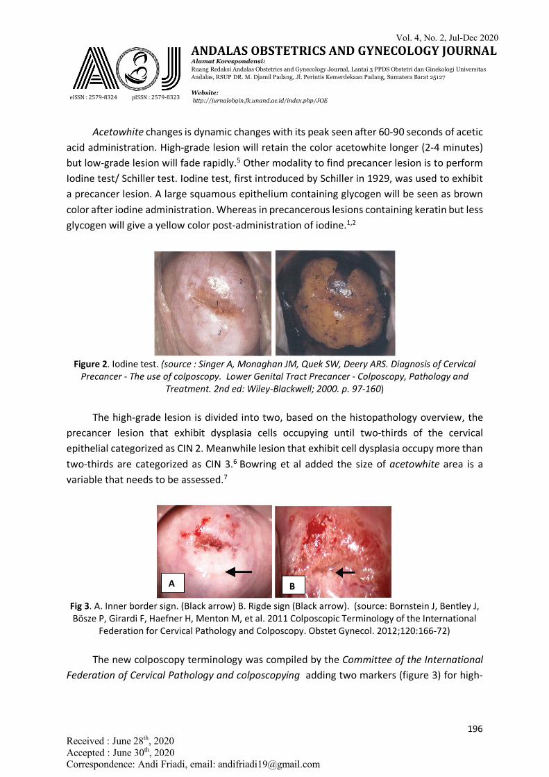
Acetowhite changes is dynamic changes with its peak seen after 60-90 seconds of acetic acid administration. High-grade lesion will retain the color acetowhite longer (2-4 minutes) but low-grade lesion will fade rapidly.5 Other modality to find precancer lesion is to perform Iodine test/ Schiller test. Iodine test, first introduced by Schiller in 1929, was used to exhibit a precancer lesion. A large squamous epithelium containing glycogen will be seen as brown color after iodine administration. Whereas in precancerous lesions containing keratin but less glycogen will give a yellow color post-administration of iodine.1,2
Figure 2. Iodine test. (source : Singer A, Monaghan JM, Quek SW, Deery ARS. Diagnosis of Cervical
Precancer - The use of colposcopy. Lower Genital Tract Precancer - Colposcopy, Pathology and Treatment. 2nd ed: Wiley-Blackwell; 2000. p. 97-160)
The high-grade lesion is divided into two, based on the histopathology overview, the
precancer lesion that exhibit dysplasia cells occupying until two-thirds of the cervical epithelial categorized as CIN 2. Meanwhile lesion that exhibit cell dysplasia occupy more than two-thirds are categorized as CIN 3.6 Bowring et al added the size of acetowhite area is a variable that needs to be assessed.7
Fig 3. A. Inner border sign. (Black arrow) B. Rigde sign (Black arrow). (source: Bornstein J, Bentley J, Bösze P, Girardi F, Haefner H, Menton M, et al. 2011 Colposcopic Terminology of the International
Federation for Cervical Pathology and Colposcopy. Obstet Gynecol. 2012;120:166-72) The new colposcopy terminology was compiled by the Committee of the International
Federation of Cervical Pathology and colposcopying adding two markers (figure 3) for high-
A B
Vol. 4, No. 2, Jul-Dec 2020
eISSN : 2579-8324 pISSN : 2579-8323
ANDALAS OBSTETRICS AND GYNECOLOGY JOURNAL Alamat Korespondensi: Ruang Redaksi Andalas Obstetrics and Gynecology Journal, Lantai 3 PPDS Obstetri dan Ginekologi Universitas Andalas, RSUP DR. M. Djamil Padang, Jl. Perintis Kemerdekaan Padang, Sumatera Barat 25127 Website: http://jurnalobgin.fk.unand.ac.id/index.php/JOE
197 Received : June 28th, 2020 Accepted : June 30th, 2020 Correspondence: Andi Friadi, email: [email protected]
grade lesion namely "inner border sign" and "Ridge sign." The Inner border sign is a strict border between the thin acetowhite region and the thick acetowhite area of the same lesion.8
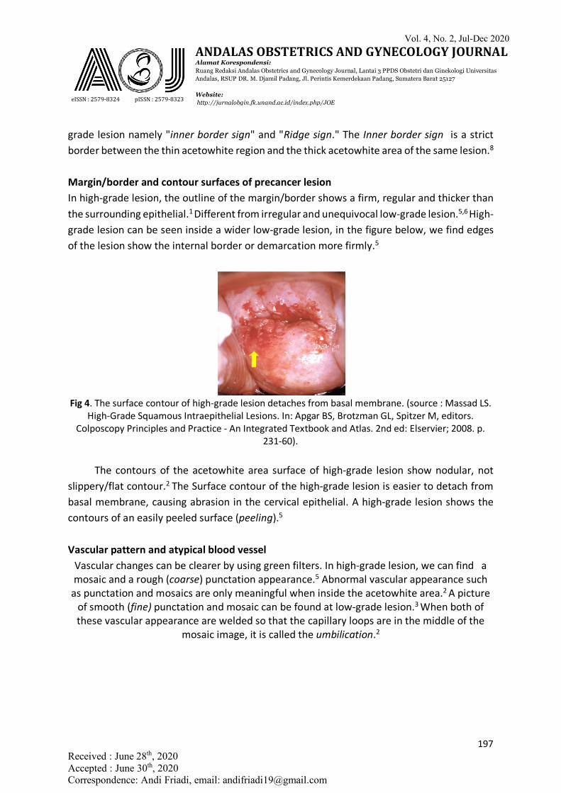
Margin/border and contour surfaces of precancer lesion In high-grade lesion, the outline of the margin/border shows a firm, regular and thicker than the surrounding epithelial.1 Different from irregular and unequivocal low-grade lesion.5,6 High-grade lesion can be seen inside a wider low-grade lesion, in the figure below, we find edges of the lesion show the internal border or demarcation more firmly.5
Fig 4. The surface contour of high-grade lesion detaches from basal membrane. (source : Massad LS.
High-Grade Squamous Intraepithelial Lesions. In: Apgar BS, Brotzman GL, Spitzer M, editors. Colposcopy Principles and Practice - An Integrated Textbook and Atlas. 2nd ed: Elservier; 2008. p.
231-60). The contours of the acetowhite area surface of high-grade lesion show nodular, not
slippery/flat contour.2 The Surface contour of the high-grade lesion is easier to detach from basal membrane, causing abrasion in the cervical epithelial. A high-grade lesion shows the contours of an easily peeled surface (peeling).5
Vascular pattern and atypical blood vessel
Vascular changes can be clearer by using green filters. In high-grade lesion, we can find a mosaic and a rough (coarse) punctation appearance.5 Abnormal vascular appearance such
as punctation and mosaics are only meaningful when inside the acetowhite area.2 A picture of smooth (fine) punctation and mosaic can be found at low-grade lesion.3 When both of these vascular appearance are welded so that the capillary loops are in the middle of the
mosaic image, it is called the umbilication.2
Vol. 4, No. 2, Jul-Dec 2020
eISSN : 2579-8324 pISSN : 2579-8323
ANDALAS OBSTETRICS AND GYNECOLOGY JOURNAL Alamat Korespondensi: Ruang Redaksi Andalas Obstetrics and Gynecology Journal, Lantai 3 PPDS Obstetri dan Ginekologi Universitas Andalas, RSUP DR. M. Djamil Padang, Jl. Perintis Kemerdekaan Padang, Sumatera Barat 25127 Website: http://jurnalobgin.fk.unand.ac.id/index.php/JOE
198 Received : June 28th, 2020 Accepted : June 30th, 2020 Correspondence: Andi Friadi, email: [email protected]
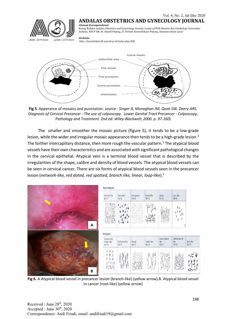
Fig 5. Apperance of mosaics and punctation. source : Singer A, Monaghan JM, Quek SW, Deery ARS. Diagnosis of Cervical Precancer - The use of colposcopy. Lower Genital Tract Precancer - Colposcopy,
Pathology and Treatment. 2nd ed: Wiley-Blackwell; 2000. p. 97-160) The smaller and smoother the mosaic picture (figure 5), it tends to be a low-grade
lesion, while the wider and irregular mosaic appearance then tends to be a high-grade lesion.3
The farther intercapillary distance, then more rough the vascular pattern.5 The atypical blood vessels have their own characteristics and are associated with significant pathological changes in the cervical epithelial. Atypical vein is a terminal blood vessel that is described by the irregularities of the shape, calibre and density of blood vessels. The atypical blood vessels can be seen in cervical cancer. There are six forms of atypical blood vessels seen in the precancer lesion (network-like, red doted, red spotted, branch like, linear, loop-like).1
Fig 6. A.Atypical blood vessel in precancer lesion (branch-like) (yellow arrow).B. Atypical blood vessel
in cancer (root-like) (yellow arrow)
A
B
Vol. 4, No. 2, Jul-Dec 2020
eISSN : 2579-8324 pISSN : 2579-8323
ANDALAS OBSTETRICS AND GYNECOLOGY JOURNAL Alamat Korespondensi: Ruang Redaksi Andalas Obstetrics and Gynecology Journal, Lantai 3 PPDS Obstetri dan Ginekologi Universitas Andalas, RSUP DR. M. Djamil Padang, Jl. Perintis Kemerdekaan Padang, Sumatera Barat 25127 Website: http://jurnalobgin.fk.unand.ac.id/index.php/JOE
199 Received : June 28th, 2020 Accepted : June 30th, 2020 Correspondence: Andi Friadi, email: [email protected]
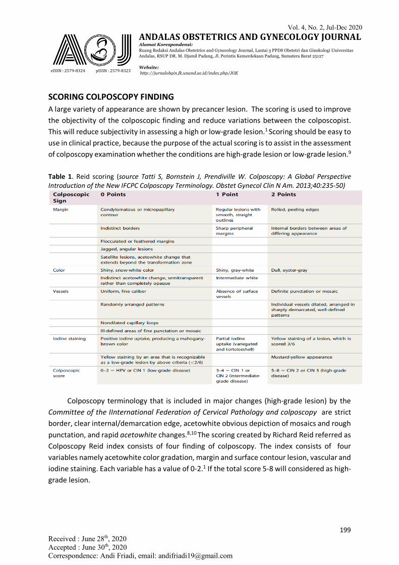
SCORING COLPOSCOPY FINDING A large variety of appearance are shown by precancer lesion. The scoring is used to improve the objectivity of the colposcopic finding and reduce variations between the colposcopist. This will reduce subjectivity in assessing a high or low-grade lesion.1 Scoring should be easy to use in clinical practice, because the purpose of the actual scoring is to assist in the assessment of colposcopy examination whether the conditions are high-grade lesion or low-grade lesion.9
Table 1. Reid scoring (source Tatti S, Bornstein J, Prendiville W. Colposcopy: A Global Perspective Introduction of the New IFCPC Colposcopy Terminology. Obstet Gynecol Clin N Am. 2013;40:235-50)
Colposcopy terminology that is included in major changes (high-grade lesion) by the
Committee of the IInternational Federation of Cervical Pathology and colposcopy are strict border, clear internal/demarcation edge, acetowhite obvious depiction of mosaics and rough punctation, and rapid acetowhite changes.8,10 The scoring created by Richard Reid referred as Colposcopy Reid index consists of four finding of colposcopy. The index consists of four variables namely acetowhite color gradation, margin and surface contour lesion, vascular and iodine staining. Each variable has a value of 0-2.1 If the total score 5-8 will considered as high-grade lesion.
Vol. 4, No. 2, Jul-Dec 2020
eISSN : 2579-8324 pISSN : 2579-8323
ANDALAS OBSTETRICS AND GYNECOLOGY JOURNAL Alamat Korespondensi: Ruang Redaksi Andalas Obstetrics and Gynecology Journal, Lantai 3 PPDS Obstetri dan Ginekologi Universitas Andalas, RSUP DR. M. Djamil Padang, Jl. Perintis Kemerdekaan Padang, Sumatera Barat 25127 Website: http://jurnalobgin.fk.unand.ac.id/index.php/JOE
200 Received : June 28th, 2020 Accepted : June 30th, 2020 Correspondence: Andi Friadi, email: [email protected]
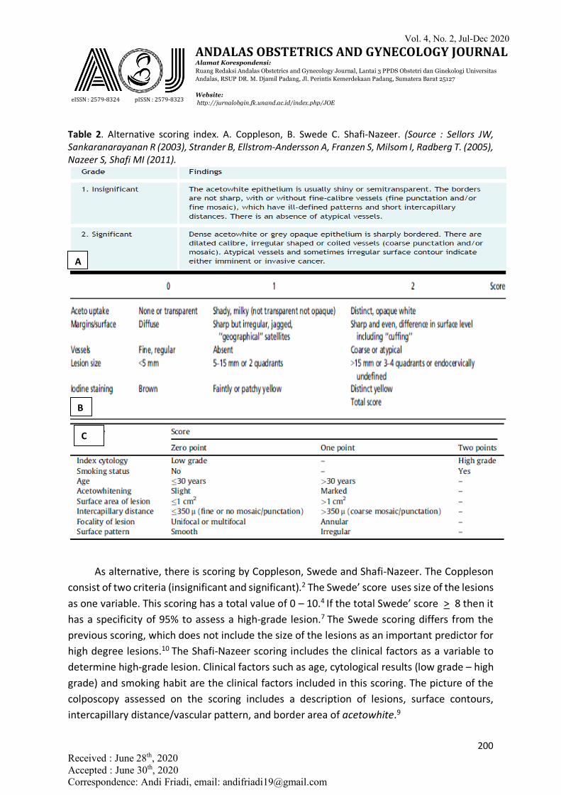
Table 2. Alternative scoring index. A. Coppleson, B. Swede C. Shafi-Nazeer. (Source : Sellors JW, Sankaranarayanan R (2003), Strander B, Ellstrom-Andersson A, Franzen S, Milsom I, Radberg T. (2005), Nazeer S, Shafi MI (2011).
As alternative, there is scoring by Coppleson, Swede and Shafi-Nazeer. The Coppleson
consist of two criteria (insignificant and significant).2 The Swede’ score uses size of the lesions as one variable. This scoring has a total value of 0 – 10.4 If the total Swede’ score > 8 then it has a specificity of 95% to assess a high-grade lesion.7 The Swede scoring differs from the previous scoring, which does not include the size of the lesions as an important predictor for high degree lesions.10 The Shafi-Nazeer scoring includes the clinical factors as a variable to determine high-grade lesion. Clinical factors such as age, cytological results (low grade – high grade) and smoking habit are the clinical factors included in this scoring. The picture of the colposcopy assessed on the scoring includes a description of lesions, surface contours, intercapillary distance/vascular pattern, and border area of acetowhite.9
A
B
C
Vol. 4, No. 2, Jul-Dec 2020
eISSN : 2579-8324 pISSN : 2579-8323
ANDALAS OBSTETRICS AND GYNECOLOGY JOURNAL Alamat Korespondensi: Ruang Redaksi Andalas Obstetrics and Gynecology Journal, Lantai 3 PPDS Obstetri dan Ginekologi Universitas Andalas, RSUP DR. M. Djamil Padang, Jl. Perintis Kemerdekaan Padang, Sumatera Barat 25127 Website: http://jurnalobgin.fk.unand.ac.id/index.php/JOE
201 Received : June 28th, 2020 Accepted : June 30th, 2020 Correspondence: Andi Friadi, email: [email protected]
CONCLUSION 1. Colposcopy followed by directional biopsy is the gold standard to diagnose of cervical
precancer lesions 2. Three main modalities to diagnose precancer lesions are cytology, colposcopy and
histopathology should be interconnected 3. Criteria that distinguish the extent of the high grade compare to low grade are
acetowhite appearance, demarcation line between the acetowhite area with the surrounding area, the blood vessel, and the description of atypical blood vessels
4. Various scoring is available to improve the objectivity of the colposcopic finding and reduce variations between the colposcopist
REFERENCES 1. Singer A, Monaghan JM, Quek SW, Deery ARS. Diagnosis of Cervical Precancer - The
use of colposcopy. Lower Genital Tract Precancer - Colposcopy, Pathology and Treatment. 2nd ed: Wiley-Blackwell; 2000. p. 97-160.
2. Sellors JW, Sankaranarayanan R. Colposcopic assessment of cervical intraepithelial neoplasia. Colposcopy and Treatment of Cervical Intraepithelial Neoplasia: A Beginners’ Manual. 1st ed: WHO-IARC; 2003. p. 55-68.
3. Walker P, Dexeus S, De Palo G, Barrasso R, Campion M, Girardi F, et al. International Terminology of Colposcopy: An Updated Report From the International Federation for Cervical Pathology and Colposcopy. Obstet Gynecol. 2003;101(1):175-7.
4. Strander B, Ellstrom-Andersson A, Franzen S, Milsom I, Radberg T. The performance of a new scoring system for colposcopy in detecting high-grade dysplasia in the uterine cervix. Acta Obstet Gynecol Scand. 2005;84:1013-7.
5. Massad LS. High-Grade Squamous Intraepithelial Lesions. In: Apgar BS, Brotzman GL, Spitzer M, editors. Colposcopy Principles and Practice - An Integrated Textbook and Atlas. 2nd ed: Elservier; 2008. p. 231-60.
6. O’Connor DM. A Tissue Basis for Colposcopic Findings. In: Rayburn WF, Waxman AG, editors. Colposcopy, Cervical Screening, and HPV. 35: Obstet Gynecol Clin N Am - Elsevier; 2008. p. 565-82.
7. Bowring J, Strander B, Young M, Evans H, Walker P. The Swede Score: Evaluation of a Scoring System Designed to Improve the Predictive Value of Colposcopy. J Lower Genit Tract Dis. 2010;14(4):301-5.
8. Bornstein J, Bentley J, Bösze P, Girardi F, Haefner H, Menton M, et al. 2011 Colposcopic Terminology of the International Federation for Cervical Pathology and Colposcopy. Obstet Gynecol. 2012;120:166-72.
Vol. 4, No. 2, Jul-Dec 2020
eISSN : 2579-8324 pISSN : 2579-8323
ANDALAS OBSTETRICS AND GYNECOLOGY JOURNAL Alamat Korespondensi: Ruang Redaksi Andalas Obstetrics and Gynecology Journal, Lantai 3 PPDS Obstetri dan Ginekologi Universitas Andalas, RSUP DR. M. Djamil Padang, Jl. Perintis Kemerdekaan Padang, Sumatera Barat 25127 Website: http://jurnalobgin.fk.unand.ac.id/index.php/JOE
202 Received : June 28th, 2020 Accepted : June 30th, 2020 Correspondence: Andi Friadi, email: [email protected]
9. Nazeer S, Shafi MI. Objective perspective in colposcopy. Best Practice & Research Clinical Obstetrics and Gynaecology. 2011;25:631-40.
10. Tatti S, Bornstein J, Prendiville W. Colposcopy: A Global Perspective Introduction of the New IFCPC Colposcopy Terminology. Obstet Gynecol Clin N Am. 2013;40:235-50.