Colposcopy in Gyn Practice
59
Colposcopy in Gyn Practice Presenter : DR Joseph G. Kimaro Facilitator: DR .Fadhlun 03/30/22 1
-
Upload
joseph-kimaro -
Category
Documents
-
view
26 -
download
0
description
Colposcopy is one of important procedure which is done to women with abnormal cervical pap smear.
Transcript of Colposcopy in Gyn Practice

Colposcopy in Gyn Practice
Presenter : DR Joseph G. KimaroFacilitator: DR .Fadhlun
04/28/23 1
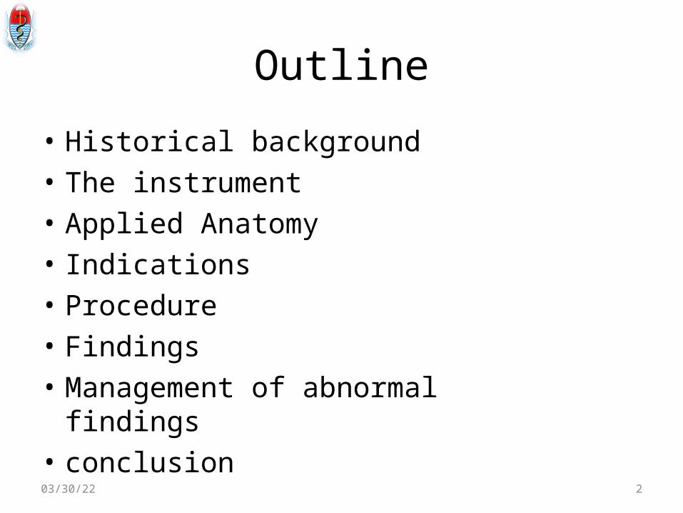
Outline
• Historical background• The instrument• Applied Anatomy• Indications• Procedure • Findings • Management of abnormal findings• conclusion04/28/23 2

Historical background
• Colposcope is a special microscope with magnification of up to 40x
• Colpo - Vagina, Scope- look into• Provides illuminated and magnified
assessment of lower genital tract• Described by Hans Hinselman in 1925• His theories were incorrect and protocol was
impractical
04/28/23 3

Ct..
• His work identified several atypical appearances
• which are still used today:– Leukoplakia– Punctation– Felderung (mosaicism)
• Abandoned until 1960 - used to confirmation and follow-up of abnormal pap smear
04/28/23 4

Ct…
• In 1970s, treatment of abnormal Pap smear was cone biopsy
• There was over treatment as most Bx revealed minor epithelial changes
• Colposcopic evaluation procedure to identify those who require surgical therapy decreased unnecessary surgeries.
04/28/23 5

Ct…
• Today colposcopy has been accepted as a diagnostic tool in evaluating abnormal pap tests
• Colposcopy elegantly identifies the location of abnormal lesions allowing the practitioner to obtain histologic samples
04/28/23 6

Colposcopy Goals
To detect abnormal epithelium,
To identify the area of epithelium with the
highest degree of disease
To take direct biopsies to that area
04/28/23 7

Indications
• Abnormal Pap smear, with no gross lesion on cervix
• Persistence of inflammatory cells despite adequate treatment
• Grossly abnormal / unhealthy cervix or vagina• Women with positive high risk HPV DNA test,
even if Pap neg.• Pos-tcoital bleeding
04/28/23 8

Ct…
• Trearment of women with CIN• Monitoring of women treated for CIN• Preop evaluation of women diagnosed with
Stage Ia or b cervcal cs on cl ex & bx – to rule out vag involvement
• Evaluation of women with anogenital condylomas
04/28/23 9

Contraindications
• No absolute contraindications to the performance of a colposcopic examination exist
• Take precautions in special circumstances, such as a pregnant patient with placenta previa.
• Ability to tolerate a standard speculum examination is the only true limiting factor.
• Active cervicitis and vulvovaginitis should be treated
04/28/23 10
Colposcope-Relevant anatomy • Cervix epithelium is composed of squamous and
columnar epithelial- ecto and endo • Pre-menarche columnar extend to cervical portio• Undergoes squamous metaplasia-physiological
change• Ring of squamous metaplasia is referred to as the
transformation zone transformation zone (TZ)• Squamocolumnar junction (Squamocolumnar junction (SCJ) is the border btn
metaplastic epithelium and endocervical colunar epithelium
04/28/23 11

Ct..
• Virus commonly infect rapidly dividing epithelium,
• Transformation zone is typically the locus of dysplastic change.
• Complete visualization of the entire transformation zone, most specifically the squamocolumnar junction is required for adequate assessment.
04/28/23 12

• Actively dividing cells have increased nuclear–cytoplasm ratio. This is especially true of HPV–infected cells
• Exposure of such tissue to dilute acetic acid results in increased light reflectivity and, therefore, visual contrast with normal tissue
04/28/23 13
• Mature squamous epithelial cells contain a large amount of glycogen, which stains a characteristic mahogany color when exposed to iodine (the Schiller reaction),
• Rapidly dividing cells, which contain relatively little glycogen, remain unstained
04/28/23 14

• There is in-growth of the perpendicular vessels into the epithelium.
• Viewed end-on under magnification, these vessels appear as small red dots “punctationpunctation”
Left is fine, right is coarse
04/28/23 15

• Vessels can become interconnected in intricate patterns that resemble a tiled floor “mosaicism”“mosaicism”
• Irregular vessels eg, sudden vessel termination, “hairpin” or “comma” type vessels, abnormal branching, or increasing diameter of a vessel is suggestive of malignant transformation
04/28/23 16
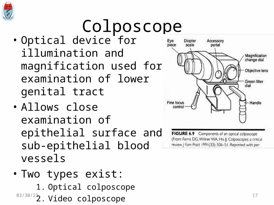
Colposcope • Optical device for
illumination and magnification used for examination of lower genital tract
• Allows close examination of epithelial surface and sub-epithelial blood vessels
• Two types exist:1. Optical colposcope2. Video colposcope04/28/23 17

Video colposcope
04/28/23 18

Colposcope • Have focal length of 200 to 350mm• Focusing is done using knob instead of moving
whole colposcope• Have powerful source of light for illumination • Green or blue filters are present for contrast• Green filters block red light hence blood
vessels appears black• Required magnification for cervix examination
is 7.5X to 15X04/28/23 19
Colposcope
• Colposcopes can be affixed to Exam. table, mounted to stand or fitted to swivel arm
• Mobile units are most practical• Three types of support available
• Centre post• Flexible articulating arm• Overhead boon type
04/28/23 20
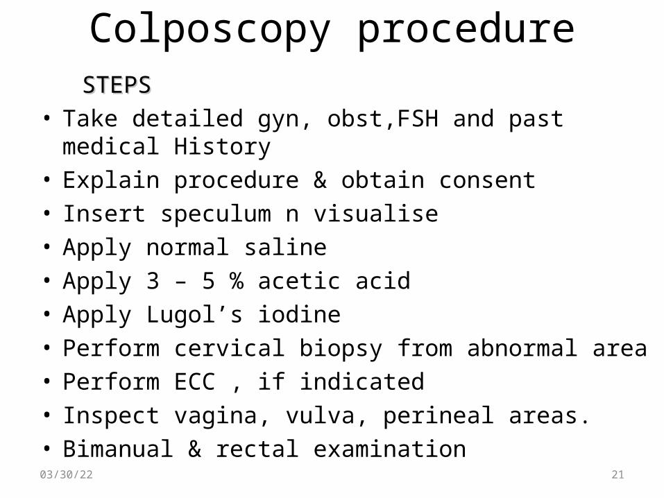
Colposcopy procedure STEPSSTEPS• Take detailed gyn, obst,FSH and past medical History• Explain procedure & obtain consent• Insert speculum n visualise• Apply normal saline• Apply 3 – 5 % acetic acid• Apply Lugol’s iodine• Perform cervical biopsy from abnormal area• Perform ECC , if indicated• Inspect vagina, vulva, perineal areas.• Bimanual & rectal examination04/28/23 21
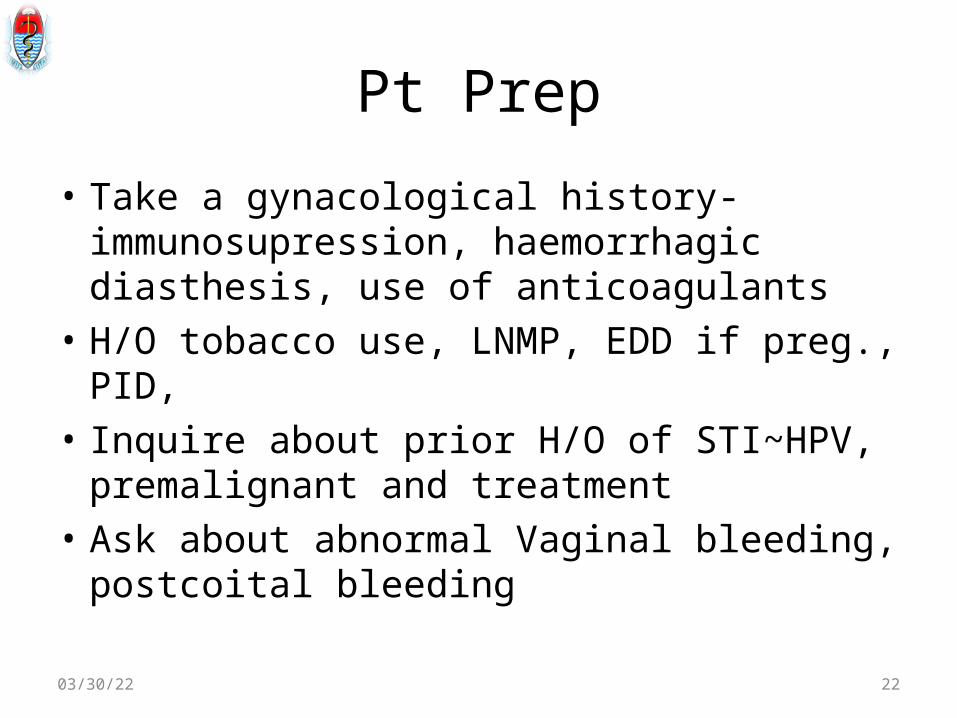
Pt Prep
• Take a gynacological history-immunosupression, haemorrhagic diasthesis, use of anticoagulants
• H/O tobacco use, LNMP, EDD if preg., PID, • Inquire about prior H/O of STI~HPV,
premalignant and treatment• Ask about abnormal Vaginal bleeding,
postcoital bleeding
04/28/23 22

When?• Menstrual cycle : day8 – day 12 as cervical
mucus abundant & clear, and ext os open.• Postmenopausal women should ideally
receive estrogen for 7-21 days, colpo performed on last day of oestrogen. Not after stopping as mucosa reverts rapidly.
• Colpo under adequate oestrogen reduces chances of unsatisfactory colpo, need for ECC
04/28/23 23

Ct..
• Systematic approach is essential• Consist of four distinct and orderly tasks
i. Visualizationii. Assessmentiii. Sampling and iv. Correlation
04/28/23 24

Colposcopy procedure• RequirementsRequirements 1. Colposcope2. Kevokian punch biopsy forceps3. Bivalve or/and Sims vagina
speculum4. Cervical speculum5. Endocervical brush6. Edocervical curette7. Normal saline, acetic acid, Lugols
Iodine8. Silver nitrate stick or monsels
solution04/28/23 25

Ct.. • Ensure equipment and supplies
availability• Position her on examination
table• Communication-patients’ co-
operation• Insert adequate size of speculum• If obese may apply glove around
speculum• Move scope to the working focal
length04/28/23 26

Technique• Orient colposcope so that a panoramic view is
obtained• Start with normal light and 2X to 10×
magnification• Entire cervix should be visualized• Clean off obscuring mucous or discharge with
normal saline• Look for blood vessels and SCJ • Apply generous amount acetic acid or Lugols
iodine04/28/23 27

Stains 15% or 3% Acetic Acid5% or 3% Acetic Acid• Works as a desiccant• The cellular cytoplasm is reduced enhancing a
prominent nucleus• The nucleus is enlarged secondary to HPV
replication• This nuclear enlargement is seen as acetowhite
changes
04/28/23 28

Stains 2
Lugol’s iodine solution (Schiller test)Lugol’s iodine solution (Schiller test)• Works by staining glycogen• Dysplastic tissue has an increased metabolic
rate thereby lowering cellular glycogen• Normal tissue stains black/brown while
dysplastic tissue appears highlighted or yellow• Useful stain for hard to see lesions
04/28/23 29

Technique – Evaluation • Evaluate the squamocolumnar junctionEvaluate the squamocolumnar junction(SCJ) or (SCJ) or
transformation zone (TZ)transformation zone (TZ)• Dysplasia originates from this boundary and
spreads lateral to this junction• Medial to this border are columnar
cells/glandular cells• A large volume of glandular cells are called
ectropion• Young women and pregnant women generally
have ectropion04/28/23 30
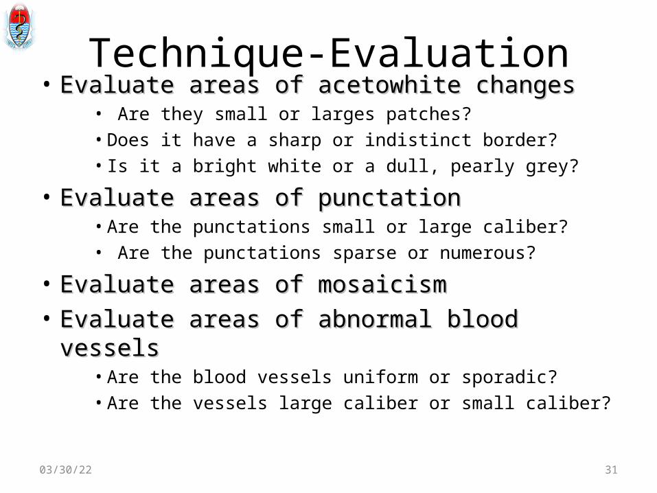
Technique-Evaluation• Evaluate areas of acetowhite changesEvaluate areas of acetowhite changes
• Are they small or larges patches?• Does it have a sharp or indistinct border?• Is it a bright white or a dull, pearly grey?
• Evaluate areas of punctationEvaluate areas of punctation• Are the punctations small or large caliber?• Are the punctations sparse or numerous?
• Evaluate areas of mosaicismEvaluate areas of mosaicism• Evaluate areas of abnormal blood vesselsEvaluate areas of abnormal blood vessels
• Are the blood vessels uniform or sporadic?• Are the vessels large caliber or small caliber?
04/28/23 31

Adequacy of procedure• Visualization of entire TZ in 360
degrees360 of columnar epithelium360 squamous epithelium360 of current SCJ
• Entire lesion should be visible i.e distal an proximal margins must be colposcopically visible
• If lesion/SCJ can not entirely visualized then- unsatisfactory colposcopy may need Rx
04/28/23 32
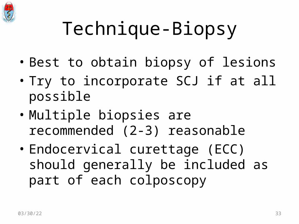
Technique-Biopsy
• Best to obtain biopsy of lesions• Try to incorporate SCJ if at all possible• Multiple biopsies are recommended (2-3)
reasonable• Endocervical curettage (ECC) should generally
be included as part of each colposcopy
04/28/23 33

Technique-ECC• It is uncomfortable portion of colposcopic
directed biopsies• Good to have patient take NSAID 30 min before
colposcopy• Apply curette at four cardinal directions• Swirl curette in fixative then take endocervical
brush to collect rest of sample• Prospective randomized trial revealed better
results with curette and brush than either alone04/28/23 34

Normal colposcopic finding
04/28/23 35

Normal colposcopic finding
04/28/23 36

Normal colposcopic finding
04/28/23 37

Normal colposcopy- SCJ
04/28/23 38

Normal histologic finding
04/28/23 39

Abnormal colposcopic findingAcetowhite changesAcetowhite changes
• Grade of lesion correlates with intensity of whiteness, surface shine, rapidity of appearance and duration of whiteness.
Low-grade LesionsLow-grade Lesions– Less dense, less extensive and less complex acetowhite
areas close to or abutting the squamocolumnar junction with well demarcated, but irregular, feathery or digitating margins
– Satellite acetowhite lesions detached (far away) from the squamocolumnar junction
– Geographical patterns (resembling geographical regions).04/28/23 40
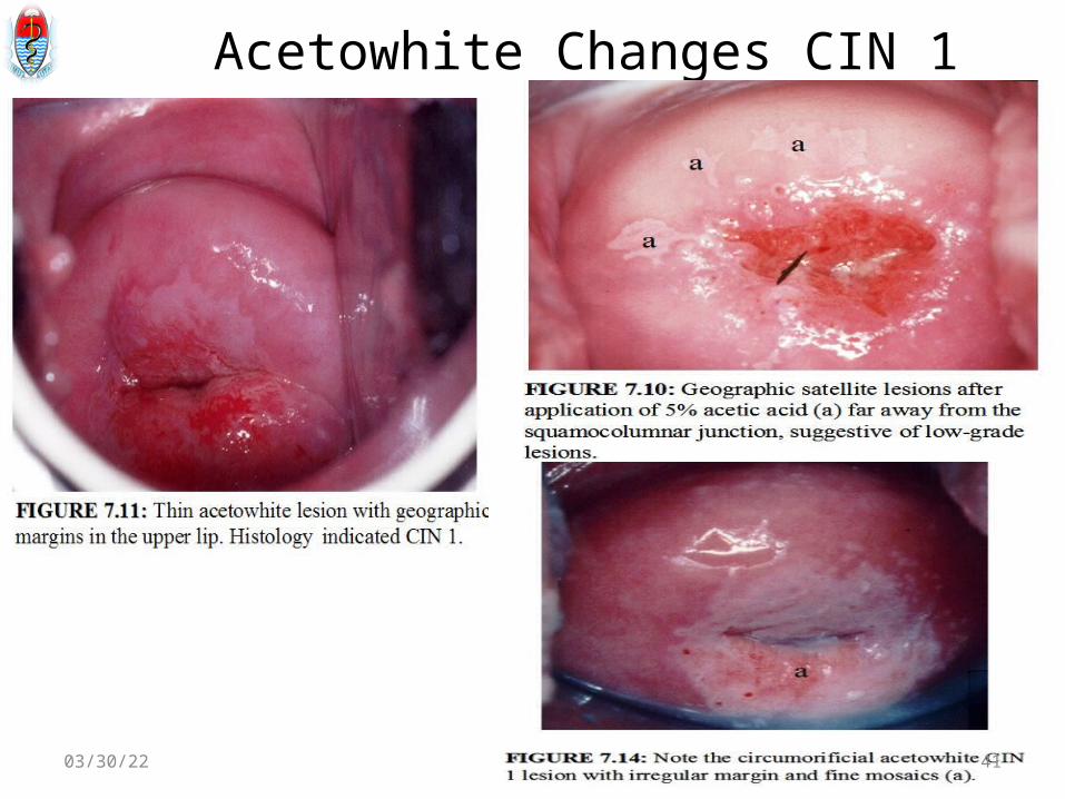
Acetowhite Changes CIN 1
04/28/23 41

High Grade Lesions dense, opaque, grey white acetowhite areas coarse punctation and/or mosaic regular and well demarcated borders Raised / rolled edge; extensive and often involve both lips and may occasionally harbour atypical vessels
04/28/23 42

HGL
04/28/23 43

HGL
A dense acetowhite, opaque, complex,circumorificial CIN 3 lesion
04/28/23 44

Ct.. Visualization of one or more
borders within an acetowhite lesion (‘lesion within lesion’) or a lesion with differing colour intensity.
The crypt openings may have thick, dense and wide acetowhite rims called cuffed crypt openings . These are whiter and wider than the mild, line-like acetowhite rings that are sometimes seen around normal crypt openings.
04/28/23 45

04/28/23 46

Mosaicim
• A mosaic is a large image pieced together by smaller usually colorful tiles
• In colposcopy it is essentially the same- a larger lesion made of small heaped epithelial islands and tiny vessels
• These islands look like a cobblestone road• Islands are separated by vessels running
parallel to the portio or colposcopist
04/28/23 47

Mosaicim
04/28/23 48

Invasive cancer• Collection of all
colposcopic finding: acetowhite, punctations, mosaicism, and vessels
• Necrotic tissue results in anaerobic odors
• Take biopsy from lesion, be ware of bleeding
04/28/23 49

Invasive carcinoma- cotton wool appearance
04/28/23 50

Abnormal colposcopic findings
04/28/23 51

Leukoplakia• Well demarcated white area prior to AA
application.
• Usually leukoplakia is idiopathic, but it may also be caused by chronic foreign body irritation, HPV infection or squamous neoplasia.
• It is not usually possible to colposcopically evaluate the vasculature beneath such an area
• No matter where the area of leukoplakia is located on the cervix, it should be biopsied to rule out high-grade CIN or malignancy.
04/28/23 52

Colposcope-Grading
04/28/23 53

04/28/23 54
Treatment modalities for abnormal findings
• Cryosugery• Loop Electricosurgical Excision Procedure
(LEEP)• Laser Ablation• Cold Knife Conization• Hysterectomy
04/28/23 55

Post colposcopy advice
• Ant pains-NSAIDS should be prescribed• Woman to avoid sex for a day or two• Counseled on findings and given the date on
when to return• Should report any excessive vagina bleeding
04/28/23 56

Relevant study• Interventions for reducing anxiety in women undergoing
colposcopy. Cochrane review of 15 papers• Galaal KA• Women experience high levels of anxiety and negative
emotional responses at all stages of cervical screening. High levels of anxiety before and during colposcopy can have adverse consequences, including pain and discomfort during the procedure and high loss to follow-up rates.
• Objectives:To compare the efficacy of various interventions aimed at reducing anxiety during colposcopic examination in women
04/28/23 57

• Finding: Listening to music during colposcopy: this intervention was associated with reduction in anxiety levels (p < 0.002). Video colposcopy was associated with reduction in anxiety levels, and the reduction in anxiety was significant (p < 0.0002 There was a reduction in anxiety levels in the intervention group compared to the control group (p < 0.00001).
Conclusion: Anxiety appears to be reduced by playing music during colposcopy, showing information videos prior to colposcopy and viewing video colposcopy during the procedure
04/28/23 58

Conclusion
• Colposcopy is important procedure in confirming abnormal cytology
• It has reduced unnecessary surgical interventions among women with abnormal cytology
• Biopsy should be taken from colposcopically abnormal areas.
04/28/23 59