Lecture 1 Cell Adaptation, Injury & Deathpbt.guc.edu.eg/Download.ashx?id=568&file=Lecture1 Cell...
27
Lecture 1 Cell Adaptation, Injury & Death Dr. Nabila Hamdi MD, PhD
-
Upload
duongthuan -
Category
Documents
-
view
223 -
download
0
Transcript of Lecture 1 Cell Adaptation, Injury & Deathpbt.guc.edu.eg/Download.ashx?id=568&file=Lecture1 Cell...

Lecture 1 Cell Adaptation, Injury & Death
Dr. Nabila Hamdi
MD, PhD
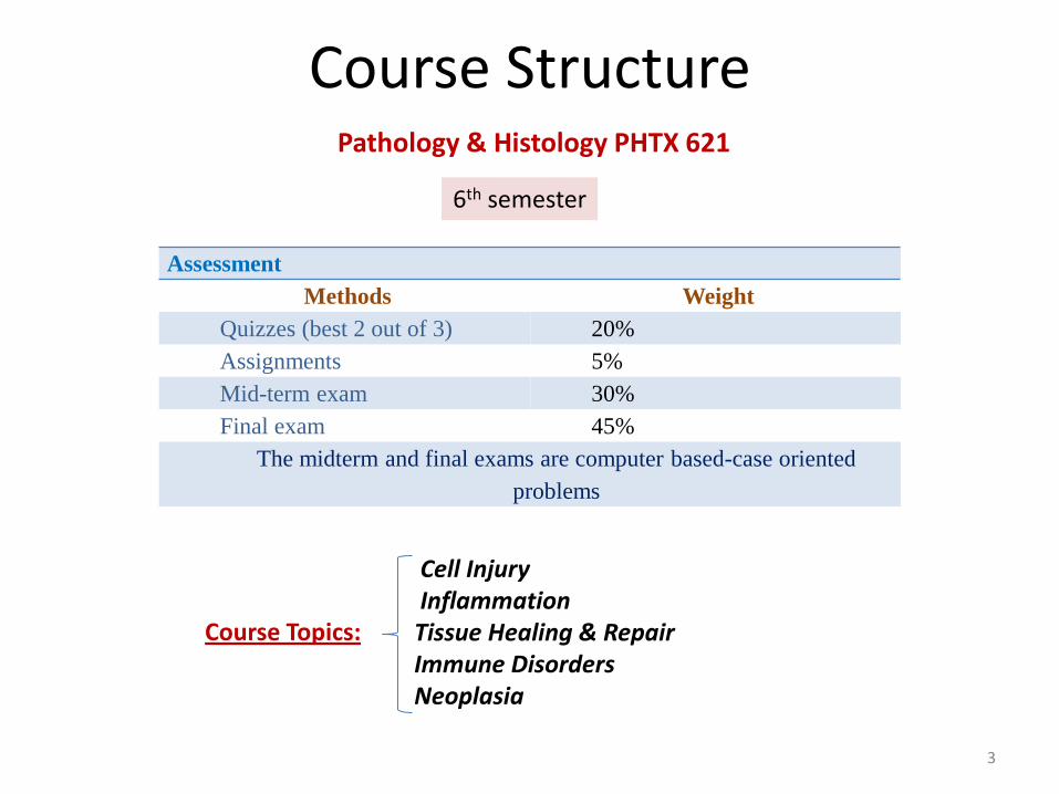
Course Structure
Assessment
Methods Weight
Quizzes (best 2 out of 3) 20%
Assignments 5%
Mid-term exam 30%
Final exam 45%
The midterm and final exams are computer based-case oriented
problems
6th semester
Pathology & Histology PHTX 621
Cell Injury Inflammation Tissue Healing & Repair Immune Disorders Neoplasia
Course Topics:
3

How to study?
Lectures
Tutorials
4

evelivesey.deviantart.com
Basic Science Cell, molecule
Clinical Practice Symptoms/Signs
Etiology (Cause)
Pathogenesis (Mechanism)
Pathology is the study of suffering
pathos logos
What is Pathology?
5

ILOs • Describe the major mechanisms whereby most injurious agents exert their effects
• Understand the examples of cell injury stated in this lecture, including their causes
and mechanisms
• Describe cell changes that occur with atrophy, hypertrophy, hyperplasia and
metaplasia, and state general conditions under which these changes occur
• State and discuss patterns of reversible/irreversible cell injury
• Differentiate cell death associated with apoptosis and necrosis
• Recognize the different patterns of necrosis
• Compare the pathogenesis of dystrophic and metastatic calcifications
• Understand subcellular responses and autophagy
• Define intracellular accumulations and cite examples
• Understand the process of aging 6
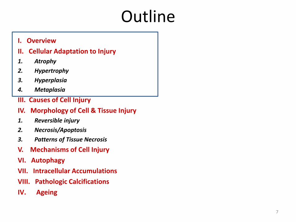
Outline I. Overview
II. Cellular Adaptation to Injury
1. Atrophy
2. Hypertrophy
3. Hyperplasia
4. Metaplasia
III. Causes of Cell Injury
IV. Morphology of Cell & Tissue Injury
1. Reversible injury
2. Necrosis/Apoptosis
3. Patterns of Tissue Necrosis
V. Mechanisms of Cell Injury
VI. Autophagy
VII. Intracellular Accumulations
VIII. Pathologic Calcifications
IV. Ageing
7
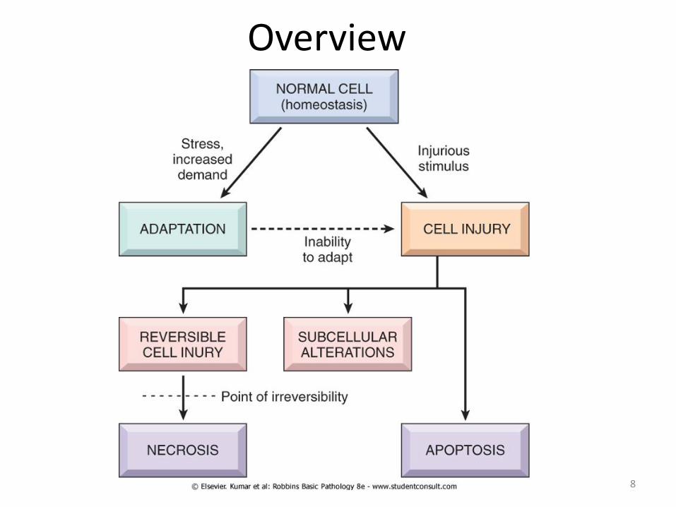
Overview
8
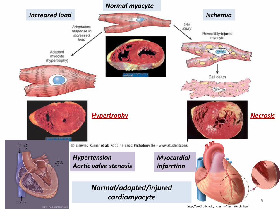
http://ww2.odu.edu/~czemlin/heartattacks.html
Ischemia
Necrosis
Increased load
Hypertension Aortic valve stenosis
Myocardial infarction
Hypertrophy
9
Normal/adapted/injured cardiomyocyte
Normal myocyte

Cellular Adaptation to Stress Atrophy
Shrinkage in the size of the cell by the loss of cell substance.
When a sufficient number of cells is involved, the entire tissue or organ diminishes in size, becoming atrophic.
Although atrophic cells may have diminished function, they are not dead.
Causes include decreased workload (immobilization), loss of innervation, decreased blood supply, inadequate nutrition, loss of endocrine function and aging.
Mechanisms include both decreased protein synthesis and increased protein degradation.
10
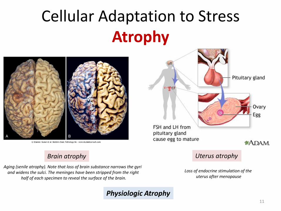
Cellular Adaptation to Stress Atrophy
Aging (senile atrophy). Note that loss of brain substance narrows the gyri and widens the sulci. The meninges have been stripped from the right
half of each specimen to reveal the surface of the brain.
Loss of endocrine stimulation of the uterus after menopause
Physiologic Atrophy
Brain atrophy Uterus atrophy
11
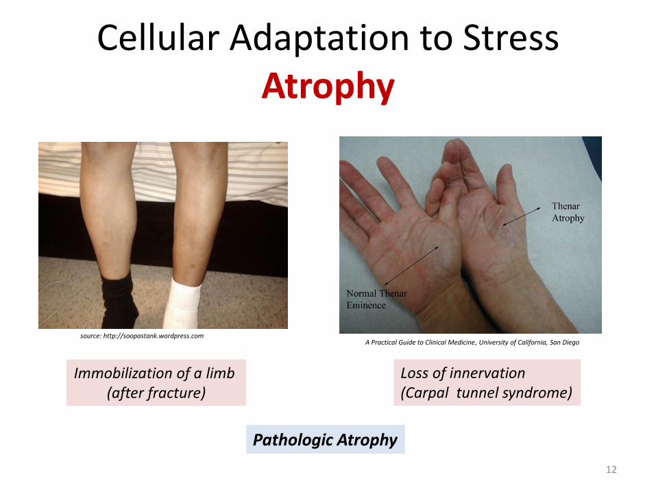
Cellular Adaptation to Stress Atrophy
Loss of innervation (Carpal tunnel syndrome)
Pathologic Atrophy
A Practical Guide to Clinical Medicine, University of California, San Diego
Immobilization of a limb (after fracture)
source: http://soopastank.wordpress.com
12
Cellular Adaptation to Stress Atrophy
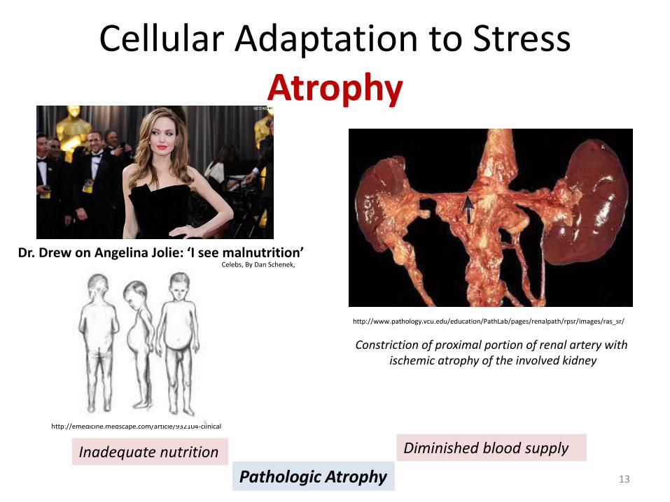
Pathologic Atrophy
Inadequate nutrition
http://emedicine.medscape.com/article/932104-clinical
Dr. Drew on Angelina Jolie: ‘I see malnutrition’ Celebs, By Dan Schenek,
Diminished blood supply
Constriction of proximal portion of renal artery with ischemic atrophy of the involved kidney
http://www.pathology.vcu.edu/education/PathLab/pages/renalpath/rpsr/images/ras_sr/
13

Cellular Adaptation to Stress Hyperplasia
Characterized by an increase in cell number.
It takes place if the cell population is capable of replication.
It can be physiologic or pathologic.
Most forms are caused by excessive hormonal or growth factor stimulation.
14

Cellular Adaptation to Stress Hyperplasia
Hormonal hyperplasia Compensatory hyperplasia
Female breast in puberty and pregnancy Liver regeneration after lobar resection
http://whydetox.net/liver-detoxification
Physiologic Hyperplasia
Growth and development of the breast during pregnancy. Ducts and glands undergo marked hyperplasia in preparation for lactation. Ductal proliferation is predominantly controlled by estrogen (E), whereas gland
differentiation is a progesterone (P) effect facilitated by estrogen.
The Breast During Pregnancy and Lactation, Julia V. Johnson and Daniel H. Riddick
Mitotic activity in the remaining cells begins as early as 12 hours later, eventually restoring the liver to its normal weight
15

Cellular Adaptation to Stress Hyperplasia
Pathologic Endometrial Hyperplasia
Excessive hormonal stimulation
(leads to abnormal menstrual bleeding)
16
Physiologic Hyperplasia during normal
menstrual cycle

Cellular Adaptation to Stress Hyperplasia
Renal cell carcinoma often secrete erythropoietin, the hormone stimulates the growth of erythrocyte precursors in the bone marrow
Pathologic Hyperplasia
Bone marrow hyperplasia Papillomaviruses cause skin warts
(Virus-induced growth factor stimulation)
Incidence of Kidney Cancer Increasing October 29, 2009 · By ASHISH V. DALAL, M
Erythropoietin
17

Cellular Adaptation to Stress Hypertrophy
Hypertrophy is an increase in the size of cells resulting in increase in the size of the organ.
There are no new cells, just bigger cells, enlarged by an increased amount of structural proteins and organelles (increase in mRNA and proteins).
It can be physiologic or pathologic and is caused either by increased functional demand or by growth factor or hormonal stimulation.
Occurs when cells are unable to divide (heart & skeletal muscle).
18

Cellular Adaptation to Stress Hypertrophy
Myocardial hypertrophy Cross-section of the heart of a patient with long-standing hypertension
shows pronounced, concentric left ventricular hypertrophy.
Cell Injury. David S. Strayer Emanuel Rubin Copyright ©2009 Lippincott Williams & Wilkins
http://aminoacidstudies.org/muscle-growth/
Skeletal muscle hypertrophy (after weightlifting)
Physiologic Hypertrophy Pathologic Hypertrophy 19

Cellular Adaptation to Stress Hypertrophy + Hyperplasia
The massive physiologic enlargement of the uterus during pregnancy occurs as a consequence of estrogen-stimulated smooth muscle hypertrophy and smooth muscle hyperplasia
Gravid uterus
20

Cellular Adaptation to Stress Metaplasia
Metaplasia is a reversible change in which one adult cell type is replaced by another adult cell type.
Cells sensitive to a particular stress are replaced by other cell types better able to withstand the adverse environment.
21

Cellular Adaptation to Stress Metaplasia
The normal ciliated columnar epithelial cells of the trachea and bronchi are focally or widely replaced by stratified squamous epithelial cells, which has survival advantages.
However, Important protective mechanisms are lost, such as mucus secretion and ciliary clearance of particulate matter.
Metaplasia of normal columnar (left) to squamous epithelium (right) in a bronchus, shown (A) schematically and (B) histologically
Cigarette smoking
22

Cellular Adaptation to Stress Metaplasia
Gastroesophageal reflux disease (GERD)
Stuart Jon Spechler, JAMA, August 14, 2013, Vol 310, No. 6
23
Cellular Adaptation to Stress
Atrophy, hypertrophy, hyperplasia and metaplasia are reversible changes!
24

Case
• On a routine visit to the physician, an otherwise healthy 51-year-
old man has a blood pressure of 150/95 mm Hg. If the patient’s
hypertension remains untreated for years, which of the following
cellular alterations will most likely be seen in the myocardium ?
a) Atrophy
b) Hyperplasia
c) Metaplasia
d) Hemosiderosis
e) Hypertrophy
25

References
ROBBINS Basic Pathology 9th Edition ROBBINS Basic Pathology 8th Edition Basic Pathology 7th Edition, by Kumar, Cotran and Robbins Source of the cover cell image http://timothyjoseph.net/kill-or-be-killed/
26

27