Cell Injury, Adaptation, and...
20
CHAPTER 14 Cell Injury, Adaptation, and Death 2 This chapter discusses the natural and pathologic life and death of cells and how they change with disease, covering biologic aging as well as distinguishing between mild and severe cell injury. BACK TO BASICS • The Origins of Cells and the Organization of Tissues • The Nucleus • The Cytoplasm • The Cell Membrane • The Cell Cycle • Cellular Communication BIOLOGIC AGING CELL INJURY AND DISEASE MILD CELL INJURY • Intracellular Accumulations • Adaptations of Cell Growth and Differentiation SEVERE CELL INJURY AND CELL DEATH After studying this chapter you should be able to: 1. Offer a brief description of the basic organization of a cell and of the organization of tissues, organs, and organ systems 2. Explain how the genetic code is written into DNA 3. Explain the role of messenger RNA 4. Explain the role of mitochondria 5. Explain how DNA replicates during cell division (mitosis) 6. Differentiate between apoptosis and necrosis 7. Explain the relationship between injury and disease 8. Explain the relationship of genes and environment in the pathogenesis of disease 9. Name the most common cause of cell injury 10. Name one cell reaction resulting from mild acute cell injury and one resulting from mild chronic injury 11. List at least two causes of cell atrophy 12. Differentiate between hypertrophy and hyperplasia 13. Define dysplasia 14. Define metaplasia and offer an example 15. Name the consequence of severe, irreversible cell injury 16. Name the most common cause of necrosis and the most common type of necrosis Learning Objectives
-
Upload
hoangkhanh -
Category
Documents
-
view
224 -
download
2
Transcript of Cell Injury, Adaptation, and...
14
C H A P T E R
14
Cell Injury, Adaptation, and Death2This chapter discusses the natural and pathologic life and death of cells and how they change with disease,
covering biologic aging as well as distinguishing between mild and severe cell injury.
BACK TO BASICS• The Origins of Cells and the Organization of Tissues• The Nucleus• The Cytoplasm• The Cell Membrane• The Cell Cycle• Cellular Communication
BIOLOGIC AGINGCELL INJURY AND DISEASEMILD CELL INJURY• Intracellular Accumulations• Adaptations of Cell Growth and DifferentiationSEVERE CELL INJURY AND CELL DEATH
After studying this chapter you should be able to:1. Offer a brief description of the basic organization of a cell and of the organization of tissues, organs, and organ
systems2. Explain how the genetic code is written into DNA3. Explain the role of messenger RNA4. Explain the role of mitochondria5. Explain how DNA replicates during cell division (mitosis)6. Differentiate between apoptosis and necrosis7. Explain the relationship between injury and disease8. Explain the relationship of genes and environment in the pathogenesis of disease9. Name the most common cause of cell injury
10. Name one cell reaction resulting from mild acute cell injury and one resulting from mild chronic injury11. List at least two causes of cell atrophy12. Differentiate between hypertrophy and hyperplasia13. Define dysplasia14. Define metaplasia and offer an example15. Name the consequence of severe, irreversible cell injury16. Name the most common cause of necrosis and the most common type of necrosis
Learning Objectives
15Chapter 2 • Cell Injury, Adaptation, and Death
BACK TO BASICS• deoxyribonucleic acid (DNA)• ribonucleic acid (RNA)• chromosomes• nucleotide bases• mitosis• meiosis• tumor suppressor genes• proto-oncogenes• hormones• homeostasis
BIOLOGIC AGING• necrosis• apoptosis
CELL INJURY AND DISEASE• hypoxia• ischemia• mutation• cytogenetic disease
MILD CELL INJURY• hydropic change• atrophy• hypertrophy• hyperplasia• metaplasia• dysplasia
SEVERE CELL INJURY AND CELL DEATH• coagulative necrosis• infarct• liquefactive necrosis
Key Terms and Concepts
On the plus side, death is one of the few things that can be done just as easilylying down.
WOODY ALLEN (B. 1935), AMERICAN FILM-MAKER AND COMEDIAN
Modern understanding of the nature of disease beganwith the great 19th century German pathologistRudolph Virchow (1821-1902), who introduced theconcept of cellular pathology and argued that injuredcells were the cause of all disease. Virchow’s observa-tions finally put to rest the ancient belief that all illnesswas an affliction of the body at large caused by one offour “humors”—phlegm, blood, black bile, or yellowbile. Virchow understood that cells collect together toform tissues, tissues collect to form organs, and organscollect into systems that compose the body. Subsequentscientists discovered the anatomic and chemical con-stituents of the cell, demonstrating that all cells havethree main elements—nucleus, cytoplasm, and cellmembrane.
THE ORIGINS OF CELLS AND THEORGANIZATION OF TISSUES
Every cell is derived from one of three primitive embry-ologic tissues: ectoderm, endoderm, and mesoderm.Ectoderm differentiates into hair, nails, and epidermis—the superficial layer of skin—and into brain and nerves.
BACK TO BASICS
The premise that all matter was formed of earth, water,air, and fire was first postulated by a Greek aristocrat,Empedocles, who lived on the island of Sicily (in the mid-dle part of the Mediterranean Sea) in the fifth centuryBC. About the same time Greek philosophers Leucippusand his student Democritus, far away in the easternMediterranean, reasoned that all things in nature areconstructed of small units, which together make up thewhole. They named the smallest unit atom, a Greek wordmeaning indivisible. Their theory was not so attractive asearth, water, air, and fire, a myth that persisted until1665 when English microscopist Robert Hooke observeda honeycomb pattern of “cells” in his study of the barkof cork trees. Later, in 1684, Hooke’s insight was con-firmed by Leeuwenhoek, who used his more powerfulmicroscope to identify red blood cells (or corpuscles, ashe called them). But it was not until 1803 that evensmaller units were discovered by Englishman John Dal-ton, who produced the first scientific evidence thatatoms actually exist.
History of Medicine
EARTH, WATER, AIR, AND FIRE

16 Part 1 • General Pathology
Endoderm differentiates into the internal lining (mu-cosa) of the intestinal and respiratory tracts and into theliver and pancreas. Mesoderm differentiates into thedeep layer of skin (dermis), bone, skeletal muscle,blood vessels, smooth muscle—including the muscularwall of the gastrointestinal tract—pleura, peritoneum,pericardium, and the urinary system and gonads.
With the exception of skin, bone, muscle, and duct-less glands (endocrine glands), all organs can be con-ceived of as hollow tubes surrounded by tissue. Even thebrain and spinal cord are hollow, tubular structures. Theventricles and canals are in the center; however, the hol-low space is small compared to total organ mass. Simi-larly, the liver and other ducted glands can be conceivedof as a network of small, hollow tubes—ducts—to whicha large number of specialized cells are attached.
Epithelium is a sheet of cells that covers a body sur-face or lines the hollow interior of an organ or its ducts.Epithelium rests on a basement membrane, a thin filmof non-cellular tissue. There are two types of epithelialcells: columnar (tall and thin) and squamous (like fishscales; from Latin squama, for scale). Gland ducts (such
as pancreatic or breast ducts) and the intestine are hol-low tubes lined by a shoulder-to-shoulder layer ofcolumnar epithelial cells. Conversely, squamous ep-ithelial cells are layered, shingle-like to form the cover-ing layer (epidermis) of skin, and they line the vagina,oral cavity, and esophagus.
The specialized cells of an organ form theparenchyma (e.g., hepatocytes in the liver, or neuronsin the brain). Parenchymal cells are held together by asupporting network of stroma—fibrocytes and collagenand elastin fibers—whose purpose is to maintain struc-tural integrity and to provide space through whichblood vessels and nerves can travel.
THE NUCLEUS
The critical parts of a cell are illustrated in Figure 2-1.Every living cell has a nucleus, with the exception of redblood cells (RBC), which expel their nucleus upon en-tering the circulation in order to have maximum roomfor hemoglobin to carry oxygen. The nucleus is orga-nized into a round mass floating in the middle of each
Golgi apparatus
Nucleolus
Phagocytosis
Nuclearmembrane
Roughendoplasmicreticulum (RER)
Cytosol
Microvilli Cilia
Lysosome
Mitochondrion
Freeribosomes
Exocytosis
Nucleus Smooth endoplasmicreticulum (SER)
Plasmamembrane
Figure 2-1 The normal cell.

17Chapter 2 • Cell Injury, Adaptation, and Death
cell and is composed of nuclear proteins, which arelarge molecules composed of multiple amino acids. Theproteins of the nucleus are deoxyribonucleic acid(DNA) and ribonucleic acid (RNA). DNA has two pur-poses: 1) to duplicate itself during cell division and 2)to code for proteins to be synthesized by elements of thecytoplasm. RNA carries DNA messages from the nu-cleus into the cytoplasm, the fluid part of the cell sur-rounding the nucleus.
DNA is constructed of building blocks known asnucleotide bases, small molecules that are strung togetherin a long chain. A gene is a segment of DNA with aspecific task: to code for a protein to be made by a cell.Many genes are combined to form a chromosome(Fig. 2-2). There are 46 chromosomes, 23 from the ovumand 23 from the sperm. In humans this parental set of 23is referred to as the haploid number. People with a nor-mal haploid set from each parent are said to be geneti-cally diploid, or euploid (chromosomally normal). Eachgene governs production of a single protein or variations ofthat protein; these proteins in turn influence every mo-lecular event in life. For example, a gene on chromosome9 governs major blood group type, determining whethera person is blood type A, B, AB, or O (Chapter 8).
DNA is a very, very long molecule composed of se-quences of four small molecules, the nucleotide bases:adenine (A), thymine (T), guanine (G), and cytosine(C). The sequence of these bases is the genetic code. A short
sequence might be . . . AAACGTGCGATC . . . ; however,the actual code is thousands of bases long. Two strandsof these molecules are twisted together like a rope toform the complete DNA molecule. As is illustrated inFigure 2-2, each nucleotide base has a “handshake” linkwith a matched companion base on the other strand ofDNA—guanine (G) and cytosine (C) always link to-gether, while thymine (T) is always matched to adenine(A) on the other side.
DNA sends its commands to the cytoplasm by syn-thesizing RNA. RNA is composed of the same nu-cleotide bases as DNA, with one exception: in RNAuracil (U) replaces the thymine (T) found in DNA.Furthermore, RNA is a single molecular strand, not atwisted double strand like DNA. There are several typesof RNA, one active in the nucleus, the others active inthe cytoplasm. As is illustrated in Figure 2-3, DNA syn-thesizes RNA and transcribes its code into it. This initialRNA is messenger RNA (mRNA), which carries the codeacross the nuclear membrane and into the cytoplasm,where it requires the help of transfer RNA (tRNA) topass the code to ribosomes composed of ribosomal RNA(rRNA), which are where proteins are made.
THE CYTOPLASM
Elements of the cytoplasm are illustrated in Figure 2-1.The fluid component of cytoplasm is the cytosol, com-
C
C
DNA segment
Nucleotide bases
Chromosome
Gene
CG
G
G
T
T
T
A
C
A
A
A
Figure 2-2 Subdivisions of a chromosome. Chromosomes are composed of genes, genes are composed of DNA, and DNA is composed of twovery long, intertwined spiral strands of four nucleotide bases: adenine (A), cytosine (C), guanine (G), and thymine (T). The bases on one strand linkacross to the other strand: adenine (A) always connects across to thymine (T), and cytosine (C) always connects across to guanine (G).

18 Part 1 • General Pathology
U
A
A
T
C
C
C
A
A
DNA
C G
C
C
C
G
G
G
G
G
CG
G
G
G
T A
A
A
A
A
A
A
Nucleus Cytoplasm mRNANuclear membrane
mRNA
T
T
T
T
T
TA
AT
T
G C
T
T
T
U
U
G U
Figure 2-3 RNA synthesis. DNA synthesizes RNA by transcribing its code to messenger RNA (mRNA). Notice that RNA is only a single strand ofbases coded from a single strand of DNA.
MITOCHONDRIA AND THE HISTORY OF HUMANKIND
Among the more interesting facts about mitochondria is that they have their own DNA, mitochondrial DNA(mDNA), which is completely independent of nuclear DNA and, stranger still, it is inherited from the motheronly—human eggs are full of mitochondria; sperm have only a few. After fertilization of the egg, paternal mito-chondria are destroyed. The result is that we get our all of our mDNA from our mother. She got it from hermother, who got it from hers—and so on back in time. This unique fact has helped answer one of the mostfundamental human questions: “Who am I?”
The immediate answer depends on knowing your ancestors, but with the passage of each generation the trailbecomes murkier and is soon lost a few generations back. DNA analysis (Chapter 7) of families and ethnic groupshas been helpful in clarifying relationships and extending genealogical trees. The analysis depends on the regu-larity of innocent mutations of mDNA that occur in every person, which produce a unique “fingerprint” that ispassed along to subsequent generations.
But of the grander question “Who are we?” mitochondrial DNA provides strong scientific evidence suggest-ing modern humans (Homo sapiens) appeared first on the east African plains between 100,000 and 200,000years ago. Mutations (changes of DNA base sequences) occur in mitochondrial DNA as they do in nuclear DNA;and they occur at a very regular rate, so that it is possible to calculate the theoretical date at which all of thesequences merge into one “mitochondrial Eve.” It was about 175,000 years ago. Modern humans began theirworldwide spread by crossing the Red Sea from Africa into the Middle East about 50,000 years ago.
BASICS IN BRIEF 2-1þ

19Chapter 2 • Cell Injury, Adaptation, and Death
posed mainly of water, in which are floating small struc-tures—cytoplasmic organelles. The main organelles aremitochondria (see Basics in Brief 2-1), ribosomes, endo-plasmic reticulum, the Golgi apparatus, and lysosomes.Also, some cells have specialized cytoplasmic organelles;for example, glandular cells contain secretory vacuoles,and muscle cells contain contractile protein filaments.
Mitochondria produce the energy required for all meta-bolic processes. They are shaped somewhat like elon-gated, intracellular bacteria. Mitochondria are formed ofan external membrane with many internal folds, andthey are packed with enzymes that consume oxygen and
chemical foodstuffs (glucose, fatty acids, and aminoacids) to create the chemical energy that powers metab-olism. In the process, carbon dioxide, water, and heat areproduced. The latter accounts for body temperature.
Ribosomes are tiny granules composed of ribosomalRNA (rRNA). As is illustrated in Figure 2-4, ribosomesmanufacture amino acids and string them together toform proteins. Each amino acid component of a proteinis coded by sequential sets of three nucleotide bases(that is, by a particular sequence of A, C, T, or G). TheDNA code for the amino acid methionine is TAC, andfor glycine the code is CCG. Therefore, a protein con-
Figure 2-4 Protein synthesis. In the nucleus, the genetic code from DNA is transcribed into messenger RNA (mRNA), which carries the code to ri-bosomes, where the code is translated into amino acids. The amino acids are joined together in a particular sequence to form a specific protein,which is then used internally or exported from the cell into interstitial fluid.
A U UG G G C C C
T A AC C C G G G
CA U G UG G C C
M G S
Methionine Glycine Serine
DNA
mRNA
Nucleus Ribosome Amino acids
Protein
Protein
Plasma membrane
Cytoplasm
Transcription
Protein synthesis
Translation
Interstitial fluid
Interstitial fluid
Cytoplasm
Methionine
Glycine
Serine
Messenger RNA
Messenger RNA
DNA
DNA produces messengerRNA for a specific protein.
1
Messenger RNA synthesizesthe necessary amino acids.
2
The amino acids join to formthe protein.
3

20 Part 1 • General Pathology
taining methionine attached to glycine would originatewith the DNA base sequence TACCCG. In this way aprotein composed of a long string of amino acids is orig-inally coded by a long set of DNA bases, transferred bymatching sequences of messenger RNA, which carriesthe code to ribosomes that use it to synthesize protein.
The endoplasmic reticulum is a folded network ofmembranes that connect with the nucleus on one sideand the cytoplasmic membrane on the other. Rough(granular) endoplasmic reticulum (RER) has ribosomesattached to its surface. RER accepts messenger RNAfrom the nucleus and delivers packets of synthesizedproteins into either the cytoplasm or into the extracel-lular space (the interstitial fluid) for further distributionto nearby cells or into blood. For example, insulin issynthesized by the rough endoplasmic reticulum of thebeta cells of the pancreatic islets of Langerhans(Chapter 17) and is secreted into blood for distributionthroughout the body as a key ingredient in cellular glu-cose metabolism. One of the two main types of diabetesis caused by a deficiency of cellular insulin synthesis.
Smooth endoplasmic reticulum (SER) has a number ofcomplex functions, the two most important of whichare synthesis of steroids and the metabolic breakdownof drugs and other molecules. Liver cells have a largeamount of SER because they degrade and excrete drugsand products of metabolism in other parts of the body.
The Golgi apparatus is a hollow metabolic cytoplas-mic organelle somewhat like a balloon collapsed uponitself into multiple folds. It accepts packets of proteinfrom the endoplasmic reticulum, biochemically modi-fies them, stuffs them into packets, and releases theminto the cytoplasm. These free-floating, intracellularpackets may 1) remain in the cytoplasm as packets ofenzymes (lysosomes) or storage vesicles; 2) be incorpo-rated into the cell membrane; or 3) be expelled from thecell into the extracellular space. For example, lipopro-teins (Chapter 12) are formed in the Golgi complex ofliver cells and are expelled from the cell and absorbedinto blood.
Lysosomes are packets of lytic (digestive) enzymessurrounded by a membrane. They originate from theGolgi complex and may remain in the cell, either to de-stroy foreign material ingested by the cell or to metabo-lize foodstuff molecules for further cell metabolism.Lysosome activity is exemplified by neutrophils, a typeof white blood cell that accumulates quickly in injuredtissue (Chapter 3). Neutrophil cytoplasm contains lyso-somes, which by conventional microscopy are a neutral(pale) tan, hence the name. Neutrophils are phagocytes(they ingest things) that swallow bacteria and foreignmaterial to kill or digest it.
THE CELL MEMBRANE
The plasma membrane (cell membrane) is illustrated inFigure 2-5. It forms the outer surface of the cell andcontrols interaction between the cell and its environ-ment. Just as skin separates and protects the body’sinner parts from the environment, the cell membranekeeps cell cytoplasm separated from the interstitialfluid. Rupture of the membrane usually results in celldeath (necrosis). Because most of the cytoplasm and in-terstitial fluid is composed of water, the membrane iscomposed mainly of lipids (lipid means “fat soluble”),which allow limited passive diffusion of small mole-cules. Large molecules require active transport, con-trolled by membrane proteins that act as channelsthrough which some proteins leave the cell. Additionalproteins lie on the outer surface of the cell membraneand act as receptors, latching onto molecules that regu-late cell activity. Other surface proteins are enzymesthat speed up reactions on the cell surface.
In addition to the molecular-scale (microscopic) ac-tivities described above, the cell membrane engages inlarger-scale (macroscopic) actions. Phagocytosis andexocytosis are bulk transfer mechanisms. Phagocytosis(Fig. 2-1) is the ingestion of bacteria and similarly largebits of outside material through the membrane and intocytoplasm. Exocytosis is the reverse—passage of pack-ets of material from the cytoplasm into the extracellularfluid. Material expelled by exocytosis can be the re-mains of ingested material or substances synthesized inthe cell.
A cell membrane may contain specialized structures.Microvilli are tiny, closely packed, short, hair-like pro-jections of cell membrane on cells that need increasedsurface area for absorptive purposes—the internal mar-gin of intestinal epithelial cells is an example. Cilia aremuch larger than microvilli and are long hair-like struc-tures that project from the cell membrane and swaytogether with cilia of nearby cells in waves to move ma-terial from one point to another in hollow organs. Ciliaof cells lining the bronchi and trachea move mucus andinhaled particles up the tracheobronchial tree, wherethey can be coughed out or swallowed; and cilia in thefallopian tube move ova (fertilized or unfertilized)down the tube to the uterine cavity.
THE CELL CYCLE
Mitosis see Basics in Brief 2-2 is the division of onecell into two identical daughter cells. During mitosischromosomes line up single-file around the equator ofthe parent cell. The two strands of DNA unravel, andone strand goes to each daughter cell. Figure 2-6 illus-

21Chapter 2 • Cell Injury, Adaptation, and Death
Carbohydrate
ProteinsCholesterol
Transportchannel
Phospholipidbilayer
Phospholipid:
Protein:receptor
Lipid segment
Phosphatesegment
Cytoplasm
Interstitialfluid
Figure 2-5 The plasma membrane. This membrane consists primarily of phospholipid molecules oriented so that the lipid segment is in the cen-ter of the membrane, and the phosphate (water-soluble) segment faces interstitial fluid on the outside and cytoplasm on the inside. The membranealso contains proteins, carbohydrates, and cholesterol, which serve special functions.
MITOSIS VERSUS MEIOSIS
Mitosis is cell division of the type that occurs in somatic (non-germ) cells, the cells that compose every organ inthe body except some of the cells in the gonads. In mitosis every chromosome (all 46 of them) divides, half go-ing into one cell, half into the other, so that each new cell contains 46 chromosomes.
Meiosis is cell division of the type that occurs only in ovarian and testicular germ cells (the precursor cells ofova and sperm). In meiosis chromosomes line up in matched pairs and one of each goes into the new cells. Forexample, both number 21 chromosomes line up side by side—one entire chromosome destined for one offspringovum or sperm, the other destined for the other. Ova and sperm, therefore, contain 23 chromosomes, not 46.Thus, when one ovum and one sperm combine fertilization the new conceptus has a normal complement of 46chromosomes.
BASICS IN BRIEF 2-2þ

22 Part 1 • General Pathology
DNA of one chromosome
Chemical soup of nucleotide bases
New bases attach
Nucleus
A
B
C
Exact copy oforiginal DNA
D
To otherdaughtercell
DNA
To onedaughtercell
Gene
Figure 2-6 DNA replication. A. DNA before division. B. DNA begins to unravel, with each strand attracting new nucleotide bases. C. Cell divisioncontinues as new nucleotide bases attach to each strand to form a coil of new DNA. D. Cell division is complete. Each new cell contains an exactcopy of the parent chromosome’s DNA.

trates the process. After unraveling, the nucleotidebases in each strand capture a partner base in the“chemical soup” of the cytoplasm—glycine (G) grabsonto to a cytosine (C) molecule, and adenine (A) to anew thymine (T), and so on. These newly capturednucleotides then link sideways to one another to forma new strand of DNA identical to the strand lost to thenew daughter cell. The result is a newly formed DNAchain with two intertwined strands of nucleotidebases. The original DNA molecule has thus becometwo; one in each new daughter cell. The DNA in eachdaughter cell is identical to the DNA of the parent cell,which ensures perpetuation of the original geneticcode from one generation of cells to another.
Cell reproduction is either promoted or restrained bypro- or anti-growth genes. Anti-growth genes synthe-size growth inhibition proteins and are called tumorsuppressor genes because unsuppressed cell growthmay grow uncontrollably into a tumor. A very impor-tant tumor suppressor is the p53 gene. Over 50% of allcancers contain an ineffective, mutated (abnormal) p53gene, which fails to suppress cell growth. Conversely,some genes that stimulate cell growth (proto-onco-genes) are capable of mutation into genes (oncogenes)that promote uncontrolled cell overgrowth and theformation of tumors.
Cells differ in their ability to proliferate. Some cells(labile cells) reproduce continuously; some (stablecells) are quiet and reproduce very slowly until stimu-lated (by injury, for example); and others (permanentcells) never divide—they must last a lifetime. Cells ofthe epidermis and epithelial cells lining the GI tract arelabile cells, and they divide continuously and are re-newed every few days, a feature that ensures a constantsupply of fresh cells to face the harshness of the outsideenvironment and intestinal lumen. Liver, kidney, andpancreas cells, stable cells, divide slowly but can repro-duce rapidly in response to injury. Brain and musclecells are permanent cells and cannot reproduce.
CELLULAR COMMUNICATION
Normal function requires that cells influence one an-other. As is illustrated in Figure 2-7, influence is com-municated by chemicals known as hormones. There are
three varieties of hormones: autocrine, paracrine, andendocrine. Autocrine hormones act on the cell thatproduced them; paracrine hormones diffuse throughinterstitial fluid to act on nearby cells; and endocrinehormones are transported by blood to act on other or-gans at a distant site.
Hormones are essential in the maintenance of cells,tissues, organs, and organ systems in a balanced,steady state of equilibrium known as homeostasis, aword derived from Greek homoios (steady) and stasis(state). External events may upset this equilibrium ormove it to a faster or slower rate for some period oftime, during which a new steady state may exist for awhile; for example, running increases the heart rate.However, such deviations are temporary and cannot bemaintained indefinitely without injury. If demand ex-ceeds adaptive capacity, an injurious imbalance mayoccur. For example, if blood sugar rises, the pancreassecretes insulin into blood to reduce it by enablingcells to use more. However, if the patient is diabeticand lacks enough functioning beta cells in the islets ofLangerhans, demand for insulin may exceed the abilityof the pancreas to respond, and diabetic acidosis orcoma may occur.
Chapter 2 • Cell Injury, Adaptation, and Death 23
Paracrine
Endocrine
Figure 2-7 Hormones. Cells influence one another by producing hor-mones. Autocrine hormones act on the cell from which they arise.Paracrine hormones act on adjacent or nearby cells. Blood transportsendocrine hormones to act on cells at a distant site.

24 Part 1 • General Pathology
Biologic Aging
We all labour against our own cure, for death is thecure of all diseases.
SIR THOMAS BROWNE (1605–1682), ENGLISH PHYSICIAN AND AUTHOR
Cells age and die like every other living thing. It is a nor-mal, physiologic process distinct from disease. Natural,physiologic, planned cell death is apoptosis—a pro-grammed commitment to die. Many cells, mainly therapidly proliferating labile cells of the epidermis and gas-trointestinal epithelium, are genetically programmed tocommit “suicide” after a few days. Cell death caused bydisease is necrosis. Cell death, caused by either apopto-sis or necrosis, releases cell substances into blood, wheretheir concentration can be measured by laboratory tests.
It is also clear that as cells age they, like we, functionwith less efficiency. Just as a 70-year-old person cannotrun as far or as fast as a teenager can, old cells do notfunction as well as young ones do. Old cells burn energyless efficiently and do not make DNA and proteins aswell as young ones can. Cell nuclei, mitochondria, andother cell parts become deformed and less functional inold cells. As a result we and our cells adapt less effec-tively to environmental stress. For example, as we ageour heat muscle loses some of its contractile power, ourkidneys are less efficient at filtering waste, and nerveconduction (reflexes, for example) is slower. Interest-ingly, modern medicine has improved the average lifespan of humans, but the maximum life span has notchanged. It has been about 100 years for centuries.
How cells age is not completely clear, but genes playan important role. In tissue culture normal cells do notcontinue to divide much beyond 50 doublings (genera-
tions). However, cancer cells, which have abnormalDNA, divide endlessly. An interesting feature of DNAthat appears to play an important role in cell aging is thetelomere, a cap of nucleotide bases on the end of eachstrand of DNA that does not reproduce with each celldivision. Instead, it loses a few nucleotide bases witheach cell replication. Telomeres are, in effect, geneticdebit cards preloaded with a certain number of ticks.Reproduction stops when the account is emptied, andthe cell dies.
That genes are important in aging is also clear fromthe study of patients with progeria and Wernersyndrome. Both are rare genetic diseases associated withearly aging and short life span. Early in life thesepatients develop gray hair, cataracts, atherosclerosis, di-abetes, wrinkled skin, and other attributes of old age,and they die very young.
Cell Injury and Disease
All disease occurs because of injury. Severe injury causescell death (necrosis). Mild injury or stress, however, in-duces cells to alter and adapt without dying. Cellularadaptations may occur in cells pushed to physiologicextremes by unusual physiologic demand. Regardless ofthe cause, cell adaptations return to normal once thestress or injury is relieved. The process of cell injury orstress and reactions to it are depicted in Figure 2-8.
Injury may occur at the molecular level or any levelabove it—at the level of cells, tissues, or organs. Canceris an example of injury that arises at the molecular
Detecting Cell Death with Blood Tests
When a cell dies some of its contents are swept away intothe blood, where they can be detected in increased amountsby laboratory tests (enough cells die naturally that bloodnormally contains small amounts of cell contents). The mostcommon and useful tests for escaped cell contents are testsfor cell enzymes and other proteins.
For example, when heart muscle cells die in a heart attack(myocardial infarct), the dead cells release cardiac troponin,
a protein not found in any other organ. If you are suspect-ing a heart attack, finding an increased amount of cardiactroponin in blood is concrete evidence that your suspicion iscorrect.
Or, if you suspect liver disease, finding an increasedamount of liver enzyme in blood is confirmation that livercells are injured or dying.
LAB TOOLS
All disease is caused by injury.

level—injured DNA is the root cause of cancer. How-ever, injury is not confined to the level of molecules andcells, as anyone with a broken bone can testify.
Our genes influence how we react to injury. Somepeople are more predisposed than others to develop se-vere disease from a given injury. Genes may be thoughtof as the soil in which the seed of injury is planted; somesoil is fertile to certain seeds and less fertile to others.Some persons (very few) can eat all the cheeseburgersthey want and not develop high cholesterol (Chapter12), but others (most of us) cannot remain healthy andeat a lot of fatty foods because our cholesterol rises andwe develop atherosclerosis. Genes account for much ofthe difference between those who develop atherosclero-sis and those who do not.
Disease may result from the injury itself or from the re-pair process that follows. Fatal hemorrhage from a gun-
shot wound is disease resulting directly from injury. Onthe other hand, the new blood vessel growth and scarformation that occurs as the body tries to repair aninjured cornea can impair vision long after the originalinjury is resolved.
Cells can be injured in several ways:
• Inadequate oxygenation (hypoxia): Hypoxia is themost common cause of cell injury and is usuallycaused by insufficient arterial blood flow (ischemia).Ischemia usually affects a local block of tissue sup-plied by a single artery. However, generalized hy-poxia may be produced by lung disease, some kindsof poison, and other conditions. Hypoxia initiates aseries of chemical and acid-base imbalances that maybe reversible if blood flow or oxygenation is restored;however, prolonged hypoxia produces cell death.
• Direct physical action: Mechanical force disrupts or-gan tissues on a large scale, altering their structure;hemorrhage and ischemia are major consequences.Ingested acids or alkalis may so profoundly alterblood pH that death ensues. Acids, alkalis, or heatmay cause necrosis of skin, cornea, or mucosalsurfaces. Low temperature may freeze cell water inskin (frostbite), from which cell necrosis occurswhen ice crystals rupture cell membranes. Low bodytemperature may cause cardiac arrest subsequent toslowing of the heart’s intrinsic pacemaker.
• Ionizing radiation: Ionizing radiation is radiationstrong enough to break (ionize) water (H2O) into H1
(hydrogen ion) and OH– (hydroxyl ion). In acute ra-diation injury the hydroxyl ion attaches to DNA andprevents cell reproduction. For brain cells (perma-nent cells, which do not divide) and liver cells (sta-ble cells, which divide slowly) this is of little conse-quence; however, for the bone marrow andgastrointestinal epithelium (rapidly dividing labilecells, which must be replaced daily), it is a disaster.In acute radiation injury, intestinal lining cells stopreproducing, and the lining sloughs away. The whiteblood cell count falls dramatically because whiteblood cells live only a few days and must be replaceddaily. Loss of intestinal epithelium and decreasedwhite blood cell count leave the body vulnerable toinfection. Chronic radiation injury causes DNA mu-tations that may result in neoplasia.
• Toxic molecular injury: Virtually any natural or syn-thetic molecule can cause injury. Depending on thechemical, injury may occur in different organs andby different mechanisms. For example, heavy metalssuch as mercury and lead cause direct toxic injury toenzymes necessary for cell health. The effect of most
25Chapter 2 • Cell Injury, Adaptation, and Death
Severeinjury
IntracellularaccumulationsFatCholesterolProteinGlycogenPigments
AdaptationsAtrophyHypertrophyHyperplasiaMetaplasiaDysplasia
Hydropicdegeneration
Acuteinjury
Chronic injuryor stress
Relief of injury or stress
Reversionto normal
Pathologic celldeath (necrosis)
Cell injuryor stress
Mild injuryor stress
Figure 2-8 Cell reactions to injury or stress. Cells react in similarways to mild chronic injury or unusual physiologic demand (stress).
26 Part 1 • General Pathology
toxic molecules is dose related; fatal overdose ofheroin is an example.
• Microbes: Bacteria often produce toxins that interferewith cell protein synthesis or cell oxygen utilization.For example, Staphylococcus aureus growing on unre-frigerated food produces a toxin that may cause foodpoisoning. The ingested toxin damages intestinalepithelial cells. The cell wall of some bacteria containssubstances that are released into blood when thebacteria die. Typically these toxins cause vascularcollapse (shock) or widespread blood clotting insideof blood vessels (Chapter 5). Viruses invade cells andkill from within: They disrupt the cell or nuclearmembrane or incite an immune system (Chapter 8)response that, while aimed at the virus, kills the cell.
• Inflammatory and immune reactions: Inflammationand immune reactions are the result of cell injury,but they may in turn cause injury themselves. Theneutrophils of acute inflammation (Chapter 3) re-lease digestive enzymes designed to neutralize for-eign agents, but they also digest nearby tissue.Immune reactions injure cells directly by severalmechanisms, discussed in Chapter 8. A common ex-ample is an autoimmune disease such as rheumatoidarthritis, in which the immune system is fooled intobelieving that the body’s own cells (joint cells, in thecase of rheumatoid arthritis) are foreign and must beattacked.
• Nutritional imbalance: Too much or too little nutri-tion can cause disease. Obesity is an epidemic in thedeveloped world. About 65% of Americans are over-weight, and about half of these are frankly obese. Obe-sity is associated with cardiovascular disease, cancer,diabetes, and dozens of other ills. Excess intake of an-imal fat leads to atherosclerosis. Conversely, cellsmay not receive enough energy (calories) or buildingblocks (protein). Protein-calorie deficiency is a majorcause of illness and death worldwide. Specific vitaminand mineral deficiencies may induce cell injury by in-terfering with metabolic reactions necessary for cellhealth. Nutritional disease is discussed in Chapter 10.
• Genetic defects: There are two main types of geneticdefects: mutations and cytogenetic abnormalities. Amutation is a permanent change in DNA representedby an abnormal sequence of nucleotide bases.Cytogenetic disease is large-scale change in chro-mosomes and is characterized by extra or missingwhole chromosomes or parts of chromosomes.Genetic diseases are discussed in detail in Chapter 7.
• Aging: Cell aging is a progressive, mild injury thatultimately leads to cell death directly or renders cellsless able to withstand other injury.
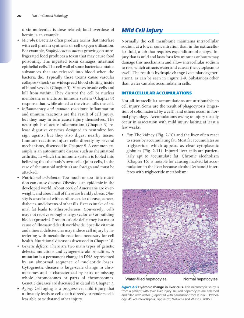
Mild Cell Injury
Normally the cell membrane maintains intracellularsodium at a lower concentration than in the extracellu-lar fluid, a job that requires expenditure of energy. In-jury that is mild and lasts for a few minutes or hours maydamage this mechanism and allow intracellular sodiumto rise, which attracts water and causes the cytoplasm toswell. The result is hydropic change (vacuolar degener-ation), as can be seen in Figure 2-9. Substances otherthan water can also accumulate in cells.
INTRACELLULAR ACCUMULATIONS
Not all intracellular accumulations are attributable tocell injury. Some are the result of phagocytosis (inges-tion of solid material by a cell), and others occur in nor-mal physiology. Accumulations owing to injury usuallyoccur in association with mild injury lasting at least afew weeks.
• Fat: The kidney (Fig. 2-10) and the liver often reactto stress by accumulating fat. Most fat accumulates astriglyceride, which appears as clear cytoplasmicglobules (Fig. 2-11). Injured liver cells are particu-larly apt to accumulate fat. Chronic alcoholism(Chapter 16) is notable for causing marked fat accu-mulation in the liver because alcohol (ethanol) inter-feres with triglyceride metabolism.
Water-filled hepatocytes Normal hepatocytes
Figure 2-9 Hydropic change in liver cells. This microscopic study isfrom a patient with toxic liver injury. Injured hepatocytes are enlargedand filled with water. (Reprinted with permission from Rubin E. Pathol-ogy. 4th ed. Philadelphia. Lippincott, Williams and Wilkins, 2005.)

27Chapter 2 • Cell Injury, Adaptation, and Death
• Cholesterol: The most extensive and most damagingintracellular accumulation is cholesterol, depositedin the cells of arteries in atherosclerosis (Chapter12). Cholesterol first appears in macrophages andsmooth muscle cells in the arterial wall and lateraccumulates into large, extracellular pools in the ar-terial wall.
• Protein: Protein accumulations can occur in cells. Animportant feature of normal proteins is that they arelong molecules that must be folded into correctshape for normal function. Microscopically visiblecytoplasmic accumulations of misfolded or other-wise abnormal proteins occur in a variety of diseases.• Alpha-1 antitrypsin deficiency is a heritable disor-
der (Chapter 14) associated with protein clumpsin hepatocytes.
• Alcoholic liver disease (Chapter 16) is associatedwith cytoplasmic protein clumps known asMallory bodies.
• Several brain diseases (Chapter 23) are associatedwith abnormal accumulations of protein in cells.Among the most notable is Alzheimer disease.
• Glycogen: Glycogen is a long chain of glucose mole-cules formed and stored in liver and muscle as a glu-cose reserve. Glycogen synthesis is regulated byblood glucose concentration. For example, patientswith diabetes (Chapter 17); have high blood glucoselevels, and, as a consequence, hepatocytes and kid-ney cells in people with diabetes are often stuffedwith glycogen.
• Pigments: The most widely occurring cell pigmentaccumulation is lipofuscin, a “wear-and-tear,”golden brown substance most notable in brain neu-rons and myocardial muscle cells, both of which arepermanent, non-reproducing cells, and in hepato-cytes, which are slow-dividing, stable cells. Melaninis a dark-brown compound that gives skin its color(Chapter 24). It is synthesized by melanocytes inthe epidermis and deposited in the cytoplasm ofcells in the basal layer of the epidermis. Inhaledcarbon particles from cigarette smoke or pollutedair are ingested by macrophages of bronchial lymphnodes (Fig. 2-12) and remain permanently withlittle damage. Hemosiderin and ferritin are brownishpigmented normal iron-storage compounds impor-tant in iron and hemoglobin metabolism (Chapter11).
A B
Liver cell with fat: Nucleus Cytoplasm
Liver cell without fat
Figure 2-11 Fatty change in liver cells. This microscopic study isfrom a patient with alcoholic liver disease.
Figure 2-10 Fatty change in the kidney. Mildly injured cells can ac-cumulate fat. A. Normal kidney. B. Fatty change in a patient with toxicinjury to the kidney.
Macrophage containingcarbon particles
Figure 2-12 Intracellular accumulation of carbon pigment. Thisphotomicrograph shows phagocytosis of carbon particles bymacrophages in a bronchial lymph node of a smoker.

28 Part 1 • General Pathology
ADAPTATIONS OF CELL GROWTH ANDDIFFERENTIATION
In addition to hydropic degeneration and intracellularaccumulations, cellular response to persistent stress orchronic mild injury may include a change in size (atro-phy or hypertrophy), an increase in number (hyperpla-sia), or alteration into another type of cell (metaplasia).
Atrophy is decreased size and function of a cell. It isan adaptive response to decreased demand or to in-creased stress; the cell shuts down its metabolicprocesses to conserve energy. Cells atrophy for severalreasons:
• Reduced functional demand. For example, muscle at-rophy occurs in a limb encased in a cast.
• Inadequate blood supply (ischemia). For example, ath-erosclerosis of the renal artery can impair blood flowenough to cause atrophy of a kidney.
• Absent or reduced neural or hormonal support. Forexample, to remain healthy, skeletal muscle cellsmust be continually stimulated by intact nerves; in-terruption of nerve supply leads to muscle atrophy(Chapter 23). Other cells require hormonal support,as do thyroid and adrenal glands, which atrophy ifthey do not receive hormonal support from the pitu-itary gland.
• Chronic inflammation associated with chronic injury.For example, chronic inflammation of the stomachlining is associated with a condition known aschronic atrophic gastritis (Chapter 15), which causesthe lining to become atrophic and very thin.
Hypertrophy is the opposite of atrophy—an in-creased size and functional capacity of a cell. It can becaused by:
• Hormonal stimulation. Cells depend on hormonalsupport. Too little and they wither; too much andthey enlarge and become overactive. For example,following delivery, women’s breasts enlarge and be-come temporarily hyperfunctional in order to pro-duce milk, a change induced by secretion of pro-lactin (a hormone) from the pituitary.
• Increased functional demand. Increased functional de-mand stresses cells and causes them to enlarge andincrease their activity. For example, a heart underthe constant strain of high blood pressure increasesin size because the individual cardiac muscle cells in-crease in size (Fig. 2-13).Hyperplasia is the enlargement of a tissue or organ
owing to an increase in the number of cells, as opposedto an increase in the cell size. It is cause by:
• Hormonal stimulation. For example, the increase ofestrogen in female puberty causes an increase in thenumber of endometrial cells.
• Increased functional demand. For example, low at-mospheric oxygen stimulates bone marrow produc-tion of RBC to carry oxygen. It is for this reason thatpeople living at high altitude have increased num-bers of circulating red blood cells (RBC).
• Chronic stress or injury. For example, the stress of ex-ceptionally high blood pressure (Chapter 12) onsmall arteries in the kidney causes cells in the arterial
Normal leftventricular wall
Hypertrophy of leftventricular wall
A BRight ventricle Right ventricle
Figure 2-13 Hypertrophy. A. Normal left ventricle. B. Hypertrophic left ventricle in a patient with severe, chronic hypertension. Ventricular wall ismarkedly thickened as a result of the increased size of individual muscle cells.
29Chapter 2 • Cell Injury, Adaptation, and Death
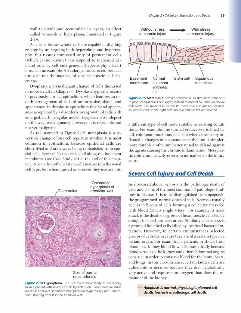
wall to divide and accumulate in layers, an effectcalled “onionskin” hyperplasia, illustrated in Figure2-14.As a rule, tissues whose cells are capable of dividing
enlarge by undergoing both hyperplasia and hypertro-phy. But tissues composed only of permanent cells(which cannot divide) can respond to increased de-mand only by cell enlargement (hypertrophy). Heartmuscle is an example: All enlarged hearts occur becausethe size, not the number, of cardiac muscle cells in-creases.
Dysplasia a premalignant change of cells discussedin more detail in Chapter 6. Dysplasia typically occursin previously normal epithelium, which features an or-derly arrangement of cells of uniform size, shape, andappearance. In dysplastic epithelium this bland appear-ance is replaced by a disorderly overgrowth of cells withenlarged, dark, irregular nuclei. Dysplasia is a mileposton the way to malignancy; however, it is reversible andnot yet malignant.
As is illustrated in Figure 2-15, metaplasia is a re-versible change of one cell type into another. It is mostcommon in epithelium, because epithelial cells areshort-lived and are always being replenished from spe-cial cells (stem cells) that reside all along the basementmembrane (see Case Study 2-1 at the end of this chap-ter). Normally epithelial stem cells mature into the usualcell type, but when injured or stressed they mature into
a different type of cell more suitable to existing condi-tions. For example, the normal endocervix is lined bytall, columnar, mucinous cells, but when chronically in-flamed it changes into squamous epithelium, a simpler,more durable epithelium better suited to defend againstthe agents causing the chronic inflammation. Metaplas-tic epithelium usually reverts to normal when the injurystops.
Severe Cell Injury and Cell Death
As discussed above, necrosis is the pathologic death ofcells and is one of the most common of pathologic find-ings in disease. It is to be distinguished from apoptosis,the programmed, normal death of cells. Necrosis usuallyoccurs in blocks of cells forming a collective mass fedwith blood from a single artery. For example, a heartattack is the death of a group of heart muscle cells fed bya single blocked coronary artery. Similarly, an abscess isa group of liquefied cells killed by localized bacterial in-fection. However, in certain circumstances selectedgroups of cells die because they are of a certain type in acertain organ. For example, in patients in shock fromblood loss, kidney blood flow falls dramatically becauseblood vessels to the kidney and other abdominal organsconstrict in order to conserve blood for the brain, heart,and lungs. In this circumstance, certain kidney cells arevulnerable to necrosis because they are metabolicallyvery active and require more oxygen than does the re-mainder of the kidney.
Glomerulus
"Onionskin"hyperplasia ofarteriolar wall
Size of normalrenal arteriole
Without stressor chronic injury
With stressor chronic injury
Basementmembrane
Normal columnarepithelialcell
Stem cell Squamousmetaplasia
Figure 2-14 Hyperplasia. This is a microscopic study of the kidneyfrom a patient with severe chronic hypertension. Blood pressure stresson renal arterioles stimulates multiplication (hyperplasia) and “onion-skin” layering of cells in the arteriolar wall.
Figure 2-15 Metaplasia. Stress or chronic injury stimulates stem cellsto produce squamous cells (right) instead of normal columnar epithelialcells (left). Columnar cells on the left have cilia and are not layered;squamous cells on the right have no cilia and are flat and layered.
Apoptosis is normal, physiologic, planned celldeath. Necrosis is pathologic cell death.
30 Part 1 • General Pathology
There are four types of necrosis:
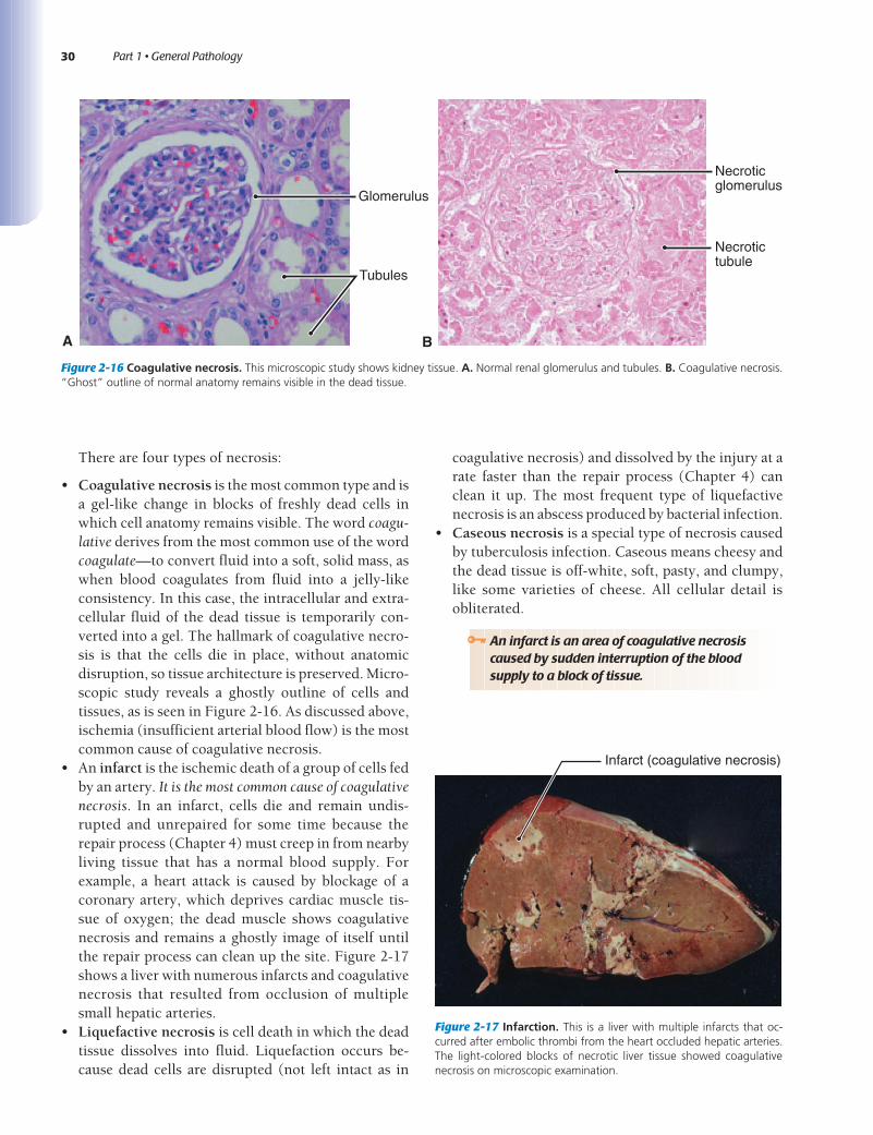
• Coagulative necrosis is the most common type and isa gel-like change in blocks of freshly dead cells inwhich cell anatomy remains visible. The word coagu-lative derives from the most common use of the wordcoagulate—to convert fluid into a soft, solid mass, aswhen blood coagulates from fluid into a jelly-likeconsistency. In this case, the intracellular and extra-cellular fluid of the dead tissue is temporarily con-verted into a gel. The hallmark of coagulative necro-sis is that the cells die in place, without anatomicdisruption, so tissue architecture is preserved. Micro-scopic study reveals a ghostly outline of cells andtissues, as is seen in Figure 2-16. As discussed above,ischemia (insufficient arterial blood flow) is the mostcommon cause of coagulative necrosis.
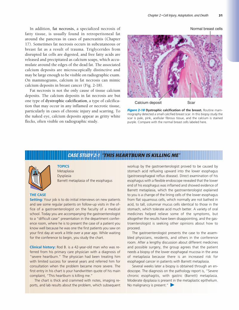
• An infarct is the ischemic death of a group of cells fedby an artery. It is the most common cause of coagulativenecrosis. In an infarct, cells die and remain undis-rupted and unrepaired for some time because therepair process (Chapter 4) must creep in from nearbyliving tissue that has a normal blood supply. Forexample, a heart attack is caused by blockage of acoronary artery, which deprives cardiac muscle tis-sue of oxygen; the dead muscle shows coagulativenecrosis and remains a ghostly image of itself untilthe repair process can clean up the site. Figure 2-17shows a liver with numerous infarcts and coagulativenecrosis that resulted from occlusion of multiplesmall hepatic arteries.
• Liquefactive necrosis is cell death in which the deadtissue dissolves into fluid. Liquefaction occurs be-cause dead cells are disrupted (not left intact as in
coagulative necrosis) and dissolved by the injury at arate faster than the repair process (Chapter 4) canclean it up. The most frequent type of liquefactivenecrosis is an abscess produced by bacterial infection.
• Caseous necrosis is a special type of necrosis causedby tuberculosis infection. Caseous means cheesy andthe dead tissue is off-white, soft, pasty, and clumpy,like some varieties of cheese. All cellular detail isobliterated.
A B
Glomerulus
Tubules
Necroticglomerulus
Necrotictubule
Figure 2-16 Coagulative necrosis. This microscopic study shows kidney tissue. A. Normal renal glomerulus and tubules. B. Coagulative necrosis.“Ghost” outline of normal anatomy remains visible in the dead tissue.
Infarct (coagulative necrosis)
Figure 2-17 Infarction. This is a liver with multiple infarcts that oc-curred after embolic thrombi from the heart occluded hepatic arteries.The light-colored blocks of necrotic liver tissue showed coagulativenecrosis on microscopic examination.
An infarct is an area of coagulative necrosiscaused by sudden interruption of the bloodsupply to a block of tissue.
31Chapter 2 • Cell Injury, Adaptation, and Death
In addition, fat necrosis, a specialized necrosis offatty tissue, is usually found in retroperitoneal fataround the pancreas in cases of pancreatitis (Chapter17). Sometimes fat necrosis occurs in subcutaneous orbreast fat as a result of trauma. Triglycerides fromdisrupted fat cells are digested, and free fatty acids arereleased and precipitated as calcium soaps, which accu-mulate around the edges of the dead fat. The associatedcalcium deposits are microscopically distinctive andmay be large enough to be visible on radiographic exam.On mammograms, calcium in fat necrosis can mimiccalcium deposits in breast cancer (Fig. 2-18).
Fat necrosis is not the only cause of tissue calciumdeposits. The calcium deposits in fat necrosis are butone type of dystrophic calcification, a type of calcifica-tion that may occur in any inflamed or necrotic tissue,particularly in cases of chronic injury and scarring. Tothe naked eye, calcium deposits appear as gritty whiteflecks, often visible on radiographic study.
Normal breast cells
ScarCalcium deposit
Figure 2-18 Dystrophic calcification of the breast. Routine mam-mography detected a small calcified breast scar. In this biopsy study thescar is pale, pink, acellular fibrous tissue, and the calcium is stainedpurple. Compare with the normal breast cells labeled here.
TOPICSMetaplasiaDysplasiaBarrett metaplasia of the esophagus
THE CASESetting: Your job is to do initial interviews on new patientsand see some regular patients on follow-up visits in the of-fice of a gastroenterologist on the faculty of a medicalschool. Today you are accompanying the gastroenterologistto a “difficult case” presentation in the department confer-ence room, where he is to present the case of a patient youknow well because he was one the first patients you saw onyour first day at work a little over a year ago. While waitingfor the conference to begin, you study the chart.
Clinical history: Rod B. is a 42-year-old man who was re-ferred from his primary care physician with a diagnosis of“severe heartburn.” The physician had been treating himwith limited success for several years and referred him forconsultation when the problem became more severe. Thefirst entry in his chart is your handwritten quote of his maincomplaint, “This heartburn is killing me.”
The chart is thick and crammed with notes, imaging re-ports, and lab results about the problem, which subsequent
workup by the gastroenterologist proved to be caused bystomach acid refluxing upward into the lower esophagus(gastroesophageal reflux disease). Direct examination of hisesophagus with a flexible endoscope revealed that the lowerend of his esophagus was inflamed and showed evidence ofBarrett metaplasia, which the gastroenterologist explainedto you is a change of the lining cells of the lower esophagusfrom flat squamous cells, which normally are not bathed inacid, to tall, columnar mucus cells identical to those in thestomach, which tolerate acid much better. A variety of oralmedicines helped relieve some of the symptoms, butaltogether the results have been disappointing, and the gas-troenterologist is seeking other opinions about how toproceed.
The gastroenterologist presents the case to the assem-bled physicians, residents, and others in the conferenceroom. After a lengthy discussion about different medicinesand possible surgery, the group agrees that the patientneeds a biopsy of the lower esophageal mucosa in the areaof metaplasia because there is an increased risk foresophageal cancer in patients with Barrett metaplasia.
Several weeks later a biopsy is obtained through an en-doscope. The diagnosis on the pathology report is, “Severechronic esophagitis, with gastric (Barrett) metaplasia.Moderate dysplasia is present in the metaplastic epithelium.No malignancy is present.” þ
CASE STUDY 2-1 “THIS HEARTBURN IS KILLING ME”

32 Part 1 • General Pathology
[Case 2-1, continued]
DISCUSSIONMetaplasia is a reversible change of one cell type into an-other. Dysplasia is a reversible, premalignant change of ep-ithelium, which can progress to cancer. In this instanceesophageal squamous epithelium was being flooded bygastric acid regurgitated upward from the stomach, whichstimulated esophageal epithelial stem cells to mature intogastric-type mucus cells that were resistant to the effect ofgastric acid. The injury was long standing and severe,enough to cause precancerous change.
Dysplasia is a well-known risk of Barrett metaplasia.Dysplasia is a mile-post on the road to malignancy, but it is
not fully malignant, nor is it irreversible. However, if thechronic injury and dysplasia persist, the dysplastic epithe-lium can become frankly malignant, invade and spreadwidely.
This patient needed vigorous treatment, perhaps includ-ing surgery, to stop the acid reflux into his esophagus andremove or destroy the dysplastic epithelium.
POINTS TO REMEMBER• Chronic injury can cause cells to change from one type to
another.• Chronic injury can cause cells to change from benign to
malignant.• Barrett metaplasia is a premalignant condition.
1. Offer a brief description of the basic organizationof a cell and of the organization of tissues, organs,and organ systems: A cell consists of a nucleus thatis surrounded by cytoplasm, which is containedwithin a cell membrane. The nucleus is composedof DNA, which is organized into the genetic code,and which controls all cell activity. The cytoplasmcarries out the metabolic instructions of nuclearDNA. Cells are organized into tissues, which are or-ganized into organs, which are organized into or-gan systems.
2. Explain how the genetic code is written into DNA:The code is very long sequence of four specializedmolecules, the DNA bases—adenine (A), thymine(T), guanine (G), and cytosine (C). The order inwhich these molecules occur is the genetic code.
3. Explain the role of messenger RNA: Messenger RNA(mRNA) carries a copy of the genetic code fromDNA in the nucleus to ribosomes in the cytoplasm,where the code is used to synthesize the proteincoded by the DNA.
4. Explain the role of mitochondria: Mitochondriaproduce the energy required for metabolicprocesses.
5. Explain how DNA replicates during cell division(mitosis): During mitosis chromosomes line up sin-gle-file around the equator of the parent cell; onestrand of DNA goes to one daughter cell; the otherstrand to the second daughter cell. To do this DNAunravels from the end into two strands like afrayed rope—one strand destined for each newdaughter cell. Bases in each unraveled strand cre-ate a “handshake” with complementary basesfrom the “chemical soup” of the cytoplasm to forma second, new strand of DNA—glycine (G) forms a
loose bond across to a new cytosine (C), and ade-nine (A) to a new thymine (T), and so on. Thesehandshakes steady the new ATCG (base) sequencesso that they can bind laterally up and down thechain to form a new helix intertwined with theother to form a new DNA molecule.
6. Differentiate between apoptosis and necrosis:Apoptosis is natural, physiologic, programmed celldeath; necrosis is pathologic death of cells becauseof injury.
7. Explain the relationship between injury and dis-ease: All disease is caused by injury.
8. Explain the relationship of genes and environmentin the pathogenesis of disease: Genes influencehow we react to injury. Some people are moredisposed, others less disposed, to develop severedisease from a given injury.
9. Name the most common cause of cell injury:Hypoxia; usually secondary to ischemia (low bloodflow).
10. Name one cell reaction resulting from mild acutecell injury and one resulting from mild chronicinjury: Acute mild injury—hydropic (vacuolar)change; chronic mild injury—intracellular accumu-lations of fat, cholesterol, protein, glycogen orpigments.
11. List at least two causes of cell atrophy: Reducedfunctional demand, inadequate blood supply, lackof hormonal or neural support, chronic injury, cellaging.
12. Differentiate between hypertrophy and hyper-plasia: Hypertrophy is tissue enlargement resultingfrom an increase in the size of individual cells.Hyperplasia is tissue enlargement resulting fromincreased number of cells.
Objectives Recap

33Chapter 2 • Cell Injury, Adaptation, and Death
13. Define dysplasia: Dysplasia is a premalignantchange of cells typically seen in epithelium, inwhich the orderly arrangement of normal cells isreplaced by a disorderly overgrowth of cells withenlarged, dark, irregular nuclei.
14. Define metaplasia and offer an example:Metaplasia is the change of one cell type into an-other following stress or chronic mild injury; forexample, the change of endocervical glandular
epithelium into squamous epithelium as a result ofchronic inflammation of the cervix (cervicitis).
15. Name the consequence of severe, irreversible cellinjury: Necrosis.
16. Name the most common cause of necrosis andthe most common type of necrosis: Coagulativenecrosis is the most common type of necrosis; it ismost often caused by ischemia (inadequate bloodflow).
1. Which of the following is composed of nucleotidebases?A. DNAB. mRNAC. tRNAD. rRNAE. All of the above
2. Which of the following is characteristic of apop-tosis?A. It is reversibleB. It is naturalC. It is caused by injuryD. It features fat accumulation in cells
3. Which of the following is the most common causeof cell injury?A. Physical actionB. Toxic molecular injuryC. Ionizing irradiationD. Hypoxia
4. True or false? Hemosiderin is a normal iron storagemolecule that may accumulate in cells.
5. True or false? Hypertrophy is increased number ofcells.
6. True or false? Metaplasia is a reversible change ofcell type.
Typical Test Questions