Lasiodiplodia species associated with dieback disease of mango ... · Lasiodiplodia species...
12
Lasiodiplodia species associated with dieback disease of mango (Mangifera indica) in Egypt A. M. Ismail & G. Cirvilleri & G. Polizzi & P. W. Crous & J. Z. Groenewald & L. Lombard Received: 27 February 2012 / Accepted: 2 August 2012 / Published online: 25 August 2012 # Australasian Plant Pathology Society Inc. 2012 Abstract Lasiodiplodia theobromae is a plurivorous pathogen of tropical and subtropical woody and fruit trees. In 2010, an investigation of mango plantations in Egypt resulted in the isolation of 26 Lasiodiplodia isolates that, based on previous reports from literature, were tentatively identified as L. theobromae. The aim of this study was to clarify the taxonomy of these isolates based on morphology and DNA sequence data (ITS and TEF1-α). In addition to L. theobromae, a new species, namely L. egyptiacae, was identified. Furthermore, L. pseudotheobromae is also newly recorded on mango in Egypt. Pathogenicity tests with all recognised species showed that they are able to cause dieback disease symptoms on mango seedlings. Keywords Botryosphaeriaceae . ITS . Lasiodiplodia . Mango . Morphology . TEF1-α Introduction Mango (Mangifera indica) is a popular fruit tree in Egypt, introduced from Bombay, India in 1825, and is cultivated along the Nile valley and some surrounding desert areas (El Tomi 1953; Abdalla et al. 2007). Most Egyptian mango cultivars, such as alphonso, balady, mabroka, pairi, succary and zebda, are polyembryonic, bearing fruit that are charac- terised by a sweet and spicy flavour, and low fibre content (Knight 1993; El-Soukkary et al. 2000). Among the wide range of destructive fungal pathogens that impact on mango fruit production are members of the Botryosphaeriaceae (Johnson 1992). Botryosphaeriaceae is a genus-rich family in the Dothidiomycetes, containing numerous species with a cosmopolitan distribution (Crous et al. 2006; Phillips et al. 2008). Some of the genera are important pathogens of fruit and woody trees causing symptoms such as leaf spot, die- back, stem-end rot, fruit rot and cankers that can result in tree mortality (Johnson et al. 1991, 1993; Ramos et al. 1991; Smith et al. 2001; Slippers et al. 2005; Damm et al. 2007). Most members of the Botryosphaeriaceae have a broad host range, and have been recognized as successful opportunistic pathogens that occasionally cause extensive disease symp- toms when their plant hosts are subjected to unfavourable conditions (Johnson 1992; Slippers and Wingfield 2007; Sakalidis et al. 2011). Various factors play significant roles in the predisposition of mango trees to attack by members of Botryosphaeriaceae such as mechanical injuries, mineral deficiencies and environmental factors (Ramos et al. 1991; Ploetz et al. 1996a, b). Lasiodiplodia theobromae, a member of Botryosphaer- iaceae, is a cosmopolitan fungus occurring predominantly A. M. Ismail Plant Pathology Research Institute, Agriculture Research Centre, 12619 Giza, Egypt G. Cirvilleri : G. Polizzi Dipartimento di Gestione dei Sistemi Agroalimentari e Ambientali Sez. Patologia Vegetale, 95123 Catania, Italy P. W. Crous : J. Z. Groenewald : L. Lombard (*) CBS-KNAW Fungal Biodiversity Centre, Uppsalalaan 8, 3584 CT Utrecht, The Netherlands e-mail: [email protected] P. W. Crous Laboratory of Phytopathology, Wageningen University and Research Centre (WUR), Droevendaalsesteeg 1, 6708 PB Wageningen, The Netherlands P. W. Crous Microbiology, Department of Biology, Utrecht University, Padualaan 8, 3584 CH Utrecht, The Netherlands Australasian Plant Pathol. (2012) 41:649–660 DOI 10.1007/s13313-012-0163-1
Transcript of Lasiodiplodia species associated with dieback disease of mango ... · Lasiodiplodia species...

Lasiodiplodia species associated with dieback diseaseof mango (Mangifera indica) in Egypt
A. M. Ismail & G. Cirvilleri & G. Polizzi & P. W. Crous &
J. Z. Groenewald & L. Lombard
Received: 27 February 2012 /Accepted: 2 August 2012 /Published online: 25 August 2012# Australasian Plant Pathology Society Inc. 2012
Abstract Lasiodiplodia theobromae is a plurivorouspathogen of tropical and subtropical woody and fruit trees.In 2010, an investigation of mango plantations in Egyptresulted in the isolation of 26 Lasiodiplodia isolates that,based on previous reports from literature, were tentativelyidentified as L. theobromae. The aim of this study was toclarify the taxonomy of these isolates based on morphologyand DNA sequence data (ITS and TEF1-α). In addition to L.theobromae, a new species, namely L. egyptiacae, wasidentified. Furthermore, L. pseudotheobromae is alsonewly recorded on mango in Egypt. Pathogenicity testswith all recognised species showed that they are able tocause dieback disease symptoms on mango seedlings.
Keywords Botryosphaeriaceae . ITS . Lasiodiplodia .
Mango .Morphology . TEF1-α
Introduction
Mango (Mangifera indica) is a popular fruit tree in Egypt,introduced from Bombay, India in 1825, and is cultivatedalong the Nile valley and some surrounding desert areas (ElTomi 1953; Abdalla et al. 2007). Most Egyptian mangocultivars, such as alphonso, balady, mabroka, pairi, succaryand zebda, are polyembryonic, bearing fruit that are charac-terised by a sweet and spicy flavour, and low fibre content(Knight 1993; El-Soukkary et al. 2000). Among the widerange of destructive fungal pathogens that impact on mangofruit production are members of the Botryosphaeriaceae(Johnson 1992). Botryosphaeriaceae is a genus-rich familyin the Dothidiomycetes, containing numerous species with acosmopolitan distribution (Crous et al. 2006; Phillips et al.2008). Some of the genera are important pathogens of fruitand woody trees causing symptoms such as leaf spot, die-back, stem-end rot, fruit rot and cankers that can result intree mortality (Johnson et al. 1991, 1993; Ramos et al. 1991;Smith et al. 2001; Slippers et al. 2005; Damm et al. 2007).Most members of the Botryosphaeriaceae have a broad hostrange, and have been recognized as successful opportunisticpathogens that occasionally cause extensive disease symp-toms when their plant hosts are subjected to unfavourableconditions (Johnson 1992; Slippers and Wingfield 2007;Sakalidis et al. 2011). Various factors play significant rolesin the predisposition of mango trees to attack by members ofBotryosphaeriaceae such as mechanical injuries, mineraldeficiencies and environmental factors (Ramos et al. 1991;Ploetz et al. 1996a, b).
Lasiodiplodia theobromae, a member of Botryosphaer-iaceae, is a cosmopolitan fungus occurring predominantly
A. M. IsmailPlant Pathology Research Institute, Agriculture Research Centre,12619 Giza, Egypt
G. Cirvilleri :G. PolizziDipartimento di Gestione dei Sistemi Agroalimentari eAmbientali Sez. Patologia Vegetale,95123 Catania, Italy
P. W. Crous : J. Z. Groenewald : L. Lombard (*)CBS-KNAW Fungal Biodiversity Centre,Uppsalalaan 8,3584 CT Utrecht, The Netherlandse-mail: [email protected]
P. W. CrousLaboratory of Phytopathology,Wageningen University and Research Centre (WUR),Droevendaalsesteeg 1,6708 PB Wageningen, The Netherlands
P. W. CrousMicrobiology, Department of Biology, Utrecht University,Padualaan 8,3584 CH Utrecht, The Netherlands
Australasian Plant Pathol. (2012) 41:649–660DOI 10.1007/s13313-012-0163-1

throughout tropical and subtropical regions (Punithalingam1980; Burgess et al. 2006). It has also been known as ahuman pathogen causing keratomycosis and phaeohypho-mycosis (Punithalingam 1976; Summerbell et al. 2004), andas a plant pathogen associated with up to 500 plant hosts(Punithalingam 1980). The fungus has been reported asmango pathogen worldwide associated with several plantdisease symptoms including decline, canker and dieback(Jacobs 2002; Khanzada et al. 2004a, b; Abdollahzadeh etal. 2010; de Oliveira Costa et al. 2010). In Egypt, the fungusis well established and has been considered as the maincausal agent of fruit rot, stem-end rot, panicle brown rotand dieback of mango (Ragab et al. 1971; Abdalla et al.2003). In addition to mango, it has also been reported tocause root rot of sugar beet (Abd-El Ghani and Fatouh2005) and dieback, canker and soft rot of other hosts suchas grapevine (El-Goorani and El Meleigi 1972), walnut(Haggag et al. 2007), maize (Diab et al. 1984), citrus(Abo-El-Dahab et al. 1992) and Annona spp. (Haggag andNofal 2006) in Egypt. The taxonomic placement of Botryos-phaeria rhodina (anamorph L. theobromae) has been com-plicated by several names associated with this fungus(Burgess et al. 2006). Punithalingam (1976) reduced severalspecies (L. nigra, L. triflorae, and L. tubericola) to synon-ymy under L. theobromae. Subsequent to this treatment,several studies have led to the identification of crypticspecies within the L. theobromae species complex (Pavlicet al. 2004, 2008; Burgess et al. 2006; Damm et al. 2007;Alves et al. 2008; Begoude et al. 2009; Abdollahzadeh et al.2010). Presently, up to 13 cryptic species are recognised inthe L. theobromae complex.
In recent years molecular DNA-based approaches havebeen widely employed in taxonomic studies of the Botryos-phaeriaceae (Crous and Groenewald 2005). Several phylo-genetic studies have relied on the sequence differences fromthe internal transcribed spacer (ITS) region of the rDNAoperon to distinguish species within Botryosphaeriaceae(Denman et al. 2000, 2003; Alves et al. 2008). However,ITS sequence data alone can obscure cryptic species diver-sity and proved to be inadequate to separate closely relatedspecies (de Wet et al. 2003; Slippers et al. 2004a; Slippersand Wingfield 2007; Marincowitz et al. 2008). Interestingly,contemporary phylogenetic studies using multiple gene ge-nealogies have increasingly revealed cryptic species in theL. theobromae complex (Pavlic et al. 2004, 2008; Burgess etal. 2006; Damm et al. 2007; Alves et al. 2008; Begoude etal. 2009; Abdollahzadeh et al. 2010).
Little is known of the aetiology of Botryosphaeriaceaediseases on mango in Egypt. By means of a morphologicaland DNA sequence data comparison, the present studyrepresents the first attempt to characterise the variabilitywithin an Egyptian collection of isolates previously treatedas L. theobromae or Botryodiplodia theobromae.
Materials and methods
Isolates
In February 2010, a routine survey was conducted in severalareas in Egypt where mango is cultivated. Isolations weremade from fresh symptomatic plant material showing twigand branch dieback and black lesions on leaves. Initially,samples were surface sterilised with a diluted potassiumhydroxide solution (5 %) and EtOH (70 %). Approximately3–5 mm diam pieces of plant material between the healthyand infected tissues were placed on 2 % Potato-DextroseAgar (PDA) supplemented with Streptomycin sulphate(0.1 g/L−1) and incubated at 25 °C in the dark. Pure cultureswere obtained by hyphal tip excision from the colonymargins on PDA, and subsequent incubation at 25 °C inthe dark. All isolates obtained from mango were depositedin the collection of the Plant Pathology Research Institute,Egypt. Representative isolates used for morphological andmolecular studies were also deposited in the collection ofCBS-KNAW Fungal Biodiversity Centre (CBS), Utrecht,the Netherlands (Table 1).
DNA isolation and amplification
Total genomic DNA was extracted from 8 to 10-day oldcultures using the Ultraclean® Microbial DNA Isolation Kit(MO–BIO Laboratories, Inc, Carlsbad, USA) according tothe manufacturer’s protocol. The ITS region of the rDNAoperon was amplified using the primers V9G (de Hoog andGerrits van den Ende 1998) and ITS4 (White et al. 1990).Partial sequence of the translation elongation factor 1-alpha(TEF-1α) gene region was amplified using primers EF1-728F (Carbone and Kohn 1999) and EF2 (O’Donnell et al.1998). For some isolates, the TEF-1α gene region wasamplified using primers EF1-688F and EF1-1251R (Alveset al. 2008). Each PCR reaction contained a final concen-tration of 0.5 U/μL of Taq polymerase, 1X buffer 2–2.5 mMMgCl2 (BIOLINE, San Diego, USA), 0.4–0.6 mM of eachdNTP and 0.12–0.2 μm of each primer made up to a finalvolume of 12.5 μL with sterile deionized water. PCR con-ditions included the following steps: an initial step of dena-turation at 95 °C for 5 min, followed by 40 cycles of 95 °Cfor 30 s, 52 °C for 30 s and 72 °C for 1 min, with a finalelongation step at 72 °C for 7 min.
Phylogeny
The amplified fragments of the ITS gene region weresequenced in both directions using internal primers ITS4and ITS5 (White et al. 1990), whereas the TEF-1α generegion was sequenced in both directions using the sameprimer pairs for amplification. Sequencing reactions were
650 A.M. Ismail et al.
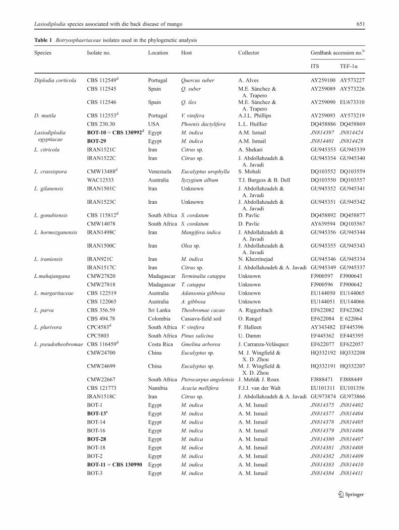
Table 1 Botryosphaeriaceae isolates used in the phylogenetic analysis
Species Isolate no. Location Host Collector GenBank accession no.b
ITS TEF-1α
Diplodia corticola CBS 112549d Portugal Quercus suber A. Alves AY259100 AY573227
CBS 112545 Spain Q. suber M.E. Sánchez &A. Trapero
AY259089 AY573226
CBS 112546 Spain Q. ilex M.E. Sánchez &A. Trapero
AY259090 EU673310
D. mutila CBS 112553d Portugal V. vinifera A.J.L. Phillips AY259093 AY573219
CBS 230.30 USA Phoenix dactylifera L.L. Huillier DQ458886 DQ458869
Lasiodiplodiaegyptiacae
BOT-10 0 CBS 130992d Egypt M. indica A.M. Ismail JN814397 JN814424
BOT-29 Egypt M. indica A.M. Ismail JN814401 JN814428
L. citricola IRAN1521C Iran Citrus sp. A. Shekari GU945353 GU945339
IRAN1522C Iran Citrus sp. J. Abdollahzadeh &A. Javadi
GU945354 GU945340
L. crassispora CMW13488d Venezuela Eucalyptus urophylla S. Mohali DQ103552 DQ103559
WAC12533 Australia Syzygium album T.I. Burgess & B. Dell DQ103550 DQ103557
L. gilanensis IRAN1501C Iran Unknown J. Abdollahzadeh &A. Javadi
GU945352 GU945341
IRAN1523C Iran Unknown J. Abdollahzadeh &A. Javadi
GU945351 GU945342
L. gonubiensis CBS 115812d South Africa S. cordatum D. Pavlic DQ458892 DQ458877
CMW14078 South Africa S. cordatum D. Pavlic AY639594 DQ103567
L. hormozganensis IRAN1498C Iran Mangifera indica J. Abdollahzadeh &A. Javadi
GU945356 GU945344
IRAN1500C Iran Olea sp. J. Abdollahzadeh &A. Javadi
GU945355 GU945343
L. iraniensis IRAN921C Iran M. indica N. Khezrinejad GU945346 GU945334
IRAN1517C Iran Citrus sp. J. Abdollahzadeh & A. Javadi GU945349 GU945337
L.mahajangana CMW27820 Madagascar Terminalia catappa Unknown FJ900597 FJ900643
CMW27818 Madagascar T. catappa Unknown FJ900596 FJ900642
L. margaritaceae CBS 122519 Australia Adansonia gibbosa Unknown EU144050 EU144065
CBS 122065 Australia A. gibbosa Unknown EU144051 EU144066
L. parva CBS 356.59 Sri Lanka Theobromae cacao A. Riggenbach EF622082 EF622062
CBS 494.78 Colombia Cassava-field soil O. Rangel EF622084 E 622064
L. plurivora CPC4583d South Africa V. vinifera F. Halleen AY343482 EF445396
CPC5803 South Africa Pinus salicina U. Damm EF445362 EF445395
L. pseudotheobromae CBS 116459d Costa Rica Gmelina arborea J. Carranza-Velásquez EF622077 EF622057
CMW24700 China Eucalyptus sp. M. J. Wingfield &X. D. Zhou
HQ332192 HQ332208
CMW24699 China Eucalyptus sp. M. J. Wingfield &X. D. Zhou
HQ332191 HQ332207
CMW22667 South Africa Pterocarpus angolensis J. Mehl& J. Roux FJ888471 FJ888449
CBS 121773 Namibia Acacia mellifera F.J.J. van der Walt EU101311 EU101356
IRAN1518C Iran Citrus sp. J. Abdollahzadeh & A. Javadi GU973874 GU973866
BOT-1 Egypt M. indica A. M. Ismail JN814375 JN814402
BOT-13c Egypt M. indica A. M. Ismail JN814377 JN814404
BOT-14 Egypt M. indica A. M. Ismail JN814378 JN814405
BOT-16 Egypt M. indica A. M. Ismail JN814379 JN814406
BOT-28 Egypt M. indica A. M. Ismail JN814380 JN814407
BOT-18 Egypt M. indica A. M. Ismail JN814381 JN814408
BOT-2 Egypt M. indica A. M. Ismail JN814382 JN814409
BOT-11 0 CBS 130990 Egypt M. indica A. M. Ismail JN814383 JN814410
BOT-3 Egypt M. indica A. M. Ismail JN814384 JN814411
Lasiodiplodia species associated with die back disease of mango 651
performed using Big Dye terminator sequencing kit v.3.1 (Perkin-Elmer Applied Bio Systems, Foster City,CA, USA) following the manufacturer’s instructionsand run using an ABI PRISM™ 3730 DNA automatedsequencer (Perkin-Elmer Applied BioSystems, Foster City,CA, USA).
The generated sequences were aligned together with othersequences obtained from GenBank using MAFFT v. 6.0(Katoh and Toh 2010). The ambiguous sequences of both 5′and 3′ ends were excluded from the final alignment and the
aligned sequences were manually checked and correctedwhere necessary. Nucleotide substitution models were deter-mined for each gene region using MrModel Test v.2.2(Nylander 2004). The model HKY+G was selected for bothITS and TEF sequence datasets. Sequences for each generegion were individually analysed for conflict using 70 %reciprocal NJ (Neighbour-Joining) bootstrap analysis and thetopology of the resulting trees were compared visually forinconsistency (Mason-Gamer and Kellogg 1996; Gueidan etal. 2007).
Table 1 (continued)
Species Isolate no. Location Host Collector GenBank accession no.b
ITS TEF-1α
BOT-17 Egypt M. indica A. M. Ismail JN814385 JN814412
BOT-12 Egypt M. indica A. M. Ismail JN814386 JN814413
BOT-24 Egypt M. indica A. M. Ismail JN814387 JN814414
BOT-26 Egypt M. indica A. M. Ismail JN814388 JN814415
BOT-27 Egypt M. indica A. M. Ismail JN814389 JN814416
BOT-22 Egypt M. indica A. M. Ismail JN814390 JN814417
BOT-15 Egypt M. indica A. M. Ismail JN814391 JN814418
BOT-25 Egypt M. indica A. M. Ismail JN814393 JN814420
BOT-21 Egypt M. indica A. M. Ismail JN814394 JN814421
L. rubropurpurea WAC12536d Australia E. grandis T. I. Burgess & G. Pegg DQ103554 DQ103572
WAC12537 Australia E. grandis T. I. Burgess & G. Pegg DQ103555 DQ103573
L. theobromae CBS 112874 South Africa V. vinifera F. Halleen EF622075 EF622055
CBS 559.70 Unknown Zea mays H. A. van der Aa EF622073 EF622053
CBS 111530 Unknown Unknown Unknown EF622074 EF622054
CMW24702 China Eucalyptus sp. M. J. Wingfield &X.D. Zhou
HQ332194 HQ332210
CMW24701 China Eucalyptus sp. M. J. Wingfield &X.D. Zhou
HQ332193 HQ332209
MUCC709 Australia Lysiphyllumcunninghamii
M. L. Sakalidis GU199367 GU199393
BOT-5 Egypt M. indica A.M. Ismail JN814376 JN814403
BOT-9 Egypt M. indica A.M. Ismail JN814392 JN814419
BOT-4 0 CBS 130989 Egypt M. indica A.M. Ismail JN814395 JN814422
BOT-7 Egypt M. indica A.M. Ismail JN814396 JN814423
BOT-6 Egypt M. indica A.M. Ismail JN814399 JN814426
BOT-23 Egypt M. indica A.M. Ismail JN814400 JN814427
L. venezuelensis CMW13513d Venezuela A. mangium S. Mohali DQ103549 DQ103570
WAC12540 Venezuela A. mangium S. Mohali DQ103548 DQ103569
Phyllostictacapitalensis
CBS 115051 Brazil Spondias mombin K.F. Rodriquez FJ538325 FJ538383
P. citricarpa CBS 102374 Brazil C. aurantium Unknown FJ538313 FJ538371
a CMW 0 culture collection of the Forestry and Agricultural Biotechnology Institute, University of Pretoria, Pretoria, South Africa; CBS 0 CBS-KNAW Fungal Biodiversity Centre, Utrecht, the Netherlands; CAA 0 A. Alves, Universidade de Aveiro, Portugal; CAP 0A.J.L. Phillips, Lisbon,Portugal. WAC0Department of Agriculture Western Australia, Plant Pathogen Collection; BOT 0A. M. Ismail, Plant Pathology Research Institute,Egypt; CPC 0 P.W. Crous working collection, maintained at CBSbGenBank accession numbers in italics were generated in this studyc Isolate numbers in bold were selected for pathogenicity testd Ex-type cultures
652 A.M. Ismail et al.

Bayesian analyses were performed with MrBayes v. 3.1.1(Ronquist and Huelsenbeck 2003) using the Markov ChainMonte Carlo (MCMC) (Larget and Simon 1999) algorithmto generate trees with Bayesian probability values. Fourchains were run simultaneously from a random tree topolo-gy and ended at 1,000,000 generations, and trees were savedevery 100th generation. The burn-in value was graphicallyestimated from the likelihood scores and therefore, the first1,000 trees were discarded from the analysis as the burn-inphase and the consensus tree was constructed from theremaining trees. Trees were rooted using Phyllosticta capi-talensis (CBS 115051) and P. citricarpa (CBS 102374)(Glienke et al. 2011).
All the sequence datasets were also analysed to determinepossible phylogenetic relationship among taxa using PAUP(Phylogenetic Analysis Using Parsimony) v. 4.0b10(Swofford 2001). Maximum parsimony (MP) tests wereconducted using the heuristic search option with randomstepwise addition using 1,000 replicates, tree bisection andreconnection (TBR) as branch swapping algorithms, andrandom taxon addition sequences for the construction ofmaximum parsimony trees. Maxtrees was set to 10,000branches of zero length were collapsed, and all multipleequally parsimonious trees were saved. In the analysis, allcharacters were unordered and had equal weight; gaps weretreated as missing data. Values calculated for parsimonyincluded tree length (TL), consistency index (CI), rescaledconsistency index (RC) and the retention index (RI).Bootstrap support values were evaluated using 1,000 boot-strap replicates (Hillis and Bull 1993). All sequencesgenerated in this study were deposited in GenBank (Table 1).The aligned sequences were deposited in TreeBASE(S12897).
Morphological characterisation
Sporulation was induced by plating representative isolatesonto 2 % (w/v) water agar with sterilised pine needles(WAP) and incubated at 25±2 °C under near-ultraviolet(UV) light for 2 weeks. Plates were observed every 2 daysfor the formation of pycnidia. Gross morphological charac-teristics were determined by mounting fungal structures inclear lactic acid. Measurements of 50 conidia and at least 30other fungal structures for each representative isolate weredetermined at ×1,000 magnification. Sections were madethrough pycnidia using a Leica CM1100 cryostat at −20 °Cand the 10 μm sections were mounted in lactic acid. Grossmorphological characteristics were observed as above. Forthe conidia, the 95 % confidence levels were calculated of30 observations, with extremes given in parentheses. Onlythe extremes are indicated for the other fungal structures.Colony characteristics were determined after 7 days on PDAin the dark at 25 °C, using the colour charts of Rayner
(1970). Optimal growth temperatures were determined foreach selected isolate on PDA at 10–35 °C in 5 °C intervalsin the dark, with three plates per isolate at each temperature.Descriptions, nomenclature and illustrations were depositedin MycoBank (Crous et al. 2004).
Pathogenicity test
Ten isolates representing three species of Lasiodiplodia(Table 1) were used for pathogenicity trials on mango seed-lings cv. “Kensington Pride”. The plants were 3–4-monthold, 40–60 cm tall, and maintained in a greenhouse underartificial light (10/14 h light-and-dark cycles) at 25±2 °Cand 70–80 % relative humidity (RH). Four plants for eachisolate and the controls were used and arranged in a rando-mised design. The epidermis of the stem was disinfectedwith 70 % ethanol, washed with sterile distilled water andleft to dry. A 5-mm cut was made into the epidermis,between two nodes and below the apex of the stem. A 5-mm diam mycelial PDA plug was removed from the edge ofactively growing cultures, and placed onto the stem wounds,with the mycelium facing the cambium. The inoculatedwounds were wrapped with Parafilm®, (Laboratory Film,Chicago, IL, USA) to prevent desiccation and contamina-tion. Control plants were inoculated with sterile PDAplugs. Six weeks after inoculation the bark lesion lengthsas well as the length of cambium discolouration weremeasured to assess the pathogenicity of the tested isolates.Re-isolation of the tested isolates was done from themargins of the necrotic lesions on PDA to prove Koch’sPostulates.
Results
Phylogeny
Amplicons of approx. 570 bp were generated for ITS usingprimer pairs ITS5 and ITS4 and approx. 500 bp for TEF-1αwere obtained using the EF1-728F and EF2 primers pairs.Amplicons of approx. 700 bp was obtained using primersEF1-688 and EF1-1252. The 70 % reciprocal NJ bootstrapanalysis indicated congruence in the tree topology of bothITS and TEF-1α trees. The combined data set consisted of69 taxa including the outgroup taxa composed of 920 char-acters including gaps, of which 589 were constant, 91 werevariable and parsimony uninformative and 240 were parsi-mony informative. Maximum parsimony analysis resulted inone most parsimonious tree (TL01774 steps, CI00.581,RI00.756, RC00.894) presented in Fig. 1. In this tree, theLasiodiplodia-like isolates obtained in this study fell intofour distinct clades. The majority of isolates (BOT-1, BOT-2, BOT-3, BOT-11, BOT-12, BOT-13, BOT-14, BOT-15,
Lasiodiplodia species associated with die back disease of mango 653
BOT-16, BOT-17, BOT-18, BOT-21, BOT-22, BOT-24,BOT-25, BOT-26, BOT-27 and BOT-28) clustered togetherin a large clade containing L. pseudotheobromae (CBS116459, culture ex-type) supported by a bootstrap (BP)value of 98 and a Bayesian posterior probability (BPP) valueof 1.0. A second well-supported clade (BS/BPP: 76/0.99)
accommodated two Lasiodiplodia-like isolates (CBS 130992and BOT-29), possibly representing a novel phylogeneticspecies. A further six isolates (BOT-4, BOT-5, BOT-6, BOT-7, BOT-9 and BOT-23) clustered together with L. theobromae(CMW 24701, CMW 24702, CBS 111530; Chen et al. 2011),with low support (BS/BPP: 54/0.58).
Fig. 1 The most parsimonious trees obtained from the maximumparsimony analysis using heuristic search with 1,000 random additionsof the combined ITS and TEF-1α sequence alignments. Scale bar
shows ten changes and bootstrap support and Bayesian posterior prob-ability values are indicated at the nodes. The tree was rooted to P.capitalensis CBS 115051 and P. citricarpa CBS 102374
654 A.M. Ismail et al.
Morphological characterisation
In this study a total of 26 isolates representing species ofBotryosphaeriaceae were obtained from mango trees. Ofthese, 12 isolates were obtained from branches, 11 fromleaves and three from twigs. No teleomorph structures wereobserved in this study. Based on cultural and conidial char-acteristics isolates were considered to belong to Lasiodiplo-dia. All isolates were included in the phylogenetic analysis.Based on DNA sequence data and conidial morphologythree species were identified which included L. theobromae,L. pseudotheobromae and a new species which is describedhere.
Lasiodiplodia egyptiacae A.M. Ismail, L. Lombard &Crous, sp. nov. MycoBank MB564516, Fig. 2
Etymology: The name refers to Egypt, the country wherethis fungus was collected.
Conidiomata stromatic, pycnidial, produced on WAPwithin 12 days, mostly solitary, or aggregated, dark-greyto black, globose to subglobose, covered with dense myce-lium, papillate with centralostiole, conidiomata semi-immersed, becoming erumpent when mature, mostly multi-loculate to uni-loculate; wall of two regions: outer regioncomposed of 5–7 layers of dark brown, thick-walled cells oftextura angularis, followed by an inner region of hyaline,thin-walled cells of textura angularis. Paraphyses hyaline,subcylindrical, arising between the conidiogenous cells,aseptate, wider at base, rounded or slightly swollen at apex,up to 57 μm long, 2–3 μm wide. Conidiogenous cellsholoblastic, hyaline, thin-walled, cylindrical, sometimesslightly swollen at the base, with rounded apex, proliferatingpercurrently to produce 1–2 min annellations, 5–11×3–5 μm. Conidia oozing from pycnidia in conidial cirri, ini-tially hyaline, smooth, thick-walled, aseptate, obovoid toellipsoid, granular, mostly somewhat tapered at apex, androunded at base, becoming brown, 1-septate, with longitu-dinal striations on the inner surface of the conidia wall due
to the melanin deposits, measuring (17–)20–24(−27) ×(11–)11–12(−13) μm (av. ± SD022±2 μm long, 12±1 μmwide, L/W ratio02).
Culture characteristics: Colonies on PDA with moder-ately dense, raised mycelium mat, initially white to smoke-grey, turning greenish grey on the surface and greenish greyin reverse, becoming dark slate-blue with age. Cardinaltemperature requirements for growth; minimum 15 °C, max-imum 35 °C, optimum 25 °C.
Specimens examined: Egypt, Sharkia Province, ElMenayar, isolated from M. indica leaf, 2 Feb. 2010, A.M.Ismail, holotype CBS H-20736, culture ex-type BOT-10 0
CBS 130992; Sharkia Province, El Menayar, isolated frommango leaf, 2 Feb. 2010, A.M. Ismail, culture BOT-29.
Notes: Lasiodiplodia egyptiacae is phylogeneticallyclosely related to L. hormozganensis (Abdollahzadeh et al.2010), but it can be distinguished based on the morphologyof its conidia and paraphyses (Table 2). The conidia of L.egyptiacae are ovoid to sub-ovoid, whereas those of L.hormozganensis are ellipsoid to cylindrical. In addition,paraphyses of L. egyptiacae are aseptate and shorter (up to57 μm), whilst the paraphyses of L. hormozganensis are 1–7-septate and longer (up to 83 μm). Furthermore, L. egyp-tiacae is still distinct from L. citricola and L. parva in termsof paraphyses morphology. The paraphyses of L. egyptiacaeare aseptate, shorter and narrower (57×2–3 μm) while thoseof L. citricola and L. parva are septate, longer and wider(125×3–4 μm), (105×3–4 μm), respectively (Table 2).
Pathogenicity test
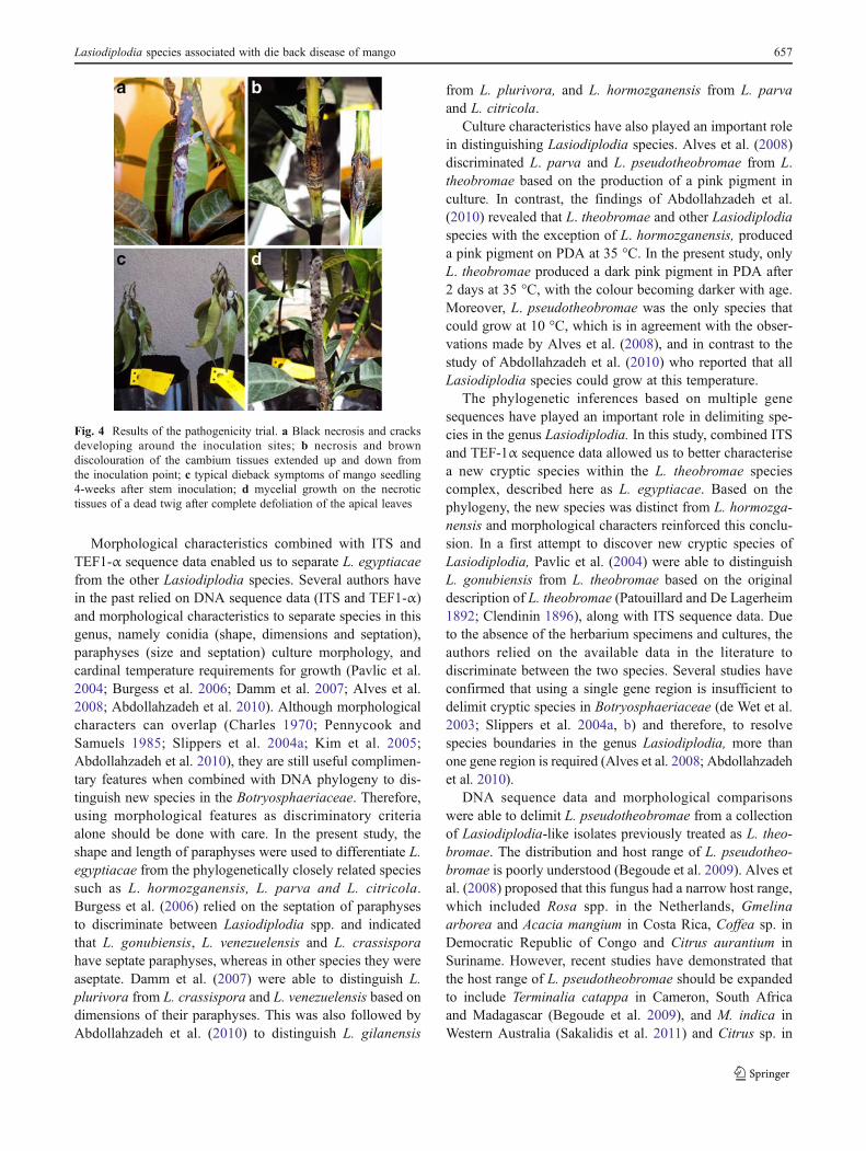
Six weeks after inoculation, all isolates displayed levels ofpathogenicity. Observed symptoms included brown, necrot-ic bark lesions around the inoculation sites extending up-wards and downwards, leading to wilting and drying of theapical as well as the terminal leaves (Fig. 4). Cracking of thestem cortex was observed for most of the isolates, and
Fig. 2 Lasiodiplodiaegyptiacae a pycnidia formedon WAP; b vertical sectionthrough pycnidia; c hyaline,aseptate paraphyses formedbetween conidiogenous cells; dconidiogenous cells; e hyalineimmature thick-walled conidia;f dark mature conidia showinglongitudinal striation. Scalebars: b020 μm; c–f010 μm
Lasiodiplodia species associated with die back disease of mango 655
fungal structures (stromatic pycnidia and mycelium) devel-oped on the necrotic lesions around the inoculation sites.Under the outer cortex, necrotic xylem vessels and browndiscolouration extended along the length of the stems(Fig. 4). Symptoms observed on the control plants couldbe due to wound reaction as no Lasiodiplodia was isolated.There was a significant difference (p<0.05) in the lesionsproduced by Lasiodiplodia isolates compared to controllesions. Isolates BOT-11 and BOT-28 (L. pseudotheobro-mae) developed the longest bark (av. 63.3 mm and 62.6 mm,respectively) and cambium (av. 64.1 mm and 63.6 mm,respectively) lesions, followed by isolate BOT-4 (L. theo-bromae), which produced a bark lesion of av. 56.5 mm andcambium lesion of av. 60.7 mm in length (Fig. 3). Thesethree isolates were the only to induce dieback symptoms
similar to those observed during the survey (Fig. 4). IsolatesCBS 130992 and BOT-29 (L. egyptiacae) produced smallerlesions (av. 38.8 mm and 35.1 mm, respectively), however,still longer than the controls (av. 25.8 mm).
Discussion
Three species of Lasiodiplodia associated with dieback andleaf lesions of mango trees were identified in the presentstudy. These were L. theobromae, L. pseudotheobromaeand the newly described L. egyptiacae. The latter newspecies is distinguished from other species of Lasiodiplodiabased on morphological characters and phylogeneticinference.
Table 2 Morphological comparison of conidia and paraphyses of Lasiodiplodia spp.
Identity Conidial size (av. μm) L/w ratio Paraphyses (μm) References
Length Width Septation
L. egyptiacae 22×12 1.8 57 2–3 Aseptate This study
L. citricola 24.5×15.4 1.6 125 3–4 Septate Abdollahzadeh et al. (2010)
L. crassispora 28.8×16 1.8 45.7 2.7 Septate Burgess et al. (2006)
L. gilanensis 31×16.6 1.9 95 2–4 Septate Abdollahzadeh et al. (2010)
L. gonubiensis 33.8×17.3 1.9 70 4 Aseptate Pavlic et al. (2004)
L. hormozganensis 21.5×12.5 1.7 83 2–4 Septate Abdollahzadeh et al. (2010)
L. iraniensis 20.7×13 1.6 127 2–4 Septate Abdollahzadeh et al. (2010)
L.mahajangana 17.5×11.5 1.4 43 3 Aseptate Begoude et al. (2009)
L. margaritaceae 15.3×11.4 1.3 37.1 2.2 Septate Pavlic et al. (2008)
L. parva 20.2×11.5 1.8 105 3–4 Septate Alves et al. (2008)
L. plurivora 29.6×15.6 1.9 130 2–5 Septate Damm et al. (2007)
L. pseudotheobromae 28×16 1.7 58 3–4 Aseptate Alves et al. (2008)
26.7×12.3 2.1 52 2–3 Aseptate This study
L. rubropurpurea 28.2×14.6 1.9 42.4 2.6 Aseptate Burgess et al. (2006)
L. theobromae 26.2×14.2 1.9 55 3–4 Septate Alves et al. (2008)
23.7×13.3 1.7 44 2–3 Septate This study
L. venezuelensis 28.4×13.5 2.1 28.3 3.5 Septate Burgess et al. (2006)
Fig. 3 Mean lengths (mm) oflog-transformed bark andcambium lesions 6 weeks afterinoculation on mango plantscv. Kensington Pride with fourspecies of Lasiodiplodia. Barsabove columns are the standarderror of the mean of bark andcambium lesions lengths
656 A.M. Ismail et al.
Morphological characteristics combined with ITS andTEF1-α sequence data enabled us to separate L. egyptiacaefrom the other Lasiodiplodia species. Several authors havein the past relied on DNA sequence data (ITS and TEF1-α)and morphological characteristics to separate species in thisgenus, namely conidia (shape, dimensions and septation),paraphyses (size and septation) culture morphology, andcardinal temperature requirements for growth (Pavlic et al.2004; Burgess et al. 2006; Damm et al. 2007; Alves et al.2008; Abdollahzadeh et al. 2010). Although morphologicalcharacters can overlap (Charles 1970; Pennycook andSamuels 1985; Slippers et al. 2004a; Kim et al. 2005;Abdollahzadeh et al. 2010), they are still useful complimen-tary features when combined with DNA phylogeny to dis-tinguish new species in the Botryosphaeriaceae. Therefore,using morphological features as discriminatory criteriaalone should be done with care. In the present study, theshape and length of paraphyses were used to differentiate L.egyptiacae from the phylogenetically closely related speciessuch as L. hormozganensis, L. parva and L. citricola.Burgess et al. (2006) relied on the septation of paraphysesto discriminate between Lasiodiplodia spp. and indicatedthat L. gonubiensis, L. venezuelensis and L. crassisporahave septate paraphyses, whereas in other species they wereaseptate. Damm et al. (2007) were able to distinguish L.plurivora from L. crassispora and L. venezuelensis based ondimensions of their paraphyses. This was also followed byAbdollahzadeh et al. (2010) to distinguish L. gilanensis
from L. plurivora, and L. hormozganensis from L. parvaand L. citricola.
Culture characteristics have also played an important rolein distinguishing Lasiodiplodia species. Alves et al. (2008)discriminated L. parva and L. pseudotheobromae from L.theobromae based on the production of a pink pigment inculture. In contrast, the findings of Abdollahzadeh et al.(2010) revealed that L. theobromae and other Lasiodiplodiaspecies with the exception of L. hormozganensis, produceda pink pigment on PDA at 35 °C. In the present study, onlyL. theobromae produced a dark pink pigment in PDA after2 days at 35 °C, with the colour becoming darker with age.Moreover, L. pseudotheobromae was the only species thatcould grow at 10 °C, which is in agreement with the obser-vations made by Alves et al. (2008), and in contrast to thestudy of Abdollahzadeh et al. (2010) who reported that allLasiodiplodia species could grow at this temperature.
The phylogenetic inferences based on multiple genesequences have played an important role in delimiting spe-cies in the genus Lasiodiplodia. In this study, combined ITSand TEF-1α sequence data allowed us to better characterisea new cryptic species within the L. theobromae speciescomplex, described here as L. egyptiacae. Based on thephylogeny, the new species was distinct from L. hormozga-nensis and morphological characters reinforced this conclu-sion. In a first attempt to discover new cryptic species ofLasiodiplodia, Pavlic et al. (2004) were able to distinguishL. gonubiensis from L. theobromae based on the originaldescription of L. theobromae (Patouillard and De Lagerheim1892; Clendinin 1896), along with ITS sequence data. Dueto the absence of the herbarium specimens and cultures, theauthors relied on the available data in the literature todiscriminate between the two species. Several studies haveconfirmed that using a single gene region is insufficient todelimit cryptic species in Botryosphaeriaceae (de Wet et al.2003; Slippers et al. 2004a, b) and therefore, to resolvespecies boundaries in the genus Lasiodiplodia, more thanone gene region is required (Alves et al. 2008; Abdollahzadehet al. 2010).
DNA sequence data and morphological comparisonswere able to delimit L. pseudotheobromae from a collectionof Lasiodiplodia-like isolates previously treated as L. theo-bromae. The distribution and host range of L. pseudotheo-bromae is poorly understood (Begoude et al. 2009). Alves etal. (2008) proposed that this fungus had a narrow host range,which included Rosa spp. in the Netherlands, Gmelinaarborea and Acacia mangium in Costa Rica, Coffea sp. inDemocratic Republic of Congo and Citrus aurantium inSuriname. However, recent studies have demonstrated thatthe host range of L. pseudotheobromae should be expandedto include Terminalia catappa in Cameron, South Africaand Madagascar (Begoude et al. 2009), and M. indica inWestern Australia (Sakalidis et al. 2011) and Citrus sp. in
Fig. 4 Results of the pathogenicity trial. a Black necrosis and cracksdeveloping around the inoculation sites; b necrosis and browndiscolouration of the cambium tissues extended up and down fromthe inoculation point; c typical dieback symptoms of mango seedling4-weeks after stem inoculation; d mycelial growth on the necrotictissues of a dead twig after complete defoliation of the apical leaves
Lasiodiplodia species associated with die back disease of mango 657

Iran (Abdollahzadeh et al. 2010). In addition, Zhao et al.(2010) recently reported L. pseudotheobromae on Mangi-fera sylvatica and on other tropical and subtropical trees inChina. This study represents the first report of L. pseudo-theobromae on mango in Egypt associated with severe twigand branch dieback, leading to tree mortality. In Egypt, L.theobromae was the second most dominant species isolatedduring the survey with mango trees showing symptoms oftwig and branch dieback. This fungus has a cosmopolitandistribution occurring on a broad spectrum of woody planthosts, especially in temperate climates (Punithalingam1980; Burgess et al. 2006; Begoude et al. 2009). In additionto Egypt, this fungus is a well-known mango pathogenassociated with gummosis, twig and branch dieback anddecline around the world (Jacobs 2002; Al Adawi et al.2003; Khanzada et al. 2004a, b; Abdollahzadeh et al.2010; de Oliveira Costa et al. 2010).
Results of the pathogenicity trial revealed that of the threespecies tested, L. pseudotheobromae and strains represent-ing L. theobromae were the most virulent on mango.Although previous pathogenic studies have been conductedusing L. theobromae isolates (Ragab et al. 1971; Khanzadaet al. 2004a; Sakalidis et al. 2011), little information isavailable on the virulence of L. pseudotheobromae. Patho-genicity results revealed that some isolates of L. pseudo-theobromae were more virulent than L. theobromae onmango. The importance of L. pseudotheobromae has beenoverlooked in the past, as it was treated as L. theobromae(Begoude et al. 2009). Therefore, the expansion in hostrange of this fungus, and its importance as a pathogen ofmango should be taken in consideration when establishingcontrol strategies.
All isolates of Lasiodiplodia in this study were able tospread asymptomatically through the internal tissues aboveand below points of inoculation resulting in brown to blackdiscolouration of vascular tissues. Previous studies (Ramoset al. 1991; Ploetz et al. 1996a; Khanzada et al. 2004a)support these findings, namely that inoculation of mangoplants with Lasiodiplodia species can manifest various ex-ternal and internal symptoms such as bark necrosis, vasculardiscolouration, defoliation, apical dieback and gummosis.However, no gummosis was observed in the present study.The upward and downward progress inside the apparentlyhealthy tissues along the mango stem can reflect thewell-known endophytic nature of these fungi (Ploetz2004; Ploetz et al. 1996a; Slippers and Wingfield2007). Hence, the external and internal symptoms thatdeveloped after inoculation reveal the capacity of rec-ognised species to cause disease and to spread rapidlythrough the vascular tissues even if their hosts are notsubjected to stress.
Lasiodiplodia egyptiacae has been isolated at low fre-quency from plant material showing brown to black leaf
lesions and branch dieback. Limited information is availableregarding its ecology and distribution in mango plantationsin Egypt, and whether it possibly originates from alternativehosts in close proximity to the surveyed mango plantations.However, the ability of the newly described species to causelesions on mango reveals that it could pose a potential threatto mango plantations elsewhere. Further surveys from dif-ferent geographical areas and additional pathological studiesare required to determine its potential threat to the Egyptianmango industry.
Acknowledgments This work was partially funded by the CBS-KNAW Fungal Biodiversity Centre (CBS), Utrecht, the Netherlands,University of Catania, Italy, and the Plant Pathology Research Institute,Giza, Egypt. The first author would like to thank all the staff at theCBS for their guidance and technical support.
References
Abdalla MA, Safie MH, El-Boghdady MM, Soltan HHM (2003) Fruitcoating with certain plant oils for controlling post-harvest diseasesof mangoes with special reference to stem end rot. Egypt J ApplSci 18:116–136
Abdalla AEM, Darwish SM, Ayad EHE, El-Hamahmy RM (2007)Egyptian mango by-product 1. Compositional quality of mangoseed kernel. Food Chem 103:1134–1140
Abd-El Ghani HS, Fatouh HM (2005) First record of sugar beet rootrot disease caused by Botryodiplodia theobromae in Egypt. EgyptJ Phytopathol 33(1):107–108
Abdollahzadeh J, Javadi A, Mohammadi Goltapeh E, Zare R, PhillipsAJL (2010) Phylogeny and morphology of four new species ofLasiodiplodia from Iran. Persoonia 25:1–10
Abo-El-Dahab MK, El-Kazazz SA, Shoeib AA, El-Sheikh MA (1992)Biochemical changes in citrus fruits infected with Botryodiplodiatheobromae. J Agric Sci Mansoura Univ 17:3525–3532
Al Adawi AO, Deadman ML, Al Rawahi AK, Khan AJ, AlMaqbali YM (2003) Diplodia theobromae associated withsudden decline of mango in the Sultanate of Oman. PlantPathol 52:419
Alves A, Crous PW, Correia A, Phillips AJL (2008) Morphologicaland molecular data reveal cryptic speciation in Lasiodiplodiatheobromae. Fungal Divers 28:1–13
Begoude BAD, Slippers B, Wingfield MJ, Roux J (2009)Botryosphaeriaceae associated with Terminalia catappa inCameron, South Africa and Madagascar. Mycol Prog 9:101–123
Burgess TI, Barber A, Mohali S, Pegg G, De Beer W, Wingfield MJ(2006) Three new Lasiodiplodia spp. from the tropics, recognizedbased on DNA sequence comparisons and morphology.Mycologia 98(2):423–435
Carbone I, Kohn LM (1999) A method for designing primer sets forspeciation studies in filamentous ascomycetes. Mycologia 91(3):553–556
Charles ML (1970) Effects of temperatures on conidium characteristicsof Ulocladium chartum and Stemphylium floridanum. Mycologia62(5):1071–1076
Chen SF, Pavlic D, Roux J, Slippers B, Xie YJ, Wingfield MJ, ZhouXD (2011) Characterization of Botryosphaeriaceae fromplantation-grown Eucalyptus species in South China. PlantPathol 60:739–751
Clendinin L (1896) Lasiodiplodia Ellis. & Everh. n. gen. Bot Gaz21:92–93
658 A.M. Ismail et al.

Crous PW, Groenewald JZ (2005) Hosts, species and genotypes:opinions versus data. Australas Plant Pathol 34:463–470
Crous PW, Gams W, Stalpers JA, Robert V, Stegehuis G (2004)MycoBank: an online initiative to launch mycology into the21st century. Stud Mycol 50:19–22
Crous PW, Slippers B, Wingfield MJ, Rheeder J, Marasas FO, PhillipsAJL, Alves A, Burgess T, Barber P, Groenewald JZ (2006)Phylogenetic lineages in the Botryosphaeriaceae. Stud Mycol55:235–253
Damm U, Crous PW, Fourie PH (2007) Botryosphaeriaceae as poten-tial pathogens of Prunus species in South Africa, with descrip-tions of Diplodia africana and Lasiodiplodia plurivora sp. nov.Mycologia 99(5):664–680
de Hoog GS, Gerrits van den Ende AHG (1998) Molecular diagnosticsof clinical strains of filamentous basidiomycetes. Mycoses41:183–189
de Oliveira Costa VS, Michereff SJ, Martins RB, Gava CAT, MizubutiESG, Câmara MPS (2010) Species of Botryosphaeriaceae associ-ated on mango in Brazil. Eur J Plant Pathol 127:509–519
de Wet J, Burgess T, Slippers B, Preisig O, Wingfield BD, WingfieldMJ (2003) Multiple gene genealogies and microsatellite markersreflect relationships between morphotypes of Sphaeropsis sapineaand distinguish a new species of Diplodia. Mycol Res 107:557–566
Denman S, Crous PW, Taylor JE, Ka JC, Pacose I, Wingfield MJ(2000) An overview of the taxonomic history of Botryosphaeriaand re-evaluation of its anamorphs based on morphology and ITSrDNA phylogeny. Stud Mycol 45:129–140
Denman S, Crous PW, Groenewald JZ, Slippers B, Wingfield BD,Wingfield MJ (2003) Circumscription of Botryosphaeria speciesassociated with Proteaceae based on morphology and DNA se-quence data. Mycologia 95:294–307
Diab MM, Kahlil I, Dawood NA, El-Assiuty EM (1984) Ear and grainrot of maize caused by Botryodiplodia theobromae pathogens inEgypt. Minufiya J Agric Res 9:129–138
El Tomi AL (1953) Subtropical fruit industry in Egypt. Proc Fla StateHortic Soc 66:195–198
El-Goorani MA, El Meleigi MA (1972) Dieback of grapevine byBotryodiplodia theobromae Pat. in Egypt. Phytopathol Mediterr11:210–211
El-Soukkary FAH, El-Sahn MA, Mohamed HMA (2000) Physico-chemical and nutritional evaluation of mango seed kernel and itsutilization for pan bread supplementation. Zagazig J Agric Res27:1319–1342
Glienke C, Pereira OL, Stringari D, Fabris J, Kava-Cordeiro V, Galli-Terasawa L, Cunnington J, Shivas RG, Groenewald JZ, CrousPW (2011) Endophytic and pathogenic Phyllosticta species, withreference to those associated with Citrus Black Spot. Persoonia26:47–56
Gueidan C, Roux C, Lutzoni F (2007) Using multigene phylogenyanalysis to assess generic delineation and character evolution inVerrucariaceae (Verrucariales, Ascomycota). Mycol Res111:1145–1168
Haggag WM, Nofal MA (2006) Improving the biological control ofBotryodiplodia disease on some Annona cultivars using single ormulti-bioagents in Egypt. Biol Control 38:341–349
Haggag WM, AbouRayya MSM, Kasim NE (2007) First report of acanker disease of walnut caused by Botryodiplodia theobromae inEgypt. Plant Dis 91:226
Hillis DM, Bull JJ (1993) An empirical test of bootstrapping as amethod for assessing confidence in phylogenetic analysis. SystBiol 42:182–192
Jacobs R (2002) Characterization of Botryosphaeria species frommango in South Africa. Dissertation, University of Pretoria
Johnson GI (1992) Biology and control of stem end rot pathogens ofmango. Dissertation, University of Queensland
Johnson GI, Cooke AW, Mead AJ, Wells IA (1991) Stem end-rot ofmango in Australia: causes and control. Acta Hortic 219:288–295
Johnson GI, Cooke T, Mead A (1993) Infection and quiescent ofmango stem-end rot pathogens. Acta Hortic 341:329–336
Katoh K, Toh H (2010) Parallelization of the MAFFT multiple se-quence alignment program. Bioinformatics 26:1899–1900
Khanzada MA, Lodhi A, Shahzad S (2004a) Mango dieback andgummosis in Sindh, Pakistan caused by Lasiodiplodia theobro-mae. Plant Health Prog. doi:10.1094/PHP-2004-0302-01-DG
Khanzada MA, Lodhi AM, Shahzad S (2004b) Pathogenicity ofLasiodiplodia theobromae and Fusarium solani on mango. PakJ Bot 36(1):181–189
Kim YK, Xiao CL, Rogers JD (2005) Influence of culture media andenvironmental factors on mycelial growth and pycnidial produc-tion of Sphaeropsis pyriputrescens. Mycologia 97(1):25–32
Knight RJ (1993) Evaluating important fruit characters in mangogermplasm. Fruit Var J 47(1):25–30
Larget B, Simon D (1999) Markov chain Monte Carlo algorithms forthe Bayesian analysis of phylogenetic trees. Mol Biol Evol16:750–759
Marincowitz S, Groenewald JZ, Wingfield MJ, Crous PW (2008)Species of Botryosphaeriaceae occurring on Proteaceae.Persoonia 21:111–118
Mason-Gamer R, Kellogg E (1996) Testing for phylogenetic conflictamong molecular datasets in the tribe Tiriceae (Graminae). SystBiol 45:524–545
Nylander JAA (2004) MrModeltest v2. Program distributed by theauthor. Evolutionary Biology Centre, Uppsala University
O’Donnell K, Kistler HC, Cigelnik E, Ploetz RC (1998) Multipleevolutionary origins of the fungus causing Panama disease ofbanana: concordant evidence from nuclear and mitochondrialgene genealogies. Proc Natl Acad Sci U S A 95:2044–2049
Patouillard N, De Lagerheim G (1892) Champignons de l’equateur(Pugillus II). Bull Soc Mycol Fr 8:113–140
Pavlic D, Slippers B, Coutinho TA, Gryzenhout M, Wingfield MJ(2004) Lasiodiplodia gonubiensis sp. nov., a new Botryosphaeriaanamorph from native Syzygium cordatum in South Africa. StudMycol 50:313–322
Pavlic D, Wingfield MJ, Barber P, Slippers B, Hardy GESJ, Burgess TI(2008) Seven new species of the Botryosphaeriaceae from baobaband other native trees in Western Australia. Mycologia 100(6):851–866
Pennycook SR, Samuels GJ (1985) Botryosphaeria and Fusicoccumspecies associated with ripe fruit rot of Actinidia deliciosa(kiwifruit) in New Zealand. Mycotaxon 24:445–458
Phillips AJL, Alves A, Pennycook SR, Johnston PR, Ramaley A,Akulov A, Crous PW (2008) Resolving the phylogenetic andtaxonomic status of dark-spored teleomorph genera in theBotryosphaeriaceae. Persoonia 21:29–55
Ploetz RC (2004) The major diseases of mango: strategies and potentialfor sustainable management. Proc Mango Acta Hort 645:137–150
Ploetz RC, Benscher D, Vázquez A, Colls A, Nagel J, Schaffer B(1996a) Mango decline: research in Florida on an apparentlywidespread disease complex. Proceeding of International MangoSymposium. Acta Hortic 455:547–553
Ploetz RC, Benscher D, Vázquez A, Colls A, Nagel J, Schaffer B(1996b) A re-examination of mango decline in Florida. Plant Dis80:664–668
Punithalingam E (1976) Botryodiplodia theobromae. CMI descriptionsof pathogenic fungi and bacteria, No.519. CommonwealthMycological Institute, Key, Surrey, England
Punithalingam E (1980) Plant diseases attributed to Botryodiplodiatheobromae Pat. J. Carmer. Vaduz
Ragab MM, Sabet KA, Dawood NA (1971) Botryodiplodia theobro-mae Pat. The cause of fruit rot and die back of mango in A.R.E.Agric Res Rev 49:81–97
Lasiodiplodia species associated with die back disease of mango 659
Ramos LJ, Lara SP, McMillan RT, Narayanan KR (1991) Tip die backof mango (Mangifera indica) caused by Botryosphaeria ribis.Plant Dis 75:315–318
Rayner RW (1970) A mycological colour chart. CMI and BritishMycological Society, Kew, Surrey, UK
Ronquist F, Huelsenbeck JP (2003) MRBAYES 3: Bayesian phyloge-netic inference under mixed models. Bioinformatics 19:1572–1574
Sakalidis ML, Ray JD, Lanoiselet V, Hardy GES, Burgess TI (2011)Pathogenic Botryosphaeriaceae associated with Mangifera indicain the Kimberley region of Western Australia. Eur J Plant Pathol130:379–391
Slippers B, Wingfield MJ (2007) Botryosphaeriaceae as endophytesand latent pathogens of woody plants: diversity, ecology and theirimpact. Fungal Biol Rev 21:90–106
Slippers B, Crous PW,Denman S, Coutinho TA,Wingfield BD,WingfieldMJ (2004a) Combined multiple gene genealogies and phenotypiccharacters differentiate several species previously identified asBotryosphaeria dothidea. Mycologia 96(1):83–101
Slippers B, Fourie G, Crous PW, Coutinho TA, Wingfield BD,Wingfield MJ (2004b) Multiple gene sequences delimitBotryosphaeria australis sp. nov. from B. lutea. Mycologia 96(5):1030–1041
Slippers B, Johnson GI, Crous PW, Coutinho TA, Wingfield B,Wingfield MJ (2005) Phylogenetic and morphological re-evolution of the Botryosphaeria species causing diseases ofMangifera indica. Mycologia 97(1):99–110
Smith H, Crous PW, Wingfield MJ, Coutinho TA, Wingfield BD(2001) Botryosphaeria eucalyptorum sp. nov., a new species inthe B. dothidea-complex on Eucalyptus in South Africa.Mycologia 93(2):277–285
Summerbell RC, Krajden S, Levine R, Fuksa M (2004) Subcutaneousphaeohyphomycosis caused by Lasiodiplodia theobromae andsuccessfully treated surgically. Med Mycol 42:543–547
Swofford DL (2001) PAUP*. Phylogenetic analysis using parsi-mony (*and other methods). Version 4. Sinaur Associates,Sunderland
White TJ, Bruns T, Lee S, Taylor J (1990) Amplification and directsequencing of fungal ribosomal RNA genes for phylogenetics. In:Innis MA, Gelfand DH, Sninsky JJ, White TJ (eds) PCR proto-cols: a guide to methods and applications. San Diego, California,pp 315–322
Zhao JP, Lu Q, Liang J, Decock C, Zhang XY (2010) Lasiodiplodiapseudotheobromae, a new record of pathogenic fungus from somesubtropical and tropical trees in southern China. CryptogamMycol 31:431–439
660 A.M. Ismail et al.