JOINT INJECTION - Equine Veterinarianequineveterinarian.com/images/pdf/eji-samples-2.pdf · and...
4
38 39 e radiocarpal and intercarpal joints can be entered with ease. e carpometacarpal joint communicates with the intercarpal joint and, therefore, does not require separate entry. Using the dorsal approach, enter the radio- carpal (antebrachiocarpal) or the intercarpal joints with the limb held and the carpus flexed. Locate the radiocarpal joint by palpating the medial aspect of the distal edge of the radius and the proximal edge of the radial carpal bone. Insert the needle midway between these two structures and medial to the medial edge of the palpable tendon of the extensor carpi radialis muscle. e joint capsule is penetrated at a depth of about 0.5 inch (1.3 cm). Locate the intercarpal joint by palpating the distal edge of the radial carpal bone and the medial aspect of the proximal edge of the third carpal bone. e technique of needle insertion is similar to that for the radiocarpal joint. It is important to point out that Ford et al 47 and Moyer et al 48 showed that the palmar out- pouchings of the carpometacarpal joint capsule extend into the fibers of the proximal portion of the suspensory ligament. erefore, one should assume that aſter injecting local anesthetic solu- tion into the intercarpal joint, the solution en- ters the carpometacarpal joint and anesthetizes the origin of the suspensory ligament. Needle: 1 to 1.5 in. (2.5 to 3.8 cm), 20 ga Volume: 7 to 10 mL for each joint Degree of difficulty: 1/3 JOINT INJECTION Radius (distal medial edge) Extensor carpi radialis tendon Radial carpal bone (proximal edge) Intercarpal joint capsule Right carpus, dorsomedial view CARPUS DORSAL APPROACH Insert the needle into the radiocarpal joint, medial to the palpable tendon of the extensor carpi radialis muscle. Note: Needle is in the right carpus. Palpate the radiocarpal and intercarpal joints medial to the palpable tendon of the extensor carpi radialis muscle. Note: The right carpus is being palpated. Insert the needle into the intercarpal joint, medial to the palpable tendon of the extensor carpi radialis muscle. Note: Needle is in the right carpus. Radiocarpal joint capsule Carpometacarpal joint capsule Third carpal bone
Transcript of JOINT INJECTION - Equine Veterinarianequineveterinarian.com/images/pdf/eji-samples-2.pdf · and...
38 39
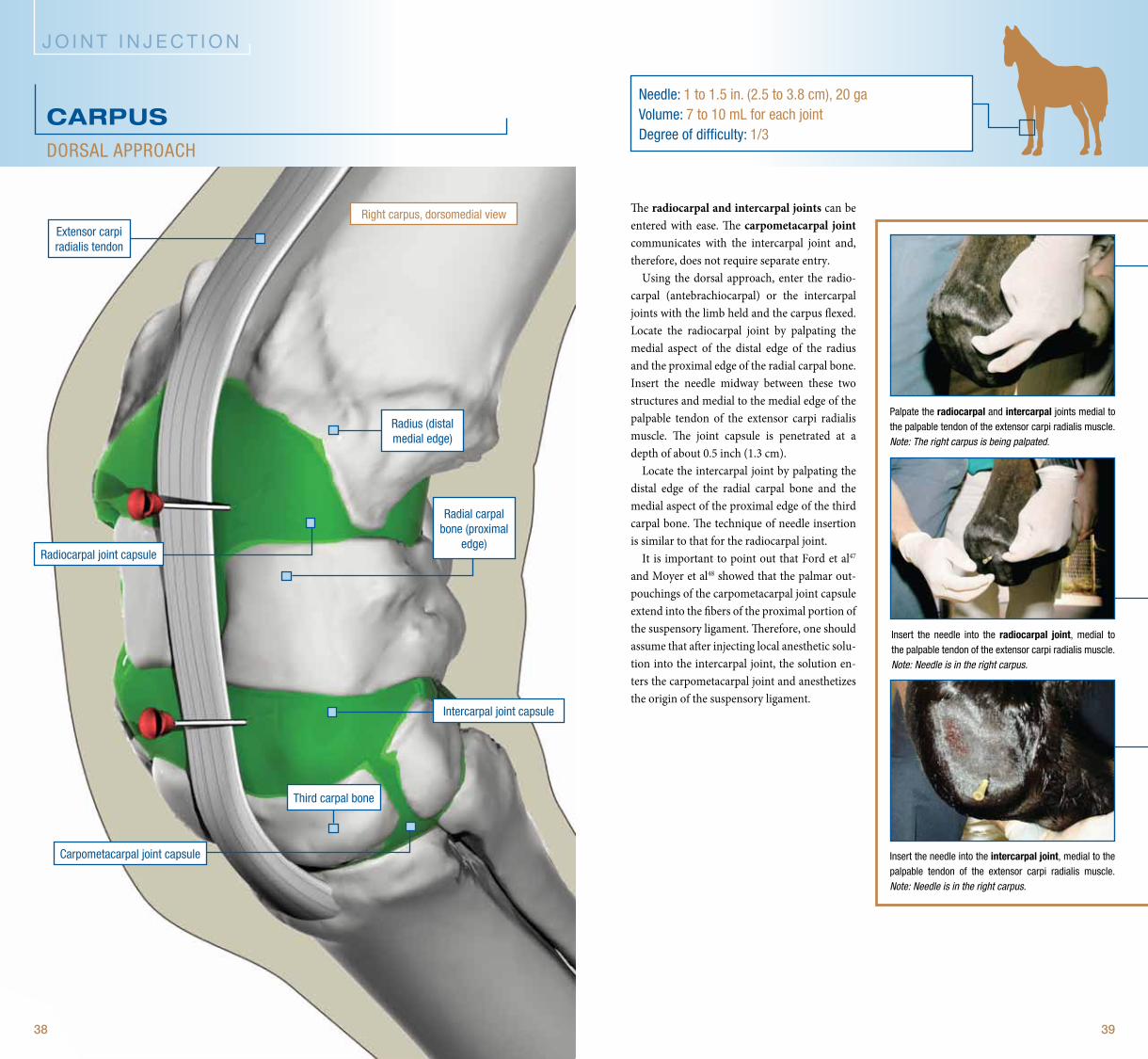
The radiocarpal and intercarpal joints can be entered with ease. The carpometacarpal joint communicates with the intercarpal joint and, therefore, does not require separate entry.
Using the dorsal approach, enter the radio-carpal (antebrachiocarpal) or the intercarpal joints with the limb held and the carpus flexed. Locate the radiocarpal joint by palpating the medial aspect of the distal edge of the radius and the proximal edge of the radial carpal bone. Insert the needle midway between these two structures and medial to the medial edge of the palpable tendon of the extensor carpi radialis muscle. The joint capsule is penetrated at a depth of about 0.5 inch (1.3 cm).
Locate the intercarpal joint by palpating the distal edge of the radial carpal bone and the medial aspect of the proximal edge of the third carpal bone. The technique of needle insertion is similar to that for the radiocarpal joint.
It is important to point out that Ford et al47
and Moyer et al48 showed that the palmar out-pouchings of the carpometacarpal joint capsule extend into the fibers of the proximal portion of the suspensory ligament. Therefore, one should assume that after injecting local anesthetic solu-tion into the intercarpal joint, the solution en-ters the carpometacarpal joint and anesthetizes the origin of the suspensory ligament.
needle: 1 to 1.5 in. (2.5 to 3.8 cm), 20 gavolume: 7 to 10 ml for each jointDegree of difficulty: 1/3
J O I N T I N J E C T I O N
radius (distal medial edge)
extensor carpi radialis tendon
radial carpal bone (proximal
edge)
intercarpal joint capsule
right carpus, dorsomedial view
CARPusDorsal approach
insert the needle into the radiocarpal joint, medial to the palpable tendon of the extensor carpi radialis muscle. Note: Needle is in the right carpus.
palpate the radiocarpal and intercarpal joints medial to the palpable tendon of the extensor carpi radialis muscle.Note: The right carpus is being palpated.
insert the needle into the intercarpal joint, medial to the palpable tendon of the extensor carpi radialis muscle. Note: Needle is in the right carpus.
radiocarpal joint capsule
carpometacarpal joint capsule
third carpal bone
42 43
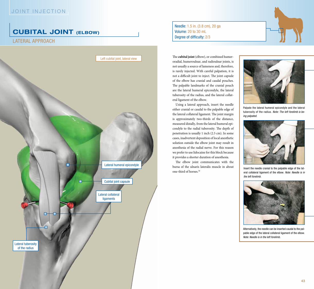
The cubital joint (elbow), or combined humer-oradial, humeroulnar, and radioulnar joints, is not usually a source of lameness and, therefore, is rarely injected. With careful palpation, it is not a difficult joint to inject. The joint capsule of the elbow has cranial and caudal pouches. The palpable landmarks of the cranial pouch are the lateral humeral epicondyle, the lateral tuberosity of the radius, and the lateral collat-eral ligament of the elbow.
Using a lateral approach, insert the needle either cranial or caudal to the palpable edge of the lateral collateral ligament. The joint margin is approximately two-thirds of the distance, measured distally, from the lateral humeral epi-condyle to the radial tuberosity. The depth of penetration is usually 1 inch (2.5 cm). In some cases, inadvertent deposition of local anesthetic solution outside the elbow joint may result in anesthesia of the radial nerve. For this reason we prefer to use lidocaine for this block because it provides a shorter duration of anesthesia.
The elbow joint communicates with the bursa of the ulnaris lateralis muscle in about one-third of horses.50
needle: 1.5 in. (3.8 cm), 20 gavolume: 20 to 30 mlDegree of difficulty: 2/3
J O I N T I N J E C T I O N
lateral collateral ligaments
lateral humeral epicondyle
cubital joint capsule
lateral tuberosity of the radius
CuBITAL JOINT (ELBOW)
lateral approach
insert the needle cranial to the palpable edge of the lat-eral collateral ligament of the elbow. Note: Needle is in the left forelimb.
palpate the lateral humeral epicondyle and the lateral tuberosity of the radius. Note: The left forelimb is be-ing palpated.
alternatively, the needle can be inserted caudal to the pal-pable edge of the lateral collateral ligament of the elbow. Note: Needle is in the left forelimb.
left cubital joint, lateral view
64 65
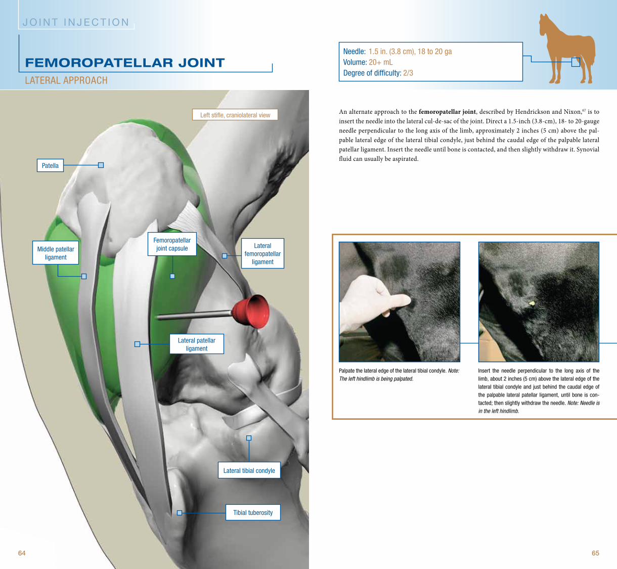
An alternate approach to the femoropatellar joint, described by Hendrickson and Nixon,67 is to insert the needle into the lateral cul-de-sac of the joint. Direct a 1.5-inch (3.8-cm), 18- to 20-gauge needle perpendicular to the long axis of the limb, approximately 2 inches (5 cm) above the pal-pable lateral edge of the lateral tibial condyle, just behind the caudal edge of the palpable lateral patellar ligament. Insert the needle until bone is contacted, and then slightly withdraw it. Synovial fluid can usually be aspirated.
needle: 1.5 in. (3.8 cm), 18 to 20 gavolume: 20+ mlDegree of difficulty: 2/3
J O I N T I N J E C T I O N
Middle patellar ligament
tibial tuberosity
lateral patellar ligament
lateral tibial condyle
FEmOROPATELLAR JOINTlateral approach
insert the needle perpendicular to the long axis of the limb, about 2 inches (5 cm) above the lateral edge of the lateral tibial condyle and just behind the caudal edge of the palpable lateral patellar ligament, until bone is con-tacted; then slightly withdraw the needle. Note: Needle is in the left hindlimb.
palpate the lateral edge of the lateral tibial condyle. Note: The left hindlimb is being palpated.
patella
left stifle, craniolateral view
femoropatellar joint capsule lateral
femoropatellar ligament