Investigation of bovine abortion and stillbirth/perinatal ...
13
REVIEW Open Access Investigation of bovine abortion and stillbirth/perinatal mortality - similar diagnostic challenges, different approaches John F. Mee Abstract This pracademic paper reviews current bovine foetopathy (abortion and stillbirth) case definitions, reporting and triage, and causes and time-of-death and proposes veterinary practitioner-focused investigative standard operating procedures (SOPs). Issues of under- and over-triage and intra-institutional SOP harmonisation are also discussed. It is proposed that an ‘observable abortion’ (120–260 days of gestation) is a more practitioner-friendly definition of abortion for reporting and benchmarking purposes and that the term ‘peristillbirth’ can replace stillbirth and perinatal mortality. Diagnosis of bovine foetopathy involves an investigative triad of the farmer, veterinary practitioner and the veterinary diagnostic laboratory. However, the poor sensitivity of abortion reporting undermines the value of currently adopted scanning/passive surveillance; parallel active surveillance/sentinel herd models should also be employed. The approach to abortion investigation differs from that of peristillbirth. The former should include collecting a herd and case history, examination and sampling of dam and cohorts and sampling of the foetus and placenta. A sample selection decision tree is provided to assist test selection. In peristillbirths, non-infectious and periparturient causes-of-death are more important hence the anamnesis must focus on peristillbirth risk factors and calving management. The foetopsy, while including the sampling menu appropriate to aborted foetuses, must also include a detailed internal and external examination of the carcass for lesions indicative of periparturient causes-of-death. In addition, for aborted foetuses the time-of-death is not important as the foetus is generally not viable; however, for the peristillbirth the time-of-death is critical as it provides useful information for the farmer to address modifiable risk factors and to alter their perinatal management. Reporting of the ultimate cause-of-death is more useful to prevent future abortions and peristillbirths though the proximate cause-of-death is often reported in the absence of a complete clinical anamnesis. Finally, the common reasons for diagnosis not reached (DNR) and the limitations of current investigative approaches are discussed. Keywords: Cattle, Abortion, Stillbirth, Perinatal mortality, Investigation, Necropsy, Cause-of-death, Ultimate, Proximate, Time-of-death © The Author(s). 2020 Open Access This article is licensed under a Creative Commons Attribution 4.0 International License, which permits use, sharing, adaptation, distribution and reproduction in any medium or format, as long as you give appropriate credit to the original author(s) and the source, provide a link to the Creative Commons licence, and indicate if changes were made. The images or other third party material in this article are included in the article's Creative Commons licence, unless indicated otherwise in a credit line to the material. If material is not included in the article's Creative Commons licence and your intended use is not permitted by statutory regulation or exceeds the permitted use, you will need to obtain permission directly from the copyright holder. To view a copy of this licence, visit http://creativecommons.org/licenses/by/4.0/. The Creative Commons Public Domain Dedication waiver (http://creativecommons.org/publicdomain/zero/1.0/) applies to the data made available in this article, unless otherwise stated in a credit line to the data. Correspondence: [email protected] Animal and Bioscience Research Department, Teagasc, Moorepark Research Centre, Fermoy, Co. Cork, Ireland Mee Irish Veterinary Journal (2020) 73:20 https://doi.org/10.1186/s13620-020-00172-0
Transcript of Investigation of bovine abortion and stillbirth/perinatal ...

REVIEW Open Access
Investigation of bovine abortion andstillbirth/perinatal mortality - similardiagnostic challenges, different approachesJohn F. Mee
Abstract
This pracademic paper reviews current bovine foetopathy (abortion and stillbirth) case definitions, reporting andtriage, and causes and time-of-death and proposes veterinary practitioner-focused investigative standard operatingprocedures (SOPs). Issues of under- and over-triage and intra-institutional SOP harmonisation are also discussed. It isproposed that an ‘observable abortion’ (120–260 days of gestation) is a more practitioner-friendly definition ofabortion for reporting and benchmarking purposes and that the term ‘peristillbirth’ can replace stillbirth andperinatal mortality. Diagnosis of bovine foetopathy involves an investigative triad of the farmer, veterinarypractitioner and the veterinary diagnostic laboratory. However, the poor sensitivity of abortion reportingundermines the value of currently adopted scanning/passive surveillance; parallel active surveillance/sentinel herdmodels should also be employed. The approach to abortion investigation differs from that of peristillbirth. Theformer should include collecting a herd and case history, examination and sampling of dam and cohorts andsampling of the foetus and placenta. A sample selection decision tree is provided to assist test selection. Inperistillbirths, non-infectious and periparturient causes-of-death are more important hence the anamnesis mustfocus on peristillbirth risk factors and calving management. The foetopsy, while including the sampling menuappropriate to aborted foetuses, must also include a detailed internal and external examination of the carcass forlesions indicative of periparturient causes-of-death. In addition, for aborted foetuses the time-of-death is notimportant as the foetus is generally not viable; however, for the peristillbirth the time-of-death is critical as itprovides useful information for the farmer to address modifiable risk factors and to alter their perinatalmanagement. Reporting of the ultimate cause-of-death is more useful to prevent future abortions and peristillbirthsthough the proximate cause-of-death is often reported in the absence of a complete clinical anamnesis. Finally, thecommon reasons for diagnosis not reached (DNR) and the limitations of current investigative approaches arediscussed.
Keywords: Cattle, Abortion, Stillbirth, Perinatal mortality, Investigation, Necropsy, Cause-of-death, Ultimate,Proximate, Time-of-death
© The Author(s). 2020 Open Access This article is licensed under a Creative Commons Attribution 4.0 International License,which permits use, sharing, adaptation, distribution and reproduction in any medium or format, as long as you giveappropriate credit to the original author(s) and the source, provide a link to the Creative Commons licence, and indicate ifchanges were made. The images or other third party material in this article are included in the article's Creative Commonslicence, unless indicated otherwise in a credit line to the material. If material is not included in the article's Creative Commonslicence and your intended use is not permitted by statutory regulation or exceeds the permitted use, you will need to obtainpermission directly from the copyright holder. To view a copy of this licence, visit http://creativecommons.org/licenses/by/4.0/.The Creative Commons Public Domain Dedication waiver (http://creativecommons.org/publicdomain/zero/1.0/) applies to thedata made available in this article, unless otherwise stated in a credit line to the data.
Correspondence: [email protected] and Bioscience Research Department, Teagasc, Moorepark ResearchCentre, Fermoy, Co. Cork, Ireland
Mee Irish Veterinary Journal (2020) 73:20 https://doi.org/10.1186/s13620-020-00172-0

BackgroundReported bovine abortion rates vary between approximately0.5 and 10% [1]. Perinatal calf mortality (0-48 h) rates varybetween approximately 2 [2] and 10% [3], internationally.These losses represent economic, welfare and societal con-cerns [4]. Abortion (20 to 50% [5];) and stillbirth (30 to 75%[6];), diagnosis rates do not appear to have improved overtime internationally despite the development of moleculardiagnostic techniques and the discovery of new foetopatho-gens, e.g. Schmallenberg virus [7]. While reasons for theoverall low diagnostic rates from foetal investigations aredetailed hereunder, this paper addresses this challengingconundrum with a view to veterinary practitioner-led im-provements in diagnostic rates. The differing proportionsof infectious and non-infectious and pre- and periparturientaetiologies of abortions and stillbirths are emphasised,hence though the diagnostic challenge is similar, the ap-proaches to diagnosis differ.The investigative triad involving the farmer, veterinary
practitioner and veterinary pathologist is critical to betterinvestigative outcomes [8]. From the farmer’s perspective,under and late reporting and placental non-submissionare significant issues which can be improved. Veterinarypractitioners can be more pro-active in awareness raisingand client education regarding index case reporting, inves-tigative thresholds, pro forma anamnesis recording andadvising clients of possible causes of foetal loss. Inaddition, with closures of many government veterinarydiagnostic laboratories, veterinary practitioners may needto upskill in foetal pathology and consider providing thisnew service for their clients where this is legally allowed.Veterinary diagnosticians can review the investigative pro-tocols they currently adopt to see where they can improvetheir investigative/necropsy/sampling/testing SOPs. Add-itionally, the educational role of veterinary diagnosticiansto both veterinary practitioners and farmers can contrib-ute substantially to improved field investigations. At aninternational level, the current lack of homogeneity in in-vestigative approaches highlights the need for proceduralstandardisation [9]. While the focus of this review is ondairy foeto-perinatal loss, the material is equally applicableto beef foeto-perinatal loss [10]. Additionally, frequent ref-erence is made to aspects of investigation of human peri-natal mortality, where relevant.
Case definitionsBoth abortion and stillbirth are variously defined in theliterature and in common usage by scientists, veterinarypractitioners and by farmers. While viability-based defi-nitions are occasionally used, e.g. nonviable foetuses areconsidered aborted irrespective of gestation length(foetal mortality), gestation-based definitions are recom-mended [11]. These definitions differ both inter- andintra-nationally.
‘Observable abortion’By scientific consensus a bovine abortion has been definedas the expulsion of a foetus between the completion of dif-ferentiation (day 42) and the limit of foetal independentviability (day 260), [11]. Day 42 also represented the typ-ical timing of manual pregnancy detection when this def-inition was formulated, whereas now with early (~ 30days) and repeat (~ 60 days) ultrasonographic pregnancydetection the late embryonic and early foetal loss (LEEFL)periods are often combined. There are numerous foetalloss definitions used internationally. For example, inFrance a hybrid definition of abortion is used encompass-ing both abortion and perinatal mortality; expulsion of afoetus from 42 days of pregnancy onwards either stillbornor dying within 48 h of birth [12]. Whilst including early(42–120 d) foetal mortality is scientifically valid, in prac-tice animals which abort this early generally return tooestrus without a foetus being expelled (resorbed) or ob-served (too small). Such returns-to-service have been ad-vocated as an indicator of first and second trimesterabortion (inferred abortion) in syndromic surveillance sys-tems [13]. The low observation rate of first trimester abor-tions was confirmed in a US study where only 9% ofabortions < 125 days were observed, while 41% of abor-tions between day 125 and 245 were observed [14].Thus, from a veterinary practitioner and their clients’
perspectives, abortion may be defined as the expulsionof a non-independently-viable foetus before full-termthat is likely to be observed (and possibly submitted to alaboratory for examination) by a farmer, i.e. an ‘observ-able abortion’. Irish regional veterinary laboratory dataindicate that the period of risk for an observable abor-tion is from 120 to 260 days of gestation; over 95% ofaborted foetuses submitted to a veterinary laboratoryover a 25 year period were from the fourth month ofpregnancy on (> 120 days), (Table 1) [15]. The foetus at120 days is the size of a small cat with a crown rumplength (CRL) of 20-30 cm and weighs ~ 1 kg. These datasupport the findings [16] that the majority of laboratory-investigated abortion cases are from the last trimester,
Table 1 Gestational age of veterinary laboratory-investigatedaborted foetuses
Trimester Month of gestation Foetuses (No.) %
First (early) 2 73 0.7
3 298 2.9
Second (mid) 4 841 8.2
5 1393 13.7
6 2069 20.3
Third (late) 7 2858 28.0
8 2661 26.1
Source: Cork Regional Veterinary Laboratory, DAFM, 10,193 foetuses over25 years (1980–2003)
Mee Irish Veterinary Journal (2020) 73:20 Page 2 of 13

while the majority of non-submitted abortions are fromthe first two trimesters; this leads to under-estimation ofreal abortion rates.
‘Peristillbirth’Perinatal mortality may be defined as death of the foetus orperinate before, during or within 48 h of calving at full term(> 260 days); it includes both stillbirth and early neonatalmortality [17]. While ‘stillbirth’ is commonly used as asynonym for perinatal mortality, particularly by farmersand their veterinary practitioners, it is better defined as thedeath of a foetus before or during calving at full term. Anovel portmanteau ‘peristillbirth’ (incorporating both still-birth and perinatal mortality) may avoid this terminologicalimprecision. These gestational and perinate age thresholdsare arbitrary and vary both intra- and inter-nationally.In cases of indeterminate gestational age (e.g. unre-
corded natural services), surrogate objective variablesmay be used as with babies, most commonly, birthweight and CRL. However, both have limitations. In cat-tle there are inadequate breed and plurality-adjusted ges-tational age body weight norm reference data to assesswhen a birth weight might be more indicative of anabortion or a peristillbirth. In order to address this issuethe data in Table 2 have been compiled from researchherds with excellent records to provide guidance on dif-ferent full-term birth weights for various common breedand crossbreed, and plurality groups. The same principleis used by the WHO to define human perinatal mortalitycases [18]. From these data the following breed andplurality-adjusted body weight norms for term calves areindicated: ‘Jerseys’ (Jersey or Jersey x dams mated toJersey or Jersey x sires); > 15 kg for singletons or twins,‘Jersey crossbreds’ [Jersey or Jersey x dams or siresmated to non-Jersey or Jersey x dams or sires, respect-ively, (i.e. only the dam or the sire is Jersey or Jersey x,not both)]; > 20 kg for singletons or twins, ‘Non-Jersey’
[other dairy breed dams (non-Jersey or Jersey x) matedto other dairy sires (non-Jersey or Jersey x) or beef sires];> 25 and > 20 kg for singletons and twins, respectively.
Reporting of foetal/perinatal mortalityBefore addressing the titular topic; investigation, theissue of reporting needs to be addressed as it is equally ifnot more important.
Under-reportingUnder-reporting is a major issue with bovine foetal/peri-natal mortality internationally (the ‘untagged calf lossphenomenon’). To some risk-averse farmers the loss of asingle valuable pregnancy may trigger a veterinary inves-tigation (threshold for investigation) while for the major-ity of farmers multiple losses need to occur before thisthreshold is exceeded. A few abortions in large herds are‘expected’, i.e. abortion is considered a ‘normal’ event aslong as it is sporadic. In a French study only 20% of beeffarmers who had an abortion or stillbirth (0.8% abortionrate) reported it, indicating the low sensitivity of event-driven or passive abortion surveillance [19]. A foetalmortality rate of > 5% is the investigation threshold usedby the majority of veterinary practitioners in the EU[20]. However, a cluster of cases is a more important‘tipping point’ for investigation in seasonal calving herds,than percentage of pregnancies aborted. Scan statisticprobabilities can be used to determine what constitutesa significant clustering of abortions [1]. Given the diag-nostic gap between 42 and 120 days of pregnancy whenaborted foetuses are generally not observed or reported/submitted for examination, return-to-service has beenproposed as a proxy syndromic surveillance indicator tomonitor losses in this period [13]. Proximity to the la-boratory, number of losses, ability to identify the abortedcow, fear of a contagious abortifacient (e.g. Brucellaabortus), ‘farm blindness’ [21] and lack of understanding
Table 2 Birth weights (kg) (no. records, mean, mean + 2SD, range) and gestation length (Ges, days) of fullterm (260–300 days),singleton (n = 10,422) and twin (n = 615) calves of different breeds and crossbreeds
Dam genotype Sire genotype Singletons Twins
No.records
Birth wt.mean
Birth wt.mean + 2SD
Birth wt.range
Ges.days
No.records
Birth wt.mean
Birth wt.mean + 2SD
Birth wt.range
Ges.days
Jersey, JerseyX Jersey, JerseyX 200 26.21 16.54–35.88 14–45 281.15 6 21.83 10.92-32.74
15–30 284.66
Jersey, JerseyXor dairy
Jersey, JerseyXor dairy
919 32.63 20.31–44.95 14–51 280.34 32 26.63 22.04-31.22
15–34 277.87
Dairy (excl. Je,JeX)a
Dairy(excl. Je, JeX)
7960 41.84 29.61–54.07 11 to 76 281.62 496 34.14 23.18–45.1 18–55 277.02
Dairy(excl. Je, JeX)
Beefb 1343 42.82 30.86–54.78 18–63 283.6 81 35.07 22.45–47.69 21–51 278.86
aDairy (excl. Je, Jex) = Holstein-Friesian, Friesian, Friesianx, Ayrshire, Norwegian Red, Norwegian Redx, Swedish Red,Swedish Redx, Montbeliarde, Montbeliardex, Normande, NormandexbBeef = Aberdeen Angus, Belgian Blue, Blonde D’Aquitaine, Charolais, Hereford, Limousin, SimmentalSource: Teagasc dairy research herds
Mee Irish Veterinary Journal (2020) 73:20 Page 3 of 13

of the costs and potential consequences of abortion in-fluence the likelihood of sample submission. In addition,there is often a disconnect between the priority placedon reporting of abortion by governmental risk managersand veterinary practitioners and farmers [12].The herd-level occurrence of abortions/stillbirths fol-
lows a right-skewed distribution with most herds havingno or low losses but a minority of herds have high losses.The threshold at which loss prevalence becomes a ‘prob-lem/cluster/storm’, and hence is investigated, is usuallydefined by the herdowner. Norm-referenced thresholdsare cohort-based which may reflect the national recordedabortion or stillbirth rate. Criteria-referenced thresholdsare based on metrics other than those based on cohorts,e.g. a theoretical or empirical threshold. While the latterhas traditionally been 2% for abortions, this may only beappropriate for observable abortions (> 120 days) (in tem-perate regions) but may underestimate ‘normal’ all-cause,first through third trimester abortion rates.
Under- and over-triageThe approach to a sporadic abortion/stillbirth or anautolysed foetus in a herd without a history of foetal lossproblems will differ from that in a herd with a series ofcases or a fresh case. This is referred to as under-triage,whereby such cases are not investigated as thoroughly asfresh cases or those from multiple mortality herds. How-ever, over-triage may be warranted even for sporadiccases on farms a long distance from the local laboratoryas resubmission of material is less likely. One cannotpredict that the index abortion case is not the first ofmany in a herd outbreak (all abortion ‘storms’ start witha single case). Studies have shown that only approxi-mately 20 to 30% of abortions are observed [22] suggest-ing that in many cases the first case presented (indexcase) is not in fact the first case that occurred, merelythe first that was observed.
Veterinary practitioner-led abortion/stillbirthinvestigationWhen approached by a client with an aborted cow or astillbirth problem (as defined above) the following stand-ard operating procedure (SOP) is recommended.
SOP for abortion/stillbirth investigationThis involves three steps; collect a history, examinepregnant animals and the aborted dam and examine thefoetus/placenta. This procedure uses multiple diagnostictools to make a clinico-pathological diagnosis [23, 24].
Collect a historyThe first step is to establish the nature and extent of theproblem by investigating possible risk factors associatedwith the loss/es. Standard questions include how many
cows have aborted/had stillbirths, how many cows arepregnant (denominator-based diagnosis), is the dam ill,are most losses in heifers, and recent husbandry changes.A pro forma questionnaire is used by veterinary laboratorystaff to collect the history prior to the postmortem exam-ination; practitioners might consider using such a formatfor problem herds – exemplars for abortions and peristill-births are provided in Additional files 1 and 2. In additionto collecting information on the individual abortion/still-birth, the herd health history is important. For example,details of most recent vaccinations against foetopathogens,recent cattle purchases and bulk milk test results.
Examine/sample the pregnant and aborted/stillbirthanimalsThe pregnant cohortExamining the pregnant animals and their environment al-lows the practitioner to assess their general health, bodycondition score (BCS) and feeding management. Cohortsampling of dams (n = 5–10) in the affected group can beuseful in determining differences in exposure (presence/ab-sence, prevalence) between affected and unaffected dams(case-control); but vaccinal status needs to be known.
The damClinical examination of the dam may reveal pyrexia,diarrhoea, respiratory signs, etc. A faecal sample may beuseful where salmonellosis is suspected. Single bloodsamples from aborted cows are the most common sam-ples collected to investigate bovine abortion (sero-diag-nosis). These can have moderate utility as proxy samplesfor foetal material. For example, a single blood samplefrom the non-vaccinated dam of an aborted or stillbornfoetus can be up to 85% accurate in predicting a foetalculture-positive result for Salmonella Dublin, [25]. How-ever, the primary value of maternal serum is as an exclu-sionary test for maternal antibody, i.e. a negative resultrules out some causes, e.g. Neospora caninum.Paired sera (more than 2 weeks apart) may detect ris-
ing titres (two to four-fold) for some abortifacients (e.g.S. Dublin SAT) but not for others (e.g. Leptospira spp.,Neospora) due to the lag phase between infection andfoetal mortality. In vaccinated herds natural infectioncan still be distinguished where DIVA vaccines are used(gE-deleted BoHV-I) and where titres are much higherthan those expected from vaccination (e.g. Leptospira)this suggests current, active infection.
Examine the foetus and placentaAbortion/stillbirth foetopsy SOPAs with a clinical examination on a live animal a system-atic approach means that important findings are notmissed due to focusing on the obvious lesion/s; this in-volves three steps - external and internal examination
Mee Irish Veterinary Journal (2020) 73:20 Page 4 of 13

and sampling the carcass. While for the aborted foetusfew lesions are visible grossly, in the stillborn foetus le-sions indicative of periparturient causes-of-death aremore likely to be detected.
External examinationThe external examination will confirm the animal identi-fication (assuming it is tagged), foetal preservation, ma-turity, size for date/plurality, and detect defects,discharges and predation. The stage of development ofthe foetus may be estimated from its body weight or size(e.g. CRL), (Table 3). External developmental indicators(dentition, pilosity, skull doming) are less useful due totheir gradual appearance and subjectivity in assessment.Gestational age may be estimated (within plus or minusapproximately 2 weeks) from foetometric variables suchas straight CRL (sCRL) using simple formulae, for ex-ample: gestational age (GA), (days) = 68 + 2.25 x sCRL(cm) or GA = (sCRL + 21) × 2.5 or GA = 53 + (2.3 xSCRL) or more complex multi-variable formulae [26].
Internal examinationHow the carcass is opened and how the contents are ex-amined varies between pathologists. An illustrated guideto bovine foetopsy by the practitioner has recently beenpublished which details a simple method which can becarried out in a practice setting with minimal specialisedequipment [27]. It is recommended that once the carcassis opened samples for microbiological testing are col-lected first to avoid contamination during the internalorgan examination.
Sampling the carcassA sample selection algorithm for aborted/stillborn foe-tuses is outlined in Fig. 1. It is not possible to be pre-scriptive about test selection as laboratories differ in thetest menu and tiers they offer. Variations between la-boratories in sample and test selection both inter andintra-institutionally indicate the need for SOP harmon-isation both nationally and internationally. Test selectionwill also be determined by the anamnesis, degree ofcarcass autolysis and costs. Rather than ‘necropsy by al-gorithm’ the decision-making of the practitioner or path-ologist still determines whether and which samples to
collect. Additionally, samples can be discarded if col-lected in the early stages of the necropsy where subse-quent examination reveals the likely cause does notrequire laboratory testing.
Microbiology samplesAbomasal contents can be sampled aseptically by searingthe abomasal serosa with a heated scalpel blade and as-pirating a sample into a plain vaccutainer tube. In gen-eral, tissue samples are preferable to swabbed samplesand surface swabbed samples are preferable to fluidswab samples. If an abomasal sample is unobtainable(due to scavenging or colostrum ingestion) or if septi-caemia is suspected, lung, liver or brain samples are suit-able alternatives, though not as sensitive [28]. Brainsampling is of particular value in severely scavenged andmummified foetuses. The presences of a pure growth ofa pathogen with associated lesions in the absence ofother causes of death are usually accepted as diagnosticcriteria. However, recent research on variations inpathogen virulence (e.g. Trueperella pyogenes) suggestthat in future it may be necessary to determine the pres-ence of virulence factors also [29].
Serology samplesSerological sampling can be useful from the second tri-mester (> 120 days) when the foetus is immunocompe-tent as antibodies indicate foetal infection (but notnecessarily foetopathy, e.g. Neopsora congenital infec-tion), assuming placental competence. However, relianceon foetal serology alone may grossly underestimatefoetal infection rates [30]. Recently, other biomarkers(acute phase proteins and non-pathogen specific immu-noglobulins) have also been shown to be detectable inbovine foetal infection [31].
Trace element samplesExamination of the foetal thyroid gland for absolutegoitre (thyroid enlarged relative to a criterion-referencedthreshold thyroid weight, e.g. > 30 g) or relative goitre(thyroid enlarged relative to a criterion-referencedthreshold thyroid g: kg ratio with body weight, e.g. >0.80) and submission of a fresh (I2 content) and formali-nised lobe (histopathology) will detect dietary iodine
Table 3 Guideline physical characteristics of foetuses in the last two trimesters of gestation
Month of gestation Body weight (kg) sCRLa (cm) Pilosity (trunk/abdomen) Teeth eruption (incisors)
6 5 50 Extremities only Not erupted
7 15 70 Light hair coat Partially erupted
8 30 90 Full hair coat Partially or fully erupted
9 35 95 Full hair coat Fully eruptedasCRL = straight crown-rump lengthSource: Teagasc research, Irish dairy herds
Mee Irish Veterinary Journal (2020) 73:20 Page 5 of 13

imbalance. Where selenium deficiency is suspected (pos-sibly in conjunction with iodine deficiency) a sample ofthe foetal liver preferably [32, 33] or kidneys should besubmitted. Given the associations between micronutrientimbalances and bovine abortion [34] and peristillbirth[35] examination of micronutrient status should be in-cluded in the diagnostic panel when investigating bovinefoetopathy.
Histopathology samplesSamples for histopathology should include normal and ab-normal tissue and should not be greater than 1 cm thickand 2 cm long as they need to fit into processing cassettes(3.5 × 2.5 × 0.5 cm); most samples collected by practi-tioners are too thick. Pathologists routinely sample lung,liver, thyroid, heart and brain. All samples can be submit-ted in a single pottle. As the brain is of particular value inthe histopathological diagnosis of neosporosis, submissionof the skull or entire brain is recommended as histopa-thologists vary in the sections they prefer to examine.
Standard abortion packagePractitioners will be guided by their local laboratory aboutwhich samples they prefer to use routinely for each test(‘standard abortion package’) and which tests are non-routine (e.g. PCR, histopathology). While for sporadic casesthe basic sampling package may suffice (Table 4), in abortion
storms, tiered sample escalation is advised as it provides theoption of sampling, storing and testing as deemed necessary.Increasingly, genomic tests (e.g. dense SNPs) are being devel-oped for developmental disorders which require specialistsampling, e.g. hair follicles, skin, muscle or liver.
Photo-documentationPhoto-documentation can be useful for inexperienced prac-titioners and for unusual lesions [36]. In future practitioners(and possibly farmers) may be able to use digital imagingtechnologies to enhance their PME yield, as currently oc-curs on North American feedlots (remote digital PME).
Sample submissionIf samples are collected on a day when the laboratory isclosed the samples should be stored for culture, serologyand histopathology in a fridge (4 °C) and those for PCRin a freezer (− 20 °C). Practitioners need to be cognisantof postal regulations pertaining to packaging and label-ling for biological substances. Completion of the receiv-ing laboratory submission sheet and inclusion with thesamples will facilitate timely processing of samples.
Examining the placentaIf the placenta has not yet been expelled at examination,a sample of the retained placentomes or a vaginal swabcan be obtained. A normal placenta at term weighs
Fig. 1 Practitioner-focused necropsy sample selection decision tree for aborted/stillborn foetuses (COD = cause-of-death, CD = congenital defect,DIU = dead in utero)
Mee Irish Veterinary Journal (2020) 73:20 Page 6 of 13

approximately 5 kg, has 75–125 red cotyledons and hasthin, translucent intercotyledonary tissue that sometimescontains adventitious placentation. Placentitis may mani-fest as discoloured, necrotic, exudative cotyledons andopaque, erythematous, thickened, oedematous intercoty-ledonary tissue. Ideally three abnormal cotyledons/inter-cotyledonary tissue samples should be submitted (prefer-ably macroscopically abnormal and uncontaminated) forhistopathological and microbiological examination(Gram smear, MZN, culture, PCR).
Time-of-deathBefore attributing a cause-of-death, the time-of-death(TOD) should be determined as the latter influences thediagnosis of the former. Time-of-death is classified aspre-, intra- and postpartum. Foetuses which die beforebirth will have varying degrees of autolysis; corneal opa-city, excess body cavity sero-sanguinous transudate,decomposed organs (especially kidneys, liver, spleen andbrain), haemoglobin imbibition, flaccid musculature,subserosal and subcutaneous serosanguinous oedema/emphysema and atelectatic lungs. Autolytic lesions de-velop within approximately 12 h of death in utero andbecome more advanced over time. Foetuses which werealive at the beginning of calving but die during birth orwithin an hour of calving will not have signs of autolysisand may have signs of breathing (partial lung inflation),
bradytocia (localised peripheral oedema) or traumotocia(trauma lesions with haemorrhage). Perinates which diemore than an hour after calving will not have signs ofautolysis and will have signs of breathing (complete lunginflation, tracheal froth), and of umbilical cord rupturewhile alive [large (> 4 mm) diameter, pointed umbilicalthrombi], possibly attempting to stand or standing (worneponychia/foetal hoof ‘slippers’, especially hind) and col-ostrum consumption (colostrum in abomasum).
Cause-of-deathCause-of-death (COD) may be defined as ultimate/underlying (UCOD), (the first initiating event in thechain of events resulting in death; this often includes theclinical history, e.g. dystocia), intermediate and proxim-ate/terminal (PCOD), (final event/mechanism causingdeath; often from the necropsy examination, e.g. frac-tured spine or asphyxia). The UCOD answers the ques-tion ‘why did the calf die?’, and the PCOD answers asubtly different question ‘how did the calf die? The fun-damental question about the aetiology of perinatal mor-tality is ‘why did the calf die?’ i.e. what was the ultimatecause of death? For stillborn babies, the WHO recom-mends recording of the main single, underlying disease/condition causing death on the WHO Death Certificate[37]. In contrast, in veterinary studies the proximateCOD from necropsy results is usually cited, as the
Table 4 Standard and additional samples to collect from aborted and stillborn foetuses* for the investigation of infectious,nutritional and genetic causes of death
For investigation of Standard samples Ancillary samples Comments
Failure of passive transfer of immunoglobulins Perinate blood NA** Only test calves > 24 h old, e.g. ZSTT
Foetopathogenic bacteria and fungi (e.g. Aspergillusspp., B. licheniformis, L. monocytogenes, T. pyogenes,S. Dublin)
Foetal stomachcontents (FSC),Placenta
Foetal lung, liver, gallbladder, kidney, brain,eyelid.Dam vaginal swab,placentome, blood.
Ancillary samples where FSC/placenta unavailable/contaminated.
Neospora caninum Foetal brain,serum
Foetal heart.Placenta.Dam/cohort bloods
Fresh brain/placenta for PCR, fixed brain or heart/placenta for histopathology if PCR positive
Leptospira Hardjo Foetal kidney,serum
Dam/cohort bloods Foetal sample dependent upon laboratory tests
BVDv Foetal ear, spleen,thymus, serum
Foetal kidney. Dam/cohort bloods
Foetal sample dependent upon laboratory tests
BHV-I Foetal liver,serum.
Foetal kidney.Placenta. Dam/cohortbloods
Foetal PCR/histopathology preferred tests
Micronutrient deficiencies Foetal thyroid,liver, heart, rib
Foetal kidney. Dam/cohort bloods
Thyroid for iodine assay/histopath; liver/kidney forselenium assay, heart for histopath; bone formanganese assay
Gross lesions (e.g. foetal pneumonia) Affected foetalorgan
As required As appropriate (e.g. bacteriology, histopath)
Genetic congenital defect Foetal muscle,skin
Dam hair follicles Test for infectious teratogens also (e.g. BVDv, SBV)
*Standard and ancillary testing protocols are dependent upon local laboratory SOPs. Bacteriology/mycology (culture, stains, wet preparations) and serology aregenerally routine tests for sporadic cases while other tests (e.g. histopathology, PCR, FAT, IHC, micronutrient, DNA assay) can be added for multiple losses or at thediscretion of the pathologist. Maternal vaccinal status affects use and choice of serology tests, ** NA - not applicable
Mee Irish Veterinary Journal (2020) 73:20 Page 7 of 13

Table 5 Cause-of-death and diagnostic case definitions for non-infectious causes of bovine perinatal mortality
Cause-of-death Sub-category Case definition Diagnostic criteria
Accident Colostrumaspiration(iatrogenic)
Administration of colostrum by oro-oesophageal feederor bottle into the trachea with subsequent calf clinicalsigns (e.g. bawling, dyspnoea, weakness, depression,recumbence).
History and presence of colostrum in the trachea,bronchial tree and lungs, pulmonary oedema/congestion/consolidation/pneumonia and foreignbasophilic deposit in airways and alveoli
Non-parturienttrauma
Trauma independent of calving such as stood on orlaid on by cow or attacked by cow (assault injury) orotherwise fatally injured, e.g. by automatic scraper incubicle house, other machinery.
History and/or fatal traumatic lesions (e.g. antemortemfractured ribs and/or legs, hepatic rupture) usually inthe absence of subcutaneous bruising.
Oesophagealrupture(iatrogenic)
Rupture of oesophagus while administering colostrumusing an oro-oesophageal feeder with subsequent calfclinical signs (e.g. bawling, dyspnoea, weakness, depres-sion, recumbence, cervical oedema).
History and traumatic tear in oesophagus withdischarge of colostrum and cellulitis if in cervical regionresulting in a swollen neck.
Co-mortality Presence of more than one cause-of-death Listed for each cause-of-death
Congenitaldefect
Lethalcongenitaldefect
Defect present at birth incompatible with life. Wherethe cause of the defect/s is diagnosed, e.g. BVDv, bothan ultimate and proximate COD can be reported.
Most diagnosed defects are grossly visible structuraldefects. Examples include hydranencephaly,hydrocephalus, schistosomus reflexus and multipledefects. Some defects are economically lethal – the calfmay survive following remediation but this iseconomically prohibitive hence euthanasia follows.
Economically-lethalcongenitaldefect
Grossly visible structural defect incompatible withindependent life and with economic viability of the calf(e.g. surgery may be possible but cost-prohibitive andpoor prognosis) necessitating euthanasia
Examples include intestinal atresia, vestigial limbs,palatoschisis and arthrogryposis.
Dystociaa Bradytocia Prolonged stage one or two of calving History of prolonged stage one (e.g. milk fever, ‘slowcalving syndrome’, disturbance during calving, uterinetorsion) and/or prolonged stage two (e.g. foetaloversize) with moderate/severe peripheralsubcutaneous antemortem oedema (e.g. lower legs,tongue, submandibular, head, neck)
Dystoxia Dystocia with anoxia/asphyxia lesions Moderate/severe calving assistance with atelectasis andmoderate/severe meconium staining/aspiration (hair,trachea, lungs, abomasum), mucosal/serosalhaemorrhages (e.g. trachea, heart, pleura, thymus,abomasum, adrenals, sclera, conjuctiva), organcongestion and thoracic/abdominal serous transudate.
Maldistoxia Maldisposition with anoxia/asphyxia lesions Malpresentation or malposition with moderate/severemeconium staining/aspiration (hair, trachea, lungs,abomasum), mucosal/serosal haemorrhages (e.g.trachea, heart, pleura, thymus, abomasum, adrenals,sclera, conjuctiva), organ congestion, thoracic/abdominal serous transudate and atelectasis.
Traumotocia Fatal trauma to the calf at assisted calving Severe antemortem (haemorrhage at the site) acutelesions consistent with history of iatrogenic parturienttrauma (e.g. fractured/dislocated spine, ribs, limbs,moderate/severe subcutaneous thoracic and lower limbhaemorrhage/bruising, traumatic diaphragmatic hernia,hepatic rupture, moderate/severe haemothorax,haemoperitoneum, haemarthrosis, or polytrauma)
Eutoxia Eutocia with anoxia/asphyxia lesions, e.g. umbilical cordaccidents, placental insufficiency, placentitis, ‘non-clinical dystocia’.
History of no (or slight) calving assistance with some orall of the following: moderate/severe amniotic fluid ormeconium staining/aspiration [e.g. lungs (multifocalkeratinocytes, exfoliated epithelia, yellow/browngranular material, eosinophilic material in alveoli andbronchioles, incipient inflammatory reaction), hair,trachea, abomasum], mucosal/serosal petechialhaemorrhages (e.g. trachea, heart, pleura, thymus,abomasum, adrenals, sclera, conjuctiva), organcongestion, thoracic/abdominal serous transudate andatelectasis (dark purple, moist, congested, heavy fluid-filled, round-bordered lungs; negative on floatation test)
Haemorrhage/anaemia
Anaemia Generalised pallor in the absence of visiblehaemorrhage
Diffuse severe pallor (e.g. conjunctiva, gingiva, skeletalmuscles, thymus, trachea, liver, heart, lungs, brain,
Mee Irish Veterinary Journal (2020) 73:20 Page 8 of 13
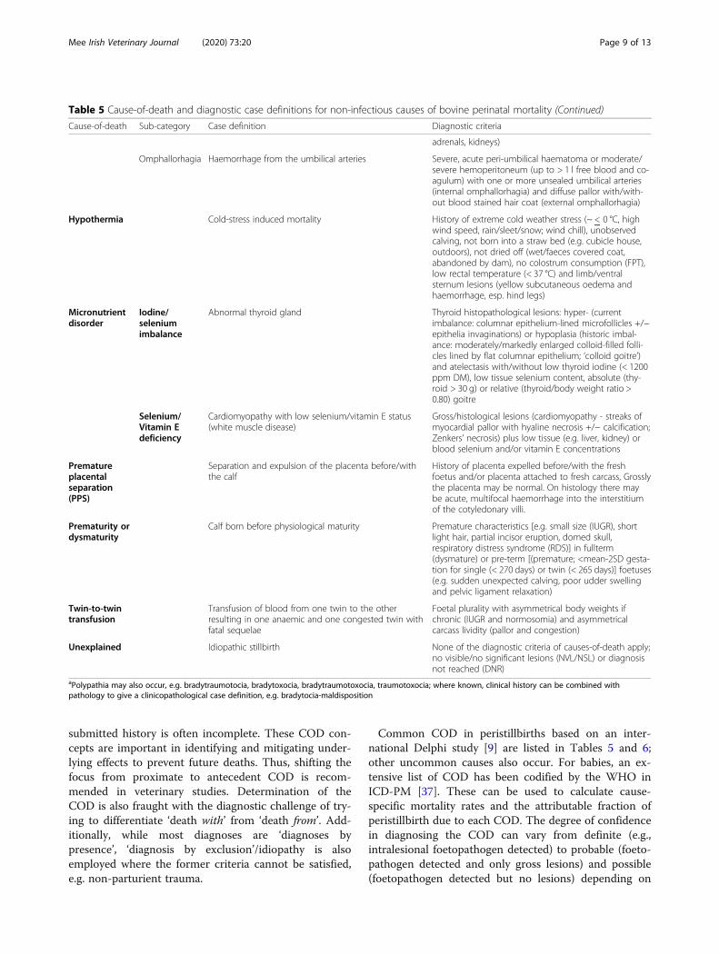
submitted history is often incomplete. These COD con-cepts are important in identifying and mitigating under-lying effects to prevent future deaths. Thus, shifting thefocus from proximate to antecedent COD is recom-mended in veterinary studies. Determination of theCOD is also fraught with the diagnostic challenge of try-ing to differentiate ‘death with’ from ‘death from’. Add-itionally, while most diagnoses are ‘diagnoses bypresence’, ‘diagnosis by exclusion’/idiopathy is alsoemployed where the former criteria cannot be satisfied,e.g. non-parturient trauma.
Common COD in peristillbirths based on an inter-national Delphi study [9] are listed in Tables 5 and 6;other uncommon causes also occur. For babies, an ex-tensive list of COD has been codified by the WHO inICD-PM [37]. These can be used to calculate cause-specific mortality rates and the attributable fraction ofperistillbirth due to each COD. The degree of confidencein diagnosing the COD can vary from definite (e.g.,intralesional foetopathogen detected) to probable (foeto-pathogen detected and only gross lesions) and possible(foetopathogen detected but no lesions) depending on
Table 5 Cause-of-death and diagnostic case definitions for non-infectious causes of bovine perinatal mortality (Continued)
Cause-of-death Sub-category Case definition Diagnostic criteria
adrenals, kidneys)
Omphallorhagia Haemorrhage from the umbilical arteries Severe, acute peri-umbilical haematoma or moderate/severe hemoperitoneum (up to > 1 l free blood and co-agulum) with one or more unsealed umbilical arteries(internal omphallorhagia) and diffuse pallor with/with-out blood stained hair coat (external omphallorhagia)
Hypothermia Cold-stress induced mortality History of extreme cold weather stress (~ < 0 °C, highwind speed, rain/sleet/snow; wind chill), unobservedcalving, not born into a straw bed (e.g. cubicle house,outdoors), not dried off (wet/faeces covered coat,abandoned by dam), no colostrum consumption (FPT),low rectal temperature (< 37 °C) and limb/ventralsternum lesions (yellow subcutaneous oedema andhaemorrhage, esp. hind legs)
Micronutrientdisorder
Iodine/seleniumimbalance
Abnormal thyroid gland Thyroid histopathological lesions: hyper- (currentimbalance: columnar epithelium-lined microfollicles +/−epithelia invaginations) or hypoplasia (historic imbal-ance: moderately/markedly enlarged colloid-filled folli-cles lined by flat columnar epithelium; ‘colloid goitre’)and atelectasis with/without low thyroid iodine (< 1200ppm DM), low tissue selenium content, absolute (thy-roid > 30 g) or relative (thyroid/body weight ratio >0.80) goitre
Selenium/Vitamin Edeficiency
Cardiomyopathy with low selenium/vitamin E status(white muscle disease)
Gross/histological lesions (cardiomyopathy - streaks ofmyocardial pallor with hyaline necrosis +/− calcification;Zenkers’ necrosis) plus low tissue (e.g. liver, kidney) orblood selenium and/or vitamin E concentrations
Prematureplacentalseparation(PPS)
Separation and expulsion of the placenta before/withthe calf
History of placenta expelled before/with the freshfoetus and/or placenta attached to fresh carcass, Grosslythe placenta may be normal. On histology there maybe acute, multifocal haemorrhage into the interstitiumof the cotyledonary villi.
Prematurity ordysmaturity
Calf born before physiological maturity Premature characteristics [e.g. small size (IUGR), shortlight hair, partial incisor eruption, domed skull,respiratory distress syndrome (RDS)] in fullterm(dysmature) or pre-term [(premature; <mean-2SD gesta-tion for single (< 270 days) or twin (< 265 days)] foetuses(e.g. sudden unexpected calving, poor udder swellingand pelvic ligament relaxation)
Twin-to-twintransfusion
Transfusion of blood from one twin to the otherresulting in one anaemic and one congested twin withfatal sequelae
Foetal plurality with asymmetrical body weights ifchronic (IUGR and normosomia) and asymmetricalcarcass lividity (pallor and congestion)
Unexplained Idiopathic stillbirth None of the diagnostic criteria of causes-of-death apply;no visible/no significant lesions (NVL/NSL) or diagnosisnot reached (DNR)
aPolypathia may also occur, e.g. bradytraumotocia, bradytoxocia, bradytraumotoxocia, traumotoxocia; where known, clinical history can be combined withpathology to give a clinicopathological case definition, e.g. bradytocia-maldisposition
Mee Irish Veterinary Journal (2020) 73:20 Page 9 of 13

the extent of the investigation and the findings [38].Additionally, current understanding on COD is con-stantly being informed by new research, for example, thedetection of foetopathogens in the pregnant uterus ofhealthy cows in the absence of inflammation [39].The relative proportions of various COD can be influ-
enced by the surveillance model used to collect data; anactive surveillance model gives an accurate picture ofwhole-herd mortality while scanning/passive surveillancemay give a biased picture of preselected cases and anunderestimation of low incidence cases.Diagnosis rates in aborted and stillborn foetuses are
generally between 20 to 50% [5] and 30 to 75%, [6], re-spectively. The more carcasses and the fresher the car-casses that are examined the higher the herd diagnosisrate. In aborted foetuses the COD are often attributed to
Table 6 Cause-of-death and diagnostic case definitions for infectious causes of bovine perinatal mortality
Cause-of-death
Sub-category Case definition Diagnostic criteria
Infection Foetopathogenicbacteria
Infection with a significant bacterium: Bacillus licheniformis,Campylobacter foetus, Listeria monocytogenes, SalmonellaDublin, Trueperella pyogenes, etc
Pure heavy growth in culture of pathogenic species fromabomasal contents/foetal tissues or nearly pure/mixedgrowth with associated lesions consistent with foetalsepsis/placentitis (in particular for potential contaminants,e.g. E. coli). If isolated from placenta only, associated intra-lesional bacterial placentitis.
Chlamydophilia/Parachlamydiacae Detection of organism in the placenta, foetal tissue orabomasal contents (e.g. MZN smear, PCR, IHC) confirmedby histopathology
Coxiella burnetii Detection of organism in the placenta, foetal tissue orabomasal contents (e.g. MZN smear, PCR, IHC) confirmedby histopathological lesions (e.g. necrotising placentitis);Detection of the organism in the absence of lesionsindicates very acute recent infection.
Leptospira spp (e.g. Hardjo, Grippotyphosa, Australis) Detection of pathogenic antigen (e.g. PCR/FAT/IHC-positive) in foetal tissues (e.g. kidney, spleen) withaccompanying lesions or detection of high foetal antibodytitre indicating recent infection
Foetopathogenicfungi
Aspergillus app, zygomycetes, yeasts, etc Detection of fungal hyphea in abomasal contents/placenta(e.g. wet prep, culture) with associated gross/histopathology lesions (e.g. foetal dermatitis, IUGR,placentitis, hepatomegaly)
Foetopathogenicparasite
Neospora caninum Detection of histological lesions (characteristicneuropathology, myocardial necrosis, multifocal placentitis;cause) and the parasite antigen in foetal tissues (e.g. PCR;infection) +/− foetal antibodies (exposure)
Foetopathogenicviruses
BoHV-I, 4 Detection of viral antigen in foetal tissues (e.g. liver,spleen, adrenal) +/− lesions, e.g. focal necrotising hepatitis,placentitis
BVDv Detection of viral antigen in foetal tissues (e.g. spleen,thymus, adrenal, ear) and gross/histopathology lesions
SBV Detection of two or more syndromic gross lesions(arthrogryposis, hydranencephaly, torticollis, scoliosis,kyphosis, brachygnathia inferior) and foetal antigen (e.g.PCR).
Infectious lesions Compelling lesions indicative of infection Gross/histological lesions consistent with exposure/response to infection e.g. pericarditis,meningoencephalitis, enteritis omphalo-peritonitis, pleuro-pneumonia, lymphadenomegaly, systemic sepsis (lesionsin at least two organs) and placentitis.
Table 7 Detection of foetopathogens in Irish aborted andstillborn foetuses and placentae
Organism %
Trueperella pyogenes 6.8
Bacillius licheniformis 5.3
Salmonella Dublin 4.2
Neospora caninum 4.0a
Listeria monocytogenes 2.2
Aspergillus spp 0.6
Secondary bacterial and fungal spp. 7.4anot all foetuses are tested for Leptospira hardjo (foetal ELISA) and Neosporacaninum (foetal ELISA +/− histology), hence no figures are recorded for theformer and those for the latter pathogen do not represent the full samplingframe (n = 1970 cases)Source: Sanchez-Miguel, (2019), [44]
Mee Irish Veterinary Journal (2020) 73:20 Page 10 of 13

events which occurred days or weeks before foetal deathso few COD lesions will be grossly visible; microbio-logical sampling is more important. However, in still-born calves the COD is more likely to be periparturient[40] rather than infection-related [41] though pre-existing congenital defects [42] and precalving nutrition[43] can also contribute to perinatal mortality. Hencegross examination can often reveal more than laboratorytesting. Foetopathogenic infections (Table 7) and dys-tocia (abnormal calving), (Table 8) continue to be themost important diagnosable causes of bovine abortionand stillbirth, respectively. The predominant infectiousagents causing abortion vary by country, with Neosporacaninum frequently cited as the most common pathogeninternationally [45]. The predominant types of dystociacausing stillbirth are foetal maldisposition (of presenta-tion or posture), bradytocia (prolonged stage one or twoof calving), and traumotocia (calving-associated foetaltrauma). Foetal mortality in many cases of dystocia iscaused by perinatal asphyxia. Co-mortality (polypathia)is not uncommon in stillbirths and should be consid-ered; in some cases the main and other causes may bedistinguishable and in other cases not. For example,immunocompromise in goitrous perinates leading to in-creased susceptibility to septicaemia [46]. The WHOrecommends recording of other significant conditionscontributing to perinatal mortality [37].
Diagnosis not reached (DNR)The common reasons for DNR are insufficient, poor qualityor incorrect sample submission (especially non-submissionof the placenta), diagnoses not attainable from PME, poorPME technique, and inadequate or unavailable laboratorytests. In recording DNR data it is useful to append the pos-sible reason for the DNR to improve investigative yields infuture studies. In the case of aborted foetuses and ‘unex-plained stillbirths’ non-infectious and non-dystocial causes,respectively, are likely to be the major reasons for DNR. Forexample, recent studies showed a significantly higher abor-tion rate in cows fed diets with high mycotoxin
concentrations [47] and a chromosomal deletion has beenshown to be significantly associated with increased stillbirthin cattle [48]; these causes are not detectable in diagnostic(as opposed to research) veterinary laboratories. WhileDNR may appear to a client or a veterinary practitioner asa failure of investigation, it can be viewed as a successfulrule-out of common causes of foetopathy.
ConclusionsFrom this thematic review it may be concluded that wecan improve and standardise our case definitions of bo-vine foetopathy, we can conduct better case investiga-tions by adopting pro forma to collect information onthe anamnesis, pregnant cohort, affected dam and herfoetus/es, we can increase our necropsy yield by per-forming a standardised external and internal examin-ation and sampling protocol while being cognisant ofthe need to establish the time-of-death relative to abor-tion or calving and the primacy of determining the ul-timate, as opposed to the proximate cause of death,diagnoses not reached/reachable, notwithstanding.
Supplementary informationSupplementary information accompanies this paper at https://doi.org/10.1186/s13620-020-00172-0.
Additional file 1. Peristillbirth questionnaire.
Additional file 2. Abortion questionnaire.
AcknowledgementsThis paper is based on an invited keynote presentation at the World BuiatricsCongress, 3-8th July 2016 in Dublin. The author acknowledges the confer-ence organisers for the invitation and the Irish Veterinary Journal for the re-view invitation. The author thanks Mr. Jonathon Kenneally, Mr. ThomasCondon and Mr. John Heffernan of Teagasc, the staff at the Cork RegionalVeterinary Laboratory, in particular, Dr. Cosme Sanchez-Miguel, the numer-ous undergraduate veterinary students from UCD, international colleaguesand the farmers who contributed to the research work cited in this paper.
Author’s contributionsJFM conceived the idea for this paper, carried out some of the citedresearch, presented the material as a plenary lecture at the World BuiatricsCongress and drafted and re-drafted and submitted the final peer-reviewedmanuscript. The author(s) read and approved the final manuscript.
Table 8 Causes of parturient mortality (%) in dairy calves by calving assistance category
Cause of calf death(%)
Unobservedcalving
Observed, no calvingassistance
Easy calvingassistance
Moderate calvingdifficulty
Severe calvingdifficulty
Maldisposition 0 2 18 29 28
Bradytocia 21 14 30 26 24
Congenital defects 8 8 4 9 18
Eutoxia 23 33 21 0 0
Infections 8 8 2 7 7
Other 29 44 22 27 16
No significantfindings
11 6 3 2 7
Source: Teagasc research, Irish dairy herds
Mee Irish Veterinary Journal (2020) 73:20 Page 11 of 13

Authors’ informationNot applicable.
FundingNot applicable.
Availability of data and materialsAll data generated or analysed during this study are included in thispublished article.
Ethics approval and consent to participateNot applicable.
Consent for publicationNot applicable.
Competing interestsThe author declares that he has no competing interests.
GlossaryBradytocia
prolonged stage one or stage two of calvingCo-mortality
more than one cause of mortality in the same calfEutocia
normal calvingDystocia
abnormal, but not necessarily traumatic/difficult, calvingFoetal maldisposition
foetal malpresentation, malposture or malpositionObservable abortion
expulsion of a foetus which is likely to be observed, and hencesubmitted to a veterinary diagnostic laboratory; between day 120 andthe limit of independent foetal viability (< 260 days)
Perinatal asphyxiaa combination of periparturient hypoxia, hypercapnia and metabolicand respiratory acidosis induced by foetal hypoperfusion/ischaemia(Greek; asphyxia = absence of pulse); synonym anoxia
Perinatal mortalitydeath of the foetus or perinate before, during or within 48 h of calvingat full term (> 260 days); it includes both stillbirth (death of a foetusbefore or during calving at full term) and early neonatal mortality.Peristillbirth incorporates both stillbirth and perinatal mortality
Proximate/terminal cause-of-deathfinal event/mechanism causing death. This is often diagnosed from thenecropsy examination based on pathological lesions, e.g. fracturedspine.
Traumotociatrauma of the foetus or dam during calving
Ultimate/underlying cause-of-deaththe first initiating event in the chain of events resulting in death.Diagnosis often includes the clinical history and the necropsyexamination, i.e. a clinico-pathological diagnosis, e.g. dystocia.
Received: 18 June 2020 Accepted: 20 August 2020
References1. Kinsel ML. An epidemiologic approach to investigating abortion problems
in dairy herds. Compendium. 2002;24:S34–9.2. Bleul U. Risk factors and rates of perinatal and postnatal mortality in cattle
in Switzerland. Livestock Sci. 2011;135:257–64.3. Hoedemaker M, Ruddat I, Teltscher M, Essmeyer K, Kreienbrock L. Influence
of animal, herd, and management factors on perinatal mortality in dairycattle - a survey in Thuringia, Germany. Berl Münch Tierärztl Wochenschr.2010;123:130–6.
4. Mee JF. Why do so many calves die on modern dairy farms and what canwe do about calf welfare in the future? Animals. 2013;3:1036–57.
5. Wheelhouse N, Mearns R, Willoughby K, Wright E, Turnbull D, LongbottomD. Evidence of members of the Chlamydiales in bovine abortions inEngland and Wales. Vet Rec. 2015. https://doi.org/10.1136/vr.103075.
6. Mee JF. Investigating stillbirths in cattle – what should a practitioner do?Cattle Pract. 2015;23:114–21.
7. Collins A, Doherty M, Barrett D, Mee JF. Schmallenberg virus: a systematicinternational literature review (2011-2019) from an Irish perspective. Ir Vet J.2019;72:1–22.
8. Mee JF. Bovine foetopathy – a veterinary practice investigation SOP. Proc.36th World Vet. Assoc. Congress, Auckland, New Zealand; 2020;1:145–7.
9. Mee JF, Sanchez-Miguel C, Doherty M. An international Delphi study of thecauses of death and the criteria used to assign cause of death in bovineperinatal mortality. Reprod Dom Anim. 2013;48:651–9.
10. Norquay R, Orr J, Norquay B, Ellis K, Mee JF, Reeves A, et al. Perinatalmortality in 23 beef herds in Orkney: incidence, risk factors and aetiology.Vet Rec. 2020. https://doi.org/10.1136/vetrec-2019-105536.
11. Anon. Recommendations for standardising bovine reproductive terms.Cornell Vet. 1972;62:216–37.
12. Bronner A, Gay E, Fortane N, Palussiere M, Hendrikx P, Henaux V, et al.Quantitative and qualitative assessment of the bovine abortion surveillancesystem in France. Prev Vet Med. 2015;120:62–9.
13. Bronner A, Morignat H, Henaux V, Madouasse A, Gay E, Calavas D. Devisingan indicator to detect mid-term abortions in dairy cattle: a first step towardssyndromic surveillance of abortive diseases. PLoS One. 2015;8:e0119012.
14. Forar AL, Gay JM, Hancock DD, Gay CC. Foetal loss frequency in tenHolstein dairy herds. Theriogenology. 1996;45:1505–13.
15. Mee JF, Sanchez-Miguel C. Abortion investigations – can practitioners dobetter? Ireland: Proc. CAVI Conf; 2015. p. 91–101.
16. Thurmond M, Picanso J, Anderson M. An example of selection bias insubmissions of aborted bovine foetuses to a diagnostic laboratory. J VetDiagn Investig. 1994;6:269–71.
17. Mee JF, Sanchez-Miguel C, Doherty M. Influence of modifiable risk factorson the incidence of stillbirth/perinatal mortality in dairy cattle. The Vet J.2014;199:19–23.
18. Lawn J, Blencowe H, Pattinson R, Causens S, Kumar R, Ibiebele I, et al. Stillbirths:where? When? Why? How to make the data count? Lancet. 2011;377:1448–63.
19. Bronner A, Henaux V, Vergne T, Vinard J, Morignat H, Hendrikx P, et al.Assessing the mandatory bovine abortion notification system in Franceusing unilist capture-recapture approach. PLoS One. 2013;8:e63246.
20. Bronner A, Henaux V, Fortane N, Hendrikx P, Calavas D. Why do farmers andveterinarians not report all bovine abortions, as requested by the clinicalbrucellosis surveillance system in France? BMC Vet Res. 2014;10:93.
21. Mee JF. Denormalizing poor dairy youngstock management: dealing with“farm-blindness”. J Anim Sci. 2020;98(S1):140–9.
22. Gadicke P, Monti G. Factors related to the level of occurrence of bovineabortion in Chilean dairy herds. Prev Vet Med. 2013;110:183–9.
23. Mock T, Mee JF, Dettwiler M, Rodriguez-Campos S, Hüsler J, Michel B, et al.Evaluation of an investigative model in dairy herds with high calf perinatalmortality rates in Switzerland. Theriogenology. 2020;148:48–59.
24. Geraghty T, Murphy A, Mee JF, Orr J. How to investigate a stillbirth on farm.In: Practice; 2020. (in press).
25. Sánchez-Miguel C, Crilly J, Grant J, Mee JF. Sensitivity, specificity and predictiveprobability values of serum agglutination test titres for the diagnosis ofSalmonella Dublin culture-positive bovine abortion and stillbirth. TransboundEmerg Dis. 2017:00:1–11. https://doi.org/10.1111/tbed.12784.
26. Krog C, Agerholm J, Nielsen S. Fetal age assessment for Holstein cattle. PLoSOne. 2018;13(11):e0207682.
27. Mee JF. A practitioner’s guide to post-mortem examination of an aborted orstillborn calf. Livestock. 2016;21:38–43.
28. Clothier K, Anderson M. Evaluation of bovine abortion cases and tissuesuitability for identification of infectious agents in California diagnosticlaboratory cases from 2007 to 2012. Theriogenology. 2016;85:933–8.
29. Ibrahim M, Wagener K, Drillich M, Ehling-Schulz M, Einspanier R, Gabler C.Trueperella pyogenes isolated from a cow with clinical endometritisshowed a higher growth rate and increased mRNA expression of virulencefactors compared with a strain isolated from a healthy cow. Reprod DomAnim. 2016;51(Suppl.2):58.
30. Mee JF, Sanchez-Miguel C, Nosov M, Gilmore J. Are we misdiagnosingmany Neospora and Leptospira-positive bovine aborted and stillbornfoetuses using foetal serology? Reprod Dom Anim. 2016;51(Suppl.2):116.
31. Jawor P, Mee JF, Stefaniak T. Perinatal immuno/inflammatory response inthe presence or absence of bovine fetal infection. BMC Vet Res. 2018;14:322.
32. Herdt TH, Hoff B. The use of blood analysis to evaluate trace mineral statusin ruminant livestock. Vet Clin Food Anim. 2011;27:255–83.
Mee Irish Veterinary Journal (2020) 73:20 Page 12 of 13

33. Puls R. Selenium. In: Mineral Levels in Animal Health, Diagnostic Data. 2ndEdition. Aldergrove: Sherpa Int; 1994. p. 230–4.
34. Giadinis N, Loukopoulos P, Petridou E, Panousis N, Konstantoudaki K,Filioussis G, et al. Abortions in three beef cattle herds attributed to seleniumdeficiency. Pak Vet J. 2015;36:145–8.
35. Micheloud J, Olmos L, Garcia J, Mattioli G, Uzal F. Perinatal mortality incattle associated with goitre. Braz J Vet Pathol. 2019;12:48–52.
36. Mee JF, Szenci O. Selected pathological causes of bovine stillbirth illustratedwith photographic images. Mag Allat Lapja. 2012;134:718–25.
37. WHO. The WHO application of ICD-10 to deaths during the perinatal period.Geneva: ICD-PM; 2016. p. 1–75.
38. Dudley D, Goldenberg R, Conway D, Silver R, Saade G, Varner M, et al. Anew system for determining the causes of stillbirth. Obstet Gynecol. 2010;116:254–60.
39. Karstrup C, Klitgaard K, Jensen T, Agerholm J, Pedersen H. Presence ofbacteria in the endometrium and placentomes of pregnant cows.Theriogenology. 2017;99:41–7.
40. Krol D, Jawor P, Mee JF, Zyromska E, Nowacki W, Stefaniak T. Prevalence ofgross pathological lesions indicative of traumotocia in stillborn calves inPolish dairy herds. Dublin: Proc 29th WBC. 2016;256.
41. Jawor P, Król D, Mee JF, Soltysiak Z, Dzimira S, Larska M, et al. Infectionexposure, detection and causes of death in perinatal mortalities in Polishdairy herds. Theriogenology. 2017;103:130–6.
42. Mee JF. Congenital defects in calves – are we missing them and if so doesit matter? Cattle Pract. 2015;23:369.
43. Mee JF. Impacts of nutrition pre-calving on periparturient dairy cow healthand neonatal calf health. Recent Adv in Anim Nutr. 2014;45:37–59.
44. Sanchez-Miguel C. Bovine abortion, All-Island Animal Disease SurveillanceReport 2018, Department of Agriculture, Food & the Marine; 2019. p. 26–30.
45. Borel N, Frey C, Gottstein B, Hilbe M, Pospischil A, Franzoso F, Waldvogel A.Laboratory diagnosis of ruminant abortion in Europe. The Vet J. 2014;200:218–29.
46. Gombac M, Gruntar I, Kvapil P, Svara T. Brevundimonas vesicularissepticaemia in a kid with congenital goitre. Slov Vet Res. 2016;53:175–9.
47. Dubuc J, Tazerout N, Chorfi Y. Identification of mycotoxin concentrationsassociated with disease and reproduction in dairy cows, Proc. 29th WBC,Dublin; 2016, p. 194.
48. Sahana G, Iso-Touro T, Wu X, Nielsen U, de Koning D, Lund M, et al. A 0.5-Mbpdeletion on bovine chromosome 23 is a strong candidate for stillbirth in Nordicred cattle. Gen Sel Evol. 2016;48. https://doi.org/10.1186/s12711-016-0215-z.
Publisher’s NoteSpringer Nature remains neutral with regard to jurisdictional claims inpublished maps and institutional affiliations.
Mee Irish Veterinary Journal (2020) 73:20 Page 13 of 13