Interaction of the actin cytoskeleton with microtubules regulates ...
12
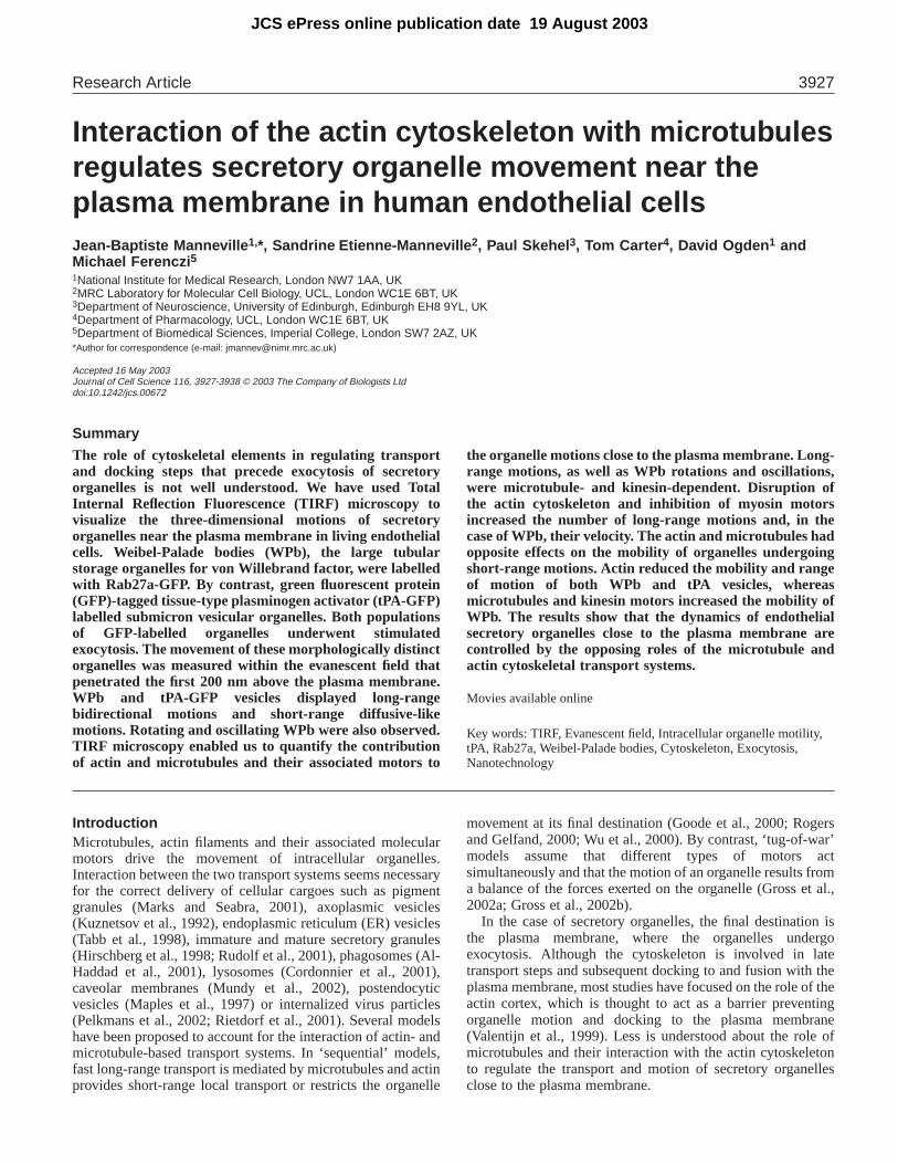
Introduction Microtubules, actin filaments and their associated molecular motors drive the movement of intracellular organelles. Interaction between the two transport systems seems necessary for the correct delivery of cellular cargoes such as pigment granules (Marks and Seabra, 2001), axoplasmic vesicles (Kuznetsov et al., 1992), endoplasmic reticulum (ER) vesicles (Tabb et al., 1998), immature and mature secretory granules (Hirschberg et al., 1998; Rudolf et al., 2001), phagosomes (Al- Haddad et al., 2001), lysosomes (Cordonnier et al., 2001), caveolar membranes (Mundy et al., 2002), postendocytic vesicles (Maples et al., 1997) or internalized virus particles (Pelkmans et al., 2002; Rietdorf et al., 2001). Several models have been proposed to account for the interaction of actin- and microtubule-based transport systems. In ‘sequential’ models, fast long-range transport is mediated by microtubules and actin provides short-range local transport or restricts the organelle movement at its final destination (Goode et al., 2000; Rogers and Gelfand, 2000; Wu et al., 2000). By contrast, ‘tug-of-war’ models assume that different types of motors act simultaneously and that the motion of an organelle results from a balance of the forces exerted on the organelle (Gross et al., 2002a; Gross et al., 2002b). In the case of secretory organelles, the final destination is the plasma membrane, where the organelles undergo exocytosis. Although the cytoskeleton is involved in late transport steps and subsequent docking to and fusion with the plasma membrane, most studies have focused on the role of the actin cortex, which is thought to act as a barrier preventing organelle motion and docking to the plasma membrane (Valentijn et al., 1999). Less is understood about the role of microtubules and their interaction with the actin cytoskeleton to regulate the transport and motion of secretory organelles close to the plasma membrane. 3927 The role of cytoskeletal elements in regulating transport and docking steps that precede exocytosis of secretory organelles is not well understood. We have used Total Internal Reflection Fluorescence (TIRF) microscopy to visualize the three-dimensional motions of secretory organelles near the plasma membrane in living endothelial cells. Weibel-Palade bodies (WPb), the large tubular storage organelles for von Willebrand factor, were labelled with Rab27a-GFP. By contrast, green fluorescent protein (GFP)-tagged tissue-type plasminogen activator (tPA-GFP) labelled submicron vesicular organelles. Both populations of GFP-labelled organelles underwent stimulated exocytosis. The movement of these morphologically distinct organelles was measured within the evanescent field that penetrated the first 200 nm above the plasma membrane. WPb and tPA-GFP vesicles displayed long-range bidirectional motions and short-range diffusive-like motions. Rotating and oscillating WPb were also observed. TIRF microscopy enabled us to quantify the contribution of actin and microtubules and their associated motors to the organelle motions close to the plasma membrane. Long- range motions, as well as WPb rotations and oscillations, were microtubule- and kinesin-dependent. Disruption of the actin cytoskeleton and inhibition of myosin motors increased the number of long-range motions and, in the case of WPb, their velocity. The actin and microtubules had opposite effects on the mobility of organelles undergoing short-range motions. Actin reduced the mobility and range of motion of both WPb and tPA vesicles, whereas microtubules and kinesin motors increased the mobility of WPb. The results show that the dynamics of endothelial secretory organelles close to the plasma membrane are controlled by the opposing roles of the microtubule and actin cytoskeletal transport systems. Movies available online Key words: TIRF, Evanescent field, Intracellular organelle motility, tPA, Rab27a, Weibel-Palade bodies, Cytoskeleton, Exocytosis, Nanotechnology Summary Interaction of the actin cytoskeleton with microtubules regulates secretory organelle movement near the plasma membrane in human endothelial cells Jean-Baptiste Manneville 1, *, Sandrine Etienne-Manneville 2 , Paul Skehel 3 , Tom Carter 4 , David Ogden 1 and Michael Ferenczi 5 1 National Institute for Medical Research, London NW7 1AA, UK 2 MRC Laboratory for Molecular Cell Biology, UCL, London WC1E 6BT, UK 3 Department of Neuroscience, University of Edinburgh, Edinburgh EH8 9YL, UK 4 Department of Pharmacology, UCL, London WC1E 6BT, UK 5 Department of Biomedical Sciences, Imperial College, London SW7 2AZ, UK *Author for correspondence (e-mail: [email protected]) Accepted 16 May 2003 Journal of Cell Science 116, 3927-3938 © 2003 The Company of Biologists Ltd doi:10.1242/jcs.00672 Research Article JCS ePress online publication date 19 August 2003
Transcript of Interaction of the actin cytoskeleton with microtubules regulates ...
IntroductionMicrotubules, actin filaments and their associated molecularmotors drive the movement of intracellular organelles.Interaction between the two transport systems seems necessaryfor the correct delivery of cellular cargoes such as pigmentgranules (Marks and Seabra, 2001), axoplasmic vesicles(Kuznetsov et al., 1992), endoplasmic reticulum (ER) vesicles(Tabb et al., 1998), immature and mature secretory granules(Hirschberg et al., 1998; Rudolf et al., 2001), phagosomes (Al-Haddad et al., 2001), lysosomes (Cordonnier et al., 2001),caveolar membranes (Mundy et al., 2002), postendocyticvesicles (Maples et al., 1997) or internalized virus particles(Pelkmans et al., 2002; Rietdorf et al., 2001). Several modelshave been proposed to account for the interaction of actin- andmicrotubule-based transport systems. In ‘sequential’ models,fast long-range transport is mediated by microtubules and actinprovides short-range local transport or restricts the organelle
movement at its final destination (Goode et al., 2000; Rogersand Gelfand, 2000; Wu et al., 2000). By contrast, ‘tug-of-war’models assume that different types of motors actsimultaneously and that the motion of an organelle results froma balance of the forces exerted on the organelle (Gross et al.,2002a; Gross et al., 2002b).
In the case of secretory organelles, the final destination isthe plasma membrane, where the organelles undergoexocytosis. Although the cytoskeleton is involved in latetransport steps and subsequent docking to and fusion with theplasma membrane, most studies have focused on the role of theactin cortex, which is thought to act as a barrier preventingorganelle motion and docking to the plasma membrane(Valentijn et al., 1999). Less is understood about the role ofmicrotubules and their interaction with the actin cytoskeletonto regulate the transport and motion of secretory organellesclose to the plasma membrane.
3927
The role of cytoskeletal elements in regulating transportand docking steps that precede exocytosis of secretoryorganelles is not well understood. We have used TotalInternal Reflection Fluorescence (TIRF) microscopy tovisualize the three-dimensional motions of secretoryorganelles near the plasma membrane in living endothelialcells. Weibel-Palade bodies (WPb), the large tubularstorage organelles for von Willebrand factor, were labelledwith Rab27a-GFP. By contrast, green fluorescent protein(GFP)-tagged tissue-type plasminogen activator (tPA-GFP)labelled submicron vesicular organelles. Both populationsof GFP-labelled organelles underwent stimulatedexocytosis. The movement of these morphologically distinctorganelles was measured within the evanescent field thatpenetrated the first 200 nm above the plasma membrane.WPb and tPA-GFP vesicles displayed long-rangebidirectional motions and short-range diffusive-likemotions. Rotating and oscillating WPb were also observed.TIRF microscopy enabled us to quantify the contributionof actin and microtubules and their associated motors to
the organelle motions close to the plasma membrane. Long-range motions, as well as WPb rotations and oscillations,were microtubule- and kinesin-dependent. Disruption ofthe actin cytoskeleton and inhibition of myosin motorsincreased the number of long-range motions and, in thecase of WPb, their velocity. The actin and microtubules hadopposite effects on the mobility of organelles undergoingshort-range motions. Actin reduced the mobility and rangeof motion of both WPb and tPA vesicles, whereasmicrotubules and kinesin motors increased the mobility ofWPb. The results show that the dynamics of endothelialsecretory organelles close to the plasma membrane arecontrolled by the opposing roles of the microtubule andactin cytoskeletal transport systems.
Movies available online
Key words: TIRF, Evanescent field, Intracellular organelle motility,tPA, Rab27a, Weibel-Palade bodies, Cytoskeleton, Exocytosis,Nanotechnology
Summary
Interaction of the actin cytoskeleton with microtubulesregulates secretory organelle movement near theplasma membrane in human endothelial cellsJean-Baptiste Manneville 1,*, Sandrine Etienne-Manneville 2, Paul Skehel 3, Tom Carter 4, David Ogden 1 andMichael Ferenczi 5
1National Institute for Medical Research, London NW7 1AA, UK2MRC Laboratory for Molecular Cell Biology, UCL, London WC1E 6BT, UK3Department of Neuroscience, University of Edinburgh, Edinburgh EH8 9YL, UK4Department of Pharmacology, UCL, London WC1E 6BT, UK5Department of Biomedical Sciences, Imperial College, London SW7 2AZ, UK*Author for correspondence (e-mail: [email protected])
Accepted 16 May 2003Journal of Cell Science 116, 3927-3938 © 2003 The Company of Biologists Ltddoi:10.1242/jcs.00672
Research Article
JCS ePress online publication date 19 August 2003

3928
Endothelial cells secrete by exocytosis several proteins thatregulate blood coagulation, blood flow and local immuneresponses. The fibrinolytic tissue-type plasminogen activator(tPA) is found in small (0.1-0.2 µm diameter) secretory vesicles(Emeis et al., 1997; Schick et al., 2001). The pro-inflammatoryadhesive protein von Willebrand factor (vWF) is stored inunique large tubular organelles (1-3 µm long, 0.1-0.2 µmdiameter) called Weibel-Palade bodies (WPb) (van Mouriket al., 2002; Weibel and Palade, 1964). Disruption ofmicrotubules blocks the regulated secretion of both tPA(Santell et al., 1992) and vWF (Sinha and Wagner, 1987;Vischer et al., 2000), suggesting that the microtubulecytoskeleton plays a central role in the processing and/ortransport of tPA and vWF. WPb are members of the family oflysosome-related organelles, which includes the pigmentgranules of melanocytes (Marks and Seabra, 2001). Studies inmelanocytes indicate that pigment granules are transported viamicrotubules to the cell periphery where they are trapped intothe actin cytoskeleton through the myosin Va-receptor Rab27a(Hammer, III and Wu, 2002; Seabra et al., 2002). However,unlike pigment granules in melanocytes, nothing is knownabout the role of microtubules and the actin cytoskeleton inregulating secretory organelle dynamics in endothelial cells.
In this study, we have visualized WPb and tPA-containingvesicles in living endothelial cells by the expression of Rab27a-GFP (green fluorescent protein) and tPA-GFP, respectively. Wehave used Total Internal Reflection Fluorescence (TIRF)microscopy for three-dimensional single-particle tracking offluorescent WPb and tPA vesicles, to quantify the movementof these morphologically distinct organelles and to study therole of the cytoskeleton and molecular motors in their transportand dynamics close to the plasma membrane.
Materials and MethodsGFP constructsThe Rab27a-EGFP construct was a gift from M. Seabra (Hume et al.,2001). The tPA-EGFP construct was obtained as follows. The fullcoding sequence of human tPA was generated by aPfu-based PCRusing the plasmid pETPFR (ATCC) as a template and theoligonucleotide primers 5′-gggaattcaatgcatggatgcaatgaagagaggg-3′and 5′-cgcggtaccgtccggtcgcatgttgtcacgaatc-3′. The resulting DNAwas digested with KpnI and ligated into the vector pEGFP-N3(Clontech) that had been digested previously with Ecl136 and KpnI.
Cell culturePrimary human umbilical vein endothelial cells (HUVECs) wereeither isolated and grown as previously described (Carter et al., 1988)or purchased as cryopreserved cells from pooled donors (ZHC-2101,TCS CellWorks, Bucks, UK). No significant difference was observedbetween cells from the two different sources. Custom cell chamberswere assembled by gluing a Teflon® frame to a glass slide. Cells weredirectly plated on the chambers without any additional coating andcultured at 37°C in a 5% CO2 atmosphere. Freshly isolated cells weregrown in M199 medium supplemented with 10% fetal calf serum(FCS), 10% newborn calf serum, 100 U/ml penicillin and 100 µg/mlstreptomycin. Cryopreserved cells were grown in endothelial growthmedium (ZHM-2953, TCS CellWorks).
Organelles and cytoskeleton labellingCells were microinjected with Rab27a-GFP or tPA-GFP cDNAs.
Rab27a-GFP cells were imaged live 48 hours after microinjection.tPA-GFP-expressing cells were imaged live 4 hours aftermicroinjection. Cells were fixed with 4% paraformaldehyde for 15minutes and permeabilized with 0.2% Triton X100 and 10% FCS for10 minutes before staining. For microtubule labelling, 0.5%glutaraldehyde (Sigma) was added during fixation. Primary antibodieswere: rabbit polyclonal anti-human vWF diluted 1/100 (A0082, Dako,Denmark) and rat monoclonal anti α-tubulin diluted 1/200 (cloneYL1/2 MCAP77; Serotec, Oxford, UK). Secondary antibodies wereRhodamine (TRITC)-conjugated donkey anti-rat, Rhodamine(TRITC)-conjugated donkey anti-rabbit, Cy2-conjugated donkey anti-rabbit (Jackson ImmunoResearch Labs, West Grove, PA). Actin wasstained with rhodamin-phalloidin (Sigma) diluted 1/500 for 45minutes. Cells were left in PBS for TIRF imaging. For colocalizationexperiments, cells were fixed immediately after live imaging tominimize cell motion, cytoskeleton deformation and fixation artefacts.A secretory organelle colocalized with a cytoskeletal element whenits fluorescence overlapped or was within one pixel from thecorresponding cytoskeletal structure.
Drug treatmentsThe microtubule cytoskeleton was depolymerized by incubating cellsin culture medium supplemented with 10 µM nocodazole (Sigma) for1 hour at 37°C, 5% CO2. The cell shape and adhesion to the glassslide remained satisfactory (data not shown). Nocodazole treatmentselectively disrupted the microtubule network but left the actincytoskeleton intact (data not shown). Microtubules repolymerized in15 minutes after cells were washed twice in culture medium. Actinfilaments were depolymerized using 5 µM latrunculin B (Calbiochem)for 10 minutes at 37°C, 5% CO2. Under these conditions, few actinbundles remained visible in TIRF images, whereas microtubules werenot significantly affected (data not shown). At higher concentrationsor longer incubation times, dramatic shape changes and loss ofadhesion of the cell to the glass substrate occurred. Kinesin motorswere blocked with 10 µM aurintricarboxylic acid (ATA, Sigma) for 2hours, whereas myosin motors were inhibited using 10 mM 2,3-butanedione monoxime (BDM, Sigma) for 30 minutes.
Dual-colour TIRF microscopyTotal internal reflection (reviewed by Axelrod, 2001; Steyer andAlmers, 2001; Toomre and Manstein, 2001) was achieved at the glassslide/culture medium interface using a trapezoidal glass prism. Therefractive index of the prism and culture medium was ni=nglass=1.52and nt=nculture medium≈ 1.336, respectively, giving a critical angle (θc)for total internal reflection of θc=61.5°. Experiments were carried outon an upright microscope (Axioplan, Zeiss, Oberkochen, Germany)using either an Argon ion laser (excitation λ=488 nm, 25 mW, Melles-Griot, Carlsbad, CA) or a Nd:YAG laser (excitation λ=532 nm, 50mW, CrystaLaser, Reno, NV). The angle of incidence of the excitationlight could be adjusted and was fixed to 68-70°, above the criticalangle. The intensity profile of the evanescent wave is exponentiallydecaying: I(z)=I0exp(–z/dP), where z is the vertical distance, I0=I(z=0)is the intensity at the interface and dP is the penetration depth givenby:
The calculated penetration depth for the Argon ion laser and for theNd:YAG laser was dP=75-85 nm and dP=85-95 nm, respectively.
GFP (respectively rhodamine) fluorescence was excited by theArgon ion (resp. Nd:YAG) laser. Differences in beam radius anddivergence were corrected by additional lenses to achieve the samespot size and position on the sample. Light from TIRF images waspassed through a dichroic filter (505DRLP02 for GFP and Cy2
λ
4πdP= .
nt2 sin2 (θ) −ni2!
Journal of Cell Science 116 (19)

3929Secretory organelle transport visualized by TIRF microscopy
fluorescence or 560DRLP for rhodamine fluorescence, OmegaOptical, VT) and an emission filter (530DF30 or 565ALP). Standardepifluorescence was achieved using a 100 W mercury lamp (excitationfilters 485DF22 or 525DF45). The temperature in the cell chamberwas maintained at 35-37oC by circulating temperature-controlledwater around the prism. 5% CO2 in O2 was blown on the cell chamberthrough a collar around the objective.
Image acquisitionThe light was collected with a 100× 1.0 NA water immersion objective(Zeiss) driven by a piezo-electric focus drive (Physik Instrumente,Waldbronn, Germany). Fluorescence images were magnified by a 0.5-2× optical zoom (Zeiss), processed by an Argus 20 image processor(Hamamatsu Photonics K.K., Hamamatsu City, Japan) and collectedwith an intensified CCD camera (Remote Head Darkstar, S25Intensifier, Photonics Science, UK). Images were digitized and storedin the memory of a Pentium III PC computer at a maximum rate of25 frames/second by a frame grabber (IC-PCI 4Mb (AMVS), ImagingTechnology, MA) and then saved to disk. Image processing wascarried out using Optimas 6.5 (Media Cybernetics L. P., Silver Spring,USA). Live time-lapse images of GFP-labelled organelles wereacquired at 0.5-2 frames/second with online two frames averagingperformed by the Argus-20 image processor. TIRF illumination waslimited to 200 frames to minimize photobleaching and phototoxicity.Dual-colour images of fixed cells were acquired with 256 framesaveraging. The pixel size was 0.1092 µm (resp. 0.0870 µm) with a1.6× (resp. 2×) optical zoom. The image size was typically 520×500pixels.
Three-dimensional organelle trackingCustomized macros were written in the C++ based language ALI(Analytical Language for Images) and run under Optimas 6.5. Theraw images (N frames, separated by the time interval δτ ) werefiltered to enhance the visibility of the organelles. A low-pass FastFourier Transform filter followed by a 3×3 pixels trimmed meanfilter was applied to remove nonuniform background. At thebeginning of the sequence, a region of interest (ROI) containing theorganelle and a threshold above which the organelle could bedetected was defined. The ‘centre of grey’ (centre of mass weightedby pixel intensities) of the organelle was detected and thefluorescence intensity of the organelle Ipart was measured from thecorresponding raw image in a 5×5 pixels box cantered on the centreof grey. The local background fluorescence intensity Ibkg wasmeasured in another ROI close to the previous one but containingno organelle. The 2D position of the centre of grey yielded the x andy coordinates of the organelles, whereas the z coordinate (relative tothe initial position and corrected for local background brightnessvariations) was given by:
Long-range directed motions and short-range diffusive-likemotionsWhen visualized under the TIRF microscope, organelles displayedtwo types of motions: long-range directed motions and short-rangediffusive-like motions. Long-range motions were defined as being>1 µm in distance and with a maximum velocity >0.05 µm/second.All other motions were considered as short-range motions. Long-range motions were identified by thresholding and accumulating theimages from a time-lapse sequence (e.g. Fig. 3A) or using frame toframe subtraction. Averaging an entire sequence enabled staticorganelles undergoing short-range diffusive motions to be identified(e.g. Fig. 4A).
Data analysisFrom the three-dimensional coordinates x(t), y(t), z(t) of the organelle,the mean squared displacement (MSD) travelled by the organelleduring a time interval ∆t=nδτ was calculated according to:
The analysis of the plot of the MSD as a function of the time interval∆t allowed us to define three classes of motion (Steyer and Almers,1999): simple diffusion (MSD(∆t) is a straight line), slow directedmotion superimposed on diffusion (MSD(∆t) deviates from a straightline and shows an upward curvature), restricted diffusion in a ‘cage’(MSD(∆t) shows a downward curvature). To quantify the motion ofthe vesicles, we used the following equations to fit the MSDdata (Steyer and Almers, 1999): simple diffusion with a diffusioncoefficient D was fitted with:
MSD(∆t) = 6D∆t +cst; (1)
directed diffusion at velocity v and with a diffusion coefficient D wasfitted with:
MSD(∆t) = 6D∆t +v2∆t2+cst; (2)
and restricted diffusion was fitted using:
MSD(∆t) =R2[1 −a1exp(–6a2D∆t/R2)] + 6Dcage∆t +cst, (3)
where R is the radius of the cage, Dcage is the diffusion coefficient ofthe cage, D is the diffusion coefficient of the vesicle inside the cage,and a1≈0.99, a2≈0.85 are two constants. In Eqns 1-3, cstis a numericalconstant accounting for the limited accuracy of the trackingalgorithms. Eqns 1-3 are valid only at small enough time intervals, ∆t.For a total number of N frames, the MSD data were typically cut to∆t=N/2 δτ. In the case of directed and restricted diffusion, thediffusion coefficient of the organelle was first obtained as, thenthe other parameters were fitted imposing the value of D in Eqns 2and 3.
Because the x-y spatial resolution of the microscope (approx. 0.1µm) is much lower than the vertical (z) resolution (approx. 0.01 µm),three-dimensional MSD data are dominated by 2D x-y motions. Tospecifically quantify displacements in the z direction, the verticalMSD was calculated according to:
The z-direction ‘diffusion coefficient’ was defined as 2Dz = lim∆t→0MSD(∆t) (Johns et al., 2001).
The resolution of the TIRF microscope was estimated by tracking0.1 µm diameter polystyrene beads (Molecular Probes, Eugene, OR)stuck to a glass slide in the same conditions as when cells wereobserved. The signal-to-noise ratio in the bead images was adjustedto match that in the organelle images. Motions of the immobilizedbeads were due to mechanical vibrations in the experimental set-up.The minimum detectable 3D diffusion coefficient was D=2.6±0.1 10–6
µm2/second (n=10), whereas the corresponding z-direction ‘diffusioncoefficient’ was Dz=1.2±0.2 10–7 µm2/second (n=10).
Fusion of secretory organelles with the plasma membraneSecretion was stimulated by 100 µM histamine (Sigma). Cells wereimaged at 2 frames/second before and during stimulation. Individualfusion events were analyzed by fitting a Gaussian distribution to the
^N−n
j=1
1
N−nMSDz(∆t =nδτ) = {[ z(jδτ+nδτ) −z(jδt)]2} .
^N−n
j=1
1
N−nMSD(∆t =nδτ) = {[ x(jδτ+nδτ) −x(jδt)]2+
[y(jδτ+nδτ) −y(jδτ)]2+ [z(jδτ+nδτ) −z(jδτ)]2} .
Ipart(t) − Ibkg(t)
Ipart(0)− Ibkg(0)z(t) =−dP ln .

3930
fluorescence image. The diffusion coefficient of the fluorophore Dfluowas deduced from a linear fit of the distribution half-width Rfluo withtime according to: R2fluo(t) = 4Dfluo t + cst.
ResultsVisualization of secretory organelles close to the plasmamembrane in living HUVECsExpression of a Rab27a-GFP chimera (Hume et al., 2001)fluorescently labelled Weibel-Palade bodies, the large rod-shaped secretory organelles of vWF storage in endothelial cells(Fig. 1C) [see article in this issue (Hannah et al., 2003)]. Bycontrast, tPA-GFP localized to small-diameter vesicles. Withconventional epifluorescence excitation, the fluorescent tPA-containing and WPb organelles were mostly unresolvedbecause of background fluorescence from regions of the cellsuch as the Golgi and the ER. However, the sectioning effectof TIRF microscopy enabled us to clearly visualize bothpopulations of organelles within 200 nm close to the plasmamembrane (tPA, Fig. 1A; WPb, Fig. 1B).
Fusion of Rab27a-GFP-positive organelles appeared as afluorescent cloud spreading away from the site of fusion after100 µM histamine stimulation (Fig. 2A). Fitting the half-widthRfluo of the fluorescence distribution with time (Fig. 2C) givesthe value for the diffusion coefficient of Rab27a-GFP in theplasma membrane: Dfluo=0.24±0.16 µm2/second (n=3). By
contrast, exocytosis of tPA vesicles was observed asa transient brightening of the vesicle followed by anexponential decay of its fluorescence intensity (Fig.2B,D) with a characteristic time τ =12.3±1.6seconds (n=13). Fusion of tPA vesicles was morefrequent (n=29 in eight cells) than fusion of Rab27aorganelles (n=3 in ten cells).
WPb and tPA vesicle movements in restingHUVECsTwo types of motion were distinguished for WPband tPA vesicles in resting HUVECs: long-rangedirected motions and much slower short-rangediffusive-like motions (see Materials and Methods).Examples of time-lapse sequences are given inSupplementary Material (see Movies 1, 2;http://jcs.biologists.org/supplemental/).
Long-range directed motionsApproximately 22% of WPb within the evanescentfield exhibited long-range motions, corresponding to
a frequency, defined as the number of long-range motions pertotal number of organelles per unit time, of 1.0×10⁻³ per second.Long-range motions often involved rapid changes or reversals indirection, changes in velocity and pauses between consecutiveruns. The average velocity during a long-range run was0.16±0.02 µm/second and the maximum velocity was 0.54±0.04µm/second (n=54). Run length and run duration averaged7.2±0.8 µm and 70.5±10.1 seconds (n=54,) respectively. Areduction in WPb velocity often correlated with an increase inthe fluorescence intensity of the organelle, indicating that it wasmoving closer towards the plasma membrane, as seen in Fig. 3B,where both sets of data are plotted in time. During pausesbetween directed runs, we observed complex behaviours ofWPb, including rotations and oscillations (see Movies 3, 4;http://jcs.biologists.org/supplemental/). Rotations (e.g. Fig. 3C)occurred in either clockwise or anticlockwise directions with thesame average frequency ωrot=0.268±0.036 rad/second (n=5). Insome cases, WPb exchanged their leading end without reversingthe direction of their motion, probably as a result of combinedrotation and translation. Oscillations (Fig. 3D) occurredpreferentially during pauses and before direction reversals. Theaverage oscillation frequency was ωosc=0.141±0.005 per second(n=6).
tPA vesicles also exhibited long-range motions. Thefrequency of long runs was greater than for WPb (2.2×10⁻³ persecond), as were the average velocity, maximum velocity and
Journal of Cell Science 116 (19)
Fig. 1.Visualization of secretory organelles in livingHUVECs by TIRF microscopy. (A,B) Epifluorescence(EPI) and corresponding TIRF (TIRF) images of tPA-GFP 4 hours after microinjection (A) and Rab27a-GFP48 hours after microinjection (B). Details of the TIRFimage show small diameter tPA-GFP vesicles (A) andtubular rod-shaped Rab27a-GFP organelles (B).Arrowheads indicate the Golgi region in epifluorescenceimages. (C) Rab27a-GFP colocalizes with vWF onWeibel-Palade bodies (TIRF images; green, Rab27a-GFP; red, vWF). Bars, 10 µm.

3931Secretory organelle transport visualized by TIRF microscopy
average run length [0.67±0.07 µm/second, 1.48±0.13µm/second and 8.2±1.1 µm (n=35), respectively]. As withWPb, the velocity of tPA vesicles often decreased whenmoving closer towards the plasma membrane (not shown).
Short-range diffusive-like motionsThe majority (>70%) of WPb close to the plasma membraneundergo diffusive-like short-range motions (e.g. Fig. 4A). Plotsof the three-dimensional MSD as a function of the time interval∆t, revealed three classes of diffusive-like behaviours: simplediffusion, directed diffusion and restricted diffusion, occurringwith approximately equal frequency. MSD(∆t) plots forindividual WPb (Fig. 4A) were fitted according to Eqns 1-3(see Materials and Methods). The diffusion coefficient D, driftvelocity v (directed diffusion), cage radius Rcageand diffusioncoefficient Dcage (restricted diffusion) derived from these fitsare summarized in Table 1. The averaged MSD plots are shownin Fig. 4B.
Similar results and orders of magnitude were obtained for tPAvesicles, except that in the case of caged diffusion, the cage wasabout five times more mobile than for WPb (data not shown).
Localization of the cytoskeleton and secretoryorganelles close to the plasma membrane of HUVECsIn fixed cells, microtubules and actin filaments were found to
extend into the region illuminated bythe evanescent field, coming closestin proximity to the plasma membranein peripheral regions of the cell (Fig.5). Actin formed stress fibres withenhanced fluorescence at sites offocal adhesion contacts (Fig. 5A). Amore diffuse actin staining was alsovisible over the cell footprint and was
probably due to a more random organization of short filamentsin the actin cortex. Microtubules were generally orientedradially, with tubulin fluorescence often greater on peripheralmicrotubules, indicating that they come closer to the plasmamembrane in these regions (Fig. 5B). Quantification of dual-colour TIRF images (see Materials and Methods) showed that58.1±3.2% of WPb colocalized with microtubules (n=15 cells)and 28.5±2.7% colocalized with actin fibres (n=14 cells).Similar results were obtained for tPA-GFP vesicles (data notshown). WPb clearly aligned with microtubules, particularly atthe cell periphery near microtubule plus-ends (Fig. 5C).
Fig. 2.Exocytosis of endothelialsecretory organelles. (A,B) Individualfusion events of a Rab27a-GFP-positiveorganelle (A) and a tPA-GFP vesicle (B)stimulated by 100 µM histamine.Rab27a-GFP diffuses in the plasmamembrane, whereas tPA-GFP remains atthe fusion site after exocytosis. Numbersindicate time (in seconds) relative to themoment of fusion. Lower panels showthree-dimensional luminance plots offour successive frames starting oneframe before fusion. Bars, 1 µm.(C) Plot of the half-width Rfluo2(t)obtained by a Gaussian fit of thedistribution of fluorescence intensitiesfrom the images shown in A. A linear fit(grey line) yields the diffusioncoefficient of Rab27a-GFP in themembrane Dfluo=0.12±0.01µm2/second.(D) Time course of the fluorescenceintensity of the tPA-GFP vesicle shownin B. The characteristic decay time ofthe fluorescence is given by anexponential fit (grey line): τ =13.7±0.9 s.
Table 1. Characteristics of Weibel-Palade bodiesundergoing short-range motions in non-treated cells
Simple diffusion Proportion 44.3±3.0%Diffusion coefficient D 3.4±0.7 10−4 µm2/s
Directed diffusion Proportion 23.3±2.7%Diffusion coefficient D 1.9±0.9 10−4 µm2/sDrift velocity v 3.9±0.7 10−3 µm2/s
Restricted diffusion Proportion 32.4±5.6%Diffusion coefficient D 3.9±0.7 10−4 µm2/s‘Cage’ diffusion coefficient Dcage 8.8±2.7 10−5 µm2/s‘Cage’ radius Rcage 187±16 nm
The averaged parameters deduced by fitting the three-dimensional MSDplots according to Eqns 1-3 are shown for each class of diffusive behaviour(n=21, 11 and 16 for simple, directed and restricted diffusion respectively).

3932
Superposition of movies of WPb movements on images of themicrotubule system after fixation indicates that long-rangemotions of WPb frequently occur along microtubules (Movies5A,B; http://jcs.biologists.org/supplemental/).
Long-range dynamics depend primarily on themicrotubule cytoskeletonMicrotubule disruption caused a significant decrease in thedensity (number of organelles per µm2) of both populations ofsecretory organelles (WPb: 16.9±7.0%, n=14; tPA vesicles:39.8±14.7%, n=4), suggesting a role for the microtubulecytoskeleton in the transport of both types of organelles to thecell periphery. Disruption of microtubules almost completelyabolished long-range motions of WPb (Fig. 6A). Innocodazole-treated cells, the frequency of WPb undergoinglong-range motions dropped from 1.0×10⁻³ to 8×10⁻⁵ persecond (Fig. 6B). For the few remaining WPb undergoing long-range motion, the total displacement was just above thearbitrary threshold for the definition of long-range motion, andthe average and maximum velocities of WPb were four timesslower than in control cells (Fig. 6B). Complex dynamics suchas oscillations and rotations seen in control cells were notobserved in nocodazole-treated cells. Long-range directed
motions, oscillations and rotations reappeared whenmicrotubules repolymerized after nocodazole washout (datanot shown).
In the presence of the kinesin ATPase inhibitoraurintricarboxylic acid (ATA) (Hopkins et al., 2000), long-range motions were unidirectional and mostly directed towardsthe cell centre (78.6%, n=14). Both the average and maximumvelocities significantly increased (P<0.04), whereas the totalrun length decreased (Fig. 6B).
Disruption of actin filaments and inhibition of myosinmotors by 2,3-butanedione monoxime (BDM) (Cramerand Mitchison, 1995) had more subtle effects. Actindepolymerization did not induce any significant change inorganelle density for either WPb (4.1±3.0%, n=4) or tPAvesicles (4.7±13.7%, n=4). Latrunculin B and BDM treatmentsinduced an increase in the frequency of long-range motions ofWPb and a decrease in the total run length (Fig. 6B). Theaverage velocity increased significantly (P<0.04) on actindisruption and myosin inhibition (Fig. 6B).
Qualitatively similar results were obtained for tPA-GFPvesicles, except that the average velocity in latrunculinB-treated cells was the same as in control cells (data notshown). Taken together, these results indicate that long-rangemotions are essentially microtubule-dependent, and that actin
Journal of Cell Science 116 (19)
Fig. 3.Long-range movements of WPb in HUVECs. (A) Long-range directed motions visualized by thresholding and accumulating framesfrom a sequence of 120 images. Bar, 10 µm. (B) Example of long-range directed motion. The three-dimensional trajectory is shown in the left-hand panels. Note the difference in scale between the z-axis and the x-y axes. The initial point (t=0) is indicated by an asterisk. The right-handpanel shows plots of vertical position z (solid line, left axis in µm) and velocity (dotted line, right axis in µm/second) as a function of time, witha rolling average of three frames. The WPb slows down as it approaches the plasma membrane (arrows) and accelerates as it moves away fromthe membrane (arrowheads). (C) WPb rotation. Plot of the angle of a rotating WPb as a function of time (anticlockwise rotation, frequencyωrot=0.40 per second). Top panels show the corresponding TIRF images (Bar, 1 µm). (D) WPb oscillations. Plot of the x-y velocity of anoscillating WPb as a function of time (oscillation frequency ωosc=0.14 per second).

3933Secretory organelle transport visualized by TIRF microscopy
decreases their frequency and, in the case of WPb, theirvelocity.
Short-range diffusive motions are controlled by actin andmicrotubule elementsShort-range motions of WPb were affected by actin andmicrotubule depolymerization and by inhibition of kinesin andmyosin motors (Figs 7, 8). We pooled data from all three classes
of diffusive-like behaviours (simple, directed and restricteddiffusion) (Fig. 7). On treatment with nocodazole, the averagediffusion coefficient decreased by a factor 4.3, whereas itincreased by a factor of 4.2 with latrunculin B treatment (Fig.7A). Effects of cytoskeleton disruption also showed up on theaveraged 3D MSD plots (obtained by averaging all MSD plotstogether, as shown on Fig. 7A, right panel). Blocking kinesinmotors activity by ATA induced a decrease in the diffusioncoefficient by a factor 2.7. By contrast, treatment with the myosin
inhibitor BDM did not change the value of thediffusion coefficient. Drug treatments hadqualitatively similar effects on the vertical
Fig. 4.Short-range motions ofWPb in HUVECs. (A) Short-range diffusive motionsvisualized by averaging thesame sequence as in Fig. 3A.Examples are shown of simplediffusion (WPb 1), directeddiffusion (WPb 2) and restricteddiffusion (WPb 3). The WPbx-y trajectories are given in thecentre panels (x and y in µm).Three-dimensional MSD plots(right-hand panels) were fittedaccording to Eqs 1-3 (seeMaterials and Methods). Theparameters deduced from thefits are: WPb 1, D=1.5 10–4
µm2/second; WPb 2,D=1.05×10⁻³ µm2/second,v=1.07×10–2 µm/second; WPb3, D=1.0×10⁻⁴ µm2/second,Dcage=3.6×10–5 µm2/second,Rcage=49 nm. Bar, 10 µm.(B) Averages of simplediffusion (n=21), directeddiffusion (n=11), and restricteddiffusion (n=16) 3D MSD plots.
Fig. 5.Visualization of cytoskeletal elements inHUVECs by TIRF microscopy. (A) TIRF image ofthe actin cytoskeleton labelled with rhodamine-phalloidin. Arrowheads point to focal adhesions.The diffuse staining is probably due to the actincortex. (B) TIRF images of the microtubulecytoskeleton visualized by immunostaining of α-tubulin. Left-hand panel: cell with radially orientedmicrotubules; arrowheads show bright peripheralmicrotubules. Right-hand panel: detail of the cellperiphery in another cell. (C) WPb align withmicrotubules. Arrowheads indicate WPbcolocalizing with microtubules in two differentcells. WPb preferentially accumulate at microtubuleplus-ends (green, Rab27a-GFP; red, α-tubulin).Bars, 10 µm.

3934
diffusion coefficient Dz(Fig. 7B). The z-direction averaged MSDzplots from nocodazole- or latrunculin B-treated cells also clearlydiffered from control cells (Fig. 7B, right panel).
The distribution and characteristics of WPb diffusivemotions in each class of diffusive-like behaviours (simple,directed and restricted diffusion) were affected by thedisruption of microtubules or actin filaments (Fig. 8).Nocodazole treatment almost completely abolished restricteddiffusive behaviours, and the proportion of directed diffusionincreased compared with simple diffusion (Fig. 8A). The driftvelocity v of directed diffusive motions did not change (notshown), whereas the diffusion coefficient of the cage (Dcage)and the cage radius (Rcage) strongly decreased (Fig. 8B).Latrunculin B treatment decreased the proportion of restricteddiffusion in favour of simple diffusion. The drift velocity vincreased by a factor of 2.5 (not shown), and the cage radiussignificantly increased (P<0.04) (Fig. 8B). Kinesin and myosininhibition by ATA and BDM, respectively, did not significantlyaffect the distribution of diffusive motions among the threeclasses (Fig. 8A).
The effects of cytoskeleton disruption on the short-rangemotions of tPA vesicles differed in two respects from those onWPb movements (data not shown). Nocodazole treatment didnot modify the average value of the diffusion coefficient andlatrunculin B treatment slightly decreased the drift velocity ofdirected diffusion of tPA vesicles.
DiscussionTransport and exocytosis of WPb and tPA vesicles inliving endothelial cells visualized by TIRF microscopyWe have used TIRF microscopy to investigate the dynamics offluorescent Weibel-Palade bodies and tPA vesicles close tothe plasma membrane and their localization with respectto cytoskeletal elements. Expression of Rab27a-GFPproduced fluorescent rod-like organelles, morphologicallyindistinguishable from native WPb, that were colabelled witha specific antibody to vWF, the major WPb protein (Fig. 1C).Consistently, endogenous Rab27a also localizes to the WPbmembrane (Hannah et al., 2003). Expression of tPA-GFP inHUVECs produced small fluorescent organelles similar tothose seen in untransfected cells (Emeis et al., 1997; Schick etal., 2001; Zupancic et al., 2002). Both Rab27a-GFP and tPA-GFP organelles undergo stimulated exocytosis, as shown byindividual fusion events with the plasma membrane onhistamine stimulation (Fig. 2). After exocytosis, Rab27a-GFPdiffuses in the plasma membrane with a diffusion coefficientof 0.24±0.16 µm2/second, in agreement with values reportedpreviously for other membrane-associated proteins (Saxtonand Jacobson, 1997). By contrast, tPA-GFP brightened onfusion and then slowly dimmed at the fusion site for acharacteristic time of 12.3±1.6 s. This may be due to resealingand reacidification of the vesicles after exocytosis, as reportedrecently in PC-12 cells (Taraska et al., 2003).
Journal of Cell Science 116 (19)
Fig. 6.Effects of cytoskeleton disruption on long-range movements of WPb. (A) Trajectories of the ten most mobile WPb in a nontreated cell (NT),a nocodazole-treated cell (noco) and a latrunculin B-treated cell (latB). Bars, 10 µm. (B) Analysis of long-range motions in nontreated cells (NT,n=54 from three cells), nocodazole-treated cells (noco, n=2 from three cells), cells treated with the kinesin ATPase inhibitor ATA (n=16 from threecells), latrunculin B-treated cells (n=33 from three cells) and cells treated with the myosin ATPase inhibitor BDM (n=44 from three cells).Parameters derived from three-dimensional tracking are: frequency of long-range motions (in s–1), defined as the number of WPb undergoing long-range motions per total number of WPb per unit time, average and maximum velocities (in µm/second) and total run length (inµm).
A
B
NT latB
NT noco latBATA BDM0
0.2
0.4
0.6
0.8
NT noco latBATA BDM0
0.5 10-3
1.0 10-3
1.5 10-3
2.0 10-3
NT noco latBATA BDM0
2
4
6
8
noco
Frequency (s-1) Average and maxi. velocity (µm/s) Total run length (µm)

3935Secretory organelle transport visualized by TIRF microscopy
Immunohistochemical localization of WPb or tPA vesiclesrevealed a close association with microtubules (Fig. 5C anddata not shown), and disruption of microtubules but not of theactin cytoskeleton decreased the density of WPb and tPAvesicles in close proximity to the plasma membrane. Thisindicates that a proportion of these organelles are transportedto and/or maintained at the cell periphery via microtubule-dependent processes.
Actin interacts with microtubules during long-rangetransport of WPb and tPA vesiclesWPb were seen to move continuously over distances of up to20 µm (mean run length approx. 7 µm). Long-range motionswere saltatory and bidirectional, with velocities in the range of0.1-1.0 µm/second. These characteristics are compatible withmicrotubule-driven motility observed in vitro and in vivo(Howard, 2001). This was confirmed by the almost completeloss of long-range motions when microtubules were disruptedby nocodazole (Fig. 6). Moreover, nonspecific inhibitionof kinesin motors with the kinesin ATPase inhibitoraurintricarboxylic acid (ATA) (Hopkins et al., 2000) strongly
decreased the frequency of long-range motions of WPb (Fig.6B). Microtubule-dependent transport of WPb to the plasmamembrane is consistent with the observation that microtubuledisruption blocks vWF secretion (Sinha and Wagner, 1987;Vischer et al., 2000).
tPA vesicles moved three to four times faster than WPb andtheir run lengths were, on average, longer by 1 µm. The motorsresponsible for the motion of tPA vesicles and WPb may bedifferent. However, another hypothesis is that collectivebehaviours of motors control the motility of large organelles(Welte et al., 1998). Long-range transport of WPb may thus beslower due to pools of microtubule motors with oppositepolarity being active at the same time. Indeed, blocking kinesinactivity increased the velocity of WPb (Fig. 6B), suggestingthat competition with dynein motors slows down motion onmicrotubules. The presence of multiple motors may also giverise to the rotations and oscillations displayed by WPb (Fig.3C,D). Oscillations frequently occurred during pauses orbefore direction reversals. Bidirectionality and oscillationsdisappeared when kinesin ATPase activity was blocked.
Actin and actin-based motors also play a role in the long-range motility of WPb and tPA vesicles. Long-range motions
Fig. 7.Effects of cytoskeleton disruption on short-range motions of WPb. (A) Data from all classes of short-range diffusive behaviours (simple,directed and restricted diffusion) were pooled to calculate the average diffusion coefficient D (left panel) and to plot the averaged three-dimensional MSD (right panel) in nontreated cells (circles or NT, n=48 from three cells), nocodazole-treated cells (diamonds or noco,n=51from three cells), cells treated with the kinesin inhibitor ATA (n=48 from three cells), latrunculin B-treated cells (triangles or latB,n=60 fromthree cells) and cells treated with the myosin inhibitor BDM (n=59 from three cells). A numerical constant was added to the averaged MSDdata so that all three plots coincide on their first data point. (B) Same analysis as in A for vertical motions (in the zdirection) in nontreatedcells (n=47 from three cells), nocodazole-treated cells (n=46 from three cells), cells treated with the kinesin inhibitor ATA (n=41 from threecells), latrunculin B-treated cells (n=53 from three cells) and cells treated with the myosin inhibitor BDM (n=53 from three cells). The left-handpanel shows the z-direction diffusion coefficient Dz. Averaged one-dimensional MSDz are plotted on the right panel. A numerical constant wasadded to the averaged MSD data so that all three plots coincide on their first data point.
0
0.1
0.2
0.3
0.4
0.5
0 10 20 30 40 50
MSD
(µm
2 )
∆t (s)
A
B
0 5 10 15 20
vert
ical
MSD
(µm
2 )
∆t (s)
7.5 10-4
5.0 10-4
2.5 10-4
0
1.0 10-3
0
0.4 10-3
0.8 10-3
1.2 10-3
1.6 10-3
D (µm2/s)
3.0 10-5
2.0 10-5
1.0 10-5
0
Dz (µm2/s)
NT noco latBATA BDM
NT noco latBATA BDM

3936
of both organelles were more frequent when actin wasdepolymerized, with about half the total run length. Similareffects on the motion of WPb were observed when cells weretreated with the myosin ATPase inhibitor BDM (Cramer andMitchison, 1995). Disruption of the actin cytoskeleton ormyosin inhibition also increased the velocity of WPb (Fig. 6B).These effects may be due to a loss of a direct interaction withthe actin cytoskeleton combined with a reduction in physicalrestriction to movement imposed by friction of the organelle inthe viscous gel formed by the actin cortex. Pauses onmicrotubules did occur preferentially at sites very close to themembrane, where the actin cortex is thought to be denser. Actinand actin-based motors may also prevent WPb from droppingoff microtubules, thus increasing their total run length. Thefinding that myosin motors slow down long-range motions onmicrotubules supports a tug-of-war situation between actin andmicrotubule motors (Gross et al., 2002a; Gross et al., 2002b).
Restriction of short-range movements of WPb and tPAvesicles near the plasma membraneAbout 80% of WPb and tPA vesicles within the evanescentfield appeared to be almost immobile, undergoing short-rangediffusive-like motions (Fig. 4). The diffusion coefficient D forWPb and tPA vesicles was of the order of D≈10–4 µm2/second,as also found for diffusive motions of chromaffin granules inchromaffin cells (D≈10–4-10–2 µm2/second) (Oheim et al.,1999; Steyer and Almers, 1999; Steyer et al., 1997), and PC12cells (Han et al., 1999; Lang et al., 2000). By comparison, a100 nm diameter bead diffusing in the cytoplasm would
have a diffusion coefficient 1000 times larger(D=kT/(6πηR)≈10–1 µm2/second; taking thecytoplasm viscosity η ≈6ηwater≈6.10–3 kg/m/s).Thus, diffusive motions of secretory organellesclose to the plasma membrane are severelyrestricted. The secretory organelles may be dockedto the plasma membrane or bound to cytoskeletalelements via complexes of molecular motors. Theirmotion could also be physically but nonspecificallyhindered by the cortical actin gel.
The actin cytoskeleton and microtubules playopposing roles in short-range motions of WPband tPA vesicles close to the plasmamembrane In HUVECs, the 3D and vertical mobilities of WPbclearly increase following actin depolymerizationby latrunculin B (Fig. 7). In other cell types,conflicting results have been reported (Bi et al.,1997; Johns et al., 2001; Lang et al., 2000; Oheim
and Stühmer, 2000). In the case of restricted diffusion, thediffusion coefficient of the ‘cage’ and the ‘cage’ radius for bothorganelles increases on actin depolymerization (Fig. 8B). Theproportion of restricted diffusion is also reduced comparedwith control cells (Fig. 8A), suggesting that actin not onlydecreases the mobility of the organelles but also physicallyrestricts their range of motion. Restricted diffusion could thusrepresent motions within the cortical actin network (Lang etal., 2000), and the increase in ‘cage’ radius may reflect awidening of the network upon latrunculin B treatment. Insteadof acting as a barrier preventing the docking of secretoryorganelles to the plasma membrane (Johns et al., 2001; Rudolfet al., 2001; Valentijn et al., 1999), the actin cortex couldactually promote docking and fusion of the organelles with theplasma membrane by reducing their mobility and increasingthe probability of association of complementary dockingcomplexes. Interestingly, blocking myosin ATPase activitydoes not affect either the diffusion coefficient (Fig. 7) or theproportion of restricted diffusive motions compared withcontrol cells (Fig. 8A), further suggesting that the cortical actingel physically reduces organelle mobility at the plasmamembrane.
On microtubule depolymerization, we observed a decreasein three-dimensional and vertical diffusion coefficients (Fig. 7).The proportion of restricted diffusion behaviours is alsostrongly reduced compared with nontreated cells (Fig. 8A),suggesting that restricted diffusion may be due not only tophysical hindrance by the actin cortex but also to a tetheringof the organelles to microtubules via molecular motors. This isalso consistent with the decrease in organelle density observed
Journal of Cell Science 116 (19)
Fig. 8.Effects of cytoskeleton disruption on simple,directed and restricted diffusive behaviours of WPb. Apercentage of simple (S), directed (D) and restricted (R)diffusion in nontreated cells (NT), nocodazole-treatedcells (noco) and latrunculin-treated cells (latB).(B) Restricted diffusion. Diffusion coefficient of thecage (Dcagein µm2/second) and radius of the cage (Rcagein µm) in nontreated cells (n=16), nocodazole-treatedcells (n=1) and latrunculin-treated cells (n=11).
B
S
D
R
0.0
0.1
0.2
0.3
0.4
Rcage (µm)
NT noco latBNT noco latB
Dcage (µm2/s)1.6 10-4
1.2 10-4
0.8 10-4
0
0.4 10-4
A
0
10
20
30
40
50
60
70
NT noco latBATA BDM
S
S
S
S
D
DD D
R
R
R
R
Proportion (%)

3937Secretory organelle transport visualized by TIRF microscopy
with nocodazole treatment. Interestingly, although actin playsa qualitatively similar role in the short-range mobility of tPAvesicles and WPb, we found that the role of microtubules wasmarkedly different. The average diffusion coefficient of tPAvesicles was not modified when microtubules weredepolymerized. As discussed above, a larger number ofmicrotubule-based motors is probably recruited onto thesurface of WPb, and this may increase their mobility.Consistent with this hypothesis, inhibition of kinesins by ATAreduces the average diffusion coefficient of WPb (Fig. 7)without affecting the distribution among the three classesof diffusive behaviours (Fig. 8A). These data show thatmicrotubules increase the mobility of WPb near the plasmamembrane, at least partly via the activity of kinesin motors.
Kinesin motors are supposed to act in the recruitment ofsecretory vesicles during a step preceding an actin-dependentstep (Bi et al., 1997). Our results show that short-rangedynamics of WPb near the plasma membrane are regulated notonly by actin but also by microtubules and kinesin motors,indicating that microtubules could also play a role in laterstages, such as organelle docking or fusion. Microtubules,probably via kinesin motors, counteract the confining effect ofactin by increasing short-range mobility of WPb at the plasmamembrane. Actin and microtubules may thus play opposingroles to fine-tune the mobility and localize organelles at targetsites on the plasma membrane.
This work was supported by a Marie Curie Individual Fellowship(J.-B.M.), an EMBO Long Term Fellowship (S.E.-M.), the BritishHeart Foundation (T.C.) and the UK Medical Research Council (allothers). We thank Miguel Seabra for the gift of the Rab27a-GFPconstruct, and Matthew Hannah and Dan Cutler for carrying outpreliminary experiments and extensive sharing of data beforesubmission. We thank Chris Magnus for assistance with cell culture,and Alan Hall and Nicoleta Moisoi for stimulating discussions.
ReferencesAl-Haddad, A., Shonn, M. A., Redlich, B., Blocker, A., Burkhardt, J. K.,
Yu, H., Hammer, III, J. A., Weiss, D. G., Steffen, W., Griffiths, G. et al.(2001). Myosin Va bound to phagosomes binds to F-actin and delaysmicrotubule-dependent motility. Mol. Biol. Cell12, 2742-2755.
Axelrod, D. (2001). Total Internal Reflection Fluorescence microscopy in cellbiology. Traffic 2, 764-774.
Bi, G.-Q., Morris, R. L., Liao, G., Alderton, J. M., Sholey, J. M. andSteinhardt, R. A. (1997). Kinesin- and myosin-driven steps of vesiclerecruitment for Ca2+-regulated exocytosis. J. Cell Biol.138, 999-1008.
Carter, T. D., Hallam, T. J., Cusak, N. J. and Pearson, J. D.(1988).Regulation of P2y-purinoceptor-mediated prostacyclin release from humanendothelial cells by cytoplasmic calcium concentration. Br. J. Pharmacol.95, 1181-1190.
Cordonnier, M.-N., Dauzonne, D., Louvard, D. and Coudrier, E.(2001).Actin filaments and myosin I alpha cooperate with microtubules for themovement of lysosomes. Mol. Biol. Cell12, 4013-4029.
Cramer, L. P. and Mitchison, T. J. (1995). Myosin is involved in postmitoticcell spreading. J. Cell Biol.131, 179-189.
Emeis, J. J., van den Eijnden-Schrauwen, Y., van den Hoogen, C. M., dePriester, W., Westmuckett, A. and Lupu, F. (1997). An endothelialstorage granule for tissue-type plasminogen activator. J. Cell Biol. 139,245-256.
Goode, B. L., Drubin, D. G. and Barnes, G.(2000). Functional cooperationbetween the microtubule and actin cytoskeletons. Curr. Opin. Cell Biol.12,63-71.
Gross, S. P., Carolina Tuma, M., Deacon, S. W., Serpinskaya, A. S., Reilein,A. R. and Gelfand, V. I. (2002a). Interactions and regulation of molecularmotors in Xenopus melanophores. J. Cell Biol.156, 855-856.
Gross, S. P., Welte, M. A., Block, S. M. and Wieschaus, E. F.(2002b).
Coordination of opposite-polarity microtubule motors. J. Cell Biol. 156,715-724.
Hammer, J. A., III and Wu, X. S. (2002). Rabs grab motors: defining theconnections between Rab GTPases and motor proteins. Curr. Opin. CellBiol. 14, 69-75.
Han, W., Ng, Y.-K., Axelrod, D. and Levitan, E. S.(1999). Neuropeptiderelease by efficient recruitment of diffusing cytoplasmic secretory vesicles.Proc. Natl. Acad. Sci. USA96, 14577-14582.
Hannah, M. J., Hume, A. N., Arribas, M., Williams, R., Hewlett, L. J.,Seabra, M. C. and Cutler, D. F.(2003). Weibel-Palade bodies recruitRab27 by a content-driven, maturation-dependent mechanism that isindependent of cell type. J. Cell Sci. 116, 3939-3948.
Hirschberg, K., Miller, C. M., Ellenberg, J., Presley, J. F., Siggia, E. D.,Phair, R. D. and Lippincott-Schwartz, J. (1998). Kinetic analysis ofsecretory protein traffic and characterization of Golgi to plasma membranetransport intermediates in living cells. J. Cell Biol.143, 1485-1503.
Hopkins, S. C., Vale, R. D. and Kuntz, I. D. (2000). Inhibitors of kinesinactivity from structure-based computer screening. Biochemistry39, 2805-2814.
Howard, J. (2001). Mechanics of motor proteins and the cytoskeleton.Sunderland, Massachusetts: Sinauer Associates.
Hume, A. N., Collinson, L. M., Rapak, A., Gomes, A. Q., Hopkins, C. R.and Seabra, M. C.(2001). Rab27a regulates the peripheral distribution ofmelanosomes in melanocytes. J. Cell Biol.152, 795-808.
Johns, L. M., Levitan, E. S., Shelden, E. A., Holz, R. W. and Axelrod, D.(2001). Restriction of secretory granule motion near the plasma membraneof chromaffin cells. J. Cell Biol.153, 177-190.
Kuznetsov, S. A., Langford, G. M. and Weiss, D. G.(1992). Actin-dependentorganelle movement in squid axoplasm. Nature356, 722-725.
Lang, T., Wacker, I., Wunderlich, I., Rohrbach, A., Giese, G., Soldati, T.and Almers, W. (2000). Role of actin cortex in the subplasmalemmaltransport of secretory granules in PC-12 cells. Biophys. J.78, 2863-2877.
Maples, C. J., Ruiz, W. G. and Apodaca, G. (1997). Both microtubules andactin filaments are required for efficient postendocytic traffic of thepolymeric immunoglobulin receptor in polarized Mardin-Darby caninekidney cells. J. Biol. Chem.272, 6741-6751.
Marks, M. S. and Seabra, M. C. (2001). The melanosome: membranedynamics in black and white. Nat. Rev. Mol. Cell. Biol.2, 1-11.
Mundy, D. I., Machleidt, T., Ying, Y., Anderson, R. G. W. and Bloom, G.S. (2002). Dual control of caveolar membrane traffic by microtubules andthe actin cytoskeleton. J. Cell Sci.115, 4327-4339.
Oheim, M. and Stühmer, W.(2000). Tracking chromaffin granules on theirway through the actin cortex. Eur. Biophys. J.29, 67-89.
Oheim, M., Loerke, D., Stühmer, W. and Chow, R. H.(1999). Multiplestimulation-dependent processes regulate the size of the releasable pool ofvesicles. Eur. Biophys. J.28, 91-101.
Pelkmans, L., Püntener, D. and Helenius, A. (2002). Local actinpolymerization and dynamin recruitment in SV40-induced internalization ofcaveolae. Science296, 535-539.
Rietdorf, J., Ploubidou, A., Reckmann, I., Holmström, A., Fischknecht, F.,Zetti, M., Zimmermann, T. and Way, M. (2001). Kinesin-dependentmovement on microtubules precedes actin-based motility of vaccina virus.Nature Cell Biol.3, 992-1000.
Rogers, S. L. and Gelfand, V. I.(2000). Membrane trafficking, organelletransport, and the cytoskeleton. Curr. Opin. Cell Biol.12, 57-62.
Rudolf, R., Salm, T., Rustom, A. and Gerdes, H.-H.(2001). Dynamics ofimmature secretory granules: role of cytoskeletal elements during transport,cortical restriction, and F-actin-dependent tethering. Mol. Biol. Cell 12,1353-1365.
Santell, L., Marotti, K., Bartfeld, N. S., Baynham, P. and Levin, E. G.(1992). Disruption of microtubules inhibits the stimulation of tissueplasminogen activator expression and promotes plasminogen activatorinhibitor type 1 expression in human endothelial cells. Exp. Cell Res.201,358-365.
Saxton, M. J. and Jacobson, K.(1997). Single-particle tracking: applicationsto membrane dynamics. Annu. Rev. Biophys. Biomol. Struct.26, 373-399.
Schick, B. P., Gradowski, J. F. and San Antonio, J. D.(2001). Synthesis,secretion and subcellular localization of serglycin proteoglycan in humanendothelial cells. Blood97, 449-458.
Seabra, M. C., Mules, E. H. and Hume, A. N.(2002). Rab GTPases,intracellular traffic and disease. Trends Mol. Med.8, 23-30.
Sinha, S. and Wagner, D. D.(1987). Intact microtubules are necessary forcomplete processing, storage and regulated secretion of von Willebrandfactor by endothelial cells. Eur. J. Cell Biol.43, 377-383.

3938
Steyer, J. A. and Almers, W.(1999). Tracking single secretory granules inlive chromaffin cells by evanescent-field fluorescence microscopy. Biophys.J. 76, 2262-2271.
Steyer, J. A. and Almers, W.(2001). A real-time view of life within 100 nmof the plasma membrane. Nat. Rev. Mol. Cell. Biol.2, 268-276.
Steyer, J. A., Horstmann, H. and Almers, W.(1997). Transport, docking andexocytosis of single secretory granules in live chromaffin cells. Nature388,474-478.
Tabb, J. S., Molyneaux, B. J., Cohen, D. L., Kuznetsov, S. A. and Langford,G. M. (1998). Transport of ER vesicles on actin filaments in neurons bymyosin V. J. Cell Sci.111, 3221-3234.
Taraska, J. W., Perrais, D., Ohara-Imaizumi, M., Nagamatsu, S. andAlmers, W. (2003). Secretory granules are recaptured largely intact afterstimulated exocytosis in cultured endocrine cells. Proc. Natl. Acad. Sci. USA100, 2070-2075.
Toomre, D. and Manstein, D.(2001). Lighting up the cell surface withevanescent wave microscopy. Trends Cell Biol.11, 298-303.
Valentijn, K., Valentijn, J. A. and Jamieson, J. D.(1999). Role of actin in
regulated exocytosis and compensatory membrane retrieval: insights froman old acquaintance. Biochem. Biophys. Res. Comm.266, 652-661.
van Mourik, J. A., de Wit, T. R. and Voorberg, J. (2002). Biogenesis andexocytosis of Weibel-Palade bodies. Histochem. Cell Biol.117, 113-122.
Vischer, U. M., Barth, H. and Wollheim, C. B. (2000). Regulated vonWillebrand factor secretion is associated with agonist-specific patterns ofcytoskeletal remodeling in cultured endothelial cells. Arterioscler. Thromb.Vasc. Biol.20, 883-891.
Weibel, E. R. and Palade, G. F.(1964). New cytoplasmic components inarterial endothelial cells. J. Cell Biol.23, 101-112.
Welte, M. A., Gross, S. P., Postner, M., Block, S. M. and Wieschaus, E. F.(1998). Developmental regulation of vesicle transport in Drosophilaembryos: forces and kinetics. Cell 69, 1181-1189.
Wu, X., Jung, G. and Hammer, III, J. A. (2000). Functions of unconventionalmyosins. Curr. Opin. Cell Biol.12, 42-51.
Zupancic, G., Ogden, D., Magnus, C. J., Wheeler-Jones, C. and Carter, T.D. (2002). Differential exocytosis from human endothelial cells evoked byhigh intracellular Ca2+ concentration. J. Physiol.544, 741-755.
Journal of Cell Science 116 (19)