Inherited disorders and their management in some...
88
Swedish University of Agricultural Sciences Faculty of Veterinary Medicine and Animal Science Inherited disorders and their management in some European warmblood sport horse breeds Danica Nikolić Department of Animal Breeding and Genetics Examensarbete 305 Uppsala 2009 Master’s Thesis, 30 HEC Erasmus Mundus programme – European Master in Animal Breeding and Genetics
Transcript of Inherited disorders and their management in some...

Swedish University of Agricultural Sciences Faculty of Veterinary Medicine and Animal Science
Inherited disorders and their management in some European warmblood sport horse breeds Danica Nikolić
Department of Animal Breeding and Genetics
Examensarbete 305
Uppsala 2009
Master’s Thesis, 30 HEC
Erasmus Mundus programme – European Master in Animal Breeding and Genetics

Inherited disorders and their management in some European warmblood sport horse breeds Danica Nikolić Supervisors: Lina Jönson (ABG, SLU) Louise Lindberg (ABG, SLU) Bart Ducro (ABG, WUR)
Examiners: Jan Philipsson, SLU, Department of Animal Breeding and Genetics Johan van Arendonk, WU, Department of Animal Sciences Credits: 30 HEC Course title: Degree project in Animal Science Course code: EX0679 Programme: Erasmus Mundus programme – European Master in Animal Breeding and Genetics Level: Advanced, A2E Place of publication: Uppsala Year of publication: 2009 Cover picture: Olga Boucher
Name of series: Examensarbete 305 Department of Animal Breeding and Genetics, SLU On-line publication: http://epsilon.slu.se Key words: genetic disorders, diseases, sport horses, leg conformation, fertility, hooves


1
Contents 1. Abstract.......................................................................................................................5 2. Introduction................................................................................................................6
2.1. Background......................................................................................................6 2.2. Aim of study....................................................................................................7 2.3. Genetic disorders.............................................................................................7 2.4. Conformation and lameness.............................................................................9 3. Literature study.........................................................................................................10 3.1. Skeletal disorders............................................................................................10
3.2. Formation of bone..........................................................................................12 3.3. Cervical Vertebral Malformation (CVM)......................................................14 3.3.1. Clinical symptoms and diagnosis.......................................................15 3.3.2. Treatment...........................................................................................16 3.3.3. Prognosis............................................................................................17 3.4. Subchondral bone cysts..................................................................................18 3.4.1. Clinical symptoms and diagnosis.......................................................19 3.4.2. Treatment............................................................................................19 3.4.3. Prognosis............................................................................................19 3.5. Over- and underbite........................................................................................20 3.5.1. Clinical symptoms and diagnosis.......................................................20 3.5.2. Treatment...........................................................................................21 3.5.3. Prognosis............................................................................................21 3.6. Bench knees....................................................................................................22 3.6.1. Clinical symptoms and diagnosis.......................................................22 3.6.2. Treatment...........................................................................................23 3.6.3. Prognosis............................................................................................23 3.7. Calf knees.......................................................................................................24 3.7.1. Clinical symptoms and diagnosis.......................................................25 3.7.2. Treatment...........................................................................................25 3.7.3. Prognosis............................................................................................25 3.8. Bucked knees.................................................................................................26 3.8.1. Clinical symptoms and diagnosis......................................................26 3.8.2. Treatment...........................................................................................26 3.8.3. Prognosis............................................................................................27 3.9. Weak pastern..................................................................................................27 3.9.1. Clinical symptoms and diagnosis.......................................................28 3.9.2. Treatment...........................................................................................28 3.9.3. Prognosis............................................................................................28

2
3.10. Toe out...........................................................................................................29 3.10.1. Clinical symptoms and diagnosis.......................................................30 3.10.2. Treatment...........................................................................................30 3.10.3. Prognosis............................................................................................31 3.11. Toe in.............................................................................................................31 3.11.1. Clinical symptoms and diagnosis.......................................................32 3.11.2. Treatment...........................................................................................33 3.11.3. Prognosis............................................................................................33 3.12. Abnormal bone formations in the hock..........................................................33 3.12.1. Clinical signs and diagnosis...............................................................34 3.12.2. Treatment...........................................................................................34 3.12.3. Prognosis............................................................................................35 3.13. Outward rotation of limbs..............................................................................35 3.13.1. Clinical signs and diagnosis...............................................................35 3.13.2. Treatment...........................................................................................36 3.13.3. Prognosis............................................................................................36
4. Summary...................................................................................................................36 5. Methods and Materials............................................................................................43 6. Results.......................................................................................................................43
6.1. Management of inherited disorders in breeding stallions and young horses..................................................................................................45
6.2. Management of inherited disorders in foals and mares..............................................................................................................46
6.3. Monitoring of fertility in breeding stallions...................................................47 6.4. Management of inherited disorders in breeding stallions
and young horses during private veterinary visits..........................................48 6.5. Summarising and evaluation of collected records..........................................49 6.6. Management of specific inherited disorders
in breeding stallions – Skeleton and joints.....................................................50 6.7. Management of specific inherited disorders
in breeding stallions – Hooves........................................................................53 6.8. Management of specific inherited disorders in
breeding stallions – Reproductive...................................................................54 6.9. Management of specific inherited disorders
in breeding stallions – Other...........................................................................56

3
7. Discussion..................................................................................................................58
7.1. Management of inherited disorders in breeding stallions, young horses, mares and foals.......................................................................58
7.2. Management of specific inherited disorders in breeding stallions.................63
8. Conclusions...............................................................................................................68 9. Acknowledgments....................................................................................................69 10. References.................................................................................................................69
Appendix A: Survey.................................................................................................78

4
5
1. Abstract The main aim of this study was to determine the current strategies employed to manage inherited disorders in European warmblood sport horses. An online survey was sent to 37 breeding organisations in 29 countries, from which 11 countries replied. The breeding associations played major roles in selection, management and recording of inherited disorders. Recording of disorders in both breeding stallions and young horses was practiced in five countries; usually at official events or prior to sale. When disorders of breeding stallions and young horses were recorded during private veterinary visits, there was little obligation to report them. There was a trend for countries with smaller warmblood populations to monitor the fertility of breeding stallions and record disorders in foals. Furthermore, they were more likely to summarise and evaluate records of disorders in breeding stallions and young horses. Evaluation of collected records could be used by breeding associations for the approval of breeding stallions and additionally for advising breeders on more efficient mare-stallion matching. Involvement of all interested parties in the collection and evaluation of these records should be encouraged with the possible creation of a central database for disorders. However, international data collection may not be easy to achieve due to differing classification and diagnoses of disorders or varied scoring systems. This was reflected by the varied consideration of specific disorders in selection of breeding stallions between countries. Regarding management of disorders in breeding stallions, skeletal and joint disorders were screened for the most and muscular disorders the least. Reproductive, respiratory and degenerative joint disorders usually resulted in exclusion from breeding. Most conformational deviations were considered only when severe, or could be compensated for with good performance. These deviations were considered at the same level within countries although research implies that deviations predisposing to injury vary across breeds and sport types. There appears to be a great need for more research into which deviations are most detrimental for riding horses.

6
2. Introduction 2.1. Background Warmblood horses are mainly bred as sport or pleasure horses, excelling in disciplines such as dressage, show jumping and eventing at both national and international levels. Within Europe, most of the breeding organisations of warmblood horses have strong selection and breeding programs for sport horses, and sport horses with unfavourable conformation or known genetic disorders are excluded from breeding. This selection mostly only applies to stallions while the use of mares in breeding is less rigorously controlled. The open borders policy and free trade within Europe implemented by the European Union (EU) has increased the ease of extensive exchange of genetic material across the continent. This exchange has also been facilitated by improvements in processing of semen used for artificial insemination (AI), resulting in the ability to transport viable semen across larger distances (Koenen et al., 2004). Countries which have large horse populations, such as Germany and France, apart from utilising their own genetic resources, also share them by extensively exporting horses or semen to other countries (Koenen et al., 2004). Countries with small populations usually act as importers, and it is not unusual for them to use more foreign stallions for breeding than stallions from their own breeds. This seems to be an effective way of disseminating superior genetic material despite possibly unfavourable geographical location. Furthermore it enables superior stallions to produce large numbers of offspring in a relatively shorter time than when natural mating is employed. Despite the evident advantages of such a strategy, some concerns have also been raised. A loss of genetic diversity and increased inbreeding has been suggested in the cases of stallions that are used extensively and whose genes are over-represented in a population. Although this would be a problem if the population was small, the warmblood population is relatively large and was itself formed from diverse bloodlines (Hamann & Distl, 2008). Some warmblood studbooks still practice “open studbook” policies that allow introduction of new genes into the population (The Scottish Sports Horse Studbook Guidelines). Thus promoting the dispersal of genetic material should in turn increase genetic diversity as long as matings are carefully planned. The use of well-kept studbooks and the increased information on stallions provided by numerous numbers of their progeny have enabled inheritance patterns of simple Mendelian genetic disorders to be noticed. This, coupled with the development of the equine genome map and the recent complete sequencing of the equine genome at the Broad Institute in Massachusetts, has greatly increased the number of genetic disorders that have been identified (Spencer & Davies, 2007). For example, genome mapping has been used to identify the causative mutations for hereditary equine regional dermal asthenia (HERDA) and polysaccharide storage myopathy (PSSM) (Finno et al., 2008). Once the causative mutation is identified, a DNA test can be made based on that information. Emerging technologies such as this will also allow the identification of complex polygenic traits in the near future (Finno et al., 2008).

7
As the trade of genetic material between different warmblood studbooks is increasing, it would be interesting to know what strategy each breeding association and country has to regulate selection against disorders that are known or suspected to have an inherited origin. No overview of this subject has been compiled to date. Therefore, a survey was made in order to determine what strategies European breeding associations and countries apply in their selection programs, in order to define the main similarities or differences in their approaches. Data on this issue could enable European countries to improve their strategies for handling inherited disorders by sharing of experiences and mutual learning. Additional knowledge gained in this field would not only increase welfare and health of horses, but may also serve to strengthen trading ties within Europe. 2.2. Aims of study The first aim of this study was to review 11 inherited skeletal disorders and common conformational deviations that are observed in performance horses. The second aim of this study was to determine what types of strategies are employed by European countries and/or breeding associations to manage inherited disorders in some European warmblood sport horse breeds. 2.3. Genetic Disorders Genetic disorders in the horse can be broadly defined as defects of structure or function caused by negative mutation of one or multiple genes (Trommershausen-Smith, 1980). Some disorders are purely genetic, while others result from a combination of genetic and environmental effects. Genetic disorders include congenital disorders (disorders that are present at birth) or those having a late onset, possibly years after the birth (Trommershausen-Smith, 1980). It has been suggested that the incidence of genetic disorders is lower in horses than it is in other domestic animals (Graves, 2005), possibly due to the later domestication of the horse (Bowling & Ruvinsky, 2000). The identification of genetic disorders in the horse has been adversely affected by a long gestation period, single births, incomplete recording and sometimes frequent changes of ownership of horses making them difficult to keep track of. Characteristics of genetic disorders themselves, such as delayed onset of symptoms, incomplete penetrance and varying expressivity between individuals may have also led to difficulties in the resolution of their genetic basis (Finno et al., 2008). Accordingly, studies on the inheritance of genetic disorders in horses are made difficult, lengthy and often costly (Nicholas, 2000). Genetic disorders are undesirable, not only from an economical but also from a health and welfare point of view. They reduce the value and performance of the horse and some of them are painful and decrease the quality of life of the affected horse (Finno et al., 2008). Important technological advances have been made in cases where the genetic disorder affects a specific breed and expresses developmental, congenital or lethal traits (Finno et al., 2008). Constant improvements in molecular technology have enabled scientists to discover the

8
causes and the specific mutations of certain genetic disorders, allowing population-wide screening and selection of breeding animals that are free of these disorders. Comparative genomics and the development of an equine genome map following the complete sequencing of the horse genome have accelerated the rate of these discoveries (Finno et al., 2008). One of the first such causative mutations to be identified was that of severe combined immunodeficiency disorder (SCID), related to the Arabian breed. Mutations causing disorders mainly found in American Paint Horses and Quarter Horses have also been identified, including hyperkalemic periodic paralysis (HYPP), glycogen branching enzyme deficiency (GBED), overo lethal white foal syndrome (OLWS) and hereditary equine regional dermal asthenia (HERDA) (Graves, 2005; Finno et al., 2008). In these cases, the identification of specific mutations has been aided by the fact that the inheritance of these disorders was mostly monogenic. Simple autosomal recessive or dominant mode of inheritance made it possible to use pedigree information for tracking the disorders back to specific horses. For example, a certain bloodline has been associated with the transmission of the HYPP mutation within the Quarter Horse population. The extensive use of one sire that was well known for his impressive muscle mass caused the wide spread of this disorder. However, his muscular phenotype, considered to be very favourable, was actually a compensatory reaction to the gene defect (Bowling et al., 1996; Graves, 2005; Finno et al., 2008). Mutations have also been identified for junctional epidermolysis bullosa (JEB), malignant hyperthermia (MH) and polysaccharide storage myopathy (PSSM) (Finno et al., 2008). JEB, an autosomal recessive disorder, mainly affects Belgian draft horses, other heavy coldblooded breeds and American Saddle horses (Kohn et al., 1989; Lieto et al., 2002; Milenkovic et al., 2003). It is characterized by abnormally fragile skin in certain areas of the body, resulting in skin abrasion and ulceration in the presence of pressure (Shapiro & McEwen, 1995). MH, which is well known in pigs (Hall et al., 1966), has only recently been discovered in two Quarter Horses and is caused by an autosomal dominant mutation that results in hyperthermia and acidosis when an anaesthetic is inhaled (Aleman et al., 2004, Aleman et al., 2005). PSSM is caused by a mutation in a gene that regulates glycogen synthesis which results in disruption of glycogen synthesis and possible impairment of the aerobic glycogen metabolism. As this review will focus mainly on inherited disorders in the warmblood population, it is interesting to note that in a study conducted by McCue et al. (2006), PSSM was shown to be a common cause of neuromuscular disorders amongst warmblood horses. Almost 50% of muscle biopsies from horses with neuromuscular problems in the study tested positive for PSSM. In warmblood horses, the clinical signs usually occur between 8 and 11 years of age, commonly in the form of pain in the back and hindquarter muscles. Reluctance to collect and correctly use the hindquarters when exercising, failure to bascule over jumps, uneven gaits and muscle atrophy have also been reported (Quiroz-Rothe et al., 2002; Hunt et al., 2005; McCue et al., 2006; Finno et al., 2008). PSSM has also been associated with recurring cases of exertional rhabodymyolysis (tying-up syndrome) (Valentine & Cooper, 2005; McGowan et al., 2008) the heritable form of which has been documented in the Thoroughbred (Oki et al., 2005; Dranchak et al., 2005).
9
2.4. Conformation and lameness Conformation can be described as the form or outline of the horse (Saastamoinen & Barrey, 2000). As such, it is a result of the arrangement and function of muscles, bones and tissues (McIlwraith et al., 2003). The conformation of the modern horse is a result of a combination of natural and human selection for speed, strength and endurance (Bowling & Ruvinsky, 2000). Due to the strong relation between form and function selection, the conformation tends to vary between breeds used for different purposes, although the principles of conformation evaluation are very similar across breeds (Stashak, 2002). Traditionally, the judging of conformation and aesthetic value played a vital role in phenotypic evaluation of the horse. Although this still holds true, horses today are judged more on their athletic or working abilities which has promoted the selection of horses with healthy limbs and joints, improved soundness and correct gaits rather than the aesthetic value alone (Saastamoinen & Barrey, 2000). Even though many publications have been written in the last 200 years about correct conformation, the judging of conformation still remains largely subjective, and very little objective data is available (McIlwraith et al., 2003). According to Stashak (2002) conformation is a result of several components which include balance, proportions and curvature of the topline, head structure, leg quality (well defined limbs and “clean” joints), substance of supportive tissues and muscles, and correctness of angles and alignment of skeletal structures. Correct alignment of the skeleton is important in that it provides a solid supportive base for the attachment of muscles and other tissues. Desirable conformation includes moderate length and slope of bones with straight alignment when viewed both from front, side and back, large and unswollen joints, high quality and appropriate size of hooves, adequate dimensions of the heel and a concave sole. Although changes causing lameness are mostly located in lower limbs of the horse, the actual cause may be present in the upper limb or the body. For example, the angle of the shoulder joint usually has a large influence on the angle of the pastern as the two appear to be relatively parallel with each other. A short and upright shoulder may result in a more upright pastern and relate to a rather limited stride length (Stashak, 2002). Lameness can be defined as any alteration of the horse’s normal movement (Devereux & Morrison, 1996). As different individuals move differently, it is important to be familiar with the relation between conformation and movement in order not to misinterpret certain gait characteristics as indications of lameness. Lameness and its causative factors are the major health problem in the warmblood horse population and represent the main reasons for decreased longevity and performance of sport horses. In a study conducted on insurance data of Swedish horses, it was established that the predominant causes of culling of warmblood horses were disorders and injuries of the musculoskeletal system (approximately 55% of deaths), with significant differences between the sexes. Male horses were more likely to be culled due to musculoskeletal problems than mares. Warmblood mares were found to have a significantly longer lifespan than warmblood geldings and stallions (up to 7.5 years longer). This could be a result of mares being less extensively used in sport in favour of breeding, while for stallions and especially geldings

10
sport use may be more intense and possibly the only option for human use. Therefore male horses may have a higher probability of being culled due to musculoskeletal disorders. Within these musculoskeletal disorders, joint and skeletal diseases dominated. Undefined lameness, where the actual cause of death was unknown, was also prominent. Respiratory problems were the second largest reasons for culling of warmblood horses (Wallin et al., 2000). It was interesting to note that there were major differences in causes of culling between breeds. In coldblood horses the reasons for culling were predominated by problems with the nervous system (Wallin et al., 2000). However, data from insurance companies tends not to be representative of the whole population as only a small proportion of horses are insured against death and or loss (Stock & Distl, 2005). The study by Stock & Distl (2005) confirmed that musculoskeletal problems play major roles in losses in training of warmblood horses. This is reflected by the fact that approximately 60% of horses were reported to have had at least one lameness event which caused a relevant intermission in their training, with multiple lameness periods occurring in 35% of horses. The causes of musculoskeletal or conformational disorders that affect soundness and performance are still undefined. It has been suggested that genetics, as well as external factors such as nutrition, limb loading and trauma are the key factors, although the relationship and contribution of each of these is still poorly understood (McIlwraith et al., 2003). Genetics of conformation include a collection of many genes which are responsible for controlling bone formation and remodelling as well as development and function of all other parts of the musculoskeletal system (McIlwraith et al., 2003). In principal, a horse with good conformation is less likely to go lame but many horses with minor conformation defects are able to perform well all their life (Marks, 2000). If conformation does not allow a horse to perform well, it may still be utilised for pleasure riding, consisting of light work. In the world of equine sport, certain conformations are considered desirable for specific disciplines and it would be advantageous to know which conformational defects are undesirable for each discipline (Dyson, 2000). According to Marks (2000), there are instances where a certain defect is considered undesirable in one discipline, but is acceptable in another. As an example, the conformational defect know as “back at the knee” is seen as undesirable in North American race horses but is desired for horses ridden in the show hunter hack class. This conformation produces a leg action which is found attractive by the competition judges and drastically reduces the predisposition to lameness in this discipline (Marks, 2000). 3. Literature Study 3.1. Skeletal disorders Skeletal abnormalities include conditions of abnormal bone and cartilage growth mainly found in the limbs and vertebrae of the horse. Many musculoskeletal problems are localized in the lower limbs (Figure 1). The activity of the horse, (its use for dressage, racing, jumping

11
or eventing), influences the distribution of loading and wear on the bones and joints; thereby causing use-specific predilection sites (Stock, 2004).
Figure 1: A lateral view of the bones of the lower fore limb of the horse. Skeletal disorders may often be difficult to categorize due to confusion in definitions and terminology of the disorders. Many different terms are often used to describe the same disorder interfering with the quantification of the heritable nature of the disorder (Marks, 2000). Although the disorders may not be severe or lethal, they may significantly disturb locomotion such that health and performance of the horse are affected (Knottenbelt & Pascoe, 1994). The inheritance pattern of skeletal abnormalities is difficult to determine as they are usually multifactorial i.e. caused by a combination of genetic and environmental effects, and are strongly suspected of not fitting simple Mendelian inheritance patterns. Some congenital abnormalities, if not too severe, correct themselves over time. For example some angular limb deformities of foals may not require human intervention (McIlwraith et al., 2003; Bramlage & Auer, 2006). Other disorders are developmental, and onset of symptoms could be very late in life. For example osteochondrosis (OC) lesions develop early, within the first

12
months of life, but the horse may remain asymptomatic and clinically sound for a long period of time (Vanderperren et al., 2007). The later the onset of clinical signs, the more difficult it is to ascertain the direct cause of skeletal abnormalities. Trauma, wear and tear have been identified as important factors, but it has been shown that at least in Thoroughbred race horses, problems are most likely to occur in already compromised bones and joints (Pool & Meagher, 1990; Stover et al., 1992; Haussler & Stover, 1996). Developmental orthopaedic disease (DOD) is the name given to skeletal and joint diseases observed in the growing foal. It includes all growth disturbances found in young, growing horses. The term DOD is purposefully non-specific as the cause of many of the diseases is as yet unknown. Conversely, the term metabolic bone diseases that was previously used, implied a common cause and pathogenicity (McIlwraith, 2004). Presently, the term DOD encompasses the following disorders: ostechondritis dissecans (OCD), osteochondrosis (OC), subchondral bone cysts, angular limb deformities, physitis (inflammation of the region surrounding the growth plate in bone), flexural deformities, cuboidal bone abnormalities, juvenile osteoarthirits and cervical vertebral malformation (CVM) (McIlwraith, 2004; Priest, 2007). OC and OCD are two of the most common developmental disorders of the skeleton that affect mostly young, growing horses (Philipsson et al., 1998). Thus much time and effort has been attributed to the study of the prevalence and heritability of OC and OCD (Schougaard et al., 1987; Grøndalh & Dolvik, 1993; Sandgren et al., 1993; Philipsson et al., 1993; Winter et al., 1996; Willms et al., 1999; Pieramati et al., 2003; Stock & Distl, 2005; van Grevenhof et al., 2009). More recently extensive genomic analyses have been performed on these disorders (Wittwer et al., 2007; Dierks et al., 2007; Diesterbeck et al., 2007; Wittwer et al., 2008). OC/OCD will therefore not be discussed in detail in this study. The remaining DODs have been less intensely studied. Accordingly, it is the intention of this review to introduce and discuss some of these DODs, most notably CVM, subchondral bone cysts and abnormalities in limb formation, manifesting as common conformational deviations. The treatment sections in this review are intended to bring to light the fact that some form of treatment and correction is available for the disorders mentioned and are not intended to be overly detailed in the veterinary procedures involved. To understand skeletal disorders, it is essential to first understand the formation and growth of bone. Thus in the next section, this process will be reviewed briefly. 3.2. Formation and growth of bone Maturation of early (hyaline) cartilage into bone (endochondral ossification) occurs primarily near the end of gestation. Ossification of long bones is initiated from the centre, known as the primary ossification centre, expanding radially outwards towards the periphery (Stashak, 2002; Bramlage & Auer, 2006). The early cartilage, a precursor to bone, is subsequently broken down, calcified, reasorbed and replaced by trabecular (spongy) bone. This bone then responds to stress and loading experienced at the physis (Bramlage & Auer, 2006). The physes or metaphyseal growth plates are specialised areas found at the ends of the long bones
13
where rapid growth and elongation of the long bones takes place after birth. Growth also occurs at the epiphyses, which are initially parts of bone at the end of long bones that are separated from the rest of the bone by cartilage (Figure 2). As the ossification proceeds, the cartilage that separates the epiphyses from the physes is converted to bone, uniting the two parts (Stashak, 2002; Bramlage & Auer, 2006).
Figure 2: A schematic diagram of a mature long bone. The physes (growth plates) can be divided into zones according to the cellular activities in these layers of the cartilage. The germinal zone is the zone nearest to the epiphyses where cell division starts. Proliferation of dividing active chondrocytes (cartilage cells) occurs mostly in a longitudinal direction. A ring of “resting chondrocytes”, termed the zone of Ranvier, forms the cartilage layer that separates the epyphyses from the rest of the bone. This cartilage ring continues to grow on the epiphyseal side and is resorbed on the metaphyseal side, creating a balance between the resorption and formation of bone. The proliferative zone is the zone where most of the growth and cell division occurs in the physes. In the zone of hypertrophy, the cells stop dividing and increase in size. This is a structurally very weak region of the growth plate, and fractures and damage due to trauma usually occurs in this zone. The zone of ossification is where calcification and vascularisation occurs. The arrival
14
of blood vessels provides cellular components such as osteoblasts (bone forming cells) and chondroclasts which break down remaining cartilage. As bone elongation occurs, diphyseal bone (near the centre) is eroded by the osteoclasts at the same rate that new bone is deposited at the epiphyses. This continuous process gives the bones their shape and length (Stashak, 2002). As the formation and growth of bone is a very complex process, involving many genetically encoded cellular components and processes and depending greatly on environmental factors such as nutritional balance, loading and stress on the bones and external trauma events, there are many possibilities for things to go wrong (Bramlage, 1993). 3.3. Cervical vertebral malformation (CVM) CVM, also known as Wobbler syndrome, cervical stenotic myelopathy or cervical vertebral instability, is characterised by abnormalities of the cervical vertebrae which causes compression of the spinal cord in the neck (Figure 3). The main cause of symptoms of CVM is narrowing of the vertebral canal and compression of the spinal cord due to thickening of the soft tissues, such as ligaments and connective tissues, and proliferation of bone surrounding the affected intervertebral joints. (King & Mansmann, 2001a). The cause of these processes is as yet unknown, but is thought to be a result of a combination of genetic predisposition and environmental factors such as an imbalanced diet, rapid growth and/or physical trauma. CVM is thought to be a form of degenerative joint disease and can in some cases be caused by osteochondrosis of the cervical vertebrae, resulting in enlargement of the physes and abnormal development of the epiphyses (King & Mansmann, 2001a).
Figure 3. A schematic drawing indicating the main locations of compression of the spinal cord in CVM. The broken-line arrow indicates the area affected by dynamic compression (usually between the 3rd and 4th cervical vertebrae) and the solid-line arrow indicates the area affected by static compression (usually between the 5th and 6th cervical vertebrae). Two types of compression are possible: dynamic and static compression (Figure 3). Dynamic compression occurs only when the horse flexes or extends its neck and is caused by abnormal bone development of the first four vertebrae. It usually occurs in young horses, typically at

15
between six months and two years of age. Conversely, static compression is constant and usually affects the fifth to seventh cervical vertebrae and results from bone and soft tissue abnormalities around the affected intervertebral joints. It usually affects horses ranging from two to four years of age. Initially, it is often observed that severe symptoms develop following a traumatic accident like a collision or a fall. Although, a causative role of trauma may be assumed in such cases, it actually only tends to aggravate and cause inflammation in the already abnormal joints. As a result, the symptoms that were previously very subtle become more noticeable (Mayhew, 1989). 3.3.1. Clinical symptoms and diagnosis The clinical symptoms of CVM are those classically associated with problems in neurological function, because compression of the spinal cord prevents messages from the brain being relayed to the limbs and vice versa. Symptoms include ataxia (loss of coordination, abnormal movement of limbs such as stiff or high stepping action or crossing of limbs) and muscle weakness (trembling, stumbling and difficulty backing up or carrying weight) (Nachbar, 1990; King & Mansmann, 2001a). Due to these symptoms, affected horses have been referred to as “wobblers”. The severity of the symptoms coincides with the extent of spinal cord compression. In some horses, the signs are obvious and appear suddenly, in others they may be mild for a long time and progress slowly. Regardless of the severity of clinical signs, the symptoms tend to stabilise at a point after which the condition does not improve nor deteriorate (King & Mansmann, 2001a). The neurologic symptoms may become more prominent when the horse performs tasks requiring well-functioning coordination (walking over low obstacles, walking up or down slopes or with its head raised and neck extended). In case of dynamic compression, it is likely that all four legs are affected with most problems relating to the hind legs. Static compression tends to affect the forelegs most severely because nerves that control the forelegs split from the spinal cord at this point. The symptoms of CVM are almost always symmetrical. Minor wounds and abrasions are common in ataxic horses and usually develop when the horse has been turned out in the field to move freely. Without the control of the rider, lack of coordination may lead to accidents of different severity. The wounds and abrasions often occur on the inside of the legs, between the coronet and the knee or hock. Additionally, “wobbler’s heel” occurs in advanced stages of CVM. This is when the horse over-reaches forwards with its hind limb and lacerates the heel bulb of the fore limb, causing lameness (King & Mansmann, 2001a). Other similar disorders that cause ataxia and are believed to have a genetic component, are congenital occipitoatlantoaxial malformation in Arabian horses (Mayhew et al., 1978b) and cerebellar hypoplasia in Arabians (Palmer et al., 1973) and Gotland ponies (Björck et al., 1973). The diagnosis of CVM usually considers the history of the horse, including its age, breed, growth rate and activity as well as the presence of any of the above mentioned neurological symptoms. It has been noted that CVM is more prominent in large, fast growing horses between six months and two years of age, with Thoroughbreds being particularly susceptible and more colts being affected than fillies, possibly due to their larger size (Knottenbelt & Pascoe, 1994). As there are many conditions that can cause similar clinical symptoms, it is

16
vital that these conditions are ruled out. For example, analysis of a sample of the cerebrospinal fluid can help exclude viral and protozoal infections (Nixon et al., 1985). Sometimes, CVM can be diagnosed radiographically. On the X-rays the width of the spinal cord and the outer width of the vertebrae can be measured and related to each other. Diagnosis of CVM is less conclusive when no obvious bony abnormalities are present (King & Mansmann, 2001a). In most cases a definitive diagnosis can only be obtained via myelography (Nixon et al., 1985). However, myelography is an invasive and, under practical conditions, risky procedure which involves the injection of contrast medium into the cervical spine, followed by radiography. It nevertheless shows compression of the spinal cord not detectable in plain radiography (King & Mansmann, 2001a). The use of CT and MRI scans in the future is a possibility (van Biervliet et al., 2006). 3.3.2. Treatment In young horses that are still growing, conservative management may be most effective. Firstly, a low energy diet is implemented consisting of grass hay and vitamin and mineral supplements. Secondly, the horse is confined to its stall or a small pen which reduces the risk of trauma caused by excessive movement. This slows down the growth of the horse while reducing long-term effects on the development of the horse. This should be applied until the neurological symptoms stabilise. Pain and inflammation at the site of compression can be alleviated by the use of prescribed anti-inflammatories. Since these drugs only help treat the symptoms and not the cause, they are of limited value in the long-term (King & Mansmann, 2001a). Horses older than two years that suffer from CVM and have not been exhibiting severe symptoms for a long period of time (usually less than two weeks), are sometimes recommended for surgery (King & Mansmann, 2001a). In cases of dynamic compression, the affected intervertebral joints are surgically fused using a stainless steel implant (Bagby basket), which alleviates the pressure on the spinal cord (Figure 4). The Bagby basket was specifically created by George Bagby to be used for the stabilization of equine CVM (Bagby, 1985). It works best if only one vertebral joint is affected, although success is possible with the implantation of up to two Bagby baskets.

17
A. B. Figure 4. A. Cross-section of the cervical vertebrae showing a bony abnormality causing compression of the spinal cord (indicated by arrow). B. Cross-section of the cervical vertebrae showing where a Bagby basket has been inserted which fuses the two vertebrae together and frees the spinal cord from pressure. With static compression, it is possible to remove the top part of the abnormal vertebrae, referred to as a dorsal laminectomy. As opposed to other species such as dogs, this procedure is not routine in the horse and may involve complications. Furthermore, it has been considered that if severe compression of the spinal cord has been present for a prolonged period of time prior to surgery, it is possible that irreversible damage to the spinal cord has already occurred. Surgery will then not help to alleviate the symptoms (King & Manssmann, 2001a). Following conservative management or surgery, the horse should be entered into light exercise once the symptoms have stabilised and no further clinical improvement is anticipated. This will allow the horse to develop muscle strength and re-establish its coordination. The loss of nerve function due to damage could be partly compensated by the ability of unaffected nerves to take over some control of motor skills. However, it takes months for improvements to show following rehabilitation. Full usability of the horse is rarely achieved (King & & Manssmann, 2001a). 3.3.3. Prognosis and genetic implications The prognosis of CVM depends on the age of the horse, the number and location of vertebrae involved, the severity of the symptoms and the delay between the onset of neurological symptoms and treatment. Generally, younger horses respond better to treatment as it allows a longer time period in which growth can be manipulated (King & Manssmann, 2001a). Following surgery, improvement is most noticeable in the first six months as new myelin is formed on the still intact axons around the previous point of compression (Mayhew et al., 1978a in Nachbar, 1990). Although treatment may alleviate the symptoms, it never
18
completely cures them, and most horses with severe CVM cannot be used for sport or work purposes. If the horse compensates well for its loss of coordination, light work and pleasure riding may become possible although it is questionable weather these animals are safe to ride. It would be the responsibility of the vet to discuss the abilities of such a horse, as well as possible dangerous scenarios, with the owner and rider of the horse (Nachbar, 1990). It is debatable whether horses showing signs of CVM should be used for breeding because of the genetic component of the disorder, although the extent of the genetics involved is as yet unknown. Familial inheritance of CVM has been suspected but has not yet been proven (Trommershausen-Smith, 1980). In a study on “wobbler” parents and their progeny, to determine the heritability of CVM, no CVM was detected in the 14 foals, although osteochondrosis lesions were present in various joints of the foals (Wagner et al., 1985). Additional extensive studies following the occurrence of OC lesions in the joints of Dutch warmblood foals (examined at 5 and 11 months of age) exposed to differing exercise regimes found that the incidence of OC lesions in the cervical vertebrae was relatively high (van Weeren & Barneveld, 1999a; Barneveld & van Weeren, 1999b; Barneveld & van Weeren, 1999c). All the sires and some of the mares used in the study had OC present in the stifle or hock. It was also found that some lesions present on the articular surfaces of the cervical vertebrae were extensive and most lesions occurred between the 3rd and 6th cervical vertebrae (van Weeren & Barneveld, 1999a). This shows a possible correlation between CVM and OC and it is possible that horses with numerous OC lesions, especially in the articular surface of the cervical vertebrae, are more prone to develop CVM. Additionally, Donabedian et al. (2006) found there to be strong correlation in OC scores between limbs and cervical vertebrae, suggesting an underlying connection between OC lesions at these sites. There is little information on the heritability of CVM though it is likely that differences exist between types of CVM (dynamic vs. static compression), breeds and populations. It has been reported that Thoroughbred colts seem to be most at risk (Knottenbelt & Pascoe, 1994). In an Australian veterinary study, out of 450 horses (unspecified breed) presenting neurological symptoms, 83 were diagnosed with CVM, meaning an estimated prevalence of 18.4% (Tyler et al., 1993). 3.4. Subchondral bone cysts Subchondral bone cysts, also known as subchondral cystic lesions or osseous cyst-like lesions, are pathologies of the bone that may or may not result in lameness. They can manifest on either articular or non-articular sites (i.e. involving or not involving joint surfaces). Concerning clinical relevance, it has been found that most of the bone cysts that do cause lameness are located on the weight-bearing part of the articular surface. Non-articular lesions are usually located in the metaphyses and often remain unnoticed as they do not cause clinical symptoms and may be resolved by normal bone remodelling (Stashak, 2002). The development of cystic lesions is proposed to occur due to infolding of abnormal cartilage into the underlying bone. The infolded cartilage necrotises without calcification, preventing the migration of blood vessels and osteoclasts to the area to repair the necrosis (Stashak, 2002). Coincidence with OC/OCD has led to the assumption of some relationship between OC and

19
the cystic lesions, possibly due to similarities in causative factors (abnormalities in subchondral ossification) and their combined appearance in joints in some cases (McIlwraith, 2004). 3.4.1. Clinical symptoms and diagnosis If the cystic lesion is near an articular surface, clinical signs most often involve lameness and/or inflammation of a joint, possibly following a trauma. Many causes have been proposed, such as trauma to the cartilage or subchondral bone, leakage of synovial fluid through an articular defect, chronic osteoarthritis and inflammation in the joint (McIlwraith, 1982; von Rechenberg & Auer, 2006). The cystic lesions are most often detected radiographically, although they may be invisible and hard to detect on some projections due to their small size (Vanderperren et al., 2007). Cystic lesions are most often diagnosed in the proximity of the stifle, fetlock, pastern, coffin and elbow joints (Stashak, 2002). The relationship between the occurrence of the cystic lesions and the onset of clinical symptoms is uncertain, although it seems do depend on the site of the lesion, the age at which it develops and the type of work the horse is used for. As lesions are often found in young horses, frequently at less than 3 years of age and may develop bilaterally, it has been suggested that they may be due to a developmental disorder (Stashak, 2002). However, cystic lesions can also occur in older horses as a result of some trauma to the surface cartilage (McIlwraith, 2004). In a study on 41 cases of cystic lesions, 68% of the horses were between 1 and 3 years old. The study included a mixture of breeds, predominantly Quarter Horses and Arabians, some Thoroughbreds and one Holsteiner (Howard et al., 1995). 3.4.2. Treatment Initially, treatment is usually in the form of intraarticular injections in conjunction with systemic joint therapies (Howard et al., 1995). Larger size of cyctic lesions requires additional surgical debridement (removal of dead tissue). Newer methods include packing the lesion with spongy bone and fibrin-laden chondrocytes which contain growth factors, following debridement. This promotes growth and repair of both bone and cartilage on the articular surface and may therewith speed up rehabilitation (Stashak, 2002). 3.4.3. Prognosis and genetic implications The prognosis depends on the location of the lesion, the age at which it occurs and what kind of activity the horse has been performing or is expected to perform in the future. In a study on 41 cases where arthroscopic surgical debridement was used to treat cystic lesions, 74% of the horses had a successful outcome (Howard et al., 1995). Information on the prevalence of subchondral bone cysts in different breeds and on the heritability of this condition is very sparse.
20
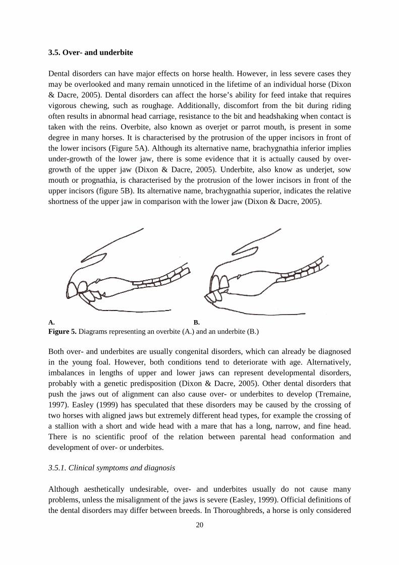
3.5. Over- and underbite Dental disorders can have major effects on horse health. However, in less severe cases they may be overlooked and many remain unnoticed in the lifetime of an individual horse (Dixon & Dacre, 2005). Dental disorders can affect the horse’s ability for feed intake that requires vigorous chewing, such as roughage. Additionally, discomfort from the bit during riding often results in abnormal head carriage, resistance to the bit and headshaking when contact is taken with the reins. Overbite, also known as overjet or parrot mouth, is present in some degree in many horses. It is characterised by the protrusion of the upper incisors in front of the lower incisors (Figure 5A). Although its alternative name, brachygnathia inferior implies under-growth of the lower jaw, there is some evidence that it is actually caused by over-growth of the upper jaw (Dixon & Dacre, 2005). Underbite, also know as underjet, sow mouth or prognathia, is characterised by the protrusion of the lower incisors in front of the upper incisors (figure 5B). Its alternative name, brachygnathia superior, indicates the relative shortness of the upper jaw in comparison with the lower jaw (Dixon & Dacre, 2005).
A. B. Figure 5. Diagrams representing an overbite (A.) and an underbite (B.) Both over- and underbites are usually congenital disorders, which can already be diagnosed in the young foal. However, both conditions tend to deteriorate with age. Alternatively, imbalances in lengths of upper and lower jaws can represent developmental disorders, probably with a genetic predisposition (Dixon & Dacre, 2005). Other dental disorders that push the jaws out of alignment can also cause over- or underbites to develop (Tremaine, 1997). Easley (1999) has speculated that these disorders may be caused by the crossing of two horses with aligned jaws but extremely different head types, for example the crossing of a stallion with a short and wide head with a mare that has a long, narrow, and fine head. There is no scientific proof of the relation between parental head conformation and development of over- or underbites. 3.5.1. Clinical symptoms and diagnosis Although aesthetically undesirable, over- and underbites usually do not cause many problems, unless the misalignment of the jaws is severe (Easley, 1999). Official definitions of the dental disorders may differ between breeds. In Thoroughbreds, a horse is only considered
21
to be affected when there is no contact at all between the upper and lower incisors. Thus over- and underbites of some degree are allowed and not recorded. Other breeds have stricter regulations and a disorder is considered to be present in any case of misalignment of incisors (Tremaine, 1997). Diagnosis of the disorder is easy and only requires thorough visual inspection. Quantification may be facilitated by radiography of the head and measurement of jaw misalignment. Overbites can lead to restrictions when chewing and result in abnormal molar wear. The development of cheek teeth disorders such as sharp enamel hooks is common in these cases, which could cause abrasions in the mouth and problems with bitting. Some clinical symptoms of problems with cheek teeth could be slow eating, weight loss and discomfort caused by the bit (Tremaine, 1997). As these changes in cheek teeth occur deep in the mouth, the expertise of a veterinarian or an equine dentist would then be required for both diagnosis and therapy. Horses with overbites may be more prone to incisor related periodontal disease (Baker, 1999). In severe cases, the downward growth of the upper incisors can stun the growth of the lower jaw. Contact of the lower incisors with the upper palate may interfere with further extension of the lower jaw. Overbite and overbite-related problems may then worsen (Easley, 1999). Although underbite occurs less often and has been less intensely studied, similar functional effects and symptoms can be expected as for overbite. 3.5.2. Treatment In less severe cases, simple rasping of the protruding incisors and correcting corresponding molar abnormalities is sufficient. In cases of severe congenital overbite in young foals, it may be necessary to employ orthodontic therapy by using corrective orthodontic braces placed between the cheek teeth and the incisors (Dixon & Dacre, 2005). Inhibiting the growth of the protruding upper jaw in the young foal gives the lower jaw a chance to “catch up” (Baker, 1999). Severe cases can only be readjusted early in the life of the horse, during the phase of rapid growth and development. However, success of orthognathic surgery has been limited in the few cases referred to in the literature. Management should include prevention and correction of abnormal growth and wear of the teeth (Easley, 1999). 3.5.3. Prognosis and genetic implications Over- and underbite have been reported to more frequently occur in small horse breeds such as miniature horses and ponies, but relatively rarely in large horse breeds (Baker, 1999). Both these disorders are mainly cosmetic in nature, unless exhibited in severe forms. When the most severe forms are considered for classification, overbite has been suggested as an inherited lethal or semi-lethal disorder that should be considered as an unsoundness, although there does not seem to be agreement in this regard (Marks, 2000). Equine studies on dental abnormalities have found incidences of overbites to be in the order of 2% to 5% amongst the animals inspected (Uhlinger, 1987 and Duke, 1989 in Baker & Easely, 1999). Gift et al. (1991) found that male horses were 5.7 times more frequently affected by overbites than female horses. However, it must be taken into account that only 20 horses were included in the study. Due to the possible genetic predisposition of the dental disorders, it is debatable
22
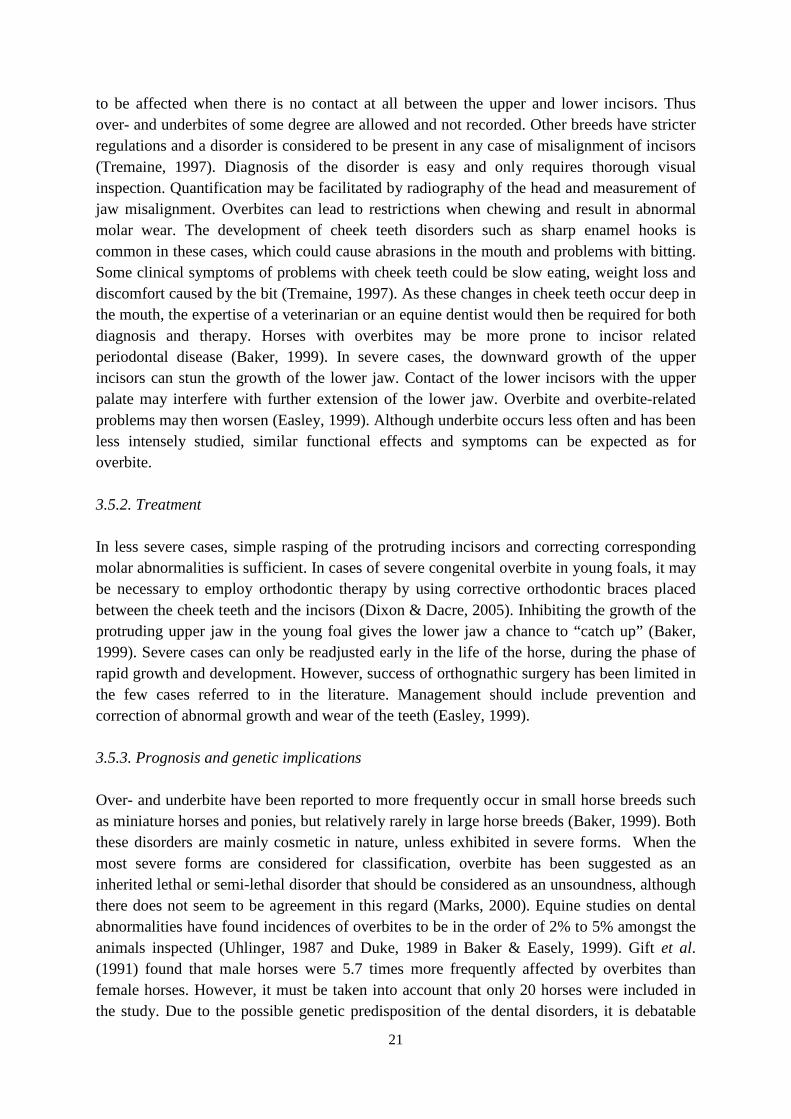
whether orthodontic treatment is ethical in animals used for breeding, without obligation of prior reporting. Some breeding associations exclude horses with this disorder from breeding, regardless of orthodontic correction having been undertaken (Dixon & Dacre, 2005). If orthodontic correction has been employed in a breeding stallion, ethical responsibilities of the stallion owner would require the dental disorder to be registered so that mare owners can assess this information when choosing a stallion to breed their mares with. 3.6. Bench knees Bench knees, also called offset knees, are characterised by the axial deviation of the carpal bones (Marks, 2000) so that the carpal bones do not align with the radius and metacarpal (cannon) bone when viewed from the front (Figure 6). However, alternative definitions exist that do not focus on the irregular conformation of the carpus, but the lacking alignment of the metacarpus with the radius (Stashak, 2002). This once again brings to light the need for standardised definitions of anatomical deviations and disorders to allow for comparisons between breeds and populations.
Figure 6: An illustration depicting correct conformation and a bench knee/offset knee conformation. 3.6.1. Clinical symptoms and diagnosis Clinical symptoms and diagnosis can be made by visual inspection of the horse’s limbs, when viewed from the front. Measurements can be taken, usually from photographs, to quantify the degree of axial rotation of the carpi. Offset measurements are usually represented as offset ratios and are calculated by drawing lines from the lateral (outside) and medial (inside) sides of the lower part of the radius and also along the lateral and medial sides of the metacarpal

23
bone (cannon bone). The distance between the two medial lines in relation to (divided by) the distance between the two lateral lines equals the offset ratio. An offset ratio greater than 1 represents bench knee conformation (McIlwraith, et al., 2003). Bench knees are most commonly found in combination with carpus valgus (knock-knees) (Stashak, 2002) or toe-in conformation (McIlwraith et al., 2003). 3.6.2. Treatment The treatment procedures depend mainly on the age of the affected horse. In young foals whose physeal growth plates have not yet closed, different therapeutic approaches exist. Alternatives include stall rest, splints and casts, hoof manipulation and finally surgery. As growth of bones is most active immediately after birth and each physeal region has an approximate and often predictable date when it closes, growth interventions must be made within this period before physeal growth plate closure. Within this period it must also be decided weather to intervene with corrective surgery or to wait and see if the deviations will resolve on their own (Auer & von Rechenberg, 2006). Stall rest can be an effective treatment of angular limb deformities (ALDs) in some cases although it is difficult to predict the outcome. Splints and casts have been successfully used on limbs of foals with incomplete ossification in carpal and tarsal bones. Corrective hoof trimming and hoof manipulation by application of foot plates or shoes with extensions is commonly used as a conservative treatment of ALDs in foals (Auer & von Rechenberg, 2006). In severe cases of ALD, in this case bench knees, corrective trimming may not represent an appropriate treatment, because it forces the limb into unnatural and uncomfortable positions which may predispose the horse to early degenerative changes in joint cartilage (Auer & von Rechenberg, 2006). Corrective surgery can be performed in different ways which generally rely on growth acceleration, growth retardation or a combination of both. Growth acceleration surgery to correct bench knee conformation involves periosteal transection and elevation (stripping) at the outside part of the lower radius and lower inside part of the third metacarpal (cannon bone) (Auer, 1985). In older horses with slight to moderate bench knee conformation, when growth cannot be manipulated any longer, adequate hoof care may be the method of choice. Regular corrective trimming or special shoes or foot plates may then be used. It has been stated that while axial deviation of the carpus alone cannot be corrected by the above mentioned methods, if axial deviation is present in combination with a varus deformity (an angular deviation), then treatment should primarily focus on the more important varus deformity (Bramlage & Auer, 2006). 3.6.3. Prognosis and genetic implications The prognosis is good if the deviation is slight to moderate but it may affect performance due to abnormal stress on other part of the supportive tissues. Bench knee conformation is considered a weak conformation as additional weight is placed on the medial splint bone which already carries more weight than the lateral splint bone in horses with regular limb conformation. The additional stress exerted on the interosseous ligaments increases the risk of the development of splints (Stashak, 2002). Several studies have found significant

24
associations between offset knee ratios and musculoskeletal injuries. Offset knees have long been associated with an increased predisposition to injuries in Thoroughbred race horses (McIlwraith et al., 2003). It was also found that for every 10% increase in the carpal offset ratio, the risk of swelling in the front fetlock joint was increased by a factor of 1.18 and the risk of front fetlock problems increased by a factor of 1.26 (McIlwraith et al., 2003; Anderson et al., 2004). It may therefore be logical to assume than when front limbs become more offset, tension or compression increases distally from the carpus and pain and swelling would occur because of the imbalance in distribution of forces acting on the limb (McIlwraith, et al., 2003). The heritability of bench knee conformation has not yet been estimated although studies on conformation have noted that in some breeds or populations, bench knee conformation is relatively common. Holmström et al. (1990) found the prevalence of bench knee conformation to be 60% in Swedish warmblood sport horses. 3.7. Calf-knees Calf knee conformation, also known as back at the knee, hyperextended knee or sheep knee, is characterised by the backward (palmar) deviation of the carpal bones (Stashak, 2002) (Figure 7). It is considered to be a weak conformation and lameness usually presents in affected limbs when heavy work is performed (Stashak, 2002).
Figure 7: An illustration depicting correct conformation and a calf knee/back at the knee conformation.
25
3.7.1. Clinical symptoms and diagnosis Clinical symptoms and diagnosis can be made from direct observations of the horse’s limbs, when viewed from the side. By measuring radiometacarpal angles when the limb is viewed from the side, calf knee conformation can be characterised by any angle of less than 180° (McIlwraith et al., 2003) (Figure 7). 3.7.2. Treatment In a study of conformational changes in young Thoroughbred horses between weaning and 3 years of age, it was found that carpal conformation tends to progressively change from back at the knee (calf knee) to slightly over in the knee (bucked knee). It is therefore likely that the conformation of a foal that is born back at the knee will improve with age (McIlwraith et al., 2003). Once again, it is therefore important to know when it is appropriate to allow self-correction to occur or when to intervene. There is sparse information available on corrective treatment possibilities for this deviation but perhaps similar strategies can be employed as mentioned in the earlier section about treatment of bench knee conformation. 3.7.3. Prognosis and genetic implications The calf knee conformation places increased strain on the ligaments supporting the carpal bones and causes compression on the dorsal section of the carpal bones (Stashak, 2002). There is a belief that calf knee conformation is especially detrimental when horses are working at speed, so that especially racing horses with this conformation are predisposed to carpal chip fractures (Marks, 2000; Stashak, 2002). In addition, Dolvik & Klemetsdal (1994) showed that there was a significant effect of calf knee conformation, amongst other conformations including standing under in front, toe out and broken hoof-pastern axis in the hind limb, with the prevalence of carpitis (arthritis of the carpal joint) in Norwegian cold-blooded trotters. However, in an English study of 21 Thoroughbred racing horses with carpal chip fractures, no significant correlation was found between calf knee conformation and carpal chip fractures (Barr, 1994). This is at odds with the experience and views of many North American racing trainers and veterinarians who consider calf knee conformation a serious problem that possibly contributes to the rather high incidence of carpal fractures and joint disease (Marks, 2000). Marks (2000) has suggested that the conflicting results may relate to the differences in American and European track surfaces, with American dirt track surfaces possibly stressing the carpi more than European turf surfaces. When estimating the heritability of conformational deviations that significantly affect the prevalence of carpitis in Norwegian cold-blooded trotters, Dolvik & Klemetsdal (1999) found that only calf knee conformation showed a significant heritability (0.42), and thus selection against this trait should be successful. However, calf knee conformation may also be acquired, as changes or injury to the soft supporting tissues at the back of the carpal joint may cause this conformation (Barr, 1994).

26
3.8. Bucked knees Bucked knee conformation, also known as over in the knees, forward at the knee, knee sprung or goat knees, is characterised by the forward (dorsal) deviation of the carpal bones (Stashak, 2002) (Figure 8). Although also considered as a weak conformation, it is generally perceived as being less detrimental than calf knee conformation (Stashak, 2002).
Figure 8: An illustration depicting correct conformation and a bucked knee/forward at the knee conformation. 3.8.1. Clinical symptoms and diagnosis Clinical symptoms and diagnosis can be made from direct observations of the horse’s limbs, when viewed from the side. When the radiometacarpal angle is measured from the side, an angle greater than 180° characterises bucked knee conformation (McIlwraith et al., 2003) (Figure 8). Congenital forms of the disorder are usually bilateral and may occur in combination with knuckling of the fetlock joints. Bucked knee conformation can be also be caused by contraction of the tendons of the carpal flexor muscles (Stashak, 2002). 3.8.2. Treatment Therapeutic approaches to bucked knee conformation are the same as for the other forms of conformational faults in the carpal joints (see sections 3.6.2 and 3.7.2).
27
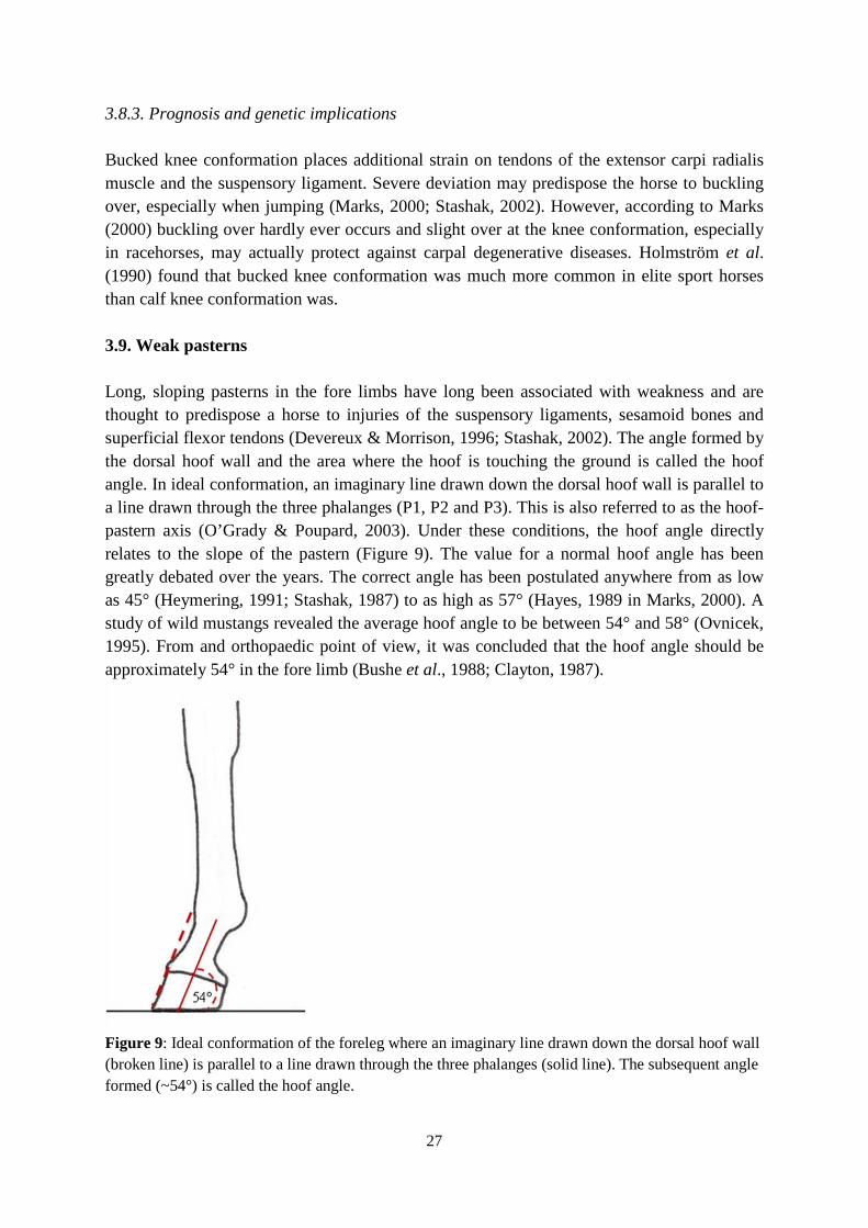
3.8.3. Prognosis and genetic implications Bucked knee conformation places additional strain on tendons of the extensor carpi radialis muscle and the suspensory ligament. Severe deviation may predispose the horse to buckling over, especially when jumping (Marks, 2000; Stashak, 2002). However, according to Marks (2000) buckling over hardly ever occurs and slight over at the knee conformation, especially in racehorses, may actually protect against carpal degenerative diseases. Holmström et al. (1990) found that bucked knee conformation was much more common in elite sport horses than calf knee conformation was. 3.9. Weak pasterns Long, sloping pasterns in the fore limbs have long been associated with weakness and are thought to predispose a horse to injuries of the suspensory ligaments, sesamoid bones and superficial flexor tendons (Devereux & Morrison, 1996; Stashak, 2002). The angle formed by the dorsal hoof wall and the area where the hoof is touching the ground is called the hoof angle. In ideal conformation, an imaginary line drawn down the dorsal hoof wall is parallel to a line drawn through the three phalanges (P1, P2 and P3). This is also referred to as the hoof-pastern axis (O’Grady & Poupard, 2003). Under these conditions, the hoof angle directly relates to the slope of the pastern (Figure 9). The value for a normal hoof angle has been greatly debated over the years. The correct angle has been postulated anywhere from as low as 45° (Heymering, 1991; Stashak, 1987) to as high as 57° (Hayes, 1989 in Marks, 2000). A study of wild mustangs revealed the average hoof angle to be between 54° and 58° (Ovnicek, 1995). From and orthopaedic point of view, it was concluded that the hoof angle should be approximately 54° in the fore limb (Bushe et al., 1988; Clayton, 1987).
Figure 9: Ideal conformation of the foreleg where an imaginary line drawn down the dorsal hoof wall (broken line) is parallel to a line drawn through the three phalanges (solid line). The subsequent angle formed (~54°) is called the hoof angle.

28
3.9.1. Clinical symptoms and diagnosis Long, sloping pasterns are characterised by hoof angles of less than 45° in the fore limb and angles of less than 50° in the hind limb, coinciding with abnormal length of the first phalanx (the long pastern bone) (Figure 10) (Devereux & Morrison, 1996). Long pasterns may also have a normal or upright conformation (Stashak, 2002). Sloping of the pasterns alone can also be attributed to long toe and collapsed heel conformation caused by infrequent or incorrect trimming of hooves (Devereux & Morrison, 1996).
Figure 10: An example of a long, sloping pastern. Note the hoof angle (<45°) and the broken hoof-pastern axis. 3.9.2. Treatment Nothing can be done to alter the abnormal length of the pastern bone. However, corrective shoeing can to some degree alleviate the slope. Trimming the toe, possibly in combination with raising the heel may be used to achieve a slightly more upright hoof angle. Excessive corrections should be avoided particularly in the adult horse in order to avoid problems caused by sudden changes of load and distribution in the distal limb (Auer & von Rechenburg, 2006). 3.9.3. Prognosis and genetic implications Long sloping pasterns have also been named as potential causes of carpal chip fractures (Barr, 1994), and long pasterns were found to increase the risk of fore limb fractures (McIlwraith et al., 2003). However, some positive aspects of long, sloping pasterns have been suggested. More intense use of the suspensory structures (such as ligaments) may be benefited by long sloping pasterns for work on hard ground (Hayes, 1952 in Marks, 2000) and may facilitate in achieving the level of suspension and cadence required of elite dressage horses (Marks, 2000). Dolvik & Klemetsdal (1999) found that the prevalence of sloping pasterns in Norwegian cold-blooded trotters was 35.2%. However, the estimated heritability

29
was very low (0.09) in this study. This could be due to this trait being more affected by environmental factors such as hoof trimming and shoeing. According to Stashak (2002), both long and short upright pasterns are also undesirable as they may predispose to traumatic arthritis of the fetlock and phalanges and to navicular disease, although the trauma to the pastern being possibly worse with short upright pastern than with long, upright pastern conformation. 3.10. Toe out Toe out or splay footed conformation is characterised by the axial rotation of the phalanges laterally (outwards), originating from the fetlock joint (Bramlage & Auer, 2006) (Figure 11). Most foals with valgus deformities (lateral deviation of the limb below the location of the deformity, also called knock knees) additionally have toe out conformation (Figure 12) (Bramlage & Auer, 2006).
Figure 11: An illustration depicting correct conformation and a toe out conformation.

30
Figure 12: An illustration depicting valgus in combination with toe out conformation. 3.10.1. Clinical symptoms and diagnosis Clinical symptoms and diagnosis can be made from direct observations of the horse’s limbs, when viewed from the front. Toe out conformation is then characterised by a pastern angle greater than 180° (McIlwraith et al., 2003). Toe out conformation may be accompanied by either base-wide or base-narrow conformation. Base-narrow conformation is present when the distance between the hooves at the ground is smaller than the distance between the upper limbs at their point of origin at the shoulder, while base-wide conformation is present when this distance is larger (Stashak, 2002). Toe out, when combined with base-narrow conformation, is one of the most detrimental types of deformities in the fore limb because it causes irregular load distribution and particularly places great strain on the structures below the fetlock (Stashak, 2002). 3.10.2. Treatment Neonatal foals often show some degree of outward rotation in both fore and hind limbs which may be accompanied by a valgus (knock kneed) conformation. This is because stabilisation of the musculoskeletal system in the newborn foal requires the stimuli of load and strain. Such deviations are usually only postural and therefore correct with age (McIlwraith et al., 2003; Bramlage & Auer, 2006). As the foal matures, gains strength and width in its chest, the conformation usually rotates inwards and correction of toe out deviation occurs (Bramlage & Auer, 2006). However, rotational deviations that occur below the carpus in combination with an inward or outward rotation of the fetlock cannot correct naturally (Stashak, 2002). In severe cases of angular deformities of the limb, such as valgus, surgical correction can be attempted at a young age. Less severe cases of toe out conformation may be managed or partially corrected by corrective trimming or shoeing in the foal (Stashak, 2002). It is

31
imperative that the hoof is properly balanced when trimming (Devereux & Morrison, 1996). Furthermore, trimming must not be so excessive that it places the growing limb in an abnormal position because this would affect the balance and regular growth of other supportive structures (Auer & von Rechenberg, 2006). 3.10.3. Prognosis and genetic implications Toe out conformation places excess strain on the medial side of the limb and may therefore predispose to the development of medial ringbone and sidebone (Devereux & Morrison, 1996). Horses with toe out conformation usually “wing” (swing their hooves in an arc inwards). Interference with the other limbs may occur, resulting in injuries (Stashak, 2002). In a study on conformation in Norwegian cold-blooded trotters, Dolvik & Klemetsdal (1999) found toe out conformation in the front limbs of 43.9% and in the hind limbs of 67.5% of the horses. They also estimated the heritabilities, which ranged from 0.04 to 0.11. In the study of conformation and musculoskeletal problems in racing Thoroughbreds, Anderson et al., (2004) found that the majority of the 115 3-year old horses used in the study had slight toe-out conformation. A study on conformation of Swedish warmblood sport horses revealed that less than 5% of the horses showed toe out conformation (Holmström et al., 1990). Both the Norwegian trotters from the study of Dolvik & Klemetsdal (1999) and the Swedish warmblood horses from the study of Holmström et al. (1990) were four years of age or older. This nicely illustrates the differences in conformation between breeds and sport horse types. 3.11. Toe in Toe in or pigeon toed conformation is characterised by the axial rotation of the phalanges medially (inwards), originating from the fetlock joint (Bramlage & Auer, 2006) (Figure 13). Most foals with varus deformities (medial deviation of the limb below the location of the deformity, also called bowlegs) (Figure 14) additionally have toe in conformation (Bramlage & Auer, 2006).

32
Figure 13: An illustration depicting correct conformation and a toe in conformation.
Figure 14: An illustration depicting varus in combination with toe in conformation. 3.11.1. Clinical symptoms and diagnosis Clinical symptoms and diagnosis can be made from direct observations of the horse’s limbs, when viewed from the front. Toe in conformation is then characterised by a pastern angle of less than 180° (McIlwraith et al., 2003). Toe in conformation is more often accompanied by base-narrow conformation than by base-wide conformation (Stashak, 2002).
33
3.11.2. Treatment In young foals, toe in conformation can be managed by corrective trimming. Later corrective shoeing of the young horse may prevent the condition from becoming worse. In young foals with an accompanying varus (bowlegged) angular limb deformity, surgery may be attempted (Stashak, 2002) because of the tendency towards inward rotation of the toe in the young foal (McIlwraith et al., 2003). In less sever cases of toe in conformation, it is again imperative that the hoof is properly balanced when trimming (Devereux & Morrison, 1996). Furthermore, trimming must not be so excessive that it places the growing limb in an abnormal position as this would affect the balance and growth of other supportive structures (Auer & von Rechenberg, 2006). 3.11.3. Prognosis and genetic implications Toe in conformation places excessive strain on the lateral side the limb and may therefore predispose to the development of lateral ringbone and sidebone (Devereux & Morrison, 1996). Horses that are toed in also tend to “paddle” (swing their hooves in an arc outwards). Horses with a combination of varus and toe in conformation, tend to actually paddle with additional inwards swinging of the hooves which may cause interference and injuries to the opposite limb, especially at the fetlock joint (Stashak, 2002). In horses that are base narrow and toed in, excessive strain occurs on the lateral supportive structures of the carpus, fetlock and phalanges (Stashak, 2002). In Norwegian cold-blooded trotters, toe in conformation was seen in 25.8% of horses studied, whereas out of 356 Swedish warmblood sport horses, 50% had mild to moderate toe in conformation, which did not seem to affect their performance (Holmström et al., 1990). 3.12. Abnormal bone formations in the hock Bony abnormalities in the hock may originate from proliferation of bone or abnormal tissue growth (Figure 15).
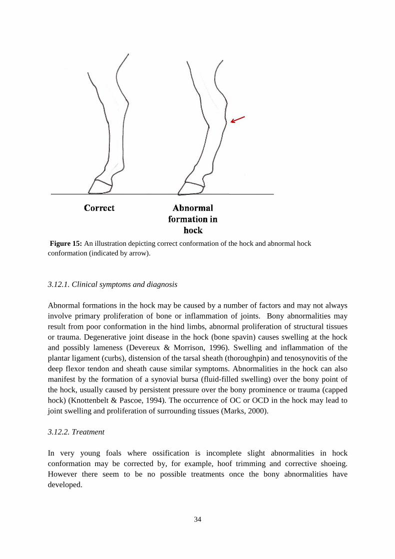
34
Figure 15: An illustration depicting correct conformation of the hock and abnormal hock conformation (indicated by arrow). 3.12.1. Clinical symptoms and diagnosis Abnormal formations in the hock may be caused by a number of factors and may not always involve primary proliferation of bone or inflammation of joints. Bony abnormalities may result from poor conformation in the hind limbs, abnormal proliferation of structural tissues or trauma. Degenerative joint disease in the hock (bone spavin) causes swelling at the hock and possibly lameness (Devereux & Morrison, 1996). Swelling and inflammation of the plantar ligament (curbs), distension of the tarsal sheath (thoroughpin) and tenosynovitis of the deep flexor tendon and sheath cause similar symptoms. Abnormalities in the hock can also manifest by the formation of a synovial bursa (fluid-filled swelling) over the bony point of the hock, usually caused by persistent pressure over the bony prominence or trauma (capped hock) (Knottenbelt & Pascoe, 1994). The occurrence of OC or OCD in the hock may lead to joint swelling and proliferation of surrounding tissues (Marks, 2000). 3.12.2. Treatment In very young foals where ossification is incomplete slight abnormalities in hock conformation may be corrected by, for example, hoof trimming and corrective shoeing. However there seem to be no possible treatments once the bony abnormalities have developed.

35
3.12.3. Prognosis and genetic implications According to Marks (2000), there are many largely unsupported statements about good or bad hock conformation. Hormones are involved to a large degree in the process of bone formation and remodelling (Amizuka et al., 1994). Combinations of hormonal, nutritional and conformational factors may therefore be involved in the development of abnormal hock formations. The role of genetics has not been quantified yet. 3.13. Outward rotation of limbs Outward rotation of the limb is present when the bones in the whole limb do not face forward, but outwards, when the horse is viewed from the front (Figure 16).
Figure 16: An illustration depicting correct conformation and outward rotation of the legs. 3.13.1. Clinical symptoms and diagnosis The outward rotation of the whole fore- or hind limb is characterised by carpal/tarsal joints, metacarpal/-tarsal bones, fetlock joints and phalanges facing outwards when the horse is viewed from the front. Affected horse are often falsely classified as having toe out conformation, although rotation of the limb occurs at the attachment point of the chest and not just at the fetlock joint (Stashak, 2002).

36
3.13.2. Treatment Although surgical treatment may be an option at a young age, before the closing of the physeal growth plates (Bramlage & Auer, 2006; Auer & von Rechenberg, 2006), complete correction of rotational deviations is unlikely to be achieved (Stashak, 2002). 3.13.3. Prognosis and genetic implications Dolvik & Klemetsdal (1999) reported that 27.7% of the Norwegian cold-blooded trotters in their study had their carpi rotated outwards, and they estimated a heritability of 0.16 for this trait. According to Marks (2000), correct conformation of the hind limbs ritually described in literature and depicted on illustrations as facing straight forwards may be very rare and almost all horses may show some outward rotation in the hind limbs. 4. Summary It can be concluded from literature that skeletal disorders result from a complex interaction between factors such as genetics, nutrition, exercise, and trauma. Furthermore, as yet unknown factors also play a role in development of normal versus abnormal conformation. In many cases, treatment or correction of skeletal disorders is possible, but because of the possible transmission to future generations, such animals should not be bred with or at least used very minimally in breeding. This is especially important if there is solid scientific evidence that the disorders predisposes a horse to injuries. Many of the conformational deviations are not lethal, but they increase the odds of fractures or injuries in limbs and joints. Although the type of work the horse is being used for impacts the definite prevalence of conformation-related injuries, breeding measures should be taken. Certain breeds and populations used for similar activities tend to share certain conformational deviations. This could be due to use-related adaptations in the skeleton in response to the activity the horse performs or direct or indirect selection for such conformations. Relationships between conformation and performance need to be investigated across breeds and sport types, in order to determine which deviations are most detrimental. A list and description of selected inherited disorders that have been described in sport horses is shown in the tables below, together with available heritabilities and frequencies of occurrence. In most cases, the prevalence, i.e. the proportion of individuals of a population that are affected by the disorder at a specific time, is given because most studies are usually performed in a population or breed of horses over a specific time period. In some cases, where the prevalence is unknown, the incidence, i.e. rate of occurrence of new cases in a population over a specific time period, is listed. Emphasis is placed on research done in warmblood horses although other breeds have been included, where this information is lacking. Degenerative joint disorders, originating from either bone or cartilage are given in Table 1. Skeletal disorders including common conformational deviations are given in Table 2. In Table 3 specific abnormalities and disorders of the hoof are given. In Table 4 disorders of the reproductive system and in Table 5 other disorders (including respiratory, skin,
37
muscular and other disorders) are mentioned. Indicated by the number of references, some disorders have been extensively studied while other disorders lack extensive scientific research. Furthermore, the amount of information on disorders shows large variation between breeds. For example, many scientific studies and research papers have been written about skeletal disorders in racing horses (both trotters and gallopers) but relatively little research has been done on riding horses. The use of multiple names for the same disorders and differences between definitions of disorders interfere with direct comparisons between study results and the composition of detailed lists of disorders with proven or suspected genetic backgrounds.

38
Table 1: A summary of the names, description and known heritability (h2), frequency (incidence or prevalence) of degenerative joint disorders Disorder Alternative name Description h2 Frequency Bone spavin Degenerative joint disease of hock; usually affects the distal intertarsal and
tarsometatarsal joints; caused by wear and tear of joints and ligaments of hock and poor conformation of the hind limbs. Causes lameness and swelling at the hock.
Estimated between 0.02-0.65 across range of breeds (1)
Patella dislocation
Patella locking, upward fixation of the patella
Temporary locking of the stifle joint resulting in the fixation of the hind limb in an extended position, ranging between a few strides to a few hours. Most common in young, unfit or poorly muscled horses. In severe cases, it can cause chronic inflammation in the stifle joint, lameness and development of degenerative joint disease.
Unknown but suspected
Osteochondrosis (OC) and Osteochondritis dissecans (OCD)
Abnormal cellular differentiation of cartilage into bone due to failure in endochondral ossification. Results in failure of bone formation and necrosis of the retained cartilage fragments, causing lesions in the cartilage. Most often occurs in epiphyseal articular cartilage and progressive degeneration of cartilage causes joint weakness, inflammation and pain. Most often affects shoulder, stifle, hock, carpus, fetlock and cervical vertebral joints. A multifactorial trait with contributing factors such as diet, exercise, growth, body size, trauma and genetics. Considered as a manifestation of progressive OC. OCD results when cartilage or bone detaches (either completely or partly) from the articular surface. If detachment is complete, the fragment may move within the joint, possibly resulting in synovitis (inflammation of synovial membrane) and pain.
Estimated between 0.02-0.64 across range of breeds (2)
Prevalence across European warmblood breeds of about 15-36% (3, 4, 5)
Cervical vertebral malformation (CVM)
Wobbler syndrome, cervical stenotic myelopathy, cervical vertebral instability
Abnormalities of the cervical vertebrae which include thickening of the soft tissues and proliferation of bone surrounding affected intervertebral joints result in narrowing of the vertebral canal and compression of the spinal cord. Problems in neurological function of the limbs manifests as ataxia. In cases of prolonged compression, irreversible damage to the spinal cord occurs.
Unknown but suspected
(1) - Winter et al., 1996; Willms et al., 1999; Bjørnsdøttir et al., 2000 (2) - Schougaard et al., 1987; Grøndalh & Dolvik, 1993; Philipsson et al., 1993; Winter et al., 1996; Willms et al., 1999; Pieramati et al., 2003; van Grevenhof et al., 2009 (3) - Studer et al., 2007 (4) - van Grevenhof et al., 2009 (5) – Stock et al., 2005
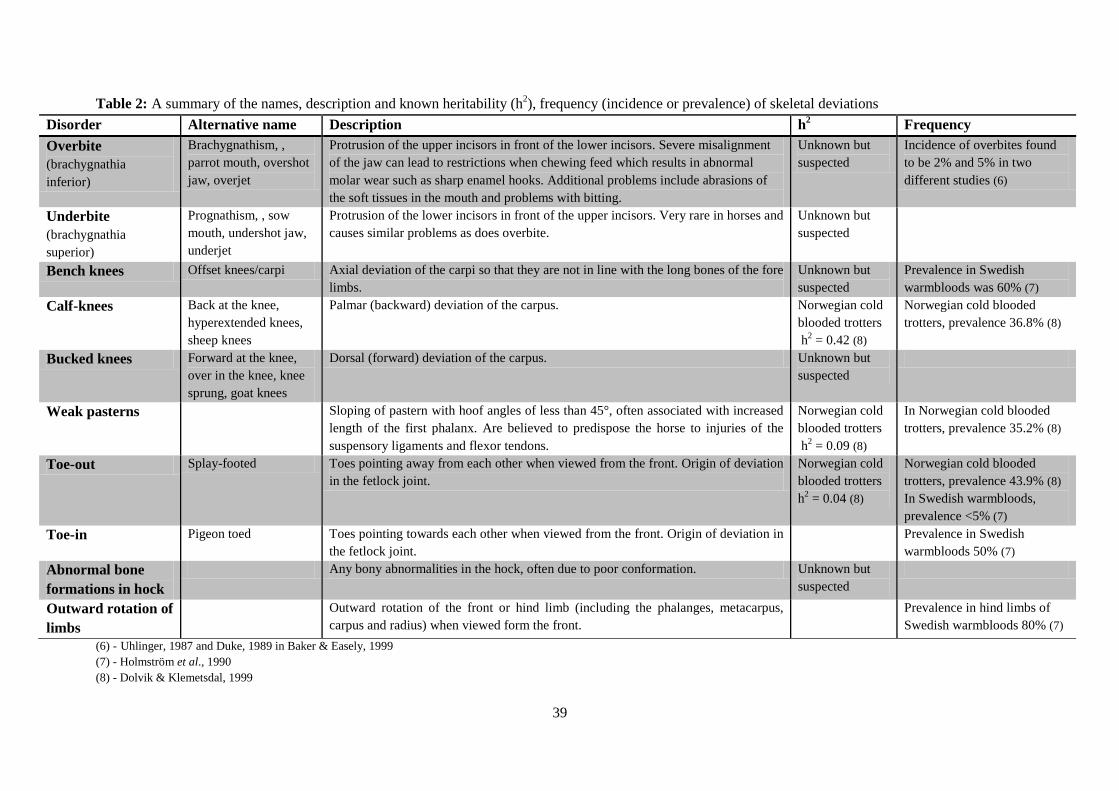
39
Table 2: A summary of the names, description and known heritability (h2), frequency (incidence or prevalence) of skeletal deviations Disorder Alternative name Description h2 Frequency Overbite (brachygnathia inferior)
Brachygnathism, , parrot mouth, overshot jaw, overjet
Protrusion of the upper incisors in front of the lower incisors. Severe misalignment of the jaw can lead to restrictions when chewing feed which results in abnormal molar wear such as sharp enamel hooks. Additional problems include abrasions of the soft tissues in the mouth and problems with bitting.
Unknown but suspected
Incidence of overbites found to be 2% and 5% in two different studies (6)
Underbite (brachygnathia superior)
Prognathism, , sow mouth, undershot jaw, underjet
Protrusion of the lower incisors in front of the upper incisors. Very rare in horses and causes similar problems as does overbite.
Unknown but suspected
Bench knees Offset knees/carpi Axial deviation of the carpi so that they are not in line with the long bones of the fore limbs.
Unknown but suspected
Prevalence in Swedish warmbloods was 60% (7)
Calf-knees Back at the knee, hyperextended knees, sheep knees
Palmar (backward) deviation of the carpus. Norwegian cold blooded trotters h2 = 0.42 (8)
Norwegian cold blooded trotters, prevalence 36.8% (8)
Bucked knees Forward at the knee, over in the knee, knee sprung, goat knees
Dorsal (forward) deviation of the carpus. Unknown but suspected
Weak pasterns Sloping of pastern with hoof angles of less than 45°, often associated with increased length of the first phalanx. Are believed to predispose the horse to injuries of the suspensory ligaments and flexor tendons.
Norwegian cold blooded trotters h2 = 0.09 (8)
In Norwegian cold blooded trotters, prevalence 35.2% (8)
Toe-out Splay-footed Toes pointing away from each other when viewed from the front. Origin of deviation in the fetlock joint.
Norwegian cold blooded trotters h2 = 0.04 (8)
Norwegian cold blooded trotters, prevalence 43.9% (8) In Swedish warmbloods, prevalence <5% (7)
Toe-in Pigeon toed Toes pointing towards each other when viewed from the front. Origin of deviation in the fetlock joint.
Prevalence in Swedish warmbloods 50% (7)
Abnormal bone formations in hock
Any bony abnormalities in the hock, often due to poor conformation. Unknown but suspected
Outward rotation of limbs
Outward rotation of the front or hind limb (including the phalanges, metacarpus, carpus and radius) when viewed form the front.
Prevalence in hind limbs of Swedish warmbloods 80% (7)
(6) - Uhlinger, 1987 and Duke, 1989 in Baker & Easely, 1999 (7) - Holmström et al., 1990 (8) - Dolvik & Klemetsdal, 1999

40
Table 3: A summary of the names, description and known heritability (h2), frequency (incidence or prevalence) of hoof disorders and abnormalities Disorder Alternative name Description h2 Frequency Abnormal hoof shape and quality
Any abnormality in the shape, size and quality of the hoof. Examples are clubfoot (shapes), crumbling, weak and cracking hooves (quality).
Hereditary nature of undesirable traits pertaining to hoof quality in Lipizzaners (9)
Navicular disease
Podotrochleitis Progressive degenerative disease that affects the navicular bone, the navicular bursa and the deep digital flexor tendon. Cause is uncertain as many factors may contribute to the disease but suggested causes include repeated compression of cartilage surrounding the navicular bone resulting in degeneration, excessive strain and inflammation of supporting ligaments and abnormal hoof formation. All results in lameness.
Estimates of h2 in warmbloods range between 0.06-0.31 (10)
Prevalence in Hanoverian warmbloods 21.7% (11)
Sidebone Ossification of collateral cartilage/hoof cartilage
Ossification of the cartilage attached to the pedal bone caused by concussion or direct injuries to the cartilage. Risk is higher in the presence of poor conformation. Inflammation initially causes pain and lameness but this usually subsides when the inflammation disappears.
Estimates in front feet of Finnhorses ranged between 0.3-0.50 (12)
Prevalence of 95% in heavy draft horses and 18% in warmboods (13) Prevalence of 80% in cold blooded Ardenner horses (14)
Laminitis Founder Changes in blood circulation in the hoof cause inflammation and congestion of the sensitive laminae surrounding the pedal bone. Lack of oxygen supply results in death of the laminae and subsequent loosening and rotation of the pedal bone within the hoof. Caused by a number of factors, including diet, toxins, exercise and management. Results in a characteristic stance with most weight on the heels and reluctance to move.
Unknown but suspected
(9) - Josseck, 1995 (10) - Winter et al., 1996; Willms et al., 1999 (11) - Stock & Distl, 2005 (12) – Ruohoniemi et al., 2003 (13) - Verschooten, 1994 (14) - Tullberg, 2006

41
Table 4: A summary of the names, description and known heritability (h2), frequency (incidence or prevalence) of reproductive disorders Disorder Alternative name Description h2 Frequency Cryptorchidism Hidden testes Failure of one or both testes to descend to the scrotum. Disturbance of the
complex process of testicular descent is considered multifactorial, although direct cause is unknown.
Heritable with references to it being dominant (15) as well as autosomal recessive although recent molecular analysis suggests a polygenic inheritance (16)
Position and texture disorders of testes
Any abnormalities in the size, position or texture of the testes. Cases such as rotation of the testes, microorchidism, anorchidism etc.
Unknown but suspected
Fertility disturbance and dysfunction
Disturbances in fertility such as low number of total motile sperm in ejaculate, abnormal sperm morphology or other dysfunctions.
Unknown but suspected. Molecular markers associated with fertility may have been found (17) Fertility has very low h2 in other species
Hernia scrotalis Scrotal hernia, inguinal hernia
Part of the intestine enters through an abnormally large inguinal canal (separates the abdominal cavity and the scrotum) and possibly slips down into the scrotum. Development may cause pain and colic-like symptoms due to compression and obstruction of the intestine. Usually also causes enlargement and edema of the scrotum.
Higher incidence in Stadardbred and Quarter horses (18)
(15) - Hayes 1986 in Lu, 2005 (16) - Diribarne et al., 2009 (17) - Hamann et al., 2007 (18) - Knottenbelt & Pascoe, 1994

42
Table 5: A summary of the name and or alternate names, the description and known heritability (h2), frequency (incidence or prevalence) of other disorders, including respiratory, skin, muscular and other disorders
Disorder Alternative name Description h2 Frequency Rhabdomyolysis (Exertional)
Tying-up syndrome, set-fast, Monday morning disease, Azoturia
Dysfunction and breakdown of muscle cells in working muscle groups such as the rump, thigh, back and shoulder muscles caused by inadequate blood flow which causes muscles to work anaerobically. Inflammation results from cell damage as well as painful muscle spasms. Causes are complex and not fully understood but include dietary, exercise, hormonal and environmental factors. Strongly related to polysaccharide storage myopathy (PSSM).
h2 = 0.42-0.43 estimated in Thoroughbreds (19) Autosomal dominant inheritance suggested (20)
Almost 50% of warmblood muscle biopsies positive for PSSM in horses with neuromuscular disorders (21)
Allergic eczema Sweet itch, culicoides hypersensitivity, summer eczema
Allergic skin condition caused by hypersensitivity to a protein in the saliva of insects from the Culicoides genus (biting midges). Results in itch mostly around the mane and tail areas and causes the horse to rub these areas on any available surface and in severe cases can result in open wounds.
Swedish-born Icelandic horses h2 = 0.26 (22) Shetland ponies and Friesian horses h2 = 0.06 (23)
Prevalence in Swedish-born Icelandic horses 8% (22)
Laryngeal hemiplegia
Hemiplegia laryngis sinistra, roaring, whistling
Degeneration of the laryngeal nerve (most commonly the left one) at the entrance of the trachea. The muscles controlled by the damaged nerve undergo atrophy and the larynx no longer opens fully. This obstructs the airflow into the trachea during fast exercise and horses make an abnormal noise when they breathe in. The noise can resemble a high pitched whistle or a roaring sound.
Estimated h2 of 0.50 in German riding horses (24)
Prevalence was 1.2% in Swiss warmbloods (25) In a German study, 25% of foals and 50% of adult horses were affected (24)
Chronic obstructive pulmonary disease (COPD)
Recurrent airway obstruction (RAO), heaves, broken wind, emphysema, chronic bronchitis, small airway disease
Hypersensitivity to inhaled fungal spores or other allergens (dust, pollen, etc.). Some of these particles are small enough to pass through to the bronchioles, leading to inflammation, increased mucus production and spasm of smooth muscles in the walls of the airways, reducing their diameter. Symptoms are progressive and include increased effort in exhaling, coughing and milky white to thick yellow nasal discharge. Severely reduces working ability.
Incidence of COPD when both parents affected 69%; only one affected parent 50%; two healthy parents 17% (26)
Abdominal wall umbilicus
Umbilical hernia Protrusion of a loop of intestine or other abdominal organs at the umbilicus. The protrusion is usually covered with skin and subcutaneous tissue. Can result in strangulation of the intestine and other intestinal problems.
Unknown but suspected
(19) - Oki et al., 2005 (20) - Dranchak et al., 2005 (21) - McCue et al., 2006 (22) - Grandinson et al., 2006 (23) – Kapell, 2005; Ruyter, 2005 (24) - Deegen et al., 1995; (25) - Studer et al., 2007 (26) - Marti et al., 1991

43
5. Methods and Materials An online survey was created to study the monitoring and management strategies of inherited disorders in breeding stallions, young horses and foals in European warmblood breeds. In November 2008 the survey (Appendix A) was sent to 37 breeding organisations, veterinarians and equine breeding specialists in 29 European countries. The main questions asked were where and which inherited disorders were recorded, where the records were kept, who the responsible organisations were for reporting disorders to record keeping organisations, how data on inherited disorders was summarised and evaluated and if such data was subsequently published. The monitoring of fertility in stallions was also considered in this context. The countries were further asked to comment on any health- or conformation-related restrictions on the registration and breeding of mares. The management of specific disorders in breeding stallions, both with a known and a suspected inheritance pattern was studied. The countries were asked if they scanned or examined for (screened for) a list of 29 specific disorders and to what level the respective disorders were considered in the selection process of breeding stallions. There were four levels of consideration of the disorders: (1) not considered at all; (2) stallion excluded from breeding only if severely affected and if phenotype affects performance; (3) considered/reported but can be compensated for with good performance and (4) stallion automatically excluded from breeding. The disorders were divided into skeletal and joint, hoof, muscular, respiratory, skin, reproductive and other disorders (see Appendix A). The results were based on replies from 11 European countries, meaning a reply rate of 38% when referring to the countires and 30% when referring to the breeding organisations contacted. Although the reply rate was not very high, the organisations that answered did represent a considerable proportion of warmblood horses in Europe, producing approximately 31 353 warmblood foals per year (Table 6). The 11 replying organisations from 11 different countries answered most of the questions in the survey. However, the rate of answering was lower in the questions referring to consideration of specific disorders in breeding stallions. 6. Results The results are divided into two sections: the first part summarises the management of inherited disorders in breeding stallions, breeding mares, young horses and foals in each country (Tables 7-10) while the second part summarises the number of countries that screened for specific inherited disorders and to what level each of the disorders is considered when registering a stallion for breeding (Figures 18-21). Figure 17 depicts an illustration of the flow of information when recording and evaluating disorders in stallions, young horses and foals occurs as well as the questions raised in this survey. The headings in italics correspond to the headings in Tables 7-10. In the second part, some countries consistently did not answer the questions, and this is represented in the figures too.
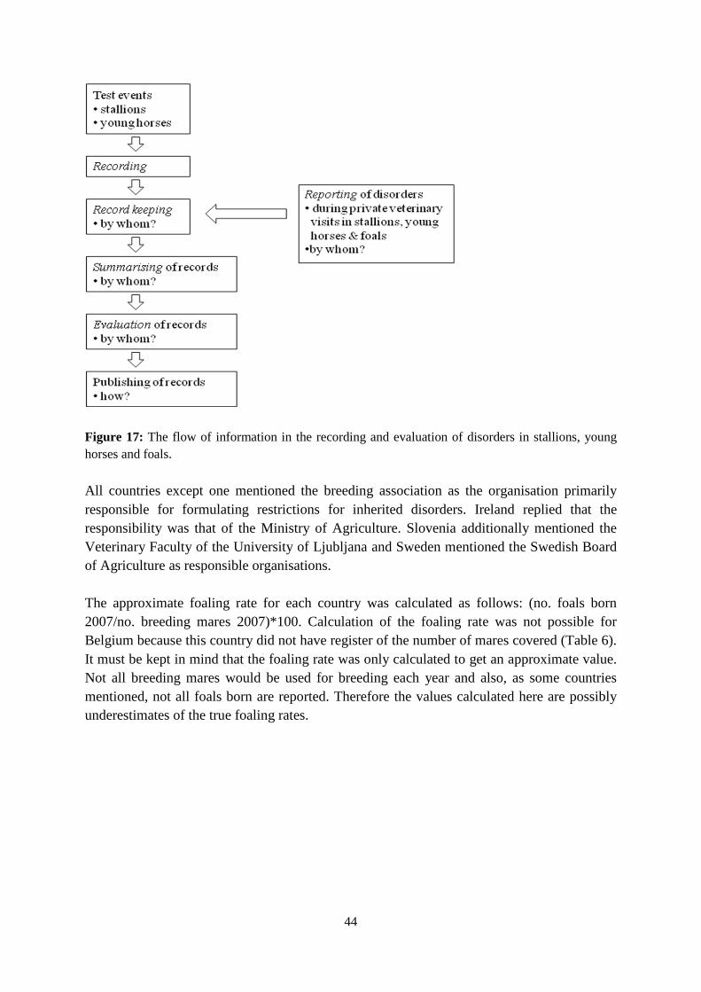
44
Figure 17: The flow of information in the recording and evaluation of disorders in stallions, young horses and foals. All countries except one mentioned the breeding association as the organisation primarily responsible for formulating restrictions for inherited disorders. Ireland replied that the responsibility was that of the Ministry of Agriculture. Slovenia additionally mentioned the Veterinary Faculty of the University of Ljubljana and Sweden mentioned the Swedish Board of Agriculture as responsible organisations. The approximate foaling rate for each country was calculated as follows: (no. foals born 2007/no. breeding mares 2007)*100. Calculation of the foaling rate was not possible for Belgium because this country did not have register of the number of mares covered (Table 6). It must be kept in mind that the foaling rate was only calculated to get an approximate value. Not all breeding mares would be used for breeding each year and also, as some countries mentioned, not all foals born are reported. Therefore the values calculated here are possibly underestimates of the true foaling rates.
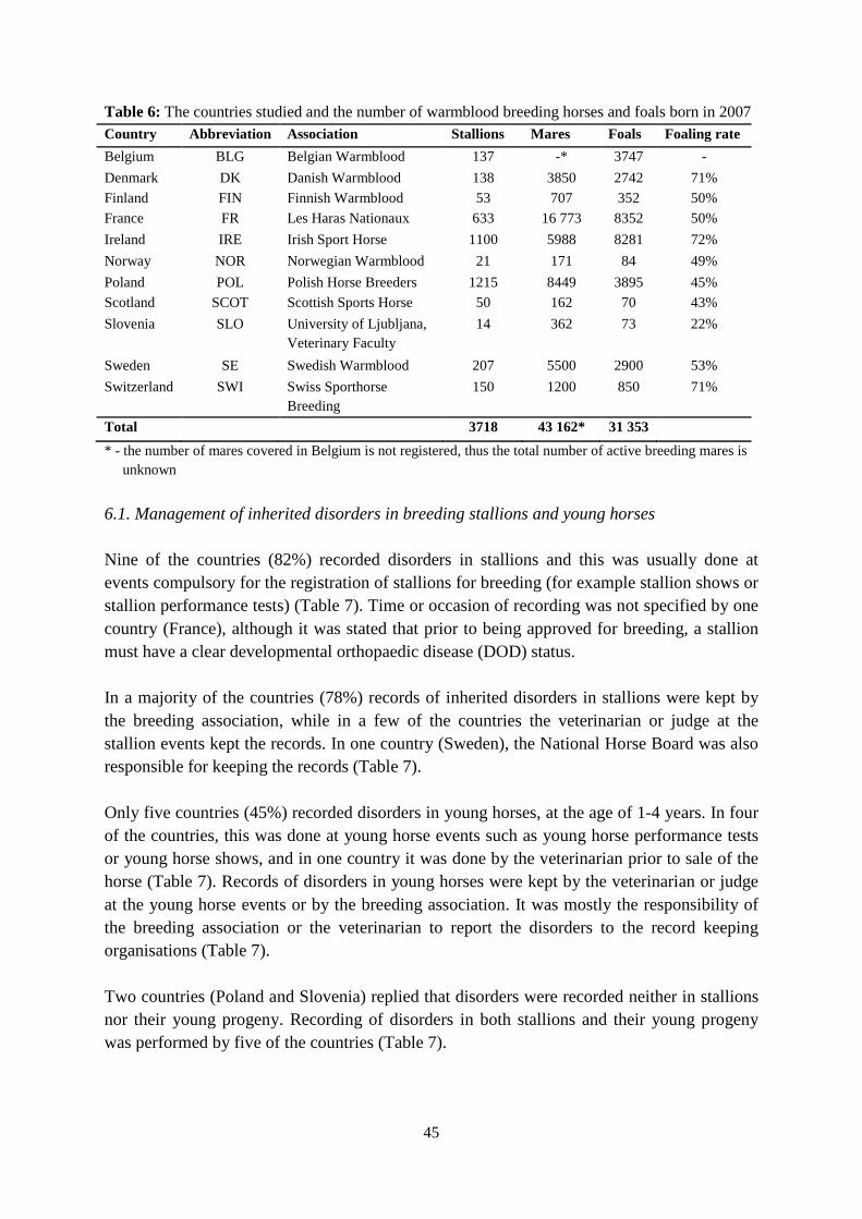
45
Table 6: The countries studied and the number of warmblood breeding horses and foals born in 2007 Country Abbreviation Association Stallions Mares Foals Foaling rate Belgium BLG Belgian Warmblood 137 -* 3747 - Denmark DK Danish Warmblood 138 3850 2742 71% Finland FIN Finnish Warmblood 53 707 352 50% France FR Les Haras Nationaux 633 16 773 8352 50% Ireland IRE Irish Sport Horse 1100 5988 8281 72% Norway NOR Norwegian Warmblood 21 171 84 49% Poland POL Polish Horse Breeders 1215 8449 3895 45% Scotland SCOT Scottish Sports Horse 50 162 70 43% Slovenia SLO University of Ljubljana,
Veterinary Faculty 14 362 73 22%
Sweden SE Swedish Warmblood 207 5500 2900 53% Switzerland SWI Swiss Sporthorse
Breeding 150 1200 850 71%
Total 3718 43 162* 31 353 * - the number of mares covered in Belgium is not registered, thus the total number of active breeding mares is unknown 6.1. Management of inherited disorders in breeding stallions and young horses Nine of the countries (82%) recorded disorders in stallions and this was usually done at events compulsory for the registration of stallions for breeding (for example stallion shows or stallion performance tests) (Table 7). Time or occasion of recording was not specified by one country (France), although it was stated that prior to being approved for breeding, a stallion must have a clear developmental orthopaedic disease (DOD) status. In a majority of the countries (78%) records of inherited disorders in stallions were kept by the breeding association, while in a few of the countries the veterinarian or judge at the stallion events kept the records. In one country (Sweden), the National Horse Board was also responsible for keeping the records (Table 7). Only five countries (45%) recorded disorders in young horses, at the age of 1-4 years. In four of the countries, this was done at young horse events such as young horse performance tests or young horse shows, and in one country it was done by the veterinarian prior to sale of the horse (Table 7). Records of disorders in young horses were kept by the veterinarian or judge at the young horse events or by the breeding association. It was mostly the responsibility of the breeding association or the veterinarian to report the disorders to the record keeping organisations (Table 7). Two countries (Poland and Slovenia) replied that disorders were recorded neither in stallions nor their young progeny. Recording of disorders in both stallions and their young progeny was performed by five of the countries (Table 7).
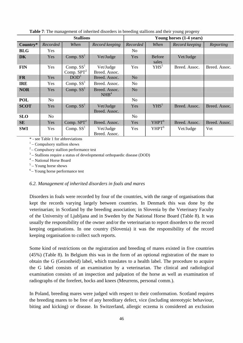
46
Table 7: The management of inherited disorders in breeding stallions and their young progeny Stallions Young horses (1-4 years) Country* Recorded When Record keeping Recorded When Record keeping Reporting BLG Yes No DK Yes Comp. SS1 Vet/Judge Yes Before
sales Vet/Judge
FIN Yes Comp. SS1
Comp. SPT2 Vet/Judge
Breed. Assoc. Yes YHS5 Breed. Assoc. Breed. Assoc.
FR Yes DOD3 Breed. Assoc. No IRE Yes Comp. SS1 Breed. Assoc. No NOR Yes Comp. SS1 Breed. Assoc.
NHB4 No
POL No No SCOT Yes Comp. SS1 Vet/Judge
Breed. Assoc. Yes YHS5 Breed. Assoc. Breed. Assoc.
SLO No No SE Yes Comp. SPT2 Breed. Assoc. Yes YHPT6 Breed. Assoc. Breed. Assoc. SWI Yes Comp. SS1 Vet/Judge
Breed. Assoc. Yes YHPT6 Vet/Judge Vet
* - see Table 1 for abbreviations 1 – Compulsory stallion shows 2 – Compulsory stallion performance test 3 – Stallions require a status of developmental orthopaedic disease (DOD) 4 – National Horse Board 5 – Young horse shows 6 – Young horse performance test 6.2. Management of inherited disorders in foals and mares Disorders in foals were recorded by four of the countries, with the range of organisations that kept the records varying largely between countries. In Denmark this was done by the veterinarian; in Scotland by the breeding association; in Slovenia by the Veterinary Faculty of the University of Ljubljana and in Sweden by the National Horse Board (Table 8). It was usually the responsibility of the owner and/or the veterinarian to report disorders to the record keeping organisations. In one country (Slovenia) it was the responsibility of the record keeping organisation to collect such reports. Some kind of restrictions on the registration and breeding of mares existed in five countries (45%) (Table 8). In Belgium this was in the form of an optional registration of the mare to obtain the G (Gezonheid) label, which translates to a health label. The procedure to acquire the G label consists of an examination by a veterinarian. The clinical and radiological examination consists of an inspection and palpation of the horse as well as examination of radiographs of the forefeet, hocks and knees (Meurrens, personal comm.). In Poland, breeding mares were judged with respect to their conformation. Scotland requires the breeding mares to be free of any hereditary defect, vice (including stereotypic behaviour, biting and kicking) or disease. In Switzerland, allergic eczema is considered an exclusion
47
criterion in breeding mares. In Slovenia, the selection of breeding mares is similar to that of stallions. Breeding stallions in Slovenia are mostly selected via progeny testing of foals. Stallions that are candidates to be approved for breeding are examined after 3 years and are judged on the basis of their exterior appearance (if they are less than 5 years old) and the results of at least 20 of their progeny. If the stallion does not have at least 20 progeny at this time, progeny evaluation is postponed. If more than 20% of the progeny are identified with errors in teeth, osteochondrosis, cryptorchidism, hernia and other constitutional problems, the stallion is excluded from breeding (Potočnik, personal comm.). 6.3. Monitoring of fertility in breeding stallions Fertility of stallions was monitored in six countries (54%) (Table 8). In Denmark, the fertility was monitored via a semen test, i.e. by counting the total number of motile and normal sperm cells. If the number of motile and normal sperm in a semen sample is below 200 000 000, the stallion is not allowed to breed that season but the test can be repeated at a later time (Christiansen, personal comm.). Similarly, stallion fertility in Norway is monitored by a semen test before the first breeding season with the minimum total number of motile sperm being set to 200 000 000 as a median from two ejaculations. In addition, the number of foals born in the season following the semen test is considered with a minimum of 50% live foals being born (Hansson, personal comm.). In Finland, fertility of stallions is monitored using the statistics of foaling and pregnancy rates, and fertility is considered low when these percentages are below 50% (Bernard, personal comm.). In Slovenia, the owner of the stallion station is asked to report each covering of a mare to the Veterinary Faculty in the University of Ljubljana where the number of foals born in relation to the number of covered mares per stallion is evaluated (Potočnik, personal comm.). In Sweden, fertility is monitored using foaling rate, i.e. the number of foals born in relation to number of mares covered. In the case of a stallion with less than 40% of coverings resulting in a foal, an investigation is carried out to check the fertility of the stallion. On the basis of his investigation it is decided whether he is allowed to be further used in breeding (Thorén-Hellsten, personal comm.). For one country (Scotland), answers indicated that fertility was monitored but they did not specify how. Four countries that recorded disorders in foals also monitored fertility of the breeding stallions (Table 8).

48
Table 8: The management of inherited disorders in foals and mares and the monitoring of fertility in stallions
* - see Table 1 for abbreviations 1 – Optional registration of mares for the G label (health label) 2 – Veterinary Faculty of University of Ljubljana, Slovenia 3 – National Horse Board of Sweden 6.4. The management of inherited disorders in breeding stallions and young horses during private veterinary visits Information from private veterinary visits was used for monitoring management of inherited disorders in four countries, all of which recorded disorders in young horses and three of which recorded disorders in both young horses and stallions (Table 9). Records from private veterinary visits of stallions in Scotland and Sweden were kept by the breeding association, although in Sweden the National Horse Hoard was also involved. However, records of young horses were kept by the veterinarian in Scotland and Denmark and by the National Horse Board only in Sweden (Table 9). France reported establishment of a monitoring system of disorders, aiming at the cooperation of researchers, equine veterinarians and the breeding association to collect data in the field and to store and analyse it (Danvy, personal comm.). It was usually the responsibility of the owner or veterinarian to report disorders of stallions and young horses found during veterinary visits although this was not obligatory. In France the responsibility was shared between the breeder/owner, the veterinarian and the national stud agents (Table 9).
Foals Mares Stallions Country* Recorded Record keeping Reporting Restrictions Fertility monitored BLG No Yes1 N DK Yes Vet No Y FIN No No Y FR No No N IRE No No N NOR No No Y POL No Yes N SCOT Yes Breed. Assoc. Breeder/owner
Vet Yes Y
SLO Yes Vet. Faculty2 Vet. Faculty2 Yes Y SE Yes NHB3 Breeder/owner No Y SWI No Yes N

49
Table 9: The management of inherited disorders in breeding stallions and their young progeny during private veterinary visits
Stallions Young horses (1-4 years) Country* Recorded Record keeping Reporting Recorded Record keeping Reporting BLG No No DK No Yes Vet FIN No No FR Yes Co-op. effort1 Breeder/owner
National stud agent Vet
Yes Co-op. effort1 Breeder/owner National stud agent
Vet IRE No No NOR No No POL No No SCOT Yes Breed. Assoc. Vet Yes Vet Breeder/owner
Vet SLO No No SE Yes Breed. Assoc.
NHB2 Breeder/owner
Yes NHB2 Breeder/owner
SWI No No
* - see Table 1 for abbreviations 1 - A cooperative effort between researchers, equine vets and breeding association to collect and evaluate data on disorders 2 - National Horse Board
6.5. Summarising and evaluation of collected records Records of disorders of stallions and/or their young progeny were summarised and evaluated by five of the countries (45%) (Table 10). In four countries (Finland, Ireland, Scotland and Sweden) the breeding association was responsible for the processing of the data, while in one country (Slovenia) the Veterinary Faculty of the University of Ljubljana was responsible (Table 10). In Ireland, the Veterinary College of Ireland, part of the University College of Dublin, was involved in summarising and evaluating the data. Information on disorders in stallions and/or their young progeny was published by four of the countries (Finland, Ireland, Slovenia and Sweden). In Slovenia, the Veterinary Faculty is responsible for using the data for selection and also for advising breeders and the breeding association in selection procedures (Potočnik, personal comm.). In Sweden, if an approved stallion is shown to have passed on a disorder to its progeny (a list of which is made by the Swedish Board of Agriculture and/or the breeding association), its breeding status is changed and it is excluded from breeding. So only this status change is published, but not the details of the investigation having caused the status change (Thorén-Hellsten, personal comm.). In Finland, the summarised form of the data is published on the website of the breeding association, although which form this data is in was not specified (Bernard, personal comm.).
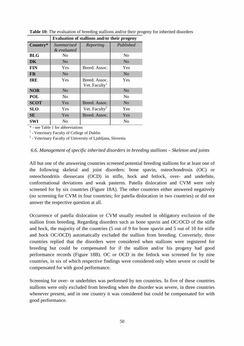
50
Table 10: The evaluation of breeding stallions and/or their progeny for inherited disorders Evaluation of stallions and/or their progeny Country* Summarised
& evaluated Reporting Published
BLG No No DK No No FIN Yes Breed. Assoc. Yes FR No No IRE Yes Breed. Assoc.
Vet. Faculty1 Yes
NOR No No POL No No SCOT Yes Breed. Assoc. No SLO Yes Vet. Faculty2 Yes SE Yes Breed. Assoc. Yes SWI No No * - see Table 1 for abbreviations 1 - Veterinary Faculty of College of Dublin 2 - Veterinary Faculty of University of Ljubljana, Slovenia 6.6. Management of specific inherited disorders in breeding stallions – Skeleton and joints All but one of the answering countries screened potential breeding stallions for at least one of the following skeletal and joint disorders: bone spavin, osteochondrosis (OC) or osteochondritis diessecans (OCD) in stifle, hock and fetlock, over- and underbite, conformational deviations and weak pasterns. Patella dislocation and CVM were only screened for by six countries (Figure 18A). The other countries either answered negatively (no screening for CVM in four countries; for patella dislocation in two countries) or did not answer the respective question at all. Occurrence of patella dislocation or CVM usually resulted in obligatory exclusion of the stallion from breeding. Regarding disorders such as bone spavin and OC/OCD of the stifle and hock, the majority of the countries (5 out of 9 for bone spavin and 5 out of 10 for stifle and hock OC/OCD) automatically excluded the stallion from breeding. Conversely, three countries replied that the disorders were considered when stallions were registered for breeding but could be compensated for if the stallion and/or his progeny had good performance records (Figure 18B). OC or OCD in the fetlock was screened for by nine countries, in six of which respective findings were considered only when severe or could be compensated for with good performance. Screening for over- or underbites was performed by ten countries. In five of these countries stallions were only excluded from breeding when the disorder was severe, in three countries whenever present, and in one country it was considered but could be compensated for with good performance.
51
Common conformational deviations such as bench/offset knees, calf knees, bucked knees, toe-out, toe-in, abnormal formations in the hock and outward rotation of the limbs were grouped together as they were considered at the same level within all the countries. Weak pastern conformation was considered differently to the other conformational faults in one country, so results have been displayed separately. Exclusion of stallions from breeding due to conformational deviations was practiced in four of the countries only when deviations were severe, in two countries whenever present and in three countries only when not compensated for with good performance. In an equal number of countries weak pastern conformation resulted in exclusion of the stallion from breeding whenever present, only when severe and only when not compensated for with good performance (Figure 18B). In some cases, like for OC/OCD and over-/underbite, two countries replied that they did screen for the disorders but did not specify how they were considered in selection of breeding stallions. One country also replied that although patella dislocation is not screened for, if found when the stallion is treated for lameness, the stallion would be automatically excluded from breeding.

52
A
0
2
4
6
8
10
12
Num
ber
of c
ount
ries
Not answered
No
Yes
B
0
1
2
3
4
5
6
Num
ber
of c
ount
ries
Not considered
Only if severe
Can be compensated
Excluded
Figure 18: A. The number of countries that screened for specific skeletal and joint disorders in breeding stallions (yes, no or did not answer the question). B. The level to which each disorder is considered with regards to registration of breeding stallions. # - Results included OC/OCD in both stifle and hock joints. * - Conformational deviations were all grouped together (excluding weak pasterns) due to the data being the same within countries and include: bench/offset knees, calf-knees/back at the knee, bucked knees/over in the knee, toe-out, toe-in, abnormal formations in hock and outward rotation of limbs.

53
6.7. Management of specific inherited disorders in breeding stallions – Hooves Most countries (9) screened for abnormal hoof shapes or bad hoof quality, which were placed in the trait group hoof abnormalities. Screening was practiced in seven countries for sidebone and in six countries for navicular disease and laminitis (Figure 19A). There was large variation between countries in how each of the hoof disorders was considered with respect to registration of stallions for breeding. Navicular disease was an exception, and most countries either excluded affected stallions from breeding or requested the disorder to be compensated for with good performance (Figure 19B). Although navicular disease is considered to be a skeletal disorder, it was placed in the trait group for hoof disorders as the symptoms are most easily noticeable in the hoof area. Despite lacking screening for navicular disease, sidebone and laminitis, affected stallions were either excluded from breeding or could compensate for with good performance in one country. In addition, the stallion’s age is considered and younger stallions with the disorders would be automatically excluded from breeding, whereas older stallions with good performance records may be allowed to continue breeding.

54
A
0
2
4
6
8
10
12
Abnormal hoof
Navicular Sidebone Laminitis
Num
ber
of c
ount
ries
Not answeredNoYes
B
0
1
2
3
4
5
Abnormal hoof
Navicular Sidebone Laminitis
Num
ber
of c
ount
ries
Not consideredOnly if severeCan be compensatedAutomatically excluded
B Figure 19: A. The number of countries that screened for hoof disorders in breeding stallions (yes, no or did not answer the question). B. The level to which each disorder is considered with regards to registration of breeding stallions. 6.8. Management of specific inherited disorders in breeding stallions – Reproductive Most of the countries screened for reproductive disorders such as cryptorchidism, disorders of the testes (including irregular size, shape or texture of the testes) and hernia scrotalis. Only five countries screened for fertility disturbances and dysfunction (Figure 20A), among which one country did not reply how these disorders were considered in breeding. Although there was some variation in how different countries considered reproductive disorders, the majority of the countries automatically excluded stallions that showed such disorders from breeding (Figure 20B). This was especially so in the case of cryptorchidism where more than half of the countries excluded stallions from breeding. There was one country replying that stallions with hernia scrotalis were only excluded from breeding if it was the inherited form and not
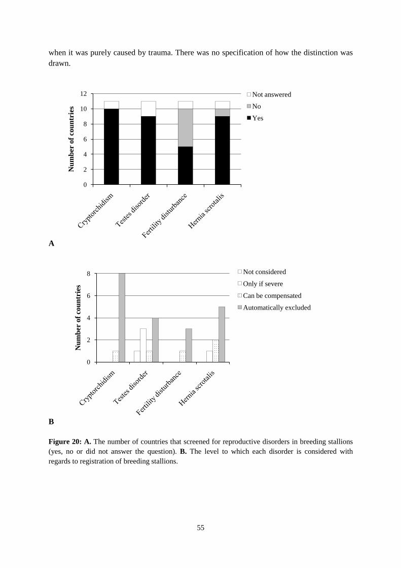
55
when it was purely caused by trauma. There was no specification of how the distinction was drawn.
A
0
2
4
6
8
10
12
Num
ber
of c
ount
ries
Not answered
No
Yes
B
0
2
4
6
8
Num
ber
of c
ount
ries
Not considered
Only if severe
Can be compensated
Automatically excluded
Figure 20: A. The number of countries that screened for reproductive disorders in breeding stallions (yes, no or did not answer the question). B. The level to which each disorder is considered with regards to registration of breeding stallions.

56
6.9. Management of specific inherited disorders in breeding stallions – Other Other disorders that did not fit into any of the major groups included respiratory (laryngeal hemiplegia or roaring; chronic obstructive pulmonary disease (COPD) or recurrent airway obstruction (RAO)), skin (allergic eczema) and muscular disorders (rhabdomyolysis or typing-up syndrome). Additionally abdominal wall umbilicus and unspecified lameness were included in this group. Abdominal wall umbilicus and the respiratory disorders were screened for to a greater extent than were muscular disorders. Unspecified lameness was screened for by seven countries and allergic eczema by four countries (Figure 21A). The majority of the countries automatically excluded stallions with respiratory disorders from breeding, especially in the case of laryngeal hemiplegia (roaring). However, there was large variation between the countries in how these disorders were considered (with the exception of roaring and allergic eczema). Allergic eczema was either not considered (2 countries) or resulted in automatic exclusion of stallions from breeding (2 countries). Most countries did not consider rhabdomyloysis in breeding stallions; one country considered it only when severe and another one automatically excluded affected stallion from breeding. It was interesting to note how differently unspecified lameness was considered in breeding stallions (Figure 21B). Some countries answered that they did screen for the specified disorders, but did specify the consequences for breeding stallions. Other countries did not mention screening but if the disorders were found, they would be considered in the breeding plan. Thus in many instances, the number of countries that examined for a disorder (Figure 21A) does not match the number of countries with available consideration details (Figure 20B).

57
A
0
2
4
6
8
10
12
Num
ber
of c
ount
ries
Not answered
No
Yes
B
0
2
4
6
Num
ber
of c
ount
ries
Not considered
Only if severe
Can be compensated
Automatically excluded
Figure 21: A. The number of countries that screened for other disorders (including respiratory, muscular and skin) in breeding stallions (yes, no or did not answer the question). B. The level to which each disorder is considered with regards to registration of breeding stallions.

58
7. Discussion This study was aimed at determining the strategies currently employed in the management of inherited disorders in European warmblood sport horses. In most countries the breeding associations play major roles in the formulation of selection strategies, including selection strategies to improve the health, soundness and performance of warmblood sport horses. This study revealed that for the warmblood horse in Europe, the breeding associations were the organisations primarily responsible for formulating restrictions in the selection against inherited disorders. In some countries, this role was shared with the Agricultural Boards and university Veterinary Faculties. Veterinarians were not extensively involved with keeping records of disorders in both breeding stallions and young progeny in most countries. Only in Switzerland the veterinarians had the responsibility to report disorders to the relevant organisation. It seems sensible to include veterinarians from a research institute in the formulation of breeding restrictions, as is done in Slovenia. In this way, data is more likely to be collected and evaluated by the students and university staff, and research into genetic aspects of disorders may be encouraged. Nicholas (2003) stated that the paradox of inherited disorders is that the more that is learned about the genetic basis of a disorder, the more likely it is that a non-genetic cure or treatment will be developed. Although such treatments would be advantageous from an animal welfare point of view, selective breeding should aim at reducing the frequency of defective alleles to a manageable level. Breeding of affected animals is therefore not viable, even if effective treatment becomes available. Selection against inherited disorders should make use of the technological advancements which facilitate the identification of defective genes and the differentiation of carriers from healthy individuals. 7.1. Management of inherited disorders in breeding stallions, young horses, mares and foals Disorders in breeding stallions were recorded in nine out of eleven countries, while only five countries recorded disorders in both breeding stallions and their young progeny. The recording of disorders in breeding stallions was usually done only on stallions that attended compulsory stallion events in order to be registered as breeding stallions, while in young progeny it was mostly done at optional young horse events. In Sweden, stallions could be examined again following the compulsory event, in case the stallion was suspected of having a disorder later in life. Young horses, usually 4-year olds, are tested at a form of YHPT called the Riding Horse Quality Test (RHQT), with both genders participating (Thorén-Hellsten, personal comm.). In Denmark, disorders in young progeny are usually recorded at auctions, prior to sale of the horse. This is the time when the horse undergoes a thorough veterinary examination, including X-rays of various joints. Denmark also commented that they did not accept stallions with any form of inherited disorder for breeding, with the only exception being OC/OCD, where a stallion’s performance in sport could compensate for the disorder. Denmark also replied that they are in the process of creating a pilot project which will
59
involve the cooperation of veterinarians and the National Department of Horse Breeding in creating an OC/OCD index for stallions using X-ray data. Since Denmark already has a central database for all horses which is linked to competition results, studies of prevalence and effects of OC/OCD can be studied (K. Christiansen, personal comm.). In France, any stallion that intends to be bred with must be screened for developmental orthpaedic diseases (DODs). This therefore may take place at some type of compulsory stallion event or auction, although this was not clarified. DOD’s most notably include ostechondritis dissecans (OCD), osteochondrosis (OC), juvenile osteoarthirits, deformities of the limbs (flexural or angular) and cervical vertebral malformation (CVM) (McIlwraith, 2004; Priest, 2007) although it must be considered that there is still considerable variation in the definition of DODs. Only a few countries (3) recorded disorders in both breeding stallions and their young progeny during private veterinary visits. It was mostly the responsibility of visiting veterinarians or the breeder/owner to report disorders to the relevant organisation. For most countries it was stated that although reporting is voluntary, it is the preferred action, but the veterinarians or the breeder/owners were under no obligations to do so. Accordingly, doubts were expressed as to how many of the cases are actually reported. It was interesting to note that records in stallions were mostly kept by the breeding association whereas for young horses records were mostly kept by the veterinarian. Only in Sweden the National Horse Board was involved in both cases. Involvement of the breeding associations in collecting records of young horses may be advantageous as it may facilitate cooperative projects to be formed, as in France, in which researchers, equine veterinarians and breed associations can benefit from joint collection and evaluation of data. Not all stallions and young horses would attend stallion and young horse events and thus studies of frequencies and heritabilities of disorders would miss these individuals. If veterinarians were encouraged to record disorders observed in horses during private veterinary visits, it would increase the statistical accuracy of estimated parameters relating to disorders. Additionally, encouraging breeders and owners to report disorders of horses that do not attend the events where recording takes place should also be encouraged. The risk here however is that reporting by breeders/owners would be subjective as for example, some would not report a disorder if they did not consider it to be a real problem. From the participating countries, two (Slovenia and Poland) replied that disorders in breeding stallions were not recorded. Slovenia seems to mainly rely on progeny testing of foals when it comes to selecting against disorders. However, it was mentioned that candidate breeding stallions must undergo some kind of veterinary examination (Potočnik, personal comm.). Progeny testing is a good strategy to employ when dealing with disorders that have low heritabilities and where selection on own performance is less efficient. The disadvantage is that it takes some time for a stallion to produce sufficient numbers of progeny having reached a certain age. It may not be an ideal strategy for disorders that have a late onset of symptoms because long duration of provisional breeding approval of stallions may lead to extensive
60
breeding use before the disorder is discovered. It would therefore be advisable to only use progeny testing of stallions in the selection against disorders that are congenital or have a young manifestation age. Poland did not comment on any strategies for recording disorders of breeding stallions, although examination for specific disorders seems to be performed in breeding stallions at some point. Less than half the countries in the study (4) recorded disorders in foals, and it was usually the responsibility of the breeder/owner of the foal or the veterinarian to report the disorder to the record keeping organisation. In Slovenia, it is the responsibility of the record keeping organisation itself, i.e. the Veterinary Faculty of the university, to collect such records. Once again, recording of disorders of foals and collection of such data would allow more accurate estimates of the frequency of disorders to be obtained. This is because foals born with certain disorders are unlikely to be taken to young horse or stallion events and thus these individuals would be excluded from disorder statistics. The best way to collect foal data may be to involve veterinarians to a greater extent. Routine veterinary visits, for example for vaccination, then provide opportunities to increase the knowledge about the occurrence of certain disorders. For some musculoskeletal disorders, it has been documented, mostly in Thoroughbred foals, that many mild forms may alleviate or correct on their own as the foal matures (McIlwraith et al., 2003; Bramlage & Auer, 2006; Auer & von Rechenberg, 2006). In these cases, active participation of the breeder/owner and the veterinarian may be particularly useful to document such changes in conformation. Most of the countries had an average foaling rate of about 56%. Only Slovenia had a very low foaling rate of 22%, which may be due to the fact that a smaller proportion of registered mares are used for breeding each year. This is probably true for the other countries too, so the foaling rate may be higher than calculated here. Foals born in Slovenia, although possibly being of same quality as foreign horses, regularly fetch lower prices, which may discourage Slovenian horse breeders (Potočnik, personal comm.). Importing of foreign genetic material has many advantages but it is important to also consider and support the national breeding program and to integrate the imported genetic material into the existing national gene pool. Ireland had the highest foaling rate of 72% although this country reported that stallions were not regularly monitored for fertility. However, stallions showing obvious fertility disturbance or dysfunction were automatically excluded from breeding. Only five countries had some kind of restrictions in the selection of mares for breeding. In Belgium an optional registration of mares has been introduced to increase the breeding status of mares having been screened for certain disorders. Such a health label, called a G label in Belgium, may be considered as an appropriate measure to increase availability of health data on a voluntary basis. Only in Slovenia was the selection of breeding mares against disorders similar to the selection of stallions. There are many more mares than breeding stallions on average in every country. Furthermore, mares often belong to many different owners whereas breeding

61
stallions are usually concentrated within a much smaller number of facilities. It is therefore more difficult to implement and control restrictions on the breeding of mares. It may be beneficial for disorders that are highly heritable to more intensely select against them in both mares and stallions. For disorders with lower heritablities it may at first be sufficient to only select against them in stallions. This is because individual stallions can be used much more extensively in their lifetimes and can sire many more offspring than an individual mare. The contribution of the stallion to the next generation is therefore on average larger than that of a mare. More than half of the countries in this study (6) monitored fertility in stallions. Most of the countries used statistical data concerting foaling and pregnancy rates to monitor fertility of stallions; one country used only a semen test (counting numbers of healthy and motile sperm) and one country used both methods. There was a trend for countries with smaller warmblood populations to monitor the fertility of stallions. Denmark and Sweden, who have relatively large populations, meant some exception in this respect. On the one hand, it may be easier to monitor a trait in smaller populations, on the other hand the need for caution to avoid negative effects of inbreeding is probably larger in a small population, especially in times before the invention of AI and efficient semen transport and preservation. Some studies in horses have shown the negative effect of inbreeding on fertility, which is known as inbreeding depression. Klemetsdal & Johnson (1989) found that the level of inbreeding had a significant effect on the rate of early abortions in Norwegian trotters. Van Eldik et al. (2006) found that inbreeding coefficients greater than 2% had significant negative effects on the motility and morphology of sperm in Shetland ponies. Fertility does not appear to be a trait of high importance in the other breeding associations. This finding was corroborated by a study conducted by Koenen et al. (2004) where out of 19 breeding organisations only 3 included fertility in their verbally expressed breeding objectives, although 10 of the 19 organisations attached a high relative importance to fertility. It is also important to consider what kind of method is used to monitor the fertility of stallions. According to Amann (2004), shortcomings of reports on stallion fertility include that the estimation of a stallion’s fertility is usually imprecise when using only information consisting of pregnancy rates or foaling rates of mares. This is due to the fact that pregnancy/foaling rates are influenced by many factors, not the fertility of the stallion alone. A successful pregnancy and eventually a live foal depends on factors such as fertility and health of the mare, the management strategies of the facility where the mare is kept, timing of artificial insemination (AI) or natural breeding as well as weather or not a mare changes stallions between inseminations. Such factors may contribute almost five times more to the outcome of a successful pregnancy than does the fertility of the stallion alone (Amann, 2004). Given the possible bias of most studies on fertility, it may be more recommendable to use counts of motile and normal sperm of a stallion, possibly together with pregnancy and foaling rates of mares. More reliable estimates of the true fertility of a stallion may then be obtained. Additionally it has been found that apart from the number of sperm, their morphology was

62
found to have a large impact on fertility in species in which it has been studied and thus this parameter should be considered in horses as well (Brito, 2007). It was interesting to note that four countries that recorded disorders in foals also monitored fertility of breeding stallions. Apart from affecting the number of foals born, abnormalities in spermatogenesis caused by disorders of the testes are suspected to be heritable. Fertility of stallion is also considered to be heritable although the heritability is assumed to be relatively low (Parlevliet et al., 1994). For cryptorchidism, high incidences of sterility and impaired fertility have been documented for some time (Scott, 1961). Other, less obvious disorders involving the size, texture and position of the testes may have similar, but more subtle effects. However, this has not been extensively studied. Among the five countries that collected, summarised and evaluated records of disorders in breeding stallions and their young progeny, were the three countries that had the smallest populations in the study. As mentioned before, this may reflect the fact that it is easier to control collection and evaluation of information in small populations. However, evaluation of smaller populations would also be statistically weaker and the estimates obtained, compared to estimates using large numbers of horses, would be less accurate. The breeding associations in Finland, Scotland and Sweden most likely have their own breeding advisors and geneticists who are able to process such data, whereas in Slovenia and Ireland the university Veterinary faculties performed these tasks. Involvement of veterinarians and especially university students and staff would be ideal as it would allow studies and evaluations on collected information to be conducted. Only four of the countries published the summarised and evaluated data, which was then available for breeders either directly or indirectly. It may be beneficial for breeders if more breeding associations published such data on stallions. Of course, for serious disorders, stallions are likely be excluded from breeding anyway, but for slight conformational faults it may be useful for mare owners to have access to this information. It would allow mare owners to match their mares to prospective stallions based on this knowledge and thus minimise conformational faults. Publication of health data of stallions and their progeny would mean an additional and objective source of information, adding to the already accessible, mostly performance related data. Many mare owners base their choices of stallions on photographs from breeding catalogues or from performance results of the stallions and do not always have a chance to see the stallion themselves, especially if they live at a great geographical distance. It would then be useful to include such information in breeding catalogues as photographs rarely show the true conformation of a horse and information is usually based solely on performance data. This information would also serve to empower breeders by making them feel more important and involved in making knowledgeable decisions with regards to their breeding plans.

63
7.2. Management of specific inherited disorders in breeding stallions There is no doubt that lameness caused by musculoskeletal disorders is a major problem in warmblood sport horses. In accordance with this, most of the countries screened for disorders relating to lameness. Disorders such as CVM and patella dislocation were screened for to a lesser extent but, when they were discovered it was most likely that the stallion would be excluded from breeding. There seems to be evidence of a connection between the presence of OC lesions in various joints, including vertebrae, and CVM (Wagner et al., 1985; van Weeren & Barneveld, 1999a; Barneveld & van Weeren, 1999b; Barneveld & van Weeren, 1999a; Donabedian et al., 2006). As this disorder is more prevalent in Thoroughbreds, the low frequencies in warmbloods may be caused by the little attention paid to this disorder in many countries. Diagnostic problems with little specificity of signs and symptoms of CVM and unforeseeable duration of asymptomatic time periods further interfere with collecting reliable information on CVM. In a review of lameness and poor performance in sports horses, Dyson (2000) mentioned that mild intermittent upward fixation or delayed release of the patella can occur especially in young warmblood horses that start training, causing discomfort and poor performance. Although this disorder in young horses if often associated with poor muscle development and is not considered to be heritable, the importance of patella fixation is yet unknown and this disorder may deserve intensified screening in warmblood stallions in the future. Half of the countries that screened for OC/OCD (5 out of 10) excluded stallions from breeding if OC/OCD was present in the stifle or hock. Only one country proceeded this way in case of OC/OCD in the fetlock. It is interesting that the consequence of the presence of the same disorder, OC/OCD, differs between joints. However, genetic analyses have implied that the genetic basis of fetlock and hock affections is not the same (Stock & Distl, 2006). Furthermore, OC/OCD alternations in the fetlock may be clinically irrelevant for a long period of time, which is rarely the case with similar alterations in hock or stifle joints. For example, plantar and palmar osteochondral fragments found in fetlocks of the hind limb seem to be mostly due to trauma and not due to heritable disturbances in endochondral ossification (Dalin et al., 1993). In Hanoverian Warmblood riding horses, it was found that the prevalence of osseous fragments was greatest in the fetlock joints while deforming arthropathy was most prevalent in hock joints (Stock & Distl, 2005). Although it was found that these radiological findings were equally prevalent throughout both competing and non-competing horses, affected horses had considerably lower mean numbers of annual entries and placings compared to unaffected horses. These results imply that besides obvious lameness, more subtle negative effects on the performance of riding horses may exist. Accordingly, more than half of the countries that screened for bone spavin (5 out of 9), automatically excluded stallions from breeding, with this disorder manifesting in the hock. One complication with diagnosing skeletal disorders is that lesions or pathological changes do not always result in lameness, and it is often difficult to determine which ones will be detrimental. Diagnoses may also be prone to subjectivity. In an extensive study following the

64
occurrence of OC lesions in warmblood foals, it was found that there was a significant reduction in the number of lesions from 5 to 11 months of age. This indicates that most lesions occur in the first 5 months after birth and are able to be repaired during the next six months, during a “window period” of repair (van Weeren & Barneveld, 1999a). The repair process is probably affected by external factors such as rearing conditions, and no regression of lesions is found after the age of one year, although his is joint-dependant (Donabedian et al., 2008). This means that when horses are screened for OC around the age of 3 years, the true frequency of lesions that occurred is unknown, although the lesions that are present at this age are more likely to be ones that cause clinical signs. Another complication is the absence of clear definitions (not only when dealing with skeletal disorders but most disorders) and the inconsistent and subjective scoring of skeletal deviations, i.e. to what degree is a skeletal deviation considered to be a problem and what degree of deviation is acceptable. Most joint disorders (except for OC/OCD in the fetlock) resulted in automatic exclusion of the stallions from breeding. Skeletal disorders mostly caused exclusions of stallions from breeding when severe so that performance was affected. It is thought that slight conformational deviations do not directly affect performance and some may even be preferred for certain disciplines. However, conformational deviations do tend to place additional strain on other parts of the supportive tissue and do predispose to certain injuries. Slight under- and overbites do not pose a serious problem to horse health and welfare and can be regularly managed by an equine dentist. However, from a genetic prospective, it may be preferable not to breed with affected animals. Within countries, there was consensus on the management of common conformational deviations (bench/offset knees, calf knees, bucked knees, toe-out, toe-in, abnormal formations in the hock and outward rotation of the limbs). This is interesting as studies have shown that certain conformational deviations in racing horses are very detrimental and predispose to injuries more than others. Unfortunately, very few studies on the relationship between conformation and performance have been conducted in warmblood horses. Most studies and literature in this area have been conducted in racehorses (either trotters or gallopers). This indicates that considering all conformational deviations at the same level in riding horses is inaccurate and more research is needed to determine which conformations are most likely to result in injuries when specific riding activities are performed. Reasons why this field of research has been dominated by studies in racing horses may be that the racing industry generates a larger amount of money, which makes funding for such studies available and also creates more commercial interest to improve performance of racing horses. Additionally, racing stables usually provide appropriate study conditions due to the large number of horses available in one location. This allows for easier control of experimental conditions. Riding horses are, however, usually dispersed over many locations with only a few horses being owned by one individual. This makes controlled experimental studies difficult. Additionally lack of funding in the riding horse industry in comparison with the racing industry also may be a limiting factor. When considering conformational deviations, certain facts also have to be taken into account. Some slight deviations are commonly seen at certain developmental phases in a young
65
growing horse. McIlwraith et al. (2003) have documented growth-related changes in conformation in Thoroughbred horses and concluded that carpal conformation changes progressively from back at the knee to slightly over at the knee between 1 and 3 years of age. Additionally, slight deviations exist that are commonly seen within a breed. A study on the variation in conformation of Swedish warmblood horses found that 60 % of the horses had bench knee (offset knee) conformation, 50 % had toe-in conformation in the fore limbs and 80 % of horses had hind limbs that rotated outwards. Most of the deviations were found to be mild to moderate, and there was a significant influence of sex and age (Holmström et al., 1990). More such information in variation of conformation within warmblood horses is needed and subsequently such information should then be related to performance and health of the horses, in order to determine which conformations are most detrimental for certain activities. Abnormal hoof shape and bad hoof quality may be important factors for the health of the equine limb because they directly impact load distribution in the respective limb. For example, there seems to be a correlation between abnormal hoof shape and navicular disease, with horses that have long toes and low heels (many Thoroughbreds) and horses with narrow upright hooves (many Quarter Horses) being more susceptible (King & Mansmann, 2001b). Dyson (2000) has also commented that the hooves of warmblood horses often have bad conformation and are not proportional in shape or size in relation to the horse’s body weight. This could relate to the fact that 21.7% of Hanoverian warmbloods were diagnosed radiologically with pathological changes in the navicular bone (Stock & Distl, 2005). Amongst diseases of the locomotor system, navicular disease was reported as the most common single diagnosis that caused premature culling in up to 9.1% of Swedish warmblood horses (Philipsson et al., 1998). It was therefore surprising to see that although most countries (9) screened for abnormal hoof quality and shape, only six countries screened for navicular disease. Stallions affected with navicular disease were automatically excluded from breeding in four countries, whereas three countries replied that the disorder could be compensated for with good performance. However, navicular disease does not appear to be a negligible problem in the warmblood breed. The little agreement on how to consider navicular disease is probably due to the continuing intense discussions about reliable diagnostic criteria and the uncertain clinical relevance of, for example, radiologically observed changes in the navicular bones. More countries screened for sidebone in warmblood stallions in relation to navicular disease. Sidebone appears to be more common in coldblooded breeds with a prevalence of ossifications of 95% in heavy draft horses (Verschooten, 1994) and 80% in Ardenner horses (Tullberg, 2006) having been determined. Prevalence in warmbloods was found to be much lower (18%) (Verschooten, 1994). Although ossification of the cartilage attached to the pedal bone may initially cause some inflammation and lameness, once ossification is complete, horses rarely remain lame (Devereux & Morrison, 1996). In one country, young stallions with sidebone were automatically excluded from breeding, whereas older stallions could compensate for the disorder with good performance and continue to breed. The consideration
66
of laminitis was more varied between countries, possibly depending on the number and severity of laminitic episodes. In the light of a genetic predisposition connected to most disorders, the discussion about possible compensation of some disorders is likely to continue. Some horses with slight forms of certain disorders may be excellent athletes and it is possible that factors such as temperament and willingness to perform may sometimes compensate in part for slight defects and vice versa; completely healthy horses may lack the temperament or willingness to perform well in sport. As long as it is not know to which extent conformational and other disorders are genetically correlated with performance traits, multiple-trait selection actually lacks a scientific basis. More studies on this issue are needed to produce sound and comprehensive breeding strategies. There was a general consensus of automatically excluding stallions from breeding which had reproductive disorders although fertility disturbance and dysfunction were the disorders with lower examination rates. This reflects that fertility problems have little weight when selecting stallions for breeding, although fertility has important economic meaning in the horse industry. However, fertility disturbances can be related to disorders in testes or other reproductive structures. Cryptorchidism, probably representing the most common abnormality of the testes (Knottenbelt & Pascoe, 1994), is examined for in most countries. Cryptorchid stallions are generally excluded from breeding. Despite this, the inheritance of cryptorchidism had not yet been determined. Various studies have proposed very different forms of inheritance, from dominant (Hayes 1986 in Lu, 2005) to autosomal recessive, although a more recent molecular study has suggested a complex polygenic inheritance (Diribarne et al., 2009). Respiratory disorders seem to be an emerging problem in riding horses, especially disorders such as COPD, which may reflect the unnatural housing conditions that most horses are exposed to. Prolonged enclosure in stalls, especially during the winter period, results in increased inhalation of irritants such as dust and fungal spores. Respiratory disorders ranked second among the main reasons for premature culling of Swedish warmblood horses (Wallin et al., 2000). In a study on Hanoverian warmblood horses, it was found that one third of the horses were reported to have respiratory problems (Stock & Distl, 2005). This was reflected by the large number of countries in this study that screened for respiratory disorders such as laryngeal hemiplegia (roaring) and COPD. Most of these countries also automatically excluded stallions with respiratory disorders from breeding. For these two respiratory disorders, a strong familial inheritance has been reported (Marti et al., 1991; Deegan et al., 1995). Heritability of laryngeal hemiplegia in a German study was estimated at 0.50 (Deegan et al., 1995). Abdominal wall umbilicus, allergic eczema and unspecified lameness were screened for in most countries, although there was large variation between countries as to how abdominal wall umbilicus and unspecified lameness were considered. The most likely reason why unspecified lameness received quite a poor response from the participant countries may be

67
that the impact on the welfare and performance of the individual horse and the breeding relevance is uncertain as long as a definitive diagnosis is missing. Nevertheless, Wallin et al. (2000) found that up to 16.8% of warmblood horses were culled due to unspecified lameness. Allergic eczema was either not considered at all or affected stallions were automatically excluded from breeding. This disorder does not appear to be a big problem in warmblood breeds as opposed to ponies (Devereux & Morrison, 1996). However, differences relating to geographic location may exist. For example, one country that excluded stallions with allergic eczema from breeding also had restrictions on breeding of affected mares, referring to allergic eczema. Very few countries (3) examined for rhabdomyolysis and only one country excluded stallions from breeding if they had the disorder. Indications of a very closely related disorder called polysaccharide storage myopathy (PSSM) that is thought to indirectly or directly cause episodes of exertional rhabdomyolysis, have been found in 50% of muscle biopsies from warmblood horses presenting neuromuscular symptoms. Additionally, very high heritabilities, ranging from 0.42 to 0.43 have been estimated by Oki et al. (2005) for extertional rhabdomyolysis in Thoroughbred horses, and an autosomal dominant inheritance has been suggested by Dranchak et al. (2005). The very high percentage of warmblood horses with signs of PSSM may be more susceptible to mild forms of rhabdomyolysis which may impair performance and possibly result in higher rates of musculoskeletal injuries. Thus in the light of riding horses as athletes, it may be beneficial to devote in depth studies to the occurrence and frequency of muscular disorders, which could be another group of emerging disorders in riding horses. Stereotypical behaviour such as cribbing (crib-biting) was considered in the selection procedures of one country, with stallions that cribbed being automatically excluded from breeding. Stereotypical behaviour seems to be more common in some families than in others, although learning of foals from their dams does not play a relevant role (Marsden, 1995). Provisional probabilities of an individual horse performing stereotypic behaviour in a certain environment were calculated, and these probabilities increased if relatives of the horse also performed stereotypical behaviour (Marsden, 1995). These results imply some direct or indirect involvement of genetic factors in the development of stereotypical behaviour in certain environments. Ultimately, equine researchers and veterinarians should try to ensure that breeders understand that the occurrence of conformational and other disorders is impossible to eradicate completely. However, high frequencies of particular disorders in certain populations should entice all people involved in horse breeding to gain as much knowledge about the occurrence, cause and genetics of such disorders as possible to take appropriate measures against them. The large size and rather high diversity of the warmblood population, which is influenced by many different breeds, acts against high frequencies of disorders that occur in other breeds. Depending on if matings are matched accordingly, there is a low risk of inbreeding.

68
However, the wide spread and extensive use of warmbloods in sports, relates to increasing problems with musculoskeletal and respiratory disorders. These problems may have arisen from an interaction of a number of factors and the unnatural way that many sport horses are managed. This can be reflected by the fact that development of many of the degenerative joint diseases includes factors such as diets that are too rich in grains and lack roughage; rapid growth that may be related to the diet or selection strategy; excessive mechanical stress of joints, bones and muscles during intensive or repetitive exercise and maybe restricted movement of horses by being stabled. Henderson (2007) has suggested that many stereotypical behaviours and the physiological well-being of horses could be dramatically improved by considering behaviour of horses in the wild. In the paper, it was also discussed how to improve the welfare of sport horses by making minimal management changes, while still taking into account the requirements of the competitive horse as an athlete. Recommendations of breeding organisations with respect to equine health should therefore consider both environmental and genetic improvements. 8. Conclusions The following suggestions for improving the current strategies for the management of disorders in the warmblood horse could be derived from this study. Horse breeding would benefit from coordinated action of all parties involved in the collection, recording and evaluation of disorders in warmblood horse populations (veterinarians, researchers, stud officials, breeding association members, etc.). This should make it possible to increase knowledge on the frequencies and genetic heritabilities of certain disorders. If such data was collected and evaluated, it could be used within the breeding associations for the approval of stallions for breeding. Furthermore, publication of health data may allow breeders to make more informed decisions about matching their mares to the approved stallions. Perhaps an option would be to create a central database for disorders, where all this information could be stored and published, starting at a national level (as some countries are in the process of doing), as international collection of data would require strict standardization in classification, diagnoses and scoring of inherited disorders. Although much needed, this standardization may not be easy to achieve. It should be ensured that health data collected is as objective as possible and an accurate representation of the horse population in question. Pre-selection of horses often poses a serious risk; both in considering average prevalence of disorders in a population, and in considering prevalence in progeny groups of breeding stallions for which this data would be used to estimate their breeding values. Additionally, further studies on the role of conformational deviations in relation to performance in warmblood horses should be conducted. This would collectively serve to ensure that the most viable breeding plans, in terms of health, longevity, performance and welfare of horses can be constructed from this information.

69
9. Acknowledgments I would like to express my deepest gratitude to the participants from the 11 countries and breeding associations for making this study possible, as well as the many veterinarians and professors who also helped me and who had to endure my endless emails. I would like to thank my supervisors, Jan Philipsson, Lina Jönson and Louise Lindberg from the Animal Breeding and Genetics Department at SLU and Bart Ducro from the Animal Breeding and Genetics Department at Wageningen University, for all the help, guidance and useful comments. A special thanks to Emma Thorèn-Hellsten for her good advice and my study coordinator Birgitta Malmfors for all her help. Thanks to the Erasmus Mundus Animal Breeding and Genetics (EM-ABG) scholarship for funding my study and stay in Sweden and my EM-ABG colleagues. An additional specials thanks to Kathrin-Friederike Stock for her time and good advice and to Olga Boucher for the photograph of her stallion Zalinero for the front cover. And lastly, I would like to thank my family and friends; especially my father Dr Nenad Nikolić, my mother Mirjana Skulić-Nikolić, my brother Andrej Nikolić, my fiancé Angus Pollard and my best friend Lara Proud for all their help, encouragement and support. 10. References Aleman, M., Brosnan, R.J., Williams, D.C., Lecouteur, R.A., Imai, A., Tharp, B.R. & Stefey,
E.P. 2005. Malignant hyperthermia in a horse anesthesized with halothane. Journal of Veterinary Internal Medicine, 19:363-366.
Aleman, M., Riehl, J., Aldridge, B.M., Lecouteur, R.A., Stott, J.L. & Pessah, I.N. 2004. Association of a mutation in the ryanodine receptor 1 gene with equine malignant hyperthermia. Muscle Nerve, 30:356-365.
Amann, R.P. 2005. Weaknesses in reports of “fertility” for horses and other species. Theriogenology, 63:698-715.
Amizuka, N., Warshawsky, H., Henderson, J.E., Goltzman, D. & Karplis, A.C. 1994. Parathyroid hormone-related peptide depleted mice show abnormal epiphyseal cartilage development and altered endochondral bone formation. The Journal of Cell Biology, 126:1611-1623.
Anderson, T.M., McIlwraith, C.W. & Douay, P. 2004. The role of conformation in musculoskeletal problems in the racing Thoroughbred. Equine Veterinary Journal, 36:571-575.
Auer, J.A. 1985. Periosteal transaction of the proximal phalanx in foals with angular limb deformities of the metacarpo/metatarsophalangeal area. Journal of the American Veterinary Medical Assocaiton, 187:496-499.
Auer, J.A. & von Rechenberg, B. 2006. Treatment of angular limb deformities in foals. Clinical Techniques in Equine Practice, 5:270-281.
Bagby, G.W. 1985. Process for fusing bone joints. United States Patent, patent number 4 501 269.

70
Baker, G.J. 1999. Abnormalities of developement and eruption. In: Baker, G.J. & Easley, J. (Eds), Equine Dentistry, 1st edition. W.B. Saunders Company Ltd, London. Pg:53-55.
Barneveld, A. & van Weeren, P.R. 1999a. Conclusions regarding the influence of exercise on the development o the equine musculoskeletal system with special reference to osteochondrosis. Equine Veterinary Journal Supplement, 31:112-119.
Barneveld, A. & van Weeren, P.R. 1999b. Early changes in the distal intertarsal joint of Dutch Warmblood foals and the influence of exercise on bone density in the third tarsal bone. Equine Veterinary Journal Supplement, 31:67-73.
Barr, A.R.S. 1994. Carpal conformation in relation to carpal chip fractures. Veterinary Records, 134:646.
van Biervliet, J., Mayhew, J. & de Lahunta, A. 2006. Cervical vertebral compressive myelopathy: diagnosis. Clinical Techniques in Equine Practice. doi:10.1053/j.ctep.2006.01.010.
Björck, G., Everz, K.E., Hansen, H.J. & Henrickson, B.1973. Congenital cerebellar ataxia in the Gotland pony breed. Reihe A-Journal of Veterinary Medicine Series, 20:341.
Bjørnsdøttir, S., Árnason, T., Axelsson, M., Eksell, P., Sigurdsson, H. & Carlsten, J., 2000. The heritability of degenerative joint disease in the distal tarsal joints of Icelandic horses.Livestock Production Science, 63:77-83.
Bramlage, L.R. & Auer, J.A. 2006. Diagnosis, assessment, and treatment strategies for angular limb deformities in the foal. Clinical Techniques in Equine Practice, 5:259-269.
Bramlage, L.R. 1993. Investigation of farm wide incidence of bone formation problems in the horse. Proceedings, 39th Annual Convention of the American Association of Equine Practitioners, San Antonio, Texas: 45-48.
Bowling, A.T. & Ruvinsky, A. 2000. The genetics of the horse. CABI Publishing, London. Pg:25-48.
Bowling, A.T., Byrns, G. & Spier, S. 1996. Evidence for a single pedigree souce of the hyperkalemic periodic paralysis susceptibility gene in quarter horses. Animal Genetics, 27:279-281.
Brito, L.F.C. 2007. Evaluation of stallion sperm morphology. Clinical Techniques in Equine Practice, 6: 249-264.
Bushe, T., Turner, T.A., Poulos, T.W. & Harwell, N.M.1988. The effects of hoof angle on coffin, pastern and fetlock joint angles. Proceedings of the American Association of Equine Practitioners, 33:729.
Clayton, H.M. 1987. Comparison of the stride of trotting horses trimmed with a normal and a broken-back hoof axis. In: Proceedings. 33rd Annual Convention of the American Associations of Equine Practitioners, 289-298.
Dalin, G., Sandgren, B. & Carlsten, J. 1993. Plantar osteochondral fragments in the metatarsophalangeal joints in Standardbred trotters; results of osteochondrosis or trauma? Equine Veterinary Journal Supplement, 16:62-65.
Deegen, E., Ohnesorge, B., Miesner, K. & Brandt, I. 1995. Hemiplegia laryngis sinistra – an hereditary disease in horses (summary). Equine Veterinary Journal, 27:412-413.
Devereux, S. & Morrison, L. 1996. The veterinary care of the horse. J.A. Allen, UK. Pg:30, 31, 36-59, 79-88, 152-158, 164-166, 195-197.
71
Dierks, C., Löhring, K., Lampe, V., Wittwer, C., Drögemüller, C. & Distl, O. 2007. Genome- wide search for markers associated with osteochondrosis in Hanoverian warmblood horses. Mammalian Genome, 18:739-747.
Diesterbeck, U.S., Hertsch, B. & Distl, O. 2007. Genome-wide search for microsatellite markers associated with radiological alterations in the navicular bone of Hanoverian warmlbood horses. Mammalian Genome, 18:373-381.
Diribarne, M., Vaiman, A., Péchayre, M., Pailhoux, E., Matta, X., Guérin, G. & Chaffaux, S. 2009. Polymorphism analysis of microsatellites associated with seven candidate genes for equine cryptorchidism. Journal of Equine Veterinary Science, 29:37-41.
Dixon, P.M. & Dacre, I. 2005. A review of equine dental disorders. The Veterinary Journal, 169:165-187.
Dolvik, N.I. & Klemetsdal, G. 1999. Conformational traits of Norwegian cold-blooded trotters: heritability and the relationship with performance. Acta Agriculturae Scandinavica, Section A – Animal Sciences, 49:156-162.
Dolvik, N.I. & Klemetsdal, G. 1994. Arthritis in the carpal joint of Norwegian trotter- heritability, effects of inbreeding and conformation. Livestock Production Science, 39:283-290.
Donabedian, M., van Weeren, P.R., Perona, G., Fleurance, G., Robert, C., Leger, S., Bergero, D., Lepage, O. & Martin-Rosset, W. 2008. Early changes in biomarkers of skeletal metabolism and their association to the occurrence of osteochondrosis (OC) in the horse. Equine Veterinary Journal, 40:253-259.
Donabedian, M., Fleurance, G., Perona, G., Robert, C., Lepage, O.M., Trillaud-Geyl, C., Léger, S., Ricard, A., Bergero, D. & Martin-Rosset, W. 2006. Effect of maximal vs. moderate growth related to nutrients intake on developmental orthopaedic diseases in horses. Animal Research, 55: 471-486.
Dranchak, P.K., Valberg, S.J., Onan, G.W., Gallant, E.M., MacLeay, J.M., McKenzie, E.C., De La Corte, F.D., Ekenstedt, K. & Mickelson, J.R. 2005. Inheritance of recurrent exertional rhabdomyolysis in thoroughbreds. Journal of the American Veterinary Medical Association, 227:762-767.
Duke, A. 1989. Equine bit analysis. Handout notes from Annual Conference of American Veterinary Dental Society, New Orleans.
Dyson, S. 2000. Lameness and poor performance in the sports horse: dressage, show jumping and horse trails (eventing). Proceedings of the Annual Convention of the AAEP, 46:308-315.
Easley, J. 1999. Basic equine orthodontics. In: Baker, G.J. & Easley, J. (Eds), Equine Dentistry, 1st edition. W.B. Saunders Company Ltd, London. Pg 206-219.
van Eldik, P., van der Waaij, E.H., Ducro, B., Kooper, A.W., Stout, T.A.E. & Colenbrander, B. 2006. Possible negative effects of inbreeding on semen quality in Shetland pony stallions. Theriogenology, 65:1159-1170.
Finno, C.J., Spier, S.J. & Valberg, S.J. 2008. Equine diseases caused by known genetic mutation. The Veterinary Journal, doi:10.1016/j.tvjl.2008.03.016.
Gift, L.J., DeBowers, R.M., Clem, M.F., Rashmir-Raven, A. & Nyrop, K.A. 1991. Brachygnathia in horses: 20 cases (1979-1989). Journal of the American Veterinary Medical Association, 200:715-719.

72
Grandison, K., Lindberg, L, Eriksson, S., Mikko, S., Broström, H., Frey, R, Sundquist, M. & Lindgren, G. 2006. Genetic parameters for allergic eczema in Icelandic horses. 8th World Congress on Genetics Applied to Livestock Production, August 13-18, 2006, Belo Horizonte, MG, Brasil.
Graves, K.T. 2005. Veterinary review - genetic disease in the horse. Lloyd’s Equine Disease Quarterly, 25:254-255.
van Grevenhof, E.M., Schurink, A., Ducro, B.J., van Weeren, P.R., van Tartwijk, J.M.F.M., Bijma, P. & van Arendonk, J.A.M. 2009. Genetic parameters of various manifestations of osteochondrosis and their correlations between and within joints in Dutch warmblood horses. Journal of Animal Science, http://jas.fass.org.
Grøndahl, A.M. & Dolvik, N.I. 1993. Heritability estimations of osteochondrosis in the tibiotarsal joint and of bony fragments in the palmar/plantar portion of the metacarpo- and metatarsophalangeal joints of horses. Journal of the American Veterinary Medical Association, 203:101-104.
Hall, L.W., Woolf, N., Bradley, J.W.P. & Jolly, D.W. 1966. Unusual reaction to suxamethonium chloride. British Medical Journal, 4:1305.
Hamann, H. & Distl, O. 2008. Genetic variability in Hanoverian warmlbood horses using pedigree analysis. Journal of Animal Science, http://jas.fass.org.
Hamann, H., Jude, R., Sieme, H., Mertens, U., Tӧpfer-Petersen, E., Distl, O. & Leeb, T. 2007. A polymorphism within the equine CRISP3 gene is associated with stallion fertility in Hanoverian warmblood horses. Animal Genetics, 38:259-264.
Haussler, K.K. & Stover, S.M. 1996. Low back pathology and pelvic stress fractures in 36 Thoroughbred racehorses. In: Proceedings, 42nd Annual Convention of the American Association of Equine Practitioners, 278-279.
Hayes, H.M. 1986. Epidemiological features of 5009 cases of equine cryptorchism. Equine Veterinary Journal, 18:467-471.
Hayes, M.H. Points of the horse, 6th edition. London: Hurst & Blackett, 1952. Henderson, A.J. 2007. Don’t fence me in: managing psychological well being for elite
performance horses. Journal of Applied Animal Welfare Science, 10:309-329. Heymering, H. 1991. The proper hoof angle. The Anvil, 16:19-26. Holmström, M., Magnusson, L.E. & Philipsson, J. 1990. Variation in conformation of
Swedish warmblood horses and conformational charachteristics of èlite sport horses. Equine Veterinary Journal, 22: 186-193.
Howard, R.D., McIlwraith, C.W. & Trotter, G.W. 1995. Arthroscopic surgery for subchondral systic lesions of the medial femoral condyle in horses: 41 cases (1988-1991). Journal of the American Veterinary Medical Association, 206:842-850.
Hunt, L.M., Valberg, S.J., Stefenhagen, K. & Bender, J.B. 2005. A retrospective study of myopathies and associated gait abnormalities in 65 Warmblood horses. Journal of Veterinary Internal Medicine, 19:428-429.
Josseck, H. 1995. Genetic aspects of hoof quality. In: Genetics and Disease in the Horse: Heinz Gerber International Workshop. Equine Veterinary Journal, 27:411-415.
Kapell, D. 2005. Student thesis, Wageningen Universtiy, The Netherlands. King, C. & Mansmann, R. 2001a. Cervical Vertebral Malformation (Wobblers). In: Wagoner,
D. (Ed), Equine Lameness. Equine Research, Inc. King, C. & Mansmann, R. 2001b.Navicular Syndrome. In: Wagoner, D. (Ed), Equine
Lameness. Equine Research, Inc.

73
Klemetsdal, G. & Johnson,1989. M. Effect of inbreeding on fertility in Norwegian trotters. Livestock Production Science, 9:743-54.
Knottenbelt, D.C. & Pascoe, R.R. 1994. Colour atlas of diseases and disorders of the horse. Mosby-Year Book Europe Limited. Pg:49, 50, 209-247, 395-401.
Koenen, E.P.C., Aldridge, L.I. & Philipsson, J. 2004. An overview of breeding objectives for warmblood sport horses. Livestock Production Science, 88:77-84.
Kohn, C.W., Johnson, G.C., Garry, F., Johnson, C.W., Martin, S. & Scott, D.W. 1989. Mechanobullous disease in two Belgian foals. Equine Veterinary Journal, 21:297-301.
Lieto, L.D., Swerczek, T.W. & Cothran, E.G. 2002. Equine epitheliogenesis imperfecta in two American saddlebred foals is a lamina defect. Veterinary Pathology, 39:576-580.
Lu, K.G. 2005. Clinical diagnosis of the cryptorchid stallion. Clinical Techniques in Equine Practice, 4:250-256.
Marks, D. 2000. Conformation and soundness. AEEP Proceedings, 46:39-45. Marsden, M.D. 2005. An investigation of the heritability of susceptibility of stereotypic
behaviour pattern – stable vices – in the horse. In: Genetics and Disease in the Horse: Heinz Gerber International Workshop. Equine Veterinary Journal, 27:411-415.
Marti, E., Gerber, H., Essich, G., Oulehla, J. & Lazary, S. 1991. The genetic basis of equine allergic diseases. 1. Chronic hypersensitivity bronchitis. Equine Veterinary Journal, 23:457-460.
Mayhew, I.G., Watson, A.G. & Heissan, J.A. 1978a. Congenital occipitoatlantoaxial malformations in horses. Equine Veterinary Journal 10:103-113.
Mayhew, I.G., deLahunta, A., Whitlock, R.H., Krook, L. & Tasker, J.B. 1978b. Spinal cord disease in the horse. Cornell Vet, 68:6.
Mayhew, I.G. 1989. Problem 10: Large Animal Neurology: In: Lea & Febiger (Eds), A Handbook for Veterinary Clinicians. Philadelphia, Pg:243-266.
McCue, M.E., Ribeiro, W.P. & Valbert, S.J. 2006. Prevalence of polysaccharide storage myopathy in horses with neuromuscular disorders. Equine Veterinary Journal Supplement, 36:340-344.
McGowan, C.M., McGowan, T.W. & Patterson-Kane, J.C. 2008. Prevalence of equine polysaccharide storage myopathy and other myopathies in two equine populations in the United Kingdom. The Veterinary Journal, 180:330-336.
McIlwraith, C.W. 2004. Developmental orthopaedic disease: Problems of limbs in young horses. Veterinary Review, 24:475-497.
McIlwraith, C.W. 1982. Subchondral cystic lesions (osteochondrosis) in the horse. Compendium on Continuing Education for the Practicing Veterinarian, 4:S394-S403.
McIlwraith, C.W., Anderson, T.M. & Sanschi, E.M. 2003. Conformation and musculoskeletal problems in the racehorse. Clinical Techniques in Equine Practice, 2:339-347.
Milenkovic, D., Chaffaux, S., Taourit, S. & Guerin, G. 2003. A mutation in the LAMC2 gene causes the Herlitz junctional epidermolysis bullosa (H-JEB) in two French draft horse breeds. Genetics Selection Evolution, 35: 249-256.
Nachbar, S.G. 1990. Cervical vertebral instability in a Tennessee Walking Horse. Senior seminar, NYS College of Veterinary Medicine, Cornell University.
74
Nicholas, F.W. 2003. Introduction to Veterinary Genetics, 2nd edition. Blackwell Publishing Ltd, U.K. Pg:254-271.
Nicholas, F.W. 2000. Genetics of morphological trains and inherited disorders. In: Bowling, A.T. & Ruvinsky, A. (Eds), The Genetics of the Horse. W.B. CABI Publishing, London. Pg:71-80.
Nixon, A.J., Stashak, T.S. & Ingram, J.T. 1985. Diagnosis of cervical vertebral malformation in the horse. Proceedings AAEP, 31:253-266.
O’Grady. S.E. & Poupard, D.A. 2003. Physiological horseshoeing – an overview. In: Equine Podiatry, Northern Virginia Equine.
Oki, H., Miyake, T., Hasegawa, T. & Sasaki, Y. 2005. Estimation of heritability for Tying-up syndrome in the Thoroughbred racehorse by Gibbs sampling. Journal of Animal Breeding and Genetics, 122:289-293.
Ovnicek, G., Erfle, J.B. & Peters, D.F. 1995. Wild horse hoof patterns offer a formula for preventing and treating lameness. In: Proceedings. 41st Annual Convention of the American Association of Equine Practitioners, 258-260.
Palmer, A.C., Blakemore, W.F., Cook, W.R., Platt, H. & Whitwell, K.E. 1973. Cerebellar hypoplasia and degeneration in the young Arab horse: clinical and neuropathological features. Veterinary Records, 93:62-66.
Parlevliet, J.M., Kemp, B. & Colenbrander. 1994. Reproductive characteristics and semen quality in maiden Dutch Warmblood stallions. Journal of Reproduction and Fertility. 101:183-187.
Philipsson, J., Andreasson, E., Sandgren, B., Dalin, G. & Carlsten, J. 1993. Osteochondrosis in the trasocrural joint and osteochondral fragments in the fetlock joints in Standardbred trotters. II. Heritability. Equine Veterinary Journal, Supplement, 16:38-41.
Philipsson, J., Brendow, E., Dalin, G. & Wallin, L. 1998. Genetic aspects of diseases and lesions in horses. Proceedings of the 6th World Congress on Genetics Applied to Livestock Production, Armidale, Australia, 24:408-415.
Pieramati, C., Pepe, M., Silvestrelli, M. & Bolla, A., 2003. Heritability estimation of osteochondrosis dissecans in Maremmano horses. Livestock Production Science, 79:249-255.
Pool, R.R. & Meagher, D.M. 1990. Pathologic findings and pathogenesis of racetrack injuries. Veterinary Clinicians of North America, 6:1-30.
Priest, E. 2007. Developmental Orthopaedic Disease: A review of nutritional and medical advances. Honours thesis in Equine Sciences, Colorado State University.
Quiroz-Rothe, E., Novales, M., guilera-Tejero, E. & Rivero, J.L. 2002. Polysaccharide storage myopathy in the M. longissimus lumborum of showjumpers and dressage horses with back pain. Equine Veterinary Journal, 34:171-176.
Ruohoniemi, M., Ahtiainen, H. & Ojala, M. 2003. Estimates of heritability for ossification of the cartilages of the front feet in the Finnhorse. Equine Veterinary Journal, 35:55-59.
Ruyter, A. 2005. Student thesis, Wageningen Universtiy, The Netherlands. Saastamoinen, M.T. & Barrey, E. 2000. Genetics of conformation, locomotion and
physiological traits. In: Bowling, A.T. & Ruvinsky, A. (Eds), The Genetics of the Horse. W.B. CABI Publishing, London. Pg:439-465.

75
Sandgren, B., Dalin, G., Carlsten, J. & Lundeheim, N. 1993. Development of osteochondrosis in the tarsocrural joint and osteochondral fragments in the fetlock joins in Standardbred tortters. II. Body measurements and clinical findings. Equine Veterinary Journal Supplement, 16:48-53.
Scott, L.S. 1961. Unilateral cryptorchidism; subsequent effects on fertility. Journal of Reproduction and Fertility, 2:54-60.
Schougaard, H., Falk-Ronne, J. & Philipsson, J., 1987: Incidence and inheritance of osteochondrosis in the sport horse. 38th Annual Meeting EAAP, 28 September to 1 October 1987, Lisboa, Portugal.
Shapiro J. & McEwen, B. 1995. Mechanobullous disease in a Belgian foal in eastern Ontario. Canadian Veterinary Journal, 36: 572.
Spencer G. & Davis, N. 2007. Horse Genome Assembled. National Institute of Health, August 2007. [online] (2009-06-12). Available http://www.genome.gov/20519480
Stashak, T.S. 2002. Adams’ lameness in horses, 5th edition. Lippincott Williams & Wilkins, USA. Pg:75-111, 401-404, 415, 416, 1140.
Stashak, T.S. 1987. Adam’s lameness in horses, 4th edition. Lea & Febiger, USA. Pg:77. Stock, K.F. 2004. Radiographic findings in the limbs of Hanoverian Warmblood horses:
Genetic analyses and relationships with performance in sport. PhD Veterinary Medicine dissertation, School of Veterinary Medicine, Hanover, Germany.
Stock, K.F. & Distl, O. 2005. Survey on the development of Hanoverian Warmblood horses selected for sale at auction in 1991 to 1998. Journal of Equine Veterinary Science, 25:210-223.
Stock, K.F. & Distl, O. 2006. Genetic correlations between osseous fragments in fetlock and hock joints, deforming arthropathy in hock joints and pathologic changes in the navicular bones of Warmblood riding horses. Livestock Science, 105:35-43.
Stock, K.F., Hamann, H. & Distl, O. 2005. Prevalence of osseous fragments in distal and proximal interphalangeal metacarpo- and metatarsophalangeal and tarsocrural joints of Hanoverian warmblood horses. Journal of Veterinary Medicine Series A, 52:388-394.
Stover, S.M., Johnson, B.J., Daft, B.M., Read, D.H., Anderson, M., Barr, B.C., Kinde, H., Moore, J., Stoltz, J., Ardans, A.A. et al. 1992. An association between complete and incomplete stress fractures of the humerus in racehorses. Equine Veterinary Journal, 24:260-263.
Studer, S., Gerber, V., Straub, R., Brehm, W., Gaillard, C., Lüth, A. & Burger, D. 2007. Prevalence of hereditary diseases in three-year-old Swiss Warmblood horses. Schweizer Archiv für Tierheilkunde, 149:161-171.
Tremaine, H. 1997. Dental care in horses. Equine Practice. Pg 186-199. Trommershausen-Smith, A. 1980. Aspects of genetics and disease in the horse. Journal of
Animal Science, 51:1087-1095. Tullberg, M. 2006. Hovbroskförbening hos Svenska Ardennerhästen. Veterinary thesis,
Swedish University of Agricultural Sciences. Tyler, C.M., Davis, R.E., Begg, A.P., Hutchins, D.R. & Hodgson, D.R. 1993. A survey of
neurological diseases in horses. Australian Veterinary Journal, 70:445-449. Uhlinger, C. 1987. Survey of selected dental abnormalities in 233 horses. Proceedings of the
33rd Annual Meeting of the American Association of Equine Practitioners, 577-583.

76
Valentine, B.A. & Cooper, B.J. 2005. Incidence of polysaccharide storage myopathy: necropsy study of 225 horses. Veterinary Pathology, 42:823–827.
Vanderperren, K., Raes, E., Van Bree, H. & Saunders, J.H. 2007. Diagnostic imaging of the equine tarsal region using radiography and ultrasonography. Part 2: Bony disorders. Veterinary Journal, doi:10.1016/j.tvjl.2007.08.025.
Verschooten, F. 1994. Osteopathy in locomotion problems of the horse. Equine Practice, 16:27-32.
Wagner, P.C., Grant, B.D., Watrous, B.J., Appell, L.H. & Blythe, L.L. 1985. A study of the heritability of cervical vertebral malformation in horses. Proc AAEP, 31:43-44.
Wallin, L., Strandberg, E., Philipsson, J. & Dalin, G. 2000. Estimates of longevity and causes of culling and death in Swedish warmblood and coldblood horses. Livestock Production Science, 63:275-289.
van Weeren, P.R. & Barneveld, A. 1999a. The effect of exercise on the distribution and manifestation of osteochondrotic lesions in the Warmblood foal. Equine Veterinary Journal Supplement, 31:16-25.
Willms, F., Röhe, R. & Kalm, E., 1999. Genetische Analyse von Merkmalskomplexen in der Reitpferdezucht unter Berücksichtigung von Gliedmaßenveränderungen. 1. Mitteilung: Züchterische Bedeutung von Gliedmaßenveränderungen. Züchtungskunde 71: 330-345.
Winter, D., Bruns, E., Glodek, P. & Hertsch, B. 1996. Genetische Disposition von Gliedmaβenerkrankungen bei reitpferden. Züchtungskunde, 68:92-108.
Wittwer, C., Löhring, K., Drögemüller, C., Hamann, H., Rosenberger, E. & Distl, O. 2007. Mapping quantitative trait loci for osetochondrosis in fetlock and hock joints and palmar/plantar osseus fragments in fetlock joints of South German Coldblood horses. Animal Genetics, 38:350-357.
Wittwer, C., Dierks, C., Hamann, H. & Distl, O. 2008. Associations between candidate gene markers at a quantitative trait locus on equine chromosome 4 responsible for osteochondrosis dissecans in fetlock joints of South German Coldblood horses. Journal of Heredity, 99:125-129.
The Scottish Sports Horse Studbook Guidelines. Homepage [online] (2009-04-01).
Available: www.scottishsportshorse.org.

77
List of participants Aird, J. Scottish Sports Horse Association. www.scottishsportshorse.org Bernard, A.M. Finnish Warmblood Association. www.hippos.fi Burger, D. Swiss Sporthorse Breeding Association. www.swisshorse.ch Christiansen, K. Danish Warmblood Association. www.varmblod.dk Danvy, S. Les Haras Nationaux Association. www.haras-nationaux.fr Farrell, C. Irish Sport Horse Association. www.irishsporthorse.com Hansson, M. Norwegian Warmblood Association. www.norskvarmblod.no Meurrens, I. Belgian Warmblood (BWP) Association. www.bwp.be Potočnik, K. Veterinary Faculty of University of Ljubljana, Slovenia. Polish Horse Breeders Association. www.pzhk.pl Thorèn-Hellsten, E. Swedish Warmblood Association. www.warmblood.se
78
Appendix A – The survey
79

80

81

82
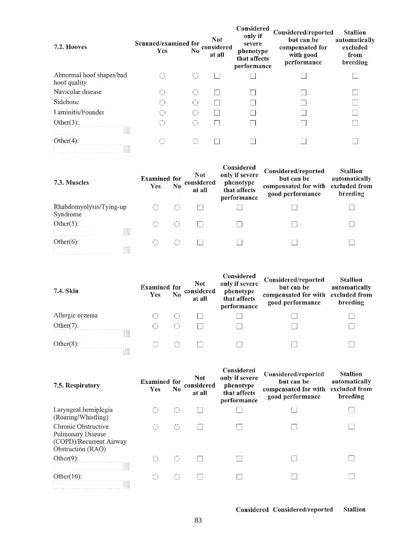
83
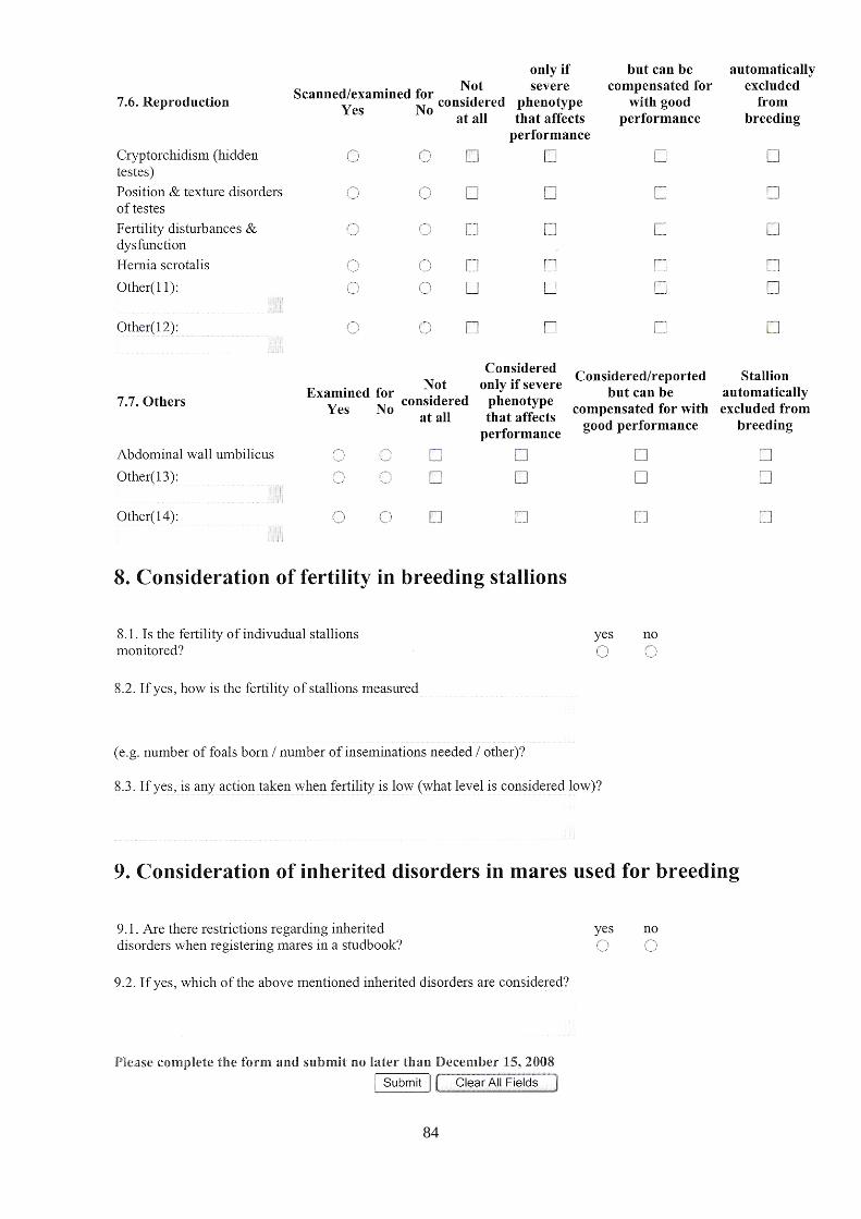
84