Imaging of Acute Invasive Fungal Rhinosinusitis in a Patient with ...
5
Hindawi Publishing Corporation Case Reports in Otolaryngology Volume 2013, Article ID 272314, 4 pages http://dx.doi.org/10.1155/2013/272314 Case Report Imaging of Acute Invasive Fungal Rhinosinusitis in a Patient with Gorlin Syndrome and Acute Lymphocytic Leukemia S. T. Donovan, 1 J. W. Thompson, 2 J. T. Sandlund, 3 E. E. Adderson, 4 E. K. Pivnick, 5 and J. H. Harreld 6 1 Department of Otolaryngology, University of Florida College of Medicine, 1600 SW Archer Road, Gainesville, FL 32608, USA 2 Department of Otolaryngology, University of Tennessee Health Sciences Center, 910 Madison Avenue, Memphis, TN 38163, USA 3 Department of Oncology, St. Jude Children’s Research Hospital, 262 Danny omas Boulevard, MS-260, Memphis, TN 38105, USA 4 Department of Infectious Diseases, St. Jude Children’s Research Hospital, 262 Danny omas Boulevard, MS-320, Memphis, TN 38105, USA 5 Departments of Pediatrics and Ophthalmology, University of Tennessee Health Sciences Center, 711 Jefferson Avenue, Suite 523, Memphis, TN 38105, USA 6 Department of Radiological Sciences, St. Jude Children’s Research Hospital, 262 Danny omas Boulevard, MS-220, Memphis, TN 38105, USA Correspondence should be addressed to J. H. Harreld; [email protected] Received 19 March 2013; Accepted 8 May 2013 Academic Editors: V. A. Resto and H. Sudhoff Copyright © 2013 S. T. Donovan et al. is is an open access article distributed under the Creative Commons Attribution License, which permits unrestricted use, distribution, and reproduction in any medium, provided the original work is properly cited. Gorlin Syndrome (GS), also known as nevoid basal cell carcinoma syndrome, is a rare autosomal dominant condition characterized by developmental abnormalities and predisposition to certain neoplasms. Acute invasive fungal rhinosinusitis (AIFRS) is an uncommon clinical entity characterized by high morbidity and mortality. In immunocompromised patients, computed tomography plays a critical role in screening for suspected AIFRS. However, due to the association between exposure to ionizing radiation and subsequent development of malignancies in patients with GS, patients with GS and suspected AIFRS present a unique and challenging clinical scenario. We present a case of a pediatric patient with GS and acute lymphocytic leukemia (ALL) diagnosed with AIFRS; to the best of our knowledge, it is the only case described in the literature. 1. Introduction Acute invasive fungal rhinosinusitis (AIFRS) is a serious con- dition characterized by high morbidity and a high mortality rate of 50%–80% in immunocompromised patients [1]. Due to the need for expeditious imaging in suspected cases to assist in diagnosis and management, CT is oſten used as the initial diagnostic modality, though magnetic resonance imaging (MRI) plays a critical role in the diagnosis of AIFRS [2]. Gorlin Syndrome (GS) is a rare autosomal dominant condition characterized by multiple basal cell carcinomas of the skin, odontogenic keratocysts, dermoid cysts, mesenteric cysts, pits of the palms and soles, eye anomalies, variable developmental delay, and skeletal abnormalities including macrocephaly, tall stature, and rib and vertebral anomalies [3, 4]. Gorlin Syndrome is due to a mutation in PTCH1, a tumor suppressor gene involved in the Sonic Hedgehog ligand- signaling pathway that plays a role in normal embryonic development [4]. Loss of heterozygosity due to this mutation predisposes patients to the characteristic features of the syndrome, as well as medulloblastomas, rhabdomyosarcomas and other cancers [4, 5]. Because exposure to ionizing radia- tion is an important cofactor for secondary malignancies in patients with GS, CT and X-ray should be used judiciously in these patients [6]. us, screening as well as diagnostic testing for fungal sinus disease presents a particular challenge in these patients. We present a case of a pediatric patient with GS and ALL diagnosed with AIFRS. We discuss the special considerations
Transcript of Imaging of Acute Invasive Fungal Rhinosinusitis in a Patient with ...
Hindawi Publishing CorporationCase Reports in OtolaryngologyVolume 2013, Article ID 272314, 4 pageshttp://dx.doi.org/10.1155/2013/272314
Case ReportImaging of Acute Invasive Fungal Rhinosinusitis in a Patientwith Gorlin Syndrome and Acute Lymphocytic Leukemia
S. T. Donovan,1 J. W. Thompson,2 J. T. Sandlund,3 E. E. Adderson,4
E. K. Pivnick,5 and J. H. Harreld6
1 Department of Otolaryngology, University of Florida College of Medicine, 1600 SW Archer Road, Gainesville, FL 32608, USA2Department of Otolaryngology, University of Tennessee Health Sciences Center, 910 Madison Avenue, Memphis, TN 38163, USA3Department of Oncology, St. Jude Children’s Research Hospital, 262 Danny Thomas Boulevard, MS-260, Memphis, TN 38105, USA4Department of Infectious Diseases, St. Jude Children’s Research Hospital, 262 Danny Thomas Boulevard, MS-320, Memphis,TN 38105, USA
5Departments of Pediatrics and Ophthalmology, University of Tennessee Health Sciences Center, 711 Jefferson Avenue,Suite 523, Memphis, TN 38105, USA
6Department of Radiological Sciences, St. Jude Children’s Research Hospital, 262 Danny Thomas Boulevard, MS-220, Memphis,TN 38105, USA
Correspondence should be addressed to J. H. Harreld; [email protected]
Received 19 March 2013; Accepted 8 May 2013
Academic Editors: V. A. Resto and H. Sudhoff
Copyright © 2013 S. T. Donovan et al. This is an open access article distributed under the Creative Commons Attribution License,which permits unrestricted use, distribution, and reproduction in any medium, provided the original work is properly cited.
Gorlin Syndrome (GS), also known as nevoid basal cell carcinoma syndrome, is a rare autosomal dominant condition characterizedby developmental abnormalities and predisposition to certain neoplasms. Acute invasive fungal rhinosinusitis (AIFRS) is anuncommon clinical entity characterized by highmorbidity andmortality. In immunocompromised patients, computed tomographyplays a critical role in screening for suspected AIFRS. However, due to the association between exposure to ionizing radiationand subsequent development of malignancies in patients with GS, patients with GS and suspected AIFRS present a unique andchallenging clinical scenario. We present a case of a pediatric patient with GS and acute lymphocytic leukemia (ALL) diagnosedwith AIFRS; to the best of our knowledge, it is the only case described in the literature.
1. Introduction
Acute invasive fungal rhinosinusitis (AIFRS) is a serious con-dition characterized by high morbidity and a high mortalityrate of 50%–80% in immunocompromised patients [1]. Dueto the need for expeditious imaging in suspected cases toassist in diagnosis and management, CT is often used asthe initial diagnostic modality, though magnetic resonanceimaging (MRI) plays a critical role in the diagnosis of AIFRS[2].
Gorlin Syndrome (GS) is a rare autosomal dominantcondition characterized by multiple basal cell carcinomas ofthe skin, odontogenic keratocysts, dermoid cysts, mesentericcysts, pits of the palms and soles, eye anomalies, variabledevelopmental delay, and skeletal abnormalities including
macrocephaly, tall stature, and rib and vertebral anomalies [3,4]. Gorlin Syndrome is due to a mutation in PTCH1, a tumorsuppressor gene involved in the Sonic Hedgehog ligand-signaling pathway that plays a role in normal embryonicdevelopment [4]. Loss of heterozygosity due to this mutationpredisposes patients to the characteristic features of thesyndrome, as well asmedulloblastomas, rhabdomyosarcomasand other cancers [4, 5]. Because exposure to ionizing radia-tion is an important cofactor for secondary malignancies inpatients with GS, CT and X-ray should be used judiciouslyin these patients [6]. Thus, screening as well as diagnostictesting for fungal sinus disease presents a particular challengein these patients.
We present a case of a pediatric patient with GS and ALLdiagnosed with AIFRS.We discuss the special considerations
2 Case Reports in Otolaryngology
of this case, in addition to the characteristics of GorlinSyndrome as they apply to the otolaryngologist, the clinicaland radiographic findings of AIFRS, and role of imaging inthe diagnosis of AIFRS.
2. Case Report
An 8-year-old Caucasian male presented to our facilitywith a 2-week history of refractory clear rhinorrhea, facialpain, jaw pain, and somnolence. The patient had previouslybeen diagnosed with sporadic GS after being found to havemultiple basal cell nevi and pitting of the palms and solesat the age of 5, as well as more recently a second relapseof ALL and chemotherapy-associated neutropenia (absoluteneutrophil count 0/mm3). Family history was negative forGS but revealed a maternal third cousin with osteosarcomaand a maternal second cousin with ALL. The patient denieddouble vision, decreased vision, and nasal obstruction but didhave one episode of epistaxis on the day of admission. Therewas no known exposure to nickel or significant secondhandsmoke exposure. At the time of evaluation he was receivingmicafungin for anti-fungal prophylaxis. On physical exami-nation there was a pale gray lesion on the palate suspicious foravascular tissue, versus underlying Gorlin-related cyst. Blackdebris in the nares was believed to be due to epistaxis; deeperexamination revealed grey/brown tissue and nasal polyposis.Because of concerns regarding ionizing radiation, evaluationwith MRI was pursued.
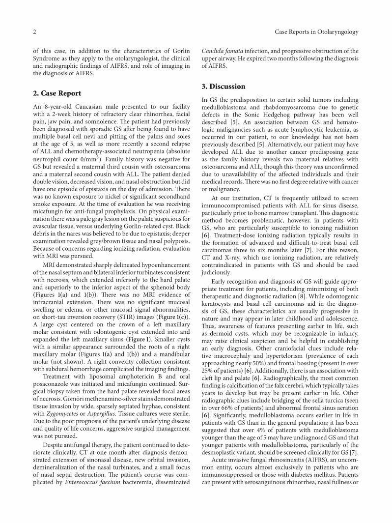
MRI demonstrated sharply delineated hypoenhancementof the nasal septumandbilateral inferior turbinates consistentwith necrosis, which extended inferiorly to the hard palateand superiorly to the inferior aspect of the sphenoid body(Figures 1(a) and 1(b)). There was no MRI evidence ofintracranial extension. There was no significant mucosalswelling or edema, or other mucosal signal abnormalities,on short-tau inversion recovery (STIR) images (Figure 1(c)).A large cyst centered on the crown of a left maxillarymolar consistent with odontogenic cyst extended into andexpanded the left maxillary sinus (Figure 1). Smaller cystswith a similar appearance surrounded the roots of a rightmaxillary molar (Figures 1(a) and 1(b)) and a mandibularmolar (not shown). A right convexity collection consistentwith subdural hemorrhage complicated the imaging findings.
Treatment with liposomal amphotericin B and oralposaconazole was initiated and micafungin continued. Sur-gical biopsy taken from the hard palate revealed focal areasof necrosis. Gomorimethenamine-silver stains demonstratedtissue invasion by wide, sparsely septated hyphae, consistentwith Zygomycetes or Aspergillus. Tissue cultures were sterile.Due to the poor prognosis of the patient’s underlying diseaseand quality of life concerns, aggressive surgical managementwas not pursued.
Despite antifungal therapy, the patient continued to dete-riorate clinically. CT at one month after diagnosis demon-strated extension of sinonasal disease, new orbital invasion,demineralization of the nasal turbinates, and a small focusof nasal septal destruction. The patient’s course was com-plicated by Enterococcus faecium bacteremia, disseminated
Candida famata infection, and progressive obstruction of theupper airway. He expired twomonths following the diagnosisof AIFRS.
3. Discussion
In GS the predisposition to certain solid tumors includingmedulloblastoma and rhabdomyosarcoma due to geneticdefects in the Sonic Hedgehog pathway has been welldescribed [5]. An association between GS and hemato-logic malignancies such as acute lymphocytic leukemia, asoccurred in our patient, to our knowledge has not beenpreviously described [5]. Alternatively, our patient may havedeveloped ALL due to another cancer predisposing geneas the family history reveals two maternal relatives withosteosarcoma and ALL, though this theory was unconfirmeddue to unavailability of the affected individuals and theirmedical records.Therewas no first degree relativewith canceror malignancy.
At our institution, CT is frequently utilized to screenimmunocompromised patients with ALL for sinus disease,particularly prior to bone marrow transplant.This diagnosticmethod becomes problematic, however, in patients withGS, who are particularly susceptible to ionizing radiation[6]. Treatment-dose ionizing radiation typically results inthe formation of advanced and difficult-to-treat basal cellcarcinomas three to six months later [7]. For this reason,CT and X-ray, which use ionizing radiation, are relativelycontraindicated in patients with GS and should be usedjudiciously.
Early recognition and diagnosis of GS will guide appro-priate treatment for patients, including minimizing of boththerapeutic and diagnostic radiation [8]. While odontogenickeratocysts and basal cell carcinomas aid in the diagno-sis of GS, these characteristics are usually progressive innature and may appear in later childhood and adolescence.Thus, awareness of features presenting earlier in life, suchas dermoid cysts, which may be recognizable in infancy,may raise clinical suspicion and be helpful in establishingan early diagnosis. Other craniofacial clues include rela-tive macrocephaly and hypertelorism (prevalence of eachapproaching nearly 50%) and frontal bossing (present in over25% of patients) [6]. Additionally, there is an association withcleft lip and palate [6]. Radiographically, the most commonfinding is calcification of the falx cerebri, which typically takesyears to develop but may be present earlier in life. Otherradiographic clues include bridging of the sella turcica (seenin over 66% of patients) and abnormal frontal sinus aeration[6]. Significantly, medulloblastoma occurs earlier in life inpatients with GS than in the general population; it has beensuggested that over 4% of patients with medulloblastomayounger than the age of 5 may have undiagnosed GS and thatyounger patients with medulloblastoma, particularly of thedesmoplastic variant, should be screened clinically for GS [7].
Acute invasive fungal rhinosinusitis (AIFRS), an uncom-mon entity, occurs almost exclusively in patients who areimmunosuppressed or those with diabetes mellitus. Patientscan present with serosanguinous rhinorrhea, nasal fullness or
Case Reports in Otolaryngology 3
(a) (b) (c)
Figure 1: (a) Coronal postcontrast fat-saturated T1-weighted images (T1WI) and (b) sagittal postcontrast T1WI demonstrate sharplydelineated hypoenhancement of the nasal septum and bilateral inferior turbinates extending inferiorly to the hard palate and superiorlyto the inferior aspect of the sphenoid body (outlined by small arrows) without significant associated mucosal edema on STIR images (c). Anodontogenic cyst centered on the crown of a left maxillary molar fills and expands the left maxillary sinus (thick arrows). A smaller maxillaryodontogenic cyst is present on the right (curved arrow).
obstruction, sinus pain, diplopia, loss of visual acuity, cranialnerve involvement, or with nonspecific signs such as fever [1,9]. Orbital involvement is typical, although it was not presentuntil late in this case [10]. Bony destruction is common [11].With an overall mortality rate of 50%–80%, prompt diagnosisand management are key [1]. The combination of promptsurgical intervention and antifungal therapy has provedsuperior to monotherapy with intravenous antifungals [11].
Previously, conventional wisdom dictated that CT wassufficient for radiographic evaluation for AIFRS. However,the sensitivity of MRI in detecting early findings of AIFRS,such as periantral fat infiltration and tissue necrosis, issuperior to that of CT; [2, 12] bony destruction detectableby CT is a late finding with respect to disease progression.MRI is therefore helpful in differentiating AIFRS from otherinflammatory entities and can make a crucial difference inpatient care. MRI is particularly suitable in circumstances ofhigh clinical suspicion for early or subtle disease, or relativecontraindication to CT, both present in this case.
Hypoenhancement and near-normal T2 signal in invasivefungal disease due to microvascular invasion of hyphae andsubsequent necrosis in the absence of an immune responsesufficient to cause substantial mucosal edema, as seen inthis case, are suggestive of AIFRS; this is distinct fromthe diffuse enhancement and T2 hyperintensity typicallyseen in other inflammatory diseases [12, 13]. A pattern ofnasal cavity, orbital, ethmoid, and maxillary involvement ishighly suggestive [14]. For the head and neck surgeon, MRIcan better delineate areas of involvement, which may guidesurgical intervention in the form of endoscopic versus opensurgical debridement and simple debridement versus moreextensive resections [15].
This report of AIFRS in a patient with GS represents anuncommonly encountered comorbidity and exhibits a uniqueand challenging clinical scenario. Because ionizing radiationshould be minimized in patients with GS, in this clinical
situation, MRI should be promptly performed for timely andaccurate diagnosis of AIFRS.
Acknowledgments
Thiswork is supported in part byGrant no. CA21765 from theNational Cancer Institute and by the American Lebanese andSyrian Associated Charities.
References
[1] M. B. Gillespie, B. W. O’Malley Jr., and H. W. Francis, “Anapproach to fulminant invasive fungal rhinosinusitis in theimmunocompromised host,” Archives of Otolaryngology, vol.124, no. 5, pp. 520–526, 1998.
[2] E. R. Groppo, I. H. El-Sayed, A. H. Aiken, and C. M. Glaston-bury, “Computed tomography andmagnetic resonance imagingcharacteristics of acute invasive fungal sinusitis,” Archives ofOtolaryngology, vol. 137, no. 10, pp. 1005–1010, 2011.
[3] R. J. Gorlin and R. W. Goltz, “Multiple nevoid basal-cellepithelioma, jaw cysts and bifid rib: a syndrome,” The NewEngland journal of medicine, vol. 262, pp. 908–912, 1960.
[4] L. L. Muzio, “Nevoid basal cell carcinoma syndrome (Gorlinsyndrome),” Orphanet Journal of Rare Diseases, vol. 3, no. 1,article 32, 2008.
[5] M. M. Cajaiba, A. E. Bale, M. Alvarez-Franco, J. McNamara,and M. Reyes-Mugica, “Rhabdomyosarcoma, Wilms tumor,and deletion of the patched gene in Gorlin syndrome,” NatureClinical Practice Oncology, vol. 3, no. 10, pp. 575–580, 2006.
[6] V. E. Kimonis, A. M. Goldstein, B. Pastakia et al., “Clinicalmanifestations in 105 persons with nevoid basal cell carcinomasyndrome,” The American Journal of Medical Genetics, vol. 69,no. 3, pp. 299–308, 1997.
[7] D. G. R. Evans, L. A. Farndon, L. D. Burnell, H. Rao Gatta-maneni, and J. M. Birch, “The incidence of Gorlin syndromein 173 consecutive cases of medulloblastoma,” British Journal ofCancer, vol. 64, no. 5, pp. 959–961, 1991.
4 Case Reports in Otolaryngology
[8] A. F. Bree and M. R. Shah, “Consensus statement from thefirst international colloquium on basal cell nevus syndrome(BCNS),”The American Journal of Medical Genetics A, vol. 155,no. 9, pp. 2091–2097, 2011.
[9] S. Abbasi, J. L. Shenep, W. T. Hughes, and P. M. Flynn,“Aspergillosis in children with cancer: a 34-year experience,”Clinical Infectious Diseases, vol. 29, no. 5, pp. 1210–1219, 1999.
[10] R. Chandrasekharan, M. Thomas, and V. Rupa, “Comparativestudy of orbital involvement in invasive and non-invasive fungalsinusitis,” Journal of Laryngology and Otology, vol. 126, no. 2, pp.152–158, 2012.
[11] F. Kasapoglu, H. Coskun, O. A. Ozmen, H. Akalin, and B.Ener, “Acute invasive fungal rhinosinusitis: evaluation of 26patients treated with endonasal or open surgical procedures,”Otolaryngology, vol. 143, no. 5, pp. 614–620, 2010.
[12] S. Safder, J. S. Carpenter, T. D. Roberts, and N. Bailey, “The“black turbinate” sign: an early MR imaging finding of nasalmucormycosis,”TheAmerican Journal ofNeuroradiology, vol. 31,no. 4, pp. 771–774, 2010.
[13] S. J. Rassi, A. E. Melkane, H. G. Rizk, and H. A. Dahoui,“Sinonasal mucormycosis in immunocompromised pediatricpatients,” Journal of Pediatric Hematology/Oncology, vol. 31, no.12, pp. 907–910, 2009.
[14] D. A. Herrera, A. B. Dublin, E. L. Ormsby, S. Aminpour, and L.P. Howell, “Imaging findings of rhinocerebral mucormycosis,”Skull Base, vol. 19, no. 2, pp. 117–126, 2009.
[15] R. C. Howells and H. H. Ramadan, “Usefulness of computedtomography and magnetic resonance in fulminant invasivefungal rhinosinusitis,” The American Journal of Rhinology, vol.15, no. 4, pp. 255–261, 2001.

Submit your manuscripts athttp://www.hindawi.com
Stem CellsInternational
Hindawi Publishing Corporationhttp://www.hindawi.com Volume 2014
Hindawi Publishing Corporationhttp://www.hindawi.com Volume 2014
MEDIATORSINFLAMMATION
of
Hindawi Publishing Corporationhttp://www.hindawi.com Volume 2014
Behavioural Neurology
EndocrinologyInternational Journal of
Hindawi Publishing Corporationhttp://www.hindawi.com Volume 2014
Hindawi Publishing Corporationhttp://www.hindawi.com Volume 2014
Disease Markers
Hindawi Publishing Corporationhttp://www.hindawi.com Volume 2014
BioMed Research International
OncologyJournal of
Hindawi Publishing Corporationhttp://www.hindawi.com Volume 2014
Hindawi Publishing Corporationhttp://www.hindawi.com Volume 2014
Oxidative Medicine and Cellular Longevity
Hindawi Publishing Corporationhttp://www.hindawi.com Volume 2014
PPAR Research
The Scientific World JournalHindawi Publishing Corporation http://www.hindawi.com Volume 2014
Immunology ResearchHindawi Publishing Corporationhttp://www.hindawi.com Volume 2014
Journal of
ObesityJournal of
Hindawi Publishing Corporationhttp://www.hindawi.com Volume 2014
Hindawi Publishing Corporationhttp://www.hindawi.com Volume 2014
Computational and Mathematical Methods in Medicine
OphthalmologyJournal of
Hindawi Publishing Corporationhttp://www.hindawi.com Volume 2014
Diabetes ResearchJournal of
Hindawi Publishing Corporationhttp://www.hindawi.com Volume 2014
Hindawi Publishing Corporationhttp://www.hindawi.com Volume 2014
Research and TreatmentAIDS
Hindawi Publishing Corporationhttp://www.hindawi.com Volume 2014
Gastroenterology Research and Practice
Hindawi Publishing Corporationhttp://www.hindawi.com Volume 2014
Parkinson’s Disease
Evidence-Based Complementary and Alternative Medicine
Volume 2014Hindawi Publishing Corporationhttp://www.hindawi.com