Orbital Metastasis of Breast Cancer Mimicking Invasive Fungal ...
5
Case Report Orbital Metastasis of Breast Cancer Mimicking Invasive Fungal Rhinosinusitis Mayara Tabai, Igor Moreira Hazboun, Emerson Taro Inoue Sakuma, Marcelo Hamilton Sampaio, and Eulalia Sakano Department of Otolaryngology Head and Neck, Faculty of Medical Sciences, University of Campinas (UNICAMP), P.O. Box 6111, 13081-970 Campinas, SP, Brazil Correspondence should be addressed to Mayara Tabai; [email protected] Received 23 May 2016; Revised 29 June 2016; Accepted 10 July 2016 Academic Editor: Rong-San Jiang Copyright © 2016 Mayara Tabai et al. is is an open access article distributed under the Creative Commons Attribution License, which permits unrestricted use, distribution, and reproduction in any medium, provided the original work is properly cited. Introduction. A range of traumatic, vascular, inflammatory, infectious, and neoplastic processes can affect the orbit and its structures. In the area of otolaryngology, the rhino-orbital-cerebral involvement of invasive fungal rhinosinusitis can affect the orbit, which may look like initially a rhinosinusitis or even mimic malignancy. Case Presentation. Female patient, 32 years old, with headache and ocular proptosis. She was using prednisone in immunosuppressive doses for a year and had breast cancer treated three years earlier. e initial CT scan showed opacification of the sphenoid and ethmoid sinuses, leſt intraorbital involvement and contrast impregnation in the cavernous sinus. e biopsy resulted positive for invasive ductal carcinoma of the breast. Discussion. e initial CT scan of our patient showed both signs of early changes of invasive fungal rhinosinusitis (IFR) and possible metastatic involvement. e intracranial extension and ocular involvement are usually the most common signs of IFR (first hypothesis). Among metastases at the orbit and the eye, breast and lung carcinomas are the most frequent. Conclusion. Although several studies on the differential diagnosis of orbital lesions exist, especially when it concerns the involvement of the nasal cavity, the diagnosis by imaging is still a challenge. 1. Introduction Paranasal sinuses and orbit are potential sites for metas- tases. Distant metastases of skull base occur in 4% of patients with cancer and the most common primary sites are breast, prostate, and lung. Occasionally, breast carcinoma can metastasize to the nasal cavity and orbit and manifest with unspecific symptoms. If the primary tumor is unknown and the metastatic deposits mimic rhinosinusitis or cause cavernous sinus syndrome, the diagnosis is usually delayed. Although metastasis to the orbit is rare, it must be considered in the differential diagnosis of any patient with history of cancer, presenting with ophthalmic symptom [1]. Orbital metastasis constitutes 3% of orbital lesions and 10% of orbital tumors. Breast cancer is the most common primary site for orbital metastasis in women with known dis- seminated disease and the ocular symptoms can appear years aſter the inicial brest cancer diagnosis [2]. Considering all types of orbital metastasis, in 19%, there is no history of cancer when the patient presents with ophthalmic symptoms and, in 10%, the primary site remains unclear, despite systemic evaluation. Computed tomography or magnetic resonance imaging can show only a thickening of the extraocular muscles, and oſten the possibility of an inflammatory process is raised [3]. A careful history and physical examination, with special attention to the orbit and eye attachments, are necessary to identify subtle orbital anomalies that might otherwise be ignored or mistakenly contributed to a nonorbital process. In this context, we can mention, in the area of otolaryngology, the rhino-orbital-cerebral involvement of invasive fungal rhi- nosinusitis, especially in immunocompromised patients [4]. Clinically, rhino-orbital involvement initially may look like a rhinosinusitis or even mimic malignancy. Also, we must consider the differential diagnosis of orbital disorders, such as migraine; chronic rhinosinusitis; preseptal and orbital Hindawi Publishing Corporation Case Reports in Otolaryngology Volume 2016, Article ID 2913241, 4 pages http://dx.doi.org/10.1155/2016/2913241
Transcript of Orbital Metastasis of Breast Cancer Mimicking Invasive Fungal ...

Case ReportOrbital Metastasis of Breast Cancer MimickingInvasive Fungal Rhinosinusitis
Mayara Tabai, Igor Moreira Hazboun, Emerson Taro Inoue Sakuma,Marcelo Hamilton Sampaio, and Eulalia Sakano
Department of Otolaryngology Head and Neck, Faculty of Medical Sciences, University of Campinas (UNICAMP),P.O. Box 6111, 13081-970 Campinas, SP, Brazil
Correspondence should be addressed to Mayara Tabai; [email protected]
Received 23 May 2016; Revised 29 June 2016; Accepted 10 July 2016
Academic Editor: Rong-San Jiang
Copyright © 2016 Mayara Tabai et al. This is an open access article distributed under the Creative Commons Attribution License,which permits unrestricted use, distribution, and reproduction in any medium, provided the original work is properly cited.
Introduction. A range of traumatic, vascular, inflammatory, infectious, and neoplastic processes can affect the orbit and its structures.In the area of otolaryngology, the rhino-orbital-cerebral involvement of invasive fungal rhinosinusitis can affect the orbit, whichmay look like initially a rhinosinusitis or even mimic malignancy. Case Presentation. Female patient, 32 years old, with headacheand ocular proptosis. She was using prednisone in immunosuppressive doses for a year and had breast cancer treated three yearsearlier. The initial CT scan showed opacification of the sphenoid and ethmoid sinuses, left intraorbital involvement and contrastimpregnation in the cavernous sinus. The biopsy resulted positive for invasive ductal carcinoma of the breast. Discussion. Theinitial CT scan of our patient showed both signs of early changes of invasive fungal rhinosinusitis (IFR) and possible metastaticinvolvement. The intracranial extension and ocular involvement are usually the most common signs of IFR (first hypothesis).Among metastases at the orbit and the eye, breast and lung carcinomas are the most frequent. Conclusion. Although several studieson the differential diagnosis of orbital lesions exist, especially when it concerns the involvement of the nasal cavity, the diagnosisby imaging is still a challenge.
1. Introduction
Paranasal sinuses and orbit are potential sites for metas-tases. Distant metastases of skull base occur in 4% ofpatients with cancer and the most common primary sitesare breast, prostate, and lung. Occasionally, breast carcinomacan metastasize to the nasal cavity and orbit and manifestwith unspecific symptoms. If the primary tumor is unknownand the metastatic deposits mimic rhinosinusitis or causecavernous sinus syndrome, the diagnosis is usually delayed.Althoughmetastasis to the orbit is rare, it must be consideredin the differential diagnosis of any patient with history ofcancer, presenting with ophthalmic symptom [1].
Orbital metastasis constitutes 3% of orbital lesions and10% of orbital tumors. Breast cancer is the most commonprimary site for orbital metastasis in women with known dis-seminated disease and the ocular symptoms can appear yearsafter the inicial brest cancer diagnosis [2]. Considering all
types of orbitalmetastasis, in 19%, there is no history of cancerwhen the patient presents with ophthalmic symptoms and,in 10%, the primary site remains unclear, despite systemicevaluation. Computed tomography or magnetic resonanceimaging can show only a thickening of the extraocularmuscles, and often the possibility of an inflammatory processis raised [3].
A careful history and physical examination, with specialattention to the orbit and eye attachments, are necessaryto identify subtle orbital anomalies that might otherwise beignored or mistakenly contributed to a nonorbital process. Inthis context, we can mention, in the area of otolaryngology,the rhino-orbital-cerebral involvement of invasive fungal rhi-nosinusitis, especially in immunocompromised patients [4].
Clinically, rhino-orbital involvement initially may looklike a rhinosinusitis or even mimic malignancy. Also, wemust consider the differential diagnosis of orbital disorders,such asmigraine; chronic rhinosinusitis; preseptal and orbital
Hindawi Publishing CorporationCase Reports in OtolaryngologyVolume 2016, Article ID 2913241, 4 pageshttp://dx.doi.org/10.1155/2016/2913241

2 Case Reports in Otolaryngology
Figure 1: CTwith contrast in the sinuses demonstrates opacificationof certain ethmoid cells (red arrow) and fluid level in the sphenoidsinus bilaterally (white arrow).
cellulitis; primary and secondary orbital tumor (metasta-sis); posttraumatic hematoma; inflammatory pseudotumor;thrombosis of the cavernous sinus; and Graves’ disease.
This paper aims to report a case of orbital metastasis ofbreast cancer mimicking invasive fungal rhinosinusitis.
2. Case Report
Female patient, 32 years old, has been complaining about aheadache for a month, with worsening pain and ocular prop-tosis a week before, associated with hyaline nasal discharge. Athrobbing headache with progression was located in the leftfrontal region, associated with fatigue and myalgia. She alsoreported proptosis on the left eye, followed by diplopia andeye pain.
Regarding her medical history, the patient had triple neg-ative breast adenocarcinoma treated with radical mastectomyand chemotherapy three years earlier. Moreover, the patienthad atopic dermatitis, which was difficult to control, diag-nosed a year before, using prednisone in immunosuppressivedoses (80mg/day in the last year), alsowith controlled asthmaand hypertension.
The physical examination on the patient showed regularcondition, normal vital signs, facies cushingoid, and leftocular proptosis. Nasal endoscopy only indicated a discretehyaline secretion in the nasal vestibule.
Facedwith this situation, we opted to performComputer-ized Tomography of the skull and sinuses.The initial CT scanrevealed discreet parenchymal atrophy, opacification of thesphenoid and ethmoid sinuses bilaterally, and left intraorbitalinvolvement with contrast impregnation that extends to theintracranial region through the cavernous sinus determiningpachymeningeal enhancement (Figures 1 and 2).
After discussion, we got the hypothesis that it was invasivefungal rhinosinusitis with involvement of the cavernoussinus; then we scheduled a biopsy and gave intravenousAmphotericinB.Oneday later, the patient developedworsen-ing of proptosis and ocular motility dysfunction and reachedoculomotor nerve palsy.
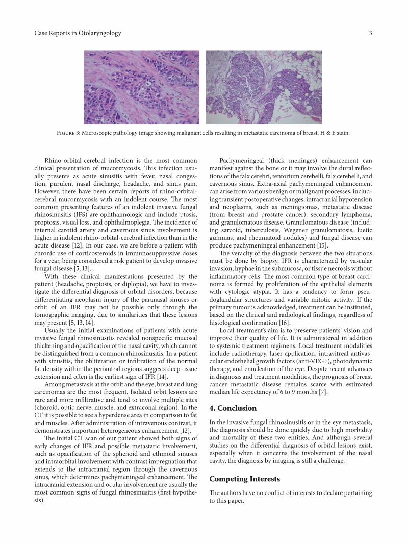
Then the case was discussed with staff of Radiology, whoguided the achievement of chest CT. In the new images werevisualized a nodule in the right breast of 1.6 cm with regularborders,mediastinal lymph nodes up to 2.2 cm, peribronchialmass (lymph node conglomerate), and atelectasis in the leftlung. The orbital and lymph nodes biopsy tested positivefor invasive ductal carcinoma of the breast (Figure 3). The
Figure 2: CT with contrast in the sinuses demonstrates left intraor-bital involvement (white arrow) that extends through the cavernoussinus determining pachymeningeal enhancement (red arrow).
patient was referred for radiotherapy and chemotherapy.After initiating chemotherapy, the patient presented clinicalinstability and died, not being possible to perform additionalimaging studies.
3. Discussion
Breast cancer may metastasize to uncommon anatomicsites, but there are no solid statistical data concerning thefrequency of head and neck metastasis in cancer patients.Some authors suggest that histologically confirmed orbitalmetastasis is diagnosed once or twice per year at largerclinical centers. These patients often complain of ocularasymmetry (noticed by the patient) or diplopia, withoutvisual field impairment. Depending on the series, breastcancers account for 29–53% of orbital metastasis and theinfiltrating lobular breast cancer is themost common subtype(87.5%) [5]. The fact that our patients present a ductal breastcancer makes our case even rarer. The presence of ocularmetastasis is a bad prognostic indicator, with survival that canrange from0 to 64months, with an average of fivemonths [6].
The extraocular muscles represent the main site ofbreast cancer orbital metastasis, causing pain, proptosis, anddiplopia. They are identified pathologically as solid depositsof the muscles. Orbital metastasis may cause exophthalmos,from mass effect, or enophthalmos [7]. Metastatic breastcancer rarely can manifest as cranial nerve palsies (incidenceof 0.13%). The most frequently affected cranial nerves are V(70%) and VII (60%). Rarely, cavernous sinus metastasis canbe the first presentation of an undetected breast cancer. Thecavernous sinus can be affected by inflammatory, vascular,or neoplastic conditions. Neoplasm may arise from adjacentstructures or from distant metastasis [8].
There have been four previous case reports of cavernoussinus syndrome as the first presentation of metastatic breastcancer. However, the initial clinical presentation of thesecases varies: Ryan et al. reported a patient who presentedwith headache and painful proptosis [9]; Martın Polo et al.reported a patient with pain and numbness over the left sideof face with occasional diplopia [10]; Fyrmpas et al. publisheda case who presented with rhino-orbital cellulites [1]; andKhaw et al. reported a patient with ptosis due to cavernoussinus syndrome as a rare presentation of advanced breastmetastasis [11].
Case Reports in Otolaryngology 3
Figure 3: Microscopic pathology image showing malignant cells resulting in metastatic carcinoma of breast. H & E stain.
Rhino-orbital-cerebral infection is the most commonclinical presentation of mucormycosis. This infection usu-ally presents as acute sinusitis with fever, nasal conges-tion, purulent nasal discharge, headache, and sinus pain.However, there have been certain reports of rhino-orbital-cerebral mucormycosis with an indolent course. The mostcommon presenting features of an indolent invasive fungalrhinosinusitis (IFS) are ophthalmologic and include ptosis,proptosis, visual loss, and ophthalmoplegia. The incidence ofinternal carotid artery and cavernous sinus involvement ishigher in indolent rhino-orbital-cerebral infection than in theacute disease [12]. In our case, we are before a patient withchronic use of corticosteroids in immunosuppressive dosesfor a year, being considered a risk patient to develop invasivefungal disease [5, 13].
With these clinical manifestations presented by thepatient (headache, proptosis, or diplopia), we have to inves-tigate the differential diagnosis of orbital disorders, becausedifferentiating neoplasm injury of the paranasal sinuses ororbit of an IFR may not be possible only through thetomographic imaging, due to similarities that these lesionsmay present [5, 13, 14].
Usually the initial examinations of patients with acuteinvasive fungal rhinosinusitis revealed nonspecific mucosalthickening and opacification of the nasal cavity, which cannotbe distinguished from a common rhinosinusitis. In a patientwith sinusitis, the obliteration or infiltration of the normalfat density within the periantral regions suggests deep tissueextension and often is the earliest sign of IFR [14].
Amongmetastasis at the orbit and the eye, breast and lungcarcinomas are the most frequent. Isolated orbit lesions arerare and more infiltrative and tend to involve multiple sites(choroid, optic nerve, muscle, and extraconal region). In theCT it is possible to see a hyperdense area in comparison to fatand muscles. After administration of intravenous contrast, itdemonstrates important heterogeneous enhancement [12].
The initial CT scan of our patient showed both signs ofearly changes of IFR and possible metastatic involvement,such as opacification of the sphenoid and ethmoid sinusesand intraorbital involvement with contrast impregnation thatextends to the intracranial region through the cavernoussinus, which determines pachymeningeal enhancement. Theintracranial extension and ocular involvement are usually themost common signs of fungal rhinosinusitis (first hypothe-sis).
Pachymeningeal (thick meninges) enhancement canmanifest against the bone or it may involve the dural reflec-tions of the falx cerebri, tentorium cerebelli, falx cerebelli, andcavernous sinus. Extra-axial pachymeningeal enhancementcan arise from various benign ormalignant processes, includ-ing transient postoperative changes, intracranial hypotensionand neoplasms, such as meningiomas, metastatic disease(from breast and prostate cancer), secondary lymphoma,and granulomatous disease. Granulomatous disease (includ-ing sarcoid, tuberculosis, Wegener granulomatosis, lueticgummas, and rheumatoid nodules) and fungal disease canproduce pachymeningeal enhancement [15].
The veracity of the diagnosis between the two situationsmust be done by biopsy. IFR is characterized by vascularinvasion, hyphae in the submucosa, or tissue necrosis withoutinflammatory cells. The most common type of breast carci-noma is formed by proliferation of the epithelial elementswith cytologic atypia. It has a tendency to form pseu-doglandular structures and variable mitotic activity. If theprimary tumor is acknowledged, treatment can be instituted,based on the clinical and radiological findings, regardless ofhistological confirmation [16].
Local treatment’s aim is to preserve patients’ vision andimprove their quality of life. It is administered in additionto systemic treatment regimens. Local treatment modalitiesinclude radiotherapy, laser application, intravitreal antivas-cular endothelial growth factors (anti-VEGF), photodynamictherapy, and enucleation of the eye. Despite recent advancesin diagnosis and treatmentmodalities, the prognosis of breastcancer metastatic disease remains scarce with estimatedmedian life expectancy of 6 to 9 months [7].
4. Conclusion
In the invasive fungal rhinosinusitis or in the eye metastasis,the diagnosis should be done quickly due to high morbidityand mortality of these two entities. And although severalstudies on the differential diagnosis of orbital lesions exist,especially when it concerns the involvement of the nasalcavity, the diagnosis by imaging is still a challenge.
Competing Interests
The authors have no conflict of interests to declare pertainingto this paper.

4 Case Reports in Otolaryngology
References
[1] G. Fyrmpas, D. Televantou, V. Papageorgiou, F. Nofal, and J.Constantinidis, “Unsuspected breast carcinoma presenting asorbital complication of rhinosinusitis,” European Archives ofOto-Rhino-Laryngology, vol. 265, no. 8, pp. 979–982, 2008.
[2] R. A. Goldberg, J. Rootman, and R. A. Cline, “Tumorsmetastatic to the orbit: a changing picture,” Survey of Ophthal-mology, vol. 35, no. 1, pp. 1–24, 1990.
[3] J. A. Shields, C. L. Shields, H. K. Brotman, C. Carvalho,N. Perez,and R. C. Eagle Jr., “Cancer metastatic to the orbit: the 2000Robert M. Curts lecture,”Ophthalmic Plastic and ReconstructiveSurgery, vol. 17, no. 5, pp. 346–354, 2001.
[4] P. Keche, A. Z. Nitnaware, M. Mair, P. Sakhare, and S. Satpute,“A study of tumours giving rise to unilateral proptosis,” IndianJournal of Otolaryngology and Head and Neck Surgery, vol. 65,no. 1, pp. 6–13, 2013.
[5] M. N. Gamaletsou, N. V. Sipsas, E. Roilides, and T. J. Walsh,“Rhino-orbital-cerebral mucormycosis,” Current Infectious Dis-ease Reports, vol. 14, no. 4, pp. 423–434, 2012.
[6] V. Ratanatharathorn, W. E. Powers, J. Grimm et al., “Eyemetastasis from carcinoma of the breast: diagnosis, radiationtreatment and results,” Cancer Treatment Reviews, vol. 18, no.4, pp. 261–276, 1991.
[7] I. Georgalas, T. Paraskevopoulos, C. Koutsandrea et al., “Oph-thalmic metastasis of breast cancer and ocular side effects frombreast cancer treatment andmanagement:mini review,”BioMedResearch International, vol. 2015, Article ID 574086, 8 pages,2015.
[8] S. M. Hall, A. U. Buzdar, and G. R. Blumenschein, “Cranialnerve palsies in metastatic breast cancer due to osseous metas-tasis without intracranial involvement,”Cancer, vol. 52, no. 1, pp.180–184, 1983.
[9] M. W. Ryan, C. H. Rassekh, and G. Chaljub, “Metastatic breastcarcinoma presenting as cavernous sinus syndrome,” Annals ofOtology, Rhinology andLaryngology, vol. 105, no. 8, pp. 666–668,1996.
[10] J. Martın Polo, M. T. Rivas Lopez, R. Martın Polo, A. OrtinCastano, J. Arcaya Navarro, and J. Cacho Gutierrez, “Cavernoussinus syndrome: an initial expression of a breast carcinoma,”Neurologia, vol. 20, no. 3, pp. 153–155, 2005.
[11] K. Khaw, N. Ramli, and K. Rahmat, “Ptosis due to cavernoussinus syndrome as a rare presentation of advanced breastmetastasis in a patient with delayed diagnosis,” MalaysianFamily Physician, vol. 7, no. 1, pp. 31–33, 2012.
[12] W. C. Harrill, M. G. Stewart, A. G. Lee, and P. Cernoch,“Chronic rhinocerebral mucormycosis,” Laryngoscope, vol. 106,no. 10, pp. 1292–1297, 1996.
[13] J. S. McNulty, “Rhinocerebral mucormycosis: predisposingfactors,” Laryngoscope, vol. 92, no. 10, pp. 1140–1143, 1982.
[14] A. T. Ilica, M. Mossa-Basha, F. Maluf, I. Izbudak, and N.Aygun, “Clinical and radiologic features of fungal diseases of theparanasal sinuses,” Journal of Computer Assisted Tomography,vol. 36, no. 5, pp. 570–576, 2012.
[15] J. G. Smirniotopoulos, F. M. Murphy, E. J. Rushing, J. H. Rees,and J. W. Schroeder, “Patterns of contrast enhancement in thebrain and meninges,” Radiographics, vol. 27, no. 2, pp. 525–551,2007.
[16] F. Laigle-Donadey, S. Taillibert, N.Martin-Duverneuil, J. Hilde-brand, and J.-Y. Delattre, “Skull-base metastases,” Journal ofNeuro-Oncology, vol. 75, no. 1, pp. 63–69, 2005.

Submit your manuscripts athttp://www.hindawi.com
Stem CellsInternational
Hindawi Publishing Corporationhttp://www.hindawi.com Volume 2014
Hindawi Publishing Corporationhttp://www.hindawi.com Volume 2014
MEDIATORSINFLAMMATION
of
Hindawi Publishing Corporationhttp://www.hindawi.com Volume 2014
Behavioural Neurology
EndocrinologyInternational Journal of
Hindawi Publishing Corporationhttp://www.hindawi.com Volume 2014
Hindawi Publishing Corporationhttp://www.hindawi.com Volume 2014
Disease Markers
Hindawi Publishing Corporationhttp://www.hindawi.com Volume 2014
BioMed Research International
OncologyJournal of
Hindawi Publishing Corporationhttp://www.hindawi.com Volume 2014
Hindawi Publishing Corporationhttp://www.hindawi.com Volume 2014
Oxidative Medicine and Cellular Longevity
Hindawi Publishing Corporationhttp://www.hindawi.com Volume 2014
PPAR Research
The Scientific World JournalHindawi Publishing Corporation http://www.hindawi.com Volume 2014
Immunology ResearchHindawi Publishing Corporationhttp://www.hindawi.com Volume 2014
Journal of
ObesityJournal of
Hindawi Publishing Corporationhttp://www.hindawi.com Volume 2014
Hindawi Publishing Corporationhttp://www.hindawi.com Volume 2014
Computational and Mathematical Methods in Medicine
OphthalmologyJournal of
Hindawi Publishing Corporationhttp://www.hindawi.com Volume 2014
Diabetes ResearchJournal of
Hindawi Publishing Corporationhttp://www.hindawi.com Volume 2014
Hindawi Publishing Corporationhttp://www.hindawi.com Volume 2014
Research and TreatmentAIDS
Hindawi Publishing Corporationhttp://www.hindawi.com Volume 2014
Gastroenterology Research and Practice
Hindawi Publishing Corporationhttp://www.hindawi.com Volume 2014
Parkinson’s Disease
Evidence-Based Complementary and Alternative Medicine
Volume 2014Hindawi Publishing Corporationhttp://www.hindawi.com