Brain Metastasis
12
Brain metastases — parenchymal metastases and lepto- meningeal metastases (BOX1) — most commonly arise from cancers of the lung, breast and skin (melanoma), but also occur at a reduced frequency in patients with diverse cancer types. The incidence of brain metastases is highest in patients with lung tumours. Approximately 10–25% of patients with lung cancer have brain metas- tases at diagnosis and another 40–50% develop them during the course of their disease, with an even greater incidence at autopsy 1 . Brain metastases conferred an inferior overall survival to patients with non-small-cell lung cancer (NSCLC), particularly to those who had a limited number of systemic (liver, bone and other organs) metastases 2 . For cancers of the breast, brain metastases occur after the diagnosis of systemic metastases. In patients with metastatic disease whose tumours fall into two categories — tumours with amplification of receptor tyrosine kinase ERBB2 (ERBB2 + ; also known as HER2 + ) or triple-negative (oestrogen receptor (ER) and pro- gesterone receptor (PR)-negative and normal levels of expression of ERBB2) tumours — the incidence of brain metastases can exceed one-third of patients. The incidence of brain metastases is lower in patients with ER-positive (ER + ) metastatic tumours 3–5 . Worryingly, brain metastases are increasingly a first site of progres- sion after treatment for metastatic disease in patients with ERBB2 + breast cancer, and this threatens to limit the survival gains made in systemic therapy 6 . For patients with triple-negative metastatic breast cancer, brain and systemic metastases often occur simulta- neously 7 . Autopsy and imaging studies indicate that an additional 15–30% of patients with metastatic breast cancer also have brain metastases that were not diagnosed 8,9 . For patients with melanoma, 50–75% have brain metastases at autopsy and two-thirds of these patients will have had symptoms and been diagnosed with brain metastases before death 10 . The prognosis of patients with melanoma who have brain metastases is poor, with a median survival of 2.8–4.0months after diagnosis. Approximately 20–55% of patients with malignant melanoma die as a result of their brain metastases 10 . Brain metastases are often indicated by symptoms, such as seizures, loss of motor and sensory function, cranial neuropathies and cognitive decline, and are con- firmed by imaging — lesions of several millimetres in size are routinely radiographically detectable. Brain metastases are expected to become more prevalent and to clinically manifest in other cancer types as systemic therapy improves, resulting in longer patient survival and the control of metastases in otherorgans. Current treatments for brain metastases are palliative and centre on surgery and radiation therapy. Surgery is a viable option for patients with only one lesion or a small number of lesions located in accessible regions of the brain and often provides rapid relief of symptoms. Two types of radiation therapy (BOX2) are commonly used for patients: stereotactic radiosurgery (SRS) or whole-brain radiotherapy (WBRT). Both the presence of brain metas- tases and their treatments cause physical and cognitive morbidities, and improvements in patient survival are still measured in weeks or months. With this in mind, this Review discusses our current understanding of the *Laboratory of Molecular Pharmacology and ‡ Radiation Oncology Branch, Center for Cancer Research, National Cancer Institute, Bethesda, Maryland 20892, USA. § Department of Pharmaceutical Sciences, Texas Tech University Health Sciences Center, Amarillo, Texas 79106, USA. Correspondence to P.S.S. e-mail: [email protected] doi:10.1038/nrc3053 Published online 7 April 2011 Parenchymal metastases Secondary tumour growth in the essential and distinctive tissue of the brain. Leptomeningeal metastases Secondary tumour growth in the linings of the brain. Cranial neuropathies Abnormal function (either sensory or motor) of one of the 12 cranial nerves. Stereotactic radiosurgery Radiation therapy in which multiple convergent beams of high energy X-rays, γ-rays or protons are delivered to a discrete lesion in the brain. Brain metastases as preventive and therapeutic targets Patricia S.Steeg*, Kevin A.Camphausen ‡ and Quentin R.Smith § Abstract | The incidence of metastasis to the brain is apparently rising in cancer patients and threatens to limit the gains that have been made by new systemic treatments. The brain is considered a ‘sanctuary site’ as the blood–tumour barrier limits the ability of drugs to enter and kill tumour cells. Translational research examining metastasis to the brain needs to be multi-disciplinary, marrying advanced chemistry, blood–brain barrier pharmacokinetics, neurocognitive testing and radiation biology with metastasis biology, to develop and implement new clinical trial designs. Advances in the chemoprevention of brain metastases, the validation of tumour radiation sensitizers and the amelioration of cognitive deficits caused by whole-brain radiation therapy are discussed. THERAPEUTIC RESISTANCE REVIEWS 352 | MAY 2011 | VOLUME 11 www.nature.com/reviews/cancer © 2011 Macmillan Publishers Limited. All rights reserved
-
Upload
sravan-kumar-muppu -
Category
Documents
-
view
34 -
download
0
description
article
Transcript of Brain Metastasis
Brain metastases — parenchymal metastases and lepto-meningeal metastases (BOX!1) — most commonly arise from cancers of the lung, breast and skin (melanoma), but also occur at a reduced frequency in patients with diverse cancer types. The incidence of brain metastases is highest in patients with lung tumours. Approximately 10–25% of patients with lung cancer have brain metas-tases at diagnosis and another 40–50% develop them during the course of their disease, with an even greater incidence at autopsy1. Brain metastases conferred an inferior overall survival to patients with non-small-cell lung cancer (NSCLC), particularly to those who had a limited number of systemic (liver, bone and other organs) metastases2.
For cancers of the breast, brain metastases occur after the diagnosis of systemic metastases. In patients with metastatic disease whose tumours fall into two categories — tumours with amplification of receptor tyrosine kinase ERBB2 (ERBB2+; also known as HER2+) or triple-negative (oestrogen receptor (ER) and pro-gesterone receptor (PR)-negative and normal levels of expression of ERBB2) tumours — the incidence of brain metastases can exceed one-third of patients. The incidence of brain metastases is lower in patients with ER-positive (ER+) metastatic tumours3–5. Worryingly, brain metastases are increasingly a first site of progres-sion after treatment for metastatic disease in patients with ERBB2+ breast cancer, and this threatens to limit the survival gains made in systemic therapy 6. For patients with triple-negative metastatic breast cancer, brain and systemic metastases often occur simulta-neously7. Autopsy and imaging studies indicate that
an additional 15–30% of patients with metastatic breast cancer also have brain metastases that were not diagnosed8,9.
For patients with melanoma, 50–75% have brain metastases at autopsy and two-thirds of these patients will have had symptoms and been diagnosed with brain metastases before death10. The prognosis of patients with melanoma who have brain metastases is poor, with a median survival of 2.8–4.0!months after diagnosis. Approximately 20–55% of patients with malignant melanoma die as a result of their brain metastases10.
Brain metastases are often indicated by symptoms, such as seizures, loss of motor and sensory function, cranial neuropathies and cognitive decline, and are con-firmed by imaging — lesions of several millimetres in size are routinely radiographically detectable. Brain metastases are expected to become more prevalent and to clinically manifest in other cancer types as systemic therapy improves, resulting in longer patient survival and the control of metastases in other!organs.
Current treatments for brain metastases are palliative and centre on surgery and radiation therapy. Surgery is a viable option for patients with only one lesion or a small number of lesions located in accessible regions of the brain and often provides rapid relief of symptoms. Two types of radiation therapy (BOX!2) are commonly used for patients: stereotactic radiosurgery (SRS) or whole-brain radiotherapy (WBRT). Both the presence of brain metas-tases and their treatments cause physical and cognitive morbidities, and improvements in patient survival are still measured in weeks or months. With this in mind, this Review discusses our current understanding of the
*Laboratory of Molecular Pharmacology and ‡Radiation Oncology Branch, Center for Cancer Research, National Cancer Institute, Bethesda, Maryland 20892, USA. §Department of Pharmaceutical Sciences, Texas Tech University Health Sciences Center, Amarillo, Texas 79106, USA.Correspondence to P.S.S.! e-mail: [email protected]:10.1038/nrc3053Published online 7 April 2011
Parenchymal metastasesSecondary tumour growth in the essential and distinctive tissue of the brain.
Leptomeningeal metastasesSecondary tumour growth in the linings of the brain.
Cranial neuropathiesAbnormal function (either sensory or motor) of one of the 12 cranial nerves.
Stereotactic radiosurgeryRadiation therapy in which multiple convergent beams of high energy X-rays, !-rays or protons are delivered to a discrete lesion in the brain.
Brain metastases as preventive and therapeutic targetsPatricia S.!Steeg*, Kevin A.!Camphausen‡ and Quentin R.!Smith§
Abstract | The incidence of metastasis to the brain is apparently rising in cancer patients and threatens to limit the gains that have been made by new systemic treatments. The brain is considered a ‘sanctuary site’ as the blood–tumour barrier limits the ability of drugs to enter and kill tumour cells. Translational research examining metastasis to the brain needs to be multi-disciplinary, marrying advanced chemistry, blood–brain barrier pharmacokinetics, neurocognitive testing and radiation biology with metastasis biology, to develop and implement new clinical trial designs. Advances in the chemoprevention of brain metastases, the validation of tumour radiation sensitizers and the amelioration of cognitive deficits caused by whole-brain radiation therapy are discussed.
T H E R A P E U T I C R E S I S TA N C E
R E V I E W S
352 | MAY 2011 | VOLUME 11 www.nature.com/reviews/cancer
© 2011 Macmillan Publishers Limited. All rights reserved
AstrocytesBrain cells that form a physical and metabolic support system for nerves while releasing communicative transmitters. When activated, astrocytes produce glial fibrillary acid protein intermediate filaments and shield neurons from damage.
biology of established brain metastases and whether recent advances in understanding the colonization of the brain by metastatic cells will enable the develop-ment of drugs that can limit the development of brain metastases. In addition, we consider the need to evaluate new drugs on the basis of whether they can treat estab-lished brain metastases or prevent them from occurring or recurring. Finally, we consider ways of improving the current standard of care (WBRT or SRS) for patients with brain metastases.
How do tumour cells colonize the brain?The colonization of the brain by metastatic cancer cells starts with a tumour cell extravasating into the brain and eventually leads to a detectable clinical metastasis (FIG.!1). This process has been deciphered using model systems. In general, tumour cell lines have been injected into the general circulation (intracardiac or intrave-nous injection) or directly upstream of the brain (intra-carotid injection). The resulting brain metastases are then harvested, expanded in culture and subjected to multiple rounds of re-injection and harvesting. Using this approach, tumour cells with a tropism for growth in the brain have been derived. Experimental models of brain metastasis have been reported for lung11–13 and breast14–20 cancers, melanoma13,21–24 and other cancer types25–27. Spontaneous mouse models of brain metas-tasis that emanate from a primary tumour have been less frequently reported20,28. The relevance of certain models to aspects of the development of human brain metastases, such as rates of proliferation or apoptosis, the neuroinflammatory response29 and drug resistance11,
have been reported. These models probably represent examples of the heterogeneity of human brain metastatic progression; additional models covering poorly under-stood facets of brain metastasis, such as chemothera-peutic resistance, cognitive dysfunction and radiation resistance, are still!needed.
Interactions between tumour cells and the brain micro-environment. In 1889 Paget described metastasis as an interaction between a tumour cell (the ‘seed’) and a congenial microenvironment (the ‘soil’)30. At least three microenvironments have been implicated in brain metastatic colonization: the perivascular niche, the brain parenchyma and the cerebrospinal fluid (CSF) or the leptomeningeal niche (BOX 1). Early after injection, breast and melanoma brain-tropic cell lines intimately associ-ate with the outside surface of a blood vessel. Tumour cells elongate their shape along the vessels, adhere to the vascular basement membrane via "1 integrins, and proliferate and invade while on top of the vascular base-ment membrane20. Similar results were reported for a brain-tropic Lewis lung carcinoma line early after carotid injection, which was followed by a brain parenchymal growth pattern12.
The second metastatic niche, the brain parenchyma, is altered by neuroinflammation29,31. Histological analysis of resected human brain metastases revealed tumour cells interdigitated with activated microglia and astrocytes29,32,33. These data indicate that metastases might form from the convergence of small micro-metastases that are encased in the brain parenchyma. Indeed, activation of astrocytes and microglia is widely evident around experimental brain metastases29,33,34. Both in!vitro and ex!vivo studies support a functional interaction of cancer cells and the neural microenvi-ronment. For example, when tumour cells embedded in matrix were cultured next to a brain slice, microglia accumulated at the point of contact, associated with the tumour cells and facilitated their invasion into the slice35. Astrocytes can enable the growth of brain-tropic tumour cell lines in co-culture experiments29,33. Seike et!al.33 have proposed a ‘vicious cycle’ in which tumour cell factors, such as macrophage inhibitory factor, interleukin-8 (IL-8) and plasminogen activator inhibitor 1, activate astrocytes that, in turn, produce proliferative factors for the tumour cells, including IL-6, IL-1" and tumour necrosis factor33.
Complex vascular changes are evident during parenchymal colonization. Although the brain has a rich supply of blood vessels, vessel density is lower in experimental metastases than in normal brain, but ves-sels are dilated and tortuous in the metastases15,20. It also seems that metastasis-specific patterns exist, as human melanoma and lung brain metastases have a lower vessel density than brain metastases from breast can-cers36. Co-option of the existing vasculature has been reported20,21, and the role of neo-angiogenesis during colonization of the parenchyma has been debated25,37,38. The role of anti-angiogenic therapy, through the inhibi-tion of the vascular endothelial growth factor (VEGF) receptor (VEGFR) has been reported in preclinical
At a glance
ERBB2 ST6GALNAC5 TCF " TGFB VEGF
Serpine1 Timp1
R E V I E W S
NATURE REVIEWS | CANCER VOLUME 11 | MAY 2011 | 353
© 2011 Macmillan Publishers Limited. All rights reserved
Iron oxide particlesIn magnetic resonance imaging, these supramagnetic particles generate a region emitting no radiofrequency signal, known as a signal void.
models and the results have been mixed. Using the Mel57-VEGF-A melanoma cell line, brain metastases became undetectable by magnetic resonance imaging (MRI) owing to permeability changes, but small non-angiogenic lesions persisted, showing evidence of vessel co-option21. In a prostatic cancer model, brain metastases demonstrated a reduced central vascular bed but retained a rim of increased blood volume25. These findings probably reflect the fact that the func-tions of VEGF and angiogenesis seem to be complex in brain metastasis. For example, the overexpression of a splice variant of VEGFA, VEGF-A165, in a melanoma cell line accelerated the invasive growth of brain metas-tases37. Central necrosis, dilation of blood vessels and vascular permeability were also evident, but sprouting angiogenesis was!absent.
Non-progressive colonization: dormancy. Dormant tumour cells have been described in the brain. Using double-contrast MRI (DC-MRI) of 231-BR breast can-cer cells expressing enhanced green fluorescent protein (EGFP) and loaded with micron-sized iron oxide particles, the fate of single metastatic cells was serially imaged in the mouse brain. Proliferation of the tumour cells divides the iron oxide particles between daughter cells, resulting in an undetectable concentration and ena-bling the detection of the fluorescent EGFP lesion. For every overt fluorescent green brain metastasis formed, three cells remained dormant39, providing a considerable pool of tumour cells to potentially awaken and lead to further relapses.
Molecular pathways mediating brain metastasis. The best evidence for gene expression changes dur-ing metastasis to the brain comes from a comparison of tissue blocks containing the primary tumour with a surgically resected brain metastasis from the same patient. Using these rare resources, differences were reported in the expression of stem cell markers40, receptor tyrosine kinases40–43, hormone receptors44,
cyclooxygenase!2 (REF.!43), proteins involved in apop-tosis43 and DNA repair enzymes45,46. The methylation of genes such as secretoglobulin family member!3A, member 1 (SCGB3A1; also known as HIN1) and retinoic acid receptor-" (RARB) was increased in metastases from the brain, as well as lung and bone47. In addition, DNA sequencing of a matched primary tumour and brain metastasis from a patient with basal-like breast cancer indicated that the metastasis and the tumour shared many mutations and that the metastasis probably developed from a few cells in the primary tumour; brain metastasis-specific DNA copy number alterations and mutations were also identified48. Among unmatched samples of primary tumours and brain metastases, reduced expression of the NM23, KISS1, KAI1, BRMS1 and MKK4 metastasis suppressor genes49, the BCL2 anti-apoptotic gene50 and the Notch-target transcription factor HES1 (REF. 51) was reported. Conversely, high expression of hexokinase 2 (HK2)52 and phosphorylated signal transducer and acti-vator of transcription!3 (STAT3)53 were seen in brain metastases. All of these trends represent potential leads for the functional modulation of brain metastatic poten-tial. To reveal additional pathways that are involved in metastasis to the brain, gene expression changes between experimental brain-tropic and parental tumour cell lines have been identified. Only a few pathways have been functionally confirmed in brain metastasis assays to date using gene overexpression or underexpression in brain-tropic cell lines. Many of these genes have pre-viously been implicated in metastasis to other organs, suggesting that brain colonization results from both general and site-specific metastatic pathways.
Overexpression of ERBB2 in the 231-BR breast cancer cell line had no effect on the number of micro-metastases per brain section, but increased the number of large metastases (comparable to a 5 mm lesion in a single dimension in a human brain) by 2.5–3-fold54. Thus, ERBB2 overexpression had no effect on the initial stages of tumour cell arrival or growth, but promoted the final steps of metastatic colonization in the brain. In lung cancer, overexpression of the receptor tyrosine kinase MET and its ligand hepatocyte growth factor (HGF) in NCI-H460 tumour cells promoted widespread metastasis, including to the!brain55.
In lung cancer, activation of the WNT pathway has been linked to bone and brain metastasis. Binding of WNT ligands to their receptor stabilizes "-catenin (encoded by CTNNB1), which binds to the transcrip-tion factors of the lymphoid enhancer-binding factor (LEF) transcription factor (TCF) family. A TCF-related gene signature predicted lung cancer metastasis-free survival but not breast cancer metastasis-free survival. Expression of dominant-negative TCFs inhibited the brain and systemic metastasis of lung cancer cell lines, and was mediated by alterations in LEF1 and homeobox protein HOXB9 (REF. 56).
A potential site-specific brain metastatic pathway involves an #-2,6-sialyltransferase ST6GALNAC5 (also known as #-N-acetylgalactosaminide). ST6GALNAC5 was identified by its overexpression in brain, but not in
Box 1 | Leptomeningeal metastases
R E V I E W S
354 | MAY 2011 | VOLUME 11 www.nature.com/reviews/cancer
© 2011 Macmillan Publishers Limited. All rights reserved

TemozolomideA brain-permeable chemotherapeutic with alkylating activity.
Partial responseAt least a 30% decrease in the sum of diameters of target lesions, taking as reference the baseline sum diameters.
Stable diseaseNeither sufficient shrinkage to qualify for partial response nor sufficient increase to qualify for progressive disease, taking as reference the smallest sum diameters while on study.
bone- or lung-tropic breast cancer cell lines — lectin staining for ST6GALNAC5 was observed in 50% of brain metastases compared with 18% of lung metas-tases. Sialyltransferases are thought to affect cell–cell interactions through the sialylation of gangliosides and glycoproteins. Knock down of ST6GALNAC5 reduced tumour cell line migration across artificial blood–brain barriers (BBBs) in!vitro and brain metastasis in animal models17.
Cytokines and their signalling pathways participate in metastatic colonization in the brain. Transforming growth factor-" (TGF") is a cytokine that has been widely reported to inhibit the initiation of tumori-genesis but to also stimulate tumour progression and metastasis. Murine B16 melanoma cells produced exclusively leptomeningeal metastases; overexpres-sion of TGF"2 induced parenchymal micro metastases but had no effect on the leptomeningeal lesions23. The STAT signalling pathway, which is downstream of many cytokines, was activated in brain metas-tases. Transfection of STAT3 into A375 brain-tropic melanoma cells increased the incidence of brain metastases, as well as their blood vessel density, and decreased the survival of the injected animals53. STAT3 promotion of melanoma brain metastasis is linked to decreased expression of the suppressor of cytokine signalling 1 (SOCS1), which is a negative regulator of cytokine signal transduction24.
Potential microenvironmental contributions to brain metastasis include the expression of proteases within the parenchyma and by the invading tumour cells. Transgenic overexpression of plasminogen activator inhibitor 1 (Serpine1) and tissue inhibitor of metallopro-teinase 1 (Timp1) in mouse brains reduced the incidence of brain metastasis26,27. Similarly, microRNA-1258 inhib-ited tumour cell heparanase expression and decreased experimental brain metastasis57.
Future investigations will no doubt identify other pathways that are essential for the colonization of the brain by metastatic!cells.
Why chemotherapy usually failsPoor chemotherapeutic permeability and efficacy. The clinical data on the responsiveness of brain metastases to standard chemotherapy and molecularly targeted drugs are unambiguously disappointing, with only a handful of clinical responses to most standard cytotoxic drugs58–64. Some clinical responses to temozolomide have been reported in patients with melanoma brain metastases65. Capecitabine (Xeloda; Roche), a nucleotide-based chemo therapeutic, has produced responses alone and in combination with other drugs in patients with breast can-cer brain metastases66,67. Disappointing results were also reported when chemotherapy was added to!WBRT68.
Epidermal growth factor receptor (EGFR) inhibi-tors produced clinical responses in 10–30% of patients with brain metastases from NSCLC69,70. However, the concentration of erlotinib (Tarceva; Genentech) in the CSF was 6% of plasma levels71. Concerns have also been raised about a high rate of brain metastases following a systemic response to EGFR inhibitors. For example, 43% of patients with a partial response to gefitinib (Iressa; AstraZeneca) developed brain metastases after a mean follow-up of 27!months72.
For patients with ERBB2+ breast cancer, the human-ized monoclonal antibody trastuzumab (Herceptin; Genentech) is the standard of care combined with chemotherapy. Considerable clinical data have accu-mulated on the incidence of brain metastases and the outcome of these patients. In one study, 50% of patients with breast cancer who had systemic metastatic ERBB2+ disease were responding to chemotherapy or had stable systemic disease when brain metastases were diagnosed, and 50% of patients died of progressive brain metas-tases6. A meta analysis of trials of trastuzumab in the adjuvant setting showed an increased relative risk of brain metastasis of 1.57 (REF. 73), indicating that treat-ment with this drug ahead of the diagnosis of distant metastatic disease seems unlikely to be able to prevent the development of brain metastases. Trastuzumab effi-cacy in the brain is probably diminished by poor pene-tration. The ratio of trastuzumab levels in the CSF and serum was 1/420 when tested at baseline, and rose to 1/49–1/76 post-radiation treatment: these levels are still considered sanctuary site levels74. Lapatinib (Tykerb; GlaxoSmithKline) was approved in combination with capecitabine in patients with ERBB2+ metastatic breast cancer who have progressed on trastuzumab and chemotherapy. Lapatinib shows restricted but improved brain uptake compared with that of trastu-zumab, reaching levels of up to one-quarter of those in plasma75. In a Phase!II trial, the shrinkage of ERBB2+ brain metastases was minimal with lapatinib or lapat-inib and capecitabine (6% and 20% partial response rates, respectively), with additional patients experienc-ing stable disease64. The fact that brain metastases are less frequent in patients with ER+ breast cancer might reflect the fact that tamoxifen, a selective ER modifier, can cross the!BBB76.
A recombinant humanized monoclonal antibody against VEGF, bevacizumab (Avastin; Genentech/Roche), was administered to patients with NSCLC brain
Box 2 | Radiation therapy for brain metastases
$
R E V I E W S
NATURE REVIEWS | CANCER VOLUME 11 | MAY 2011 | 355
© 2011 Macmillan Publishers Limited. All rights reserved

Nature Reviews | Cancer
Migration andgrowth along vessels
Micrometastases Parenchymal metastases
Tumour cell
Dormant tumour cell
Astrocyte
Microglial cell
Disease progressionAt least a 20% increase in the sum of diameters of target lesions, taking as reference the smallest sum on study (this includes the baseline sum). In addition to the relative increase of 20%, the sum must also demonstrate an absolute increase of at least 5 mm.
Facilitated diffusionThe spontaneous passage of molecules or ions across a biological membrane passing through specific transmembrane integral proteins.
metastases. Among 106 evaluable patients, two Grade!5 pulmonary haemorrhages were reported, 24.5% of parti-cipants discontinued the study owing to an adverse event and 34.9% discontinued owing to disease progression77. Inhibitors of VEGFR (and other receptor tyrosine kinases) have been tested in patients with brain metas-tases from renal cancer. In the US Food and Drug Administration expanded access programme, 4% of the patients treated with sorafenib (Nexavar; Bayer) showed a clinical response in the!brain78.
The blood–tumour barrier (BTB). Although metastatic disease is generally considered incurable, responses in the brain seem to be even lower than those at systemic sites. At least two theories may explain the disappointing chem-otherapy clinical data. First, metastatic tumour cells in the brain are more resistant to chemotherapy than systemic metastases. Resistance may result from their late devel-opment after multiple rounds of prior chemotherapies, and could reflect accumulated mutations. Second, the remnants of the BBB prohibit adequate amounts of chem-otherapy from reaching the metastases. The BBB consists of the brain vasculature and the surrounding architec-ture, which severely limits the access of many molecules to the brain (FIG.!2a). The endothelial cells of the BBB express a plethora of active transporters. Together, these transporters act as efflux pumps to send substances out of endothelial cells and back into the circulation, away from the brain parenchyma. Under normal conditions, the molecules that most readily pass from blood into the brain are small and lipophillic and are not recognized by the active efflux pumps79. These compounds diffuse across the multiple cell membranes of the BBB into the brain parenchyma. Other necessary substances, such as glucose, amino acids, vitamins, nucleic acid precur-sors and some hormones, are moved into the brain by facilitated diffusion80. Most standard chemotherapeutics have been shown to be substrates of one or more of the active efflux transporters81,82 (TABLE!1).
The brain metastasis research field has debated the extent to which metastasis disrupts the BBB, forming a BTB. Imaging studies showing a greater uptake of con-trast agents in brain metastases compared with surround-ing brain tissue have suggested that the barrier is open, whereas chemotherapeutic efficacy data suggest that, if the barrier is open, it is not open enough to permit suf-ficient drug accumulation. It is also not clear whether the pharmacokinetics of drug uptake into primary brain tumours are identical to those of brain metastases. Recent pharmacokinetic studies of two experimental brain metastasis models revealed that, although most metastases have some increased permeability compared with normal brain, heterogeneous uptake levels can occur (FIG.!2b) and only 10% had sufficient permeability to show a cytotoxic response to chemotherapy15. Median drug levels in experimental brain metastases remained a log lower than those achieved in systemic metastases. In agreement with these data, neither paclitaxel nor doxoru-bicin significantly decreased experimental brain metasta-sis in a mouse model of breast cancer. These data strongly support the conclusion that brain-permeable drugs are needed if chemotherapy is to have a prominent role in the prevention or treatment of brain metastases15.
Drug efflux pumps markedly contribute to the observed lack of brain permeability (TABLE!1). Using knockout mice for Abcb1 and Abcg2, uptake of axitinib, dasatinib (Sprycel; Bristol-Myers Squibb), erlotinib, gefit-inib, imatinib (Glivec; Novartis), lapatinib, sorafenib, sunitinib (Sutent; Pfizer) and tandutinib in the normal brain was substantially increased, with Abcb1 having a dominant role for most agents except sorafenib. Elacridar, an inhibitor of both pumps, was almost as efficacious in increasing brain sorafenib concentration as the double transporter Abcb1;Abcg2 knockout, whereas it was less potent at increasing the concentrations of gefitinib in the brain83,84. Roles of other BBB and BTB efflux pumps remain incompletely characterized and may contribute to inadequate drug permeation of brain metastases.
Figure 1 | Steps in the development of brain metastases in an animal model. Brain metastatic cancer cells traverse the vascular system and use the outside of vessels as a site of adhesion and migration13,20. Later, the tumour cells use the inflamed brain microenvironment as a niche. Tumour cells interact with activated microglia (macrophage-like cells, shown in yellow) and astrocytes (shown in orange), which provide support for neuronal function. As the metastasis expands, neuronal damage ensues. The brain microenvironment also contains damaged axons, oedema (white halo) and vascular changes (such as the disruption of the blood–brain barrier, indicated by dashed black lines). Both vessel co-option and angiogenesis have been reported in brain metastasis. Dormant solitary tumour cells can also reside in the brain39, constituting a potential source for the development of additional metastases.
R E V I E W S
356 | MAY 2011 | VOLUME 11 www.nature.com/reviews/cancer
© 2011 Macmillan Publishers Limited. All rights reserved

Nature Reviews | Cancer
Astrocyte
Active e!uxtransport
Lipophilicdi"usion
Endothelial cell
Pericyte
Basementmembrane
Intact tightjunctions
a b
c
High
Low
Dru
g ac
cum
ulat
ion
The rate of uptake of a dextran marker compared with a chemotherapeutic drug in experimental brain metas-tases was closely correlated15, suggesting that the overall architecture of the BTB contributes to altered permeability along with drug-specific transporters. Using immuno-fluorescence, the vasculature of permeable experimental brain metastases was surrounded by greater numbers of pericytes, as shown by desmin staining, whereas a drug transporter protein, P-glycoprotein (ABCB1) was compa-rably expressed in permeable and nonpermeable lesions15. The correlation of increased pericyte coverage and perme-ability was unexpected, as pericytes contribute to the BBB-protective function85,86. However, under hypoxic conditions pericytes can collaborate with astrocytes to exacerbate BBB disruption87. Another role for astrocytes in chemo-therapeutic permeability was also suggested by the adhe-sion of astrocytes to tumour cells in!vitro, thus protecting them from chemotherapeutic compounds by altering gap junctions and so resulting in calcium sequestration34.
Validation of brain-permeable compoundsIt is expected that continued mechanistic insight into the brain metastatic process will identify additional druggable targets. Multiple new approaches to prevent and treat brain metastases are now underway (TABLE!2). For drug-related approaches, preclinical data using brain-tropic model systems have addressed three general questions. First, can brain metastasis-permeable drugs be identi-fied? Increasingly, the pharmaceutical industry is now considering brain permeability when choosing a lead compound. In general, brain permeability is optimal in a compound with a low molecular mass (<450 Da), mod-erate lipophilicity (calculated logP<5), a limited number
of hydrogen bond donors (less than three) and acceptors (less than seven), neutral or basic pKa (7.5–10.5) and lim-ited polar surface area (<60–79 Å)79. Second, do brain- permeable drugs have efficacy as a treatment for estab-lished brain metastases, as assessed in ongoing clinical trials, or in the prevention of the colonization of the brain by metastatic cells? Third, can brain-permeable drugs synergize with radiation therapy?
Vorinostat (Zolinza; Merck) is a histone deacetylase inhibitor and has been approved for the treatment of recurrent cutaneous T cell lymphoma. It modulates gene expression by altering histone-dependent chro-matin conformation, and also affects the acetylation of other proteins. When injected into mice with breast cancer brain metastases, vorinostat crossed the normal BBB and exhibited heterogeneous twofold to three-fold greater uptake in metastases relative to normal brain (FIG.!2c). Administration of vorinostat on day 3 of a 25-day experiment reduced the formation of large metastases by 62%, and micrometastases by 28%, which is consistent with its brain permeability. The efficacy of vorinostat sequentially decreased to insignificant levels as the delay lengthened for administration; if the drug was started on day 14, when micrometastases and occasional large metastases had formed, it had no sig-nificant inhibitory effect88. These data highlight a dis-connection between the prevention and the treatment of a brain metastasis (discussed below). Another issue is the advancement of a drug into trials for treating brain metastases when its clinical history in the systemic meta-static setting is mixed. Vorinostat showed disappointing clinical activity against metastatic breast cancer89, and in patients with advanced lung cancer few responses were
Figure 2 | The blood–brain barrier (BBB) and its role in drug uptake. a | The BBB protects the normal brain by permitting access to only select substances. Endothelial cells are surrounded by pericytes, a basement membrane and the feet of astrocytes, all of which function as a barrier. Endothelial cells in the normal brain are tightly connected by continuous tight junctions and express multiple efflux pumps to push unwanted substances back into the bloodstream107. b | The results in mice harbouring brain metastases that were given an intravenous injection of radiolabelled drug (paclitaxel or doxorubicin) are illustrated. Drug uptake into normal brain and brain metastases was quantified by autoradiography of tissue sections. Although most brain metastases accumulated a higher concentration of the drug than cells in the normal brain, heterogeneous levels of drug uptake were observed and the highest concentration was only observed in ~10% of the lesions15. c | Vorinostat, a histone deacetylase inhibitor, was administered to mice with brain metastases (as described in part b). Drug uptake throughout the brain is evident, as well as heterogeneous increased uptake in metastases.
R E V I E W S
NATURE REVIEWS | CANCER VOLUME 11 | MAY 2011 | 357
© 2011 Macmillan Publishers Limited. All rights reserved

EpothilonesA new class of microtubule-active drugs.
observed using vorinostat as monotherapy. However, vorinostat synergized with carboplatinum and pacli-taxel to increase response rates, with a trend towards improved progression-free survival90.
We observed an additional activity of vorinostat as an inducer of DNA damage. Vorinostat induced DNA double-strand breaks in brain-tropic breast cancer cells in!vitro and in!vivo, with reduced expression of the DNA repair protein RAD52 (REF. 88), suggesting a potential syn-ergy with radiation. Mouse survival was increased using the combination of vorinostat and 5 Gy radiation follow-ing intracerebral implantation of brain-tropic breast can-cer cells91. The combination of vorinostat and radiation has progressed to a Phase!II trial (clinical trial number: NCT00838929; see ClinicalTrials.gov (see Further information)).
Lapatinib, an ERBB2 and EGFR kinase inhibitor, pre-vented the formation of metastases by brain-tropic breast cancer cells that were transfected with ERBB2 by 53% (REF. 92). Like vorinostat, lapatinib administration began soon after tumour cell injection and continued through-out the experiment. Phospho-ERBB2 staining of brain metastases was significantly reduced in lapatinib-treated
animals, confirming that the drug hit its target in!vivo. JNJ-28871063, another ERBB2 kinase inhibitor, has been reported to accumulate in the brain at higher levels than in plasma and to improve the survival of mice with intracranially implanted tumour!cells93.
A brain-permeable STAT3 inhibitor, WP1066, was tested in mice with intracerebrally inoculated melanoma metastases. The overall survival of these mice increased from 15!days to over 78!days. The drug affected the interaction of the tumour cells with the brain micro environment, reducing tumour cell pro-duction of TGF", VEGF and other chemokines. It also inhibited the proliferation of regulatory T (Treg) cells and increased cytotoxic T cell responses94. The effect of the compound on the inhibition of STAT3 activation in the tumour has not been reported.
Sagipilone is a BBB-permeable epothilone with a long half-life in the brain. It inhibited the intracerebral growth of MDA-MB-435 cancer cells approximately five-fold in contrast to the nonsignificant effect of paclitaxel. Sagipilone also significantly inhibited the intracerebral growth of Lu7187/7,466 NSCLC cells compared with the nonsignificant effects of temozolomide95.
Table 1 | Heterogeneity of drug efflux pumps for chemotherapeutic and molecular therapeutic agents
Drug class Drug P-glycoprotein (ABCB1)
BCRP (ABCG2)
MRP1–7 (ABCC1–10)
OAT OCT and OCTN
OATP ENT or CNT
Other
Vinca alkyloids Vinblastine, vincristine and vinorelbine
++* – + 7‡
Anthracyclines Doxorubicin ++ + + 1, 2, 6 and 7 + OCT6 + RALBP1
Daunorubicin ++ + + 1, 6 and 7 + RALBP1
Epidophyllotoxins Etoposide ++ + + 1, 2, 3 and 6
Taxanes Paclitaxel and docetaxel ++ – + 2 and 7 + 2 + 1B3
Tyrosine kinase Inhibitors
Axitinib, dasatinib, lapatinib, sunitinib and tandutinib
++ +
Erlotinib ++ + + 7 + 1 and 3 + 1B3
Gefitinib ++ + + 1 and 3
Imatinib ++ + + 7 + 1 and 3
Sorafenib + ++
Camptothecins Topotecan ++ + + 4 + 3
Irinotecan (SN-38) ++ + + 1, 2 and 4 + 1B1
Thiopurines 6-mercaptopurine + + 4 and 5 + 3
6-thioguanine + 4 and 5 + 3
Nucleic acid precursors
5-fluorourcil + 5 and 8 + 3
Gemcitabine + 4 and 5 + ENT1 and ENT2, and CNT2
Other Melphalan + LAT1
Cisplatin + 2, 5 and 6 + 1 and 2
Methotexate + + + 1, 2, 3 and 5 + 3 + 1B1
BCRP, breast cancer resistance protein; CNT, concentrative nucleoside transporter; ENT, equilibrative nucleoside transporter; LAT1, large neutral amino acids transporter, small subunit 1; MRP1–7, multidrug resistance-associated protein 1–7; OAT, organic anion transporter 3 (also known as SLC22A8); OATP, organic anion transporting polypeptide; OCT, organic cation transporter; OCTN, organic cation/carnitine transporter; RALBP1, RAL binding protein 1.*+ to ++ indicates the degree to which a drug is subject to efflux transport. – indicates that the drug is not transported. ‡Numerals indicate the transporter isoform at the blood–brain barrier.
R E V I E W S
358 | MAY 2011 | VOLUME 11 www.nature.com/reviews/cancer
© 2011 Macmillan Publishers Limited. All rights reserved
Pazopanib (Armala; GlaxoSmithKline), an inhibitor of VEGFR1–3, #-type platelet-derived growth factor (PDGFRA), PDGFRB and KIT, has anti-angiogenic activity and is approved for the treatment of renal can-cer. Recent experiments indicate that pazopanib also inhibits the serine/threonine protein kinase activity of BRAF, particularly wild-type BRAF that is activated by ERBB2 overexpression16. Pazopanib prevented the development of brain metastases in ERBB2-transfected 231-BR breast cancer cells by 73% and the size of brain-tropic ERBB2-transfected MCF-7 breast cancer brain metastases by twofold16. Interestingly, immunohisto-chemistry indicated that the phosphorylation levels of ERK and MEK were reduced in the pazopanib-treated brain metastases, suggesting that the inhibition of BRAF signalling was a contributing factor, but vascular density was unchanged.
Could brain-tropic viruses have a role in the treat-ment of brain metastases? Vesicular stomatitus virus attacked an intracranially implanted mouse mammary tumour, as well as a primary glioma, with the port of entry being a disrupted BTB96. Reovirus type 3 is a naturally occurring replication-competent virus that usurps the RAS signalling pathway of tumour cells with cytotoxic effects. In!vivo, reovirus inoculation into intra-cerebrally implanted breast tumour metastases reduced their size and extended survival. Side effects included a mild local inflammation and mild hydrocephalus97.
Successful chemotherapy for brain metastasisMost clinical trials for brain metastases enrol patients with diagnosed brain lesions and either test an experi-mental therapeutic in patients who have progressed after WBRT treatment, or test the therapeutic in com-bination with WBRT. Trial end points include shrink-age or stabilization of the metastases and compatibility with systemic therapy. Little effect on patient survival has been achieved. Measurements of cognition are only infrequently attempted and even fewer trials use a comprehensive battery of tests to establish cognitive side effects. Often, patients with brain metastasis are enrolled into a single trial and are not enrolled on the basis of tumour type, despite the fact that clinical and molecular features separate these diseases. The emerg-ing pharmacokinetic data suggest two avenues for future chemotherapeutic development: first, the identification
of BBB-permeable drugs with preventive or cytotoxic activity; and second, methods to increase BBB perme-ability to permit brain penetration of less permeable but effective therapeutics.
Is the prevention of brain metastases a better drug tar-get? Although arguments rage that preclinical models fail to predict clinical trial results, the data for brain metastases are currently compatible. Simply, the shrink-age of established lesions with standard cytotoxic drugs or molecularly targeted drugs has not been achieved pre-clinically, or clinically in most cases. The lack of a ther-apeutic benefit makes intuitive sense when considering the at-best partial brain permeability of most drugs, the partial permeability of the BTB, the fact that many molecular therapeutics are cytostatic not cytotoxic, the number of tumour cells in a several-millimetre lesion that must be killed to achieve a clinical response and the increased hydrostatic interstitial fluid pressure from oedema that can limit drug uptake. The most profound preclinical observation that has been reported, however, is that prevention of the outgrowth of brain metastases is partially achievable. In a prevention scenario, a brain-permeable drug could potentially reach and control the outgrowth of a more limited number of micrometa-static tumour cells. This hypothesis is supported by the time course data for vorinostat (as discussed above)88. Almost all of the preclinical compounds that have been reported to date were tested in a prevention setting. Limited clinical trial data also support the hypothesis that prevention of brain metastases is more achievable than shrinkage of an established lesion — a retrospec-tive analysis of the clinical trial data from sorafenib in patients with renal cancer brain metastases78 revealed a 75% prevention of brain metastasis development98 (compared with a 4% clinical response rate on estab-lished metastases78, as discussed above). Lapatinib exhibited low response rates in trials enrolling patients with breast cancer who had established brain metas-tases that expressed ERBB2. However, although direct comparisons cannot be made, long-term follow-up from the metastatic breast cancer (MBC) trial of lapat-inib plus capecitabine versus capecitabine alone indi-cated a significant reduction in the brain as the first site of relapse99, which is a preventive effect. A retrospec-tive review of patients with advanced NSCLC who were initially treated with gefitinib showed a 25% incidence of development of brain metastases over a median of 42!months, which is considered low by historical esti-mates and is superior to traditional response rates for established lesions100.
For many primary prevention trials, brain metastases are quantified only when they are the first site of recur-rence and later brain events are ignored, allowing conclu-sions to be drawn on the basis of partial data. Moreover, such trials are expensive and require years of patient follow-up. This underlines the need to identify patients at the highest risk of developing brain metastases for enrolment. Several methods for identifying patients who are most likely to develop brain metastases have been reported, including a WNT gene pathway signature56 and
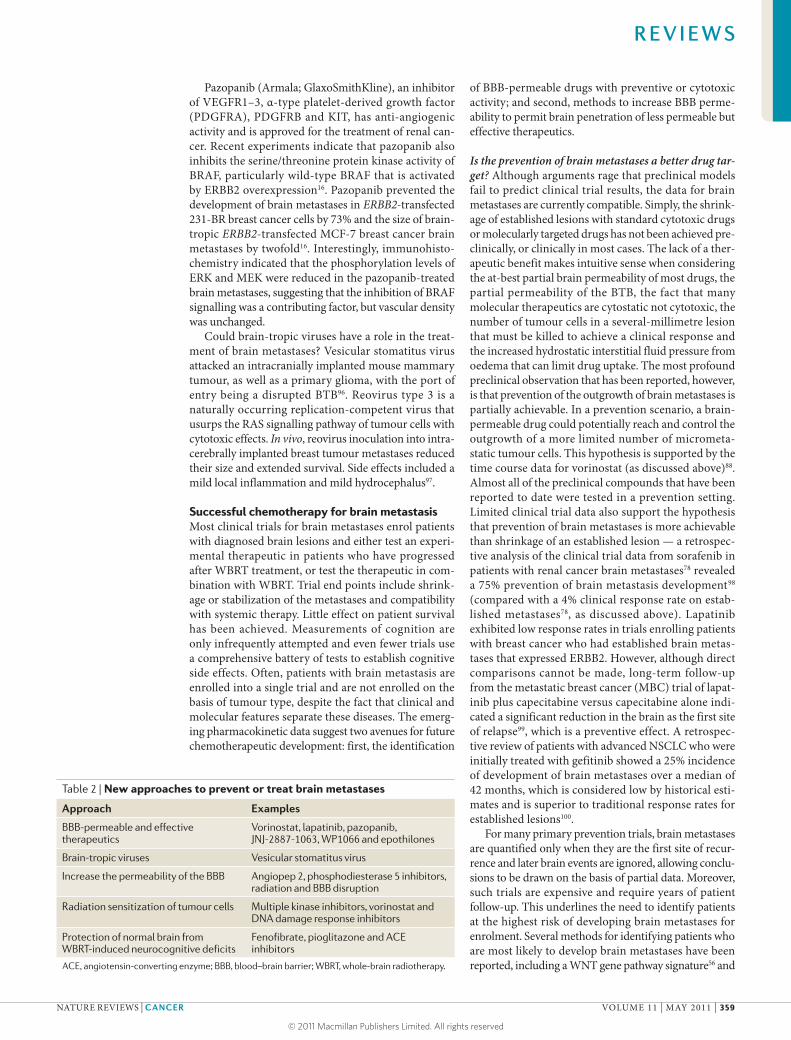
Table 2 | New approaches to prevent or treat brain metastases
Approach Examples
BBB-permeable and effective therapeutics
Vorinostat, lapatinib, pazopanib, JNJ-2887-1063, WP1066 and epothilones
Brain-tropic viruses Vesicular stomatitus virus
Increase the permeability of the BBB Angiopep 2, phosphodiesterase 5 inhibitors, radiation and BBB disruption
Radiation sensitization of tumour cells Multiple kinase inhibitors, vorinostat and DNA damage response inhibitors
Protection of normal brain from WBRT-induced neurocognitive deficits
Fenofibrate, pioglitazone and ACE inhibitors
ACE, angiotensin-converting enzyme; BBB, blood–brain barrier; WBRT, whole-brain radiotherapy.
R E V I E W S
NATURE REVIEWS | CANCER VOLUME 11 | MAY 2011 | 359
© 2011 Macmillan Publishers Limited. All rights reserved

NomogramA form of line chart showing scales for the variables involved in a particular formula so that corresponding values for each variable lie in a straight line intersecting all the scales.
Performance statusA measure of a patient’s well-being defined as the amount of normal activity that the patient can maintain.
a three-protein immunohistochemical signal101 in lung cancer and a clinical nomogram for breast cancer102, but none is in common!use.
Secondary prevention trials represent an as yet untried method for examining the efficacy of drugs at preventing brain metastases. This trial design would enrol patients who have been diagnosed with and treated (excluding WBRT) for brain metastases who are there-fore at a high risk of developing further brain metastases. Patients would receive systemic therapy and would be randomized to placebo or an investigational agent. The relevant end point would be time to the development of a new brain metastasis rather than shrinkage of the existing lesion. Other end points would include compatibility with systemic treatment, patient survival and cognitive func-tion. A graded prognostic assessment for patients with breast cancer brain metastases separated patients into groups with survival ranging from 4.2! months to 32.3!months103. The 32.3-month group, defined on the basis of performance status, ERBB2 and hormone receptor expression, and number of brain metastases, could enable the selection of longer term survivors who would be ideal candidates for secondary prevention tri-als. It remains to be determined whether drugs passing through the BBB will cause greater cognitive!losses.
Bypassing the BBB. Several interesting preclinical leads have emerged that can push non-brain-permeable drugs past the BBB. Some use the existing structure of the BBB, such as angiopep 2, a 19-amino acid pep-tide that binds the low-density lipoprotein receptor-related protein (LRP) receptors at the BBB, resulting in facilitated transport across the BBB104. Initial work with angiopep 2-conjugated paclitaxel demonstrated >50-fold enhanced delivery across the BBB104, and this agent has entered clinical trials. The role of pathologi-cal signalling in the BBB compartment is also under investigation. Phosphodiesterase 5 inhibitors, such as vardenafil (Levitra; Bayer), alter the endocytic pathway of endothelial cells. In!vivo, vardenafil increased tras-tuzumab uptake in intracranially implanted ERBB2+ breast tumour cells by twofold105. Radiation is the best studied BBB permeabilizer, although it has not yet been studied in a brain metastasis model106. Multiple pathways may mediate the radiation permeabilization of the BBB, including endothelial cell loss, reduced P-glycoprotein expression, VEGF production by activated astrocytes and binding of leukocytes to the damaged endothelia. Finally, BBB disruption is achieved by intracarotid infu-sion of a hyperosmotic agent to reversibly shrink brain endothelial cells and open their tight junctions; this strategy has been used most successfully for primary central nervous system lymphoma107.
Improving radiotherapy for brain metastasesRadiation therapy is the most commonly used procedure for the treatment of brain metastases. Overall goals for future research include optimizing the efficacy of radia-tion therapy against metastatic tumour cells compared with normal brain cells, and preventing the cognitive losses that a proportion of patients!suffer.
Radiosensitizers. Radiosensitizers are chemicals or biological agents that increase the lethal effects of radia-tion on the tumour without causing additional damage to normal tissue. Multiple drugs have been tested for radiation sensitization, including pyrimidine analogues, hypoxic cell sensitizers, traditional chemotherapeutic agents, kinase inhibitors and anti-angiogenic agents108,109. Overall, these studies have produced mixed results — some have shown a slight survival benefit but most have not shown a difference in survival — and have not been strong enough to bring any of these agents into rou-tine clinical care. Multiple molecular therapeutics have been preclinically tested in!vitro and in!vivo on a variety of cancer cell types for the sensitization of radiation with promising results, including inhibitors of MAPK, poly(ADP ribose) polymerase (PARP), and serine/threonine protein kinases PLK1, CHK1 and!CHK2
(REFS 110–113) (FIG.!3). Although it is hoped that these newer molecular therapeutics may be a long-awaited clinical advance in radiosensitization, it is also important to question why promising preclinical data on radiosen-sitizers have so far failed to translate to the clinic. One possible clue emanates from a gene expression analysis of a glioblastoma cell line grown in!vitro, as a subcutaneous tumour or intracranially. Gene expression after radia-tion therapy was dramatically different between these situations, confirming the importance of the appropriate microenvironment114. Testing of potential radiosensitizers in more relevant brain metastatic models is needed.
Radioprotectors for WBRT. A proportion of patients receiving WBRT suffer from progressive, permanent cog-nitive impairment. A recent clinical trial demonstrated a reduction in cognitive function in patients with NSCLC who were treated with WBRT, as assessed by a specific memory test115. The deleterious effects of WBRT on cog-nition have limited its use, particularly in cancer patients who have stable systemic disease and an expected pro-longed survival period. However, WBRT-induced cogni-tive decline is difficult to measure as it involves patient function at baseline (already deteriorated by the contri-butions of brain metastases and ‘chemobrain’ from sys-temic therapy), the adequacy of testing methods and the variety of drugs administered to patients.
A growing body of evidence suggests that chronic oxidative stress and inflammation have a role in cog-nitive decline116. Irradiating the adult rodent brain leads to neuroinflammation, increased oxidative stress117,118, activation of microglia119–121, and a chronic, progressive loss of both hippocampal-dependent and non-hippocampal-dependent cognitive function. A stem cell population in the vicinity of the hippocampus could be responsible for producing mature neurons, and, fol-lowing radiation injury, the inflammatory process may alter the neurogenic fate of these stem cells to a more gliogenic fate, thereby also causing memory deficits122. The cognitive effects of WBRT have been modelled in non-cancer-bearing animals. Adult rats were treated with WBRT, and cognitive function was quantified over the next year using a battery of tests, including regular and water mazes, as well as novel object recognition tests.
R E V I E W S
360 | MAY 2011 | VOLUME 11 www.nature.com/reviews/cancer
© 2011 Macmillan Publishers Limited. All rights reserved
Receptor inhibitionCetuximab, gefitiniband erlotinib
DNA repair
Cell cycleFlavopiridol andUCN-01
HDAC inhibitionVorinostat andvalproic acid
Nucleoside analoguesIUDR and BUDR
Radiation damage
MicroenvironmentBevacizumab,efaproxiral andtirapazamine
TK TK
Nucleus
Nature Reviews | Cancer
DNA repair Cell cycle Proliferation
Relative cognitive function was 73 ±6% at 12!weeks post-WBRT and decreased to 45 ±4% and 14 ±4% at 26 and 52!weeks post-WBRT, respectively, which is indicative of late, chronic and progressive cognitive impairment123.
Anti-inflammatory-based interventions have been hypothesized to prevent or ameliorate radiation-induced cognitive impairment. In non-tumour-bearing animals, the administration of pioglitazone (Actos; Takeda) (a peroxisome proliferator-activated receptor-$ (PPAR$) agonist that is prescribed for diabetes124), fenofibrate (Lipantil; Abbott Laboratories) (a PPAR# agonist that is prescribed for hypercholesterolaemia and hypertrigly-ceridaemia125) or the angiotensin type!1 receptor (AGTR1) antagonist (AT1RA) L-158809 (an angiotensin-converting enzyme (ACE) inhibitor that is typically used to treat
hypertension) significantly ameliorated WBRT-induced cognitive impairment126–128. These and similar studies suggest the intriguing hypothesis that some of the cog-nitive impairment that is associated with WBRT can be prevented using radioprotectors. Other potential radio-protectors tested in non-cancer brain diseases include melanocortins, erythropoietin, statins and antibiotics of the tetracycline and fluoroquinolone classes129. Studies in brain metastatic models are awaited in order to demon-strate the preservation of cognition, as well as the effects of the drug on metastatic colonization. Most animal models have been developed to produce brain lesions quickly, and this field will require new models permitting time for cog-nitive dysfunction to appear. Radiation-protection clinical trials would enrol newly diagnosed patients with brain metastasis who had an expected survival long enough to permit the development of cognitive sequelae; patients would be randomized to a protracted course of placebo or the investigational agent combined with WBRT. End points would be a decline in performance based on regularly administered cognitive tests, as well as quality of life, radiographic changes in brain lesions and patient survival.
ConclusionsBrain metastases cause physical and cognitive morbidi-ties and limit the survival of cancer patients, particularly those with advanced melanoma, lung cancer and breast cancer. As chemotherapy improves for other cancer types, the incidence of brain metastases is likely to rise as a sanctuary site. WBRT has efficacy as a brain metastasis-preventive therapy. New leads into the radioprotection of the normal brain to prevent cognitive loss from WBRT, and radiosensitization of tumour kill by SRS, may bring radiation therapy into safer, more effective use. Drug development can attack the problem of brain metastasis by identifying mechanistic molecular pathways, validat-ing brain-permeable inhibitors and clinically testing them in combination with systemic therapy and/or radia-tion. The currently available preclinical data suggest that chemotherapeutic drugs may be most effective in the pre-vention of brain metastases rather than the shrinkage of established lesions, which will require new trial designs. Comprehensive evaluations of patient cognition and quality of life will be essential to meaningfully progress.
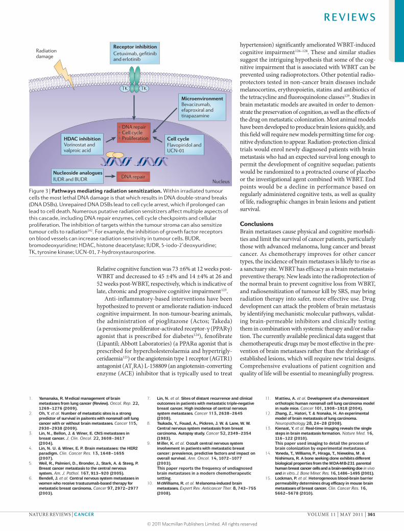
Figure 3 | Pathways mediating radiation sensitization. Within irradiated tumour cells the most lethal DNA damage is that which results in DNA double-strand breaks (DNA DSBs). Unrepaired DNA DSBs lead to cell cycle arrest, which if prolonged can lead to cell death. Numerous putative radiation sensitizers affect multiple aspects of this cascade, including DNA repair enzymes, cell cycle checkpoints and cellular proliferation. The inhibition of targets within the tumour stroma can also sensitize tumour cells to radiation142. For example, the inhibition of growth factor receptors on blood vessels can increase radiation sensitivity in tumour cells. BUDR, bromodeoxyuridine; HDAC, histone deacetylase; IUDR, 5-iodo-2’deoxyuridine; TK, tyrosine kinase; UCN-01, 7-hydroxystaurosporine.
1. Yamanaka, R. Medical management of brain metastases from lung cancer (Review). Oncol. Rep. 22, 1269–1276 (2009).
2. Oh, Y. et!al. Number of metastatic sites is a strong predictor of survival in paitents with nonsmall cell lung cancer with or without brain metastases. Cancer 115, 2930–2938 (2009).
3. Lin, N., Bellon, J. & Winer, E. CNS metastases in breast cancer. J.!Clin. Oncol. 22, 3608–3617 (2004).
4. Lin, N.!U. & Winer, E.!P. Brain metastases: the HER2 paradigm. Clin. Cancer Res. 13, 1648–1655 (2007).
5. Weil, R., Palmieri, D., Bronder, J., Stark, A. & Steeg, P. Breast cancer metastasis to the central nervous system. Am. J.!Pathol. 167, 913–920 (2005).
6. Bendell, J. et!al. Central nervous system metastases in women who receive trastuzumab-based therapy for metastatic breast carcinoma. Cancer 97, 2972–2977 (2003).
7. Lin, N. et!al. Sites of distant recurrence and clinical outcomes in patients with metastatic triple-negative breast cancer. High incidence of central nervous system metastases. Cancer 113, 2638–2645 (2008).
8. Tsukada, Y., Fouad, A., Pickren, J.!W. & Lane, W.!W. Central nervous system metastasis from breast carcinoma. Autopsy study. Cancer 52, 2349–2354 (1983).
9. Miller, K. et!al. Occult central nervous system involvement in patients with metastatic breast cancer: prevalence, predictive factors and impact on overall survival. Ann. Oncol. 14, 1072–1077 (2003).This paper reports the frequency of undiagnosed brain metastases in a modern chemotherapeutic setting.
10. McWilliams, R. et!al. Melanoma-induced brain metastases. Expert Rev. Anticancer Ther. 8, 743–755 (2008).
11. Mattieu, A. et!al. Development of a chemoresistant orthotopic human nonsmall cell lung carcinoma model in nude mice. Cancer 101, 1908–1918 (2004).
12. Zhang, Z., Hatori, T. & Nonaka, H. An experimental model of brain metastasis of lung carcinoma. Neuropathology 28, 24–28 (2008).
13. Kienast, Y. et al. Real-time imaging reveals the single steps in brain metastasis formation. Nature Med. 16, 116–122 (2010).This paper used imaging to detail the process of brain colonization by experimental metastases.
14. Yoneda, T., Williams, P., Hiraga, T., Niewolna, M. & Nishimura, R. A bone seeking clone exhibits different biological properties from the MDA-MB-231 parental human breast cancer cells and a brain-seeking cloe in!vivo and in!vitro. J.!Bone Miner. Res. 16, 1486–1495 (2001).
15. Lockman, P. et!al. Heterogeneous blood-brain barrier permeability determines drug efficacy in mouse brain metastases of breast cancer. Clin. Cancer Res. 16, 5662–5678 (2010).
R E V I E W S
NATURE REVIEWS | CANCER VOLUME 11 | MAY 2011 | 361
© 2011 Macmillan Publishers Limited. All rights reserved

This paper quantifies the heterogeneous permeability of experimental brain metastases and shows that only ~10% of lesions demonstrate sufficient chemotherapeutic permeability to produce a cytotoxic response.
16. Gril, B. et!al. Pazopanib reveals a role for tumor cell B-Raf in the prevention of breast cancer brain metastasis. Clin. Cancer Res. 17, 142–153 (2010).
17. Bos, P. et!al. Genes that mediate breast cancer metastasis to the brain. Nature 459, 1005–1010 (2009).A paper detailing the molecular characterization of breast cancer brain metastases.
18. Price, J.!E., Polyzos, A., Zhang, R.!D. & Daniels, L.!M. Tumorigenicity and metastasis of human breast carcinoma cell lines in nude mice. Cancer Res. 50, 717–721 (1990).
19. Rye, P. et!al. Brain metastasis model in athymic nude mice using a novel MUC1-secreting human breast-cancer cell line, MA11. Int. J.!Cancer 68, 682–687 (1996).
20. Carbonell, W., Ansorge, O., Sibson, N. & Muschel, R. The vascular basement membrane as “soil” in brain metastasis. PLoS ONE 4, e5857 (2009).This paper traces the colonization of the brain by brain-tropic metastatic cells and validates the contribution of integrins.
21. Leenders, W. et!al. Antiangiogenic therapy of cerebral melanoma metastases results in sustained tumor progression via vessel co-option. Clin. Cancer Res. 10, 6222–6230 (2004).This is an analysis of vascular co-option versus angiogenesis in experimental brain metastasis.
22. Cranmer, L.!D., Trevor, K.!T., Bandlamuri, S. & Hersh, E.!M. Rodent models of brain metastasis in melanoma. Melanoma Res. 15, 325–356 (2005).
23. Zhang, C., Zhang, F., Tsan, R. & Fidler, I. Transforming growth factor !2 is a molecular determinant for site specific melanoma metastasis in the brain. Cancer Res. 69, 828–835 (2009).
24. Huang, F.-J. et!al. Molecular basis of the critical role of suppressor of cytokine signaling-1 in melanoma brain metastasis. Cancer Res. 68, 9634–9642 (2008).
25. Yin, J. et!al. Noninvasive imaging of the functional effects of anti-VEGF therapy on tumor cell extravasation and regional blood volume in an experimental brain metastasis model. Clin. Exp. Metastasis 26, 403–414 (2009).
26. Kruger, A. et!al. Host TIMP-1 overexpression confers resistance to experimental brain metastasis of a fibrosarcoma cell line. Oncogene 16, 2419–2423 (1998).
27. Maillard, C. et!al. Reduction of brain metastases in plasminogen activator inhibitor-1 deficient mice with transgenic ocular tumors. Carcinogenesis 29, 2236–2242 (2008).
28. Cruz-Munoz, W., Man, S., Xu, P. & Kerbel, R. Development of a preclinical model of spontaneous human melanoma central nervous system metastasis. Cancer Res. 68, 4500–4505 (2008).
29. Fitzgerald, D. et!al. Reactive glia are recruited by highly proliferative brain metastases of breast cancer and promote tumor cell colonization. Clin. Exp. Metast. 25, 799–810 (2008).This paper validates the relevance of preclinical experimental metastasis models to resected human tissues. It describes a functional interaction between tumour cells and neuroinflammatory cells.
30. Paget, S. The distribution of secondary growths in cancer of the breast. Lancet 1, 99–101 (1889).
31. Lorger, M. & Felding-Habermann, B. Capturing changes in the brain microenvironment during intial steps of breast cancer brain metastasis. Am. J.!Pathol. 176, 2958–2971 (2010).
32. Zhang, M. & Olsson, Y. Hematogenous metastases of the human brain - characteristics of peritumoral brain changes: a review. J.!Neurooncol. 35, 81–89 (1997).
33. Seike, T. et!al. Interaction between lung cancer cells and astrocytes via specific inflammatory cytokines in the microenvironment of brain metastasis. Clin. Exp. Metastasis 28, 13–25 (2011).
34. Lin, Q. et!al. Reactive astrocytes protect melanoma cells from chemotherapy by sequestering intracellular calcium through gap junction communication channels. Neoplasia 12, 748–754 (2010).
35. Pukrop, R. et!al. Microglia promote the colonization of brain tissue by breast cancer cells in a Wnt-dependent way. Glia 58, 1477–1489 (2010).
36. Salgado, K., Toscani, N., Silva, L., Hilbig, A. & Barbosa-Coutinho, L. Immunoexpression of endoglin in brain metastasis secondary to malignant
melanoma: evaluation of angiogenesis and comparison with brain metastasis secondary to breast and lung carcinomas. Clin. Exp. Metastasis 24, 403–410 (2007).
37. Leenders, W. et!al. Vascular endothelial growth factor-A determines detectability of experimental melanoma brain metastasis in GD-DTPA-enhanced MRI. Int. J.!Cancer 105, 437–443 (2003).
38. Kim, L., Huang, S., Lu, W., Lev, D.!C. & Price, J. Vascular endothelial growth factor expression promotes the growth of breast cancer brain metastases in nude mice. Clin. Exp. Metastasis 21, 107–118 (2004).
39. Heyn, C. et!al. In!vivo magnetic resonance imaging of single cells in mouse brains with optical validation. Magn. Reson. Med. 55, 23–29 (2006).A study showing that dormant tumour cells exist in the brain using in!vivo imaging and post-mortem microscopy.
40. Silva, L.!D. et!al. HER3 and downstream pathways are involved in colonization of brain metastases from breast cancer. Breast Cancer Res. 12, 1–13 (2010).
41. Sun, M. et!al. HER family receptor abnormalities in lung cancer brain metastases and corresponding primary tumors. Clin. Cancer Res. 15, 4829–4837 (2009).
42. Koo, J. & Kim, S. EGFR and HER-2 status of non-small cell lung cancer brain metastasis and corresponding primary tumor. Neoplasma 58, 27–34 (2011).
43. Milas, I. et!al. Epidermal growth factor receptor, cyclooxygenase-2, and BAX expression in the primary non-small cell lung cancer and brain metastases. Clin. Cancer Res. 9, 1070–1076 (2003).
44. Gaedcke, J. et!al. Predominance of the basal type and HER-2/neu type in brain metastasis from breast cancer. Mod. Pathol. 20, 864–870 (2007).
45. Wu, P.-F. et!al. O6-Methylguanine-DNA methyltransferase expression and prognostic value in brain metastases of lung cancers. Lung Cancer 68, 484–490 (2010).
46. Gomez-Roca, C. et!al. Differential expression of biomarkers in primary non-small cell lung cancer and metastatic sites. J.!Thorac. Oncol. 4, 1212–1220 (2009).
47. Mehrotra, J. et!al. Very high frequency of hypermethylated genes in breast cancer metastasis to bone, brain and lung. Clin. Cancer Res. 10, 3104–3109 (2004).
48. Ding, L. et!al. Genome remodeling in a basal-like breast cancer metastasis and xenograft. Nature 464, 999–1005 (2010).
49. Stark, A., Tongers, K., Maass, N., Mehdom, H. & Held-Feidt, J. Reduced metastasis suppressor gene mRNA expression in breast cancer brain metastases. J.!Cancer Res. Clin. Oncol. 131, 191–198 (2005).
50. Stark, A. et!al. Reduced mRNA and protein expression of BCL-2 versus decreased mRNA and increased protein expression of BAX in breast cancer brain metastases: a real-time PCR and immunohistochemical evaluation. Nuerological Res. 28, 787–793 (2006).
51. Veenendaal, L. et!al. Differential notch and TGF! signaling in primary colorectal tumors and their corresponding metastases. Cell. Oncol. 30, 1–11 (2008).
52. Palmieri, D. et!al. Analyses of resected human brain metastases of breast cancer reveal the association between up-regulation of hexokinase 2 and poor prognosis. Mol. Cancer Res. 7, 1438–1445 (2009).
53. Xie, T.!X. et!al. Activation of stat3 in human melanoma promotes brain metastasis. Cancer Res. 66, 3188–3196 (2006).
54. Palmieri, D. et!al. Her-2 overexpression increases the metastatic outgrowth of breast cancer cells in the brain. Cancer Res. 67, 4190–4198 (2007).
55. Navab, R. et!al. Co-overexpression of Met and Hepatocyte growth factor promotes systemic metastasis in NCI-H460 non-small cell lung carcinoma cells. Neoplasia 11, 1292–1300 (2009).
56. Nguyen, D. et!al. WNT/TCF signaling through LEF1 and HOXB9 mediates lung adenocarcinoma metastasis. Cell 138, 51–62 (2009).
57. Zhang, L., Sullivan, P., Goodman, J., Gunaratne, P. & Marchetti, D. MicroRNA-1258 suppresses breast cancer brain metastasis by targeting heparanase. Cancer Res. 71, 645–654 (2011).
58. Cocconi, G. et al. Combination therapy with platinum and etoposide of brain metastases from breast carcinoma. Cancer Invest. 8, 327–334 (1990).
59. Boogerd, W., Dalesio, O., Bais, E. M. & van der Sande, J. J. Response of brain metastases from breast cancer to systemic chemotherapy. Cancer 69, 972–980 (1992).
60. Rosner, D., Nemoto, T. & Lane, W. W. Chemotherapy induces regression of brain metastases in breast carcinoma. Cancer 58, 832–839 (1986).
61. Oberhoff, C. et al. Topotecan chemotherapy in patients with breast cancer and brain metastases: results of a pilot study. Onkologie 24, 256–260 (2001).
62. Kurt, M., Aksoy, S., Hayran, M. & Guler, N. A retrospective review of breast cancer patients with central nervous system metastases treated with capecitabine. J.!Clin. Oncol. Abstr. 25, 1098 (2007).
63. Lin, N. et!al. Phase!II trial of lapatinib for brain metastases in patients with HER2+ breast cancer. J.!Clin. Oncol. Abstr. 24, 503 (2006).
64. Lin, N. et!al. Multicenter Phase!II study of lapatinib in patients with brain metastases from HER-2 positive breast cancer. Clin. Cancer Res. 15, 1452–1459 (2009).
65. Quirt, I. et!al. Temozolomide for the treatment of metastatic melanoma. Curr. Oncol. 14, 27–33 (2007).
66. Ekenel, M., Hormigo, A., Peak, S., DeAngelis, L. & Abrey, L. Capecitabine therapy of central nervous system metastases from breast cancer. J.!Neurooncol. 85, 223–227 (2007).
67. Rivera, E. et!al. Phase!I study of capecitabine in combination with temozolomide in the treatment of patients with brain metastases of breast cancer. Cancer 107, 1348–1354 (2006).
68. Mehta, M. et!al. The role of chemotherapy in the management of newly diagnosed brain metastases: a systematic review and evidence-based clinical practice guideline. J.!Neurooncol. 96, 71–83 (2010).
69. Ceresoli, G. et!al. Gefitinib in patients with brain metastases from non-small cell lung cancer: a prospective trial. Ann. Oncol. 15, 1042–1047 (2004).
70. Wu, C. et!al. Gefitinib as palliative therapy for lung adenocarcinoma metastatic to the brain. Lung Cancer 57, 359–364 (2007).
71. Togashi, Y. et!al. Cerebrospinal fluid concentration of erlotinib and its active metabolite OSI-420 in patients with central nervous system metastases of non-small cell lung cancer. J.!Thorac. Oncol. 5, 950–955 (2010).
72. Omuro, A.!M. et!al. High incidence of disease recurrence in the brain and leptomeninges in patients with nonsmall cell lung carcinoma after response to gefitinib. Cancer 103, 2344–2348 (2005).
73. Bria, E. et!al. Cardiotoxicity and incidence of brain metastases after adjuvant trastuzumab for early breast cancer: the dark side of the moon? A meta-analysis of the randomized trials. Breast Cancer Res. Treat. 109, 231–239 (2008).
74. Stemmler, H.-J. et!al. Ratio of trastuzumab levels in serum and cerebrospinal fluid is altered in HER2-positive breast cancer patients with brain metastases and impairment of the blood-brain barrier. Anticancer Drugs 18, 23–28 (2007).
75. Polli, J. et!al. The role of efflux and uptake transporters in N-{3-chloro-4-[(3-fluorobenzyl)oxy]phenyl}-6-[5-({[2-(methylsulfonyl)ethyl]amino}methyl)-2-furyl]-4-quinazolinamine (GWS572016, Lapatinib) disposition and drug interactions. Drug Metab. Dispos. 36, 695–701 (2008).
76. Lien, E. A., Wester, K., Lønning, P. E., Solheim, E. & Ueland, P. M. Distribution of tamoxifen and metabolites into brain tissue and brain metastases in breast cancer patients. Br. J.!Cancer 63, 641–645 (1991).
77. Socinski, M. et!al. Safety of bevicizumab in patients with non-small-cell lung cancer and brain metastases. J.!Clin. Oncol. 27, 5255–5261 (2009).
78. Stadler, W. et!al. Safety and efficacy results of the advanced renal cell carcinoma sorafenib expanded access program in north America. Cancer 116, 1272–1280 (2010).
79. Pajouhesh, H. & Lenz, G.!R. Medicinal chemical properties of successful central nervous system drugs. NeuroRx 2, 541–553 (2005).
80. Ohtsuki, S. & Terasaki, T. Contribution of carrier-mediated transport systems to the blood-brain barrier as a supporting and protecting interface for the brain; importance for CNS drug discovery and development. Pharm. Res. 24, 1745–1758 (2007).
81. Szakacs, G., Paterson, J., Ludwig, J., Booth-Genthe, C. & Gottesman, M. Targeting multidrug resistance in cancer. Nature Rev. Drug Discov. 5, 219–234 (2006).
82. Noguchi, K., Katayama, K., Mitsuhashi, J. & Sugimoto, Y. Functions of the breast cancer resistance protein (BCRP/ABCG2) in chemotherapy. Adv. Drug Deliv. Rev. 61, 26–33 (2009).
R E V I E W S
362 | MAY 2011 | VOLUME 11 www.nature.com/reviews/cancer
© 2011 Macmillan Publishers Limited. All rights reserved

83. Lagas, J. et!al. Breast cancer resistance protein and P-glycoprotein limit sorafenib brain accumulation. Mol. Cancer Ther. 9, 319–326 (2010).
84. Agarwal, S., Sane, R., Gallardo, J. L., Ohlfest, J. R. & Elmquist, W. F. Distribution of gefitinib to the brain is limited by P-glycoprotein (ABCB1) and breast cancer resistance protein (ABCG2)-mediated active efflux. J.!Pharmacol. Exp. Ther. 334, 147–155 (2010).This paper provides an excellent demonstration of roles of multiple BBB efflux transporters in restricting the brain distribution of chemotherapeutic drugs.
85. Armulik, A. et!al. Pericytes regulate the blood-brain barrier. Nature 468, 557–561 (2010).
86. Daneman, R., Zhou, L., Kebede, A. A. & Barres, B. A. Ericytes are required for blood-brain barrier integrity during embryogenesis. Nature 468, 562–566 (2010).
87. Ahmad, A., Gassmann, M. & Ogunshola, O. Maintaining blood-brain barrier integrity: pericytes perform better than astrocytes during prolonged oxygen deprivation. J.!Cell. Physiol. 218, 612–622 (2009).
88. Palmieri, D. et!al. Vorinostat inhibits brain metastatic colonization in a model of triple-negative breast cancer. Clin. Cancer Res. 15, 6148–6157 (2009).
89. Luu, T. et!al. A Phase!II trial of vorinostat (Suberoylanilide hydroxamic acid) in metastatic breast cancer: a California Cancer Consortium study. Clin. Cancer Res. 14, 7138–7142 (2008).
90. Ramalingam, S. et!al. Carboplatin and paclitaxel in combination with either vorinostat or placebo for first-line therapy of advanced non-small-cell lung cancer. J.!Clin. Oncol. 28, 56–62 (2010).
91. Baschnagel, A. et!al. Vorinostat enhances the radiosensitivity of a breast cancer brain metastatic cell line grown in!vitro and as intracranial xenografts. Mol. Cancer Ther. 8, 1589–1595 (2009).
92. Gril, B. et!al. Effect of lapatinib on the outgrowth of metastatic breast cancer cells to the brain. J.!Natl. Cancer Inst. 100, 1092–1103 (2008).This is the first demonstration that a molecular therapeutic can prevent ERRB2+ experimental brain metastases.
93. Emanuel, S. et!al. Cellular and in!vivo activity of JNJ-28871063, a nonquinazoline pan-ErbB kinase inhibitor that crosses the blood-brain barrier and displays efficacy against intracranial tumors. Mol. Pharmacol. 73, 338–348 (2008).
94. Kong, L.-Y. et!al. A novel inhibitor of signal transducers and activators of transcription 3 activation is efficaceous against established central nervous system melanoma and inhibits regulatory T!cells. Clin. Cancer Res. 14, 5759–5768 (2008).
95. Hoffmann, J. et!al. Sagopilone crosses the blood-brain barrier in!vivo to inhibit brain tumor growth and metastases. Neuro-oncology 11, 158–166 (2008).
96. Ozduman, K., Wollman, G., Piepmeier, J. & van den Pol, A. N. Systemic vesicular stomatitus virus selectively destroys multifocal glioma and metastatic carcinoma in the brain. J.!Neursci. 28, 1882–1893 (2008).
97. Yang, W. et!al. Reovirus as an experimental therapeutic for brain and leptomeningeal metastases from breast cancer. Gene Therapy 11, 1579–1590 (2004).
98. Massard, C. et!al. Incidence of brain metastases in renal cell carcinoma treated with sorafenib. Ann. Oncol. 21, 1027–1031 (2010).
99. Cameron, D. et!al. A Phase!III randomized comparison of lapatinib plus capecitabine versus capecitabine alone in women with advanced breast cancer that has progressed on trastuzumab: updated efficacy and biomarker analyses. Breast Cancer Res. Treat. 112, 533–543 (2008).
100. Heon, S. et!al. Development of central nervous system metastases in patients with advanced non-small cell lung cancer and somatic EGFR mutations treated with gefintib or erlotinib. Clin. Cancer Res. 16, 5873–5882 (2010).
101. Grinberg-Rashi, H. et!al. The expression of three genes in primary non-small cell lung cancer is associated with metastatic spread to the brain. Clin. Cancer Res. 15, 1755–1761 (2009).
102. Grasslin, O. et!al. Nomogram to predict subsequent brain metastasis in patients with metastatic breast cancer. J.!Clin. Oncol. 28, 2032–2037 (2010).
103. Sperduto, P. et al. A graded prosnostic assessment (GPA) for women with breast cancer and brain metastasssses. J.!Clin. Oncol. Suppl. Abstr. 28 1028 (2010).
104. Thomas, F. et!al. Uptake of ANG1005, a novel paclitaxel derivative, through the blood-brain barrier into brain and experimental brain metastases of breast cancer. Pharm. Res. 26, 2486–2494 (2009).
105. Hu, J. et!al. Phosphodiesterase type 5 inhibitors increase herceptin transport and treatment efficacy in mouse metastatic brain tumor models. PLoS ONE 5, e10108 (2010).
106. Yuan, H. et!al. Effects of fractionated radiation on the brain vasculature in a murine model: blood-brain barrier permeability, astrocyte proliferation, and ultrastructural changes. Int. J.!Radiat. Oncol. Biol. Phys. 66, 860–866 (2006).
107. Muldoon, L. et!al. Chemotherapy delivery issues in central nervous system malignancy: a reality check. J.!Clin. Oncol. 16, 2295–2305 (2007).
108. Rosenberg, A. & Knox, S. Radiation sensitization with redox modulators: a promising approach. Int. J.!Radiat. Oncol. Biol. Phys. 64, 343–354 (2006).
109. Francis, D., Richards, G., Forouzannia, A., Mehta, M. & Kuntia, D. Motexafin gadolinium: a novel radiosensitizer for brain tumors. Expert Opin. Pharmacother. 10, 2171–2180 (2009).
110. Russo, A. et!al. In!vitro and in!vivo radiosensitization of glioblastoma cells by the poly (ADP-Ribose) polymerase inhibitor E7016. Clin. Cancer Res. 15, 607–612 (2009).
111. Morgan, M. et!al. Mechanism of radiosensitization by the Chk1/2 inhibitor AZD7762 involves abrogation of the G2 checkpoint and inhibition of homologous recombinational DNA repair. Cancer Res. 70, 4972–4981 (2010).
112. Gerster, K. et!al. Targeting polo-like kinase 1 enhances radiation efficacy for head-and-neck squamous cell carcinoma. Int. J.!Radiat. Oncol. Biol. Phys. 77, 253–260 (2010).
113. Chung, E. et!al. In!vitro and in!vivo radiosensitization with AZD6244 (ARRY-142886), an inhibitor of mitogen-activated protein kinase/extracellular signal-regulated kinase 1/2 kinase. Clin. Cancer Res. 15, 3050–3057 (2009).
114. Camphausen, K. et!al. Orthotopic growth of human glioma cells quantitatively and qualitatively influences radiation-induced changes in gene expression. Cancer Res. 65, 10389–10393 (2005).
115. Chang, E. et al. Neurocognition in patients with brain metastases treated with radiosurgery or radiosurgery plus whole-brain irradiation: a randomized controlled trial. Lancet Oncol. 10, 1037–1044 (2009).
116. Zhao, W. & Robbins, M. Inflammation and chronic oxidative stress in radiation-induced late normal tissue injury: therapeutic implications. Curr. Med. Chem. 16, 130–143 (2009).
117. Fukuda, H. et!al. Irradiation-induced progenitor cell death in the developing brain is resistant to erythropoietin treatment and caspase inhibition. Cell Death Differ. 11, 1166–1178 (2004).
118. Limoli, C. et!al. Cell-density-dependent regulation of neural precursor cells function. Proc. Natl Acad. Sci. USA 101, 16052–16057 (2004).
119. Mildenberger, M., Beach, T., McGeer, E. & Ludgate, C. An animal model of prophylactic cranial irradiation: histologic effects at acute, early and delayed stages. Int. J.!Radiat. Oncol. Biol. Phys. 18, 1051–1060 (1990).
120. Monje, M., Toda, H. & Palmer, T. Inflammatory blockade restores adult hippocampal neurogenesis. Science 302, 1760–1765 (2003).
121. Schindler, M., Forbes, M., Robbins, M. & Riddle, D. Aging-dependent changes in the radiation response of the adult rat brain. Int. J.!Radiat. Oncol. Biol. Phys. 70, 826–834 (2008).
122. Khuntia, D., Brown, P., Li, J. & Mehta, M. Whole-brain radiotherapy in the management of brain metastasis. J.!Clin. Oncol. 24, 1295–1304 (2006).
123. Robbins, M. et!al. The AT1 receptor antagonist, L-158, 809, prevents or ameliorates factionated whole-brain irradiation-induced cognitive impairment. Int. J.!Radiat. Oncol. Biol. Phys. 73, 499–505 (2009).
124. Clar, C., Royle, P. & Waugh, N. Adding piaglitazone to insulin containing regimens in type!2 diabetes: systematic review and meta-analysis. PLoS ONE 4, e6112 (2009).
125. Abourbih, S. et!al. Effect of bibrates on lipid profiles and cardiovascular outcomes: a systematic review. Am. J.!Med. 122, 962.e1–962.e8 (2009).
126. Zhao, W. et!al. Administration of the peroxisomal proliferator-activated receptor (PPAR)g pioglitazone during fractionated brain irradiation prevents radiation-induced cognitive impariment. Int. J.!Radiat. Oncol. Biol. Phys. 67, 6–9 (2007).
127. Ramanan, S. et!al. The PPAR" agonist fenobibrate preserves hippocampal neurogenesis and inhibits microglial activation after whole-brain radiation. Int. J.!Radiat. Oncol. Biol. Phys. 75, 870–877 (2009).
128. Atwood, T. et!al. Quantitative magnetic resonance spectroscopy reveals a potential relationship between radiation-induced changes in rat brain metabolites and cognitive impairment. Radiat. Res. 168, 574–581 (2007).
129. Kim, K. et!al. High-throughput screening identifies two classes of antibiotics as radioprotectors: tetracyclines and fluoroquinolones. Clin. Cancer Res. 15, 7238–7245 (2009).
130. Gleissner, B. & Chamberlain, M. Neoplastic meningitis. Lancet Neurol. 5, 443–451 (2006).
131. Pedersen, P.-H. et!al. Leptomeningeal tissue: a barrier against brain tumor cell invasion. J.!Natl Cancer Inst. 86, 1593–1599 (1994).
132. Herrlinger, U. et!al. Leptomeningeal metastasis: survival and prognostic factors in 155 patients. J.!Neurol. Sci. 223, 167–178 (2004).
133. Kesari, S. & Batchelor, T. Leptomeningeal metastases. Neurol. Clin. 21, 25–66 (2003).
134. Borgelt, B. et al. Ultra-rapid high dose irradiation schedules for the palliation of brain metastases: final results of the first two studies by the Radiation Therapy Oncology Group. Int. J.!Radiat. Oncol. Biol. Phys. 7, 1633–1638 (1981).
135. Borgelt, B. et al. The palliation of brain metastases: final results of the first two studies by the radiation therapy oncology group. Int. J.!Radiat. Oncol. Biol. Phys. 6, 1–9 (1980).
136. Patil, C. G., Pricola, K., Garg, S. K., Bryant, A. & Black, K. L. Whole brain radiation therapy (WBRT) alone versus WBRT and radiosurgery for the treatment of brain metastases. Cochrane Database Syst. Rev. CD006121 (2010).
137. Kalkanis, S. et!al. The role of surgical resection in the management of newly diagnosed brain metastases: a systematic review and evidence-based clinical practice guideline. J.!Neurooncol. 96, 33–44 (2010).
138. Slotman, B. et!al. Prophylactic cranial irradiation in extensive small-cell lung cancer. New Engl. J.!Med. 357, 664–672 (2007).This report establishes that prophylactic cranial irradiation can be used to prevent brain metastasis.
139. Lester, J., MacBeth, F. & Coles, B. Prophylactic cranial irradiation for preventing brain metastases in patients undergoing radical treatment for non-small-cell lung cancer: a Cochrane review. Int. J.!Radiat. Oncol. Biol. Phys. 63, 690–694 (2005).
140. Sanghavi, S. et al. Radiosurgery for patients with brain metastases: a multi-institutional analysis, stratified by RTOG recursive partitioning analysis method. Int. J.!Radiat. Oncol. Biol. Phys. 51, 426–434 (2001).
141. Petrovich, Z., Yu, C., Giannotta, S. L., O’Day, S. & Apuzzo, M. L. Survival and pattern of failure in brain metastasis treated with stereotactic # knife radiosurgery. J.!Neurosurg. 97, 499–506 (2002).
142. Folkman, J. & Camphausen, K. What does radiotherapy do to endothelial cells? Science 293, 227–228 (2001).
AcknowledgementsThis work was supported by the Intramural Program, Center for Cancer Research, US National Cancer Institute, and from the Center of Excellence grant W81XWH-062-0033 from the US Department of Defense Breast Cancer Research Program. The authors regret that space restrictions did not permit all citations to be included.
Competing interests statementThe authors declare competing financial interests. See Web version for details.
FURTHER INFORMATIONPatricia S. Steeg’s homepage: http://ccr.ncifcrf.gov/staff/staff.asp?profileid=5851ClinicalTrials.gov: http://clinicaltrials.gov/
ALL LINKS ARE ACTIVE IN THE ONLINE PDF
R E V I E W S
NATURE REVIEWS | CANCER VOLUME 11 | MAY 2011 | 363
© 2011 Macmillan Publishers Limited. All rights reserved