Identification and Characterization of Novel Human Glioma- Specific
14
HUMAN GENE THERAPY 15:719–732 (August 2004) © Mary Ann Liebert, Inc. Identification and Characterization of Novel Human Glioma- Specific Peptides to Potentiate Tumor-Specific Gene Delivery IVY A.W. HO, PAULA Y.P. LAM, and KAM M. HUI ABSTRACT Glioblastomas account for approximately 20% of all primary brain tumors in adults. Glioblastoma multi- forme (GBM) is a highly malignant tumor. In spite of advances in surgery, chemotherapy, and radiotherapy, the life expectancy of the patient with glioblastoma is approximately 11 months. To enhance glioma-specific gene delivery, we employed a 12-mer phage display peptide library to isolate phages that bind specifically to human glioma cell lines. Here, we report the isolation and functional characterization of novel glioma-spe- cific peptides that target transgenes specifically to a wide array of human glioblastomas in vitro and in vivo. One of the isolated peptides, tentatively denoted as MG11, is demonstrated to be glioma specific and gives an in vitro-binding enrichment of more than 5-fold for glioma cells when compared with nonglioma cells. Intra- venous injection of phages bearing the MG11 peptide-binding motif enables the phages to home specifically to glioma xenografts. Most significantly, when Lissamine rhodamine-labeled MG11 peptide is injected intra- tumorally, it targets specifically to glioma xenografts instead of non-glioma-derived xenografts. In summary, our results suggest that the MG11 peptide is able to target specifically to tumors of glial origin, which would allow the design of applications related to the diagnosis and treatment of human gliomas. 719 OVERVIEW SUMMARY The major challenge of cancer gene therapy is to direct transgene expression to a particular tumor cell type. Iden- tification of molecules that mediate the targeted delivery of therapeutic DNA or drugs to specific cell types can signifi- cantly reduce the amount of therapeutic agent needed and, as a consequence, lessen the potential toxicity to nontarget cells. In this article, we report the isolation and character- ization of a novel human glioma-specific peptide, MG11, that targets exogenous DNA specifically to a wide array of human glioblastoma cells, in vitro and in vivo. Isolation of the MG11 peptide provides the means to conjugate thera- peutic agents for targeting. INTRODUCTION I DENTIFICATION OF MOLECULES that mediate the targeted de- livery of therapeutic DNA or drugs to specific cell types can significantly reduce the amount of therapeutic agent required, and would therefore reduce the potential toxicity to nontarget cells. Glioblastoma multiforme (GBM) is a highly malignant tumor and accounts for more than 20% of primary brain tumors in adults. In spite of advances in surgery, chemotherapy, and radiotherapy, the life expectancy of patient with glioblastoma is approximately 11 months (Cowan et al., 2000). A prominent clinical feature of GBM is its ability to infiltrate into sur- rounding brain tissues, thus rendering complete surgical removal practically impossible. One way to overcome this dif- ficulty is to design strategies that facilitate the delivery of ther- apeutic agents specifically to glioma cells. One of the common methods to enable gene expression in a selected cell population is to incorporate gene expression cas- settes under the control of a tissue-specific promoter (Shibata et al., 2000; Ueno et al., 2001). Tissue-specific promoters are generally linked with weak induction of transgene expression, and rely on inducible agents that might exert unfavorable ef- fects on the host (Agha-Mohammadi and Lotze, 2000; Nettel- beck et al., 2000). Moreover, the essential upstream control re- Division of Cellular and Molecular Research, Gene Vector Laboratory, National Cancer Centre, Singapore 169610.
Transcript of Identification and Characterization of Novel Human Glioma- Specific
HUMAN GENE THERAPY 15719ndash732 (August 2004)copy Mary Ann Liebert Inc
Identification and Characterization of Novel Human Glioma-Specific Peptides to Potentiate Tumor-Specific Gene Delivery
IVY AW HO PAULA YP LAM and KAM M HUI
ABSTRACT
Glioblastomas account for approximately 20 of all primary brain tumors in adults Glioblastoma multi-forme (GBM) is a highly malignant tumor In spite of advances in surgery chemotherapy and radiotherapythe life expectancy of the patient with glioblastoma is approximately 11 months To enhance glioma-specificgene delivery we employed a 12-mer phage display peptide library to isolate phages that bind specifically tohuman glioma cell lines Here we report the isolation and functional characterization of novel glioma-spe-cific peptides that target transgenes specifically to a wide array of human glioblastomas in vitro and in vivoOne of the isolated peptides tentatively denoted as MG11 is demonstrated to be glioma specific and gives anin vitro-binding enrichment of more than 5-fold for glioma cells when compared with nonglioma cells Intra-venous injection of phages bearing the MG11 peptide-binding motif enables the phages to home specificallyto glioma xenografts Most significantly when Lissamine rhodamine-labeled MG11 peptide is injected intra-tumorally it targets specifically to glioma xenografts instead of non-glioma-derived xenografts In summaryour results suggest that the MG11 peptide is able to target specifically to tumors of glial origin which wouldallow the design of applications related to the diagnosis and treatment of human gliomas
719
OVERVIEW SUMMARY
The major challenge of cancer gene therapy is to directtransgene expression to a particular tumor cell type Iden-tification of molecules that mediate the targeted delivery oftherapeutic DNA or drugs to specific cell types can signifi-cantly reduce the amount of therapeutic agent needed andas a consequence lessen the potential toxicity to nontargetcells In this article we report the isolation and character-ization of a novel human glioma-specific peptide MG11that targets exogenous DNA specifically to a wide array ofhuman glioblastoma cells in vitro and in vivo Isolation ofthe MG11 peptide provides the means to conjugate thera-peutic agents for targeting
INTRODUCTION
IDENTIFICATION OF MOLECULES that mediate the targeted de-livery of therapeutic DNA or drugs to specific cell types can
significantly reduce the amount of therapeutic agent requiredand would therefore reduce the potential toxicity to nontargetcells Glioblastoma multiforme (GBM) is a highly malignanttumor and accounts for more than 20 of primary brain tumorsin adults In spite of advances in surgery chemotherapy andradiotherapy the life expectancy of patient with glioblastomais approximately 11 months (Cowan et al 2000) A prominentclinical feature of GBM is its ability to infiltrate into sur-rounding brain tissues thus rendering complete surgical removal practically impossible One way to overcome this dif-ficulty is to design strategies that facilitate the delivery of ther-apeutic agents specifically to glioma cells
One of the common methods to enable gene expression in aselected cell population is to incorporate gene expression cas-settes under the control of a tissue-specific promoter (Shibataet al 2000 Ueno et al 2001) Tissue-specific promoters aregenerally linked with weak induction of transgene expressionand rely on inducible agents that might exert unfavorable ef-fects on the host (Agha-Mohammadi and Lotze 2000 Nettel-beck et al 2000) Moreover the essential upstream control re-
Division of Cellular and Molecular Research Gene Vector Laboratory National Cancer Centre Singapore 169610
gion that regulates gene expression from a particular promotercan span more than a few kilobases Cloning the whole regionwould be difficult in the construction of transcriptionally me-diated targeted vector systems (Agha-Mohammadi and Lotze2000 Nettelbeck et al 2000)
There has been much progress in the study of vector target-ing by means of various vector systems Wu and Wu (1987)were the first to report the use of the glycoprotein asialooroso-mucoid (ASOR) to specifically target the liver parenchymaRetroviral vectors have been modified to present a chimeric en-velope that targets to breast cancer cells (Tai et al 2003) About50 of GBM tumors show amplification or mutations that ac-tivate the gene encoding the epidermal growth factor receptor(EGFR) thus producing a constitutively active receptor in theabsence of the EGF ligand (Mischel and Cloughesy 2003)Vectors that target to this growth factor receptor have been de-signed Doubly ablated adenoviral vectors lacking both the cox-sackievirusndashadenovirus receptor (CAR)- and v-integrin-bind-ing capacities together with bispecific single-chain antibodiesthat recognize both the EGFR and the epithelial cell adhesionmolecule have been employed for specific gene delivery to pri-mary human brain tumors (van Beusechem et al 2002Samoylova et al 2003) Fusion proteins consisting of peptidetoxins fused to the human EGF (DAB389EGF) has been dem-onstrated to selectively kill cells that overexpress EGFR (Co-hen et al 2003)
Phage display techniques have been used for selection ofwhole cells to identify peptide ligands directed against partic-ular cell surface proteins (Parmley and Smith 1988 Barry etal 1996 Szardenings et al 1997 Campa et al 2002 Liu etal 2003) Using this technique peptides that bind to kidneylung skin pancreas intestine uterus adrenal gland retina fi-broblast cells myoblasts myotubes human neutrophils humanlaryngeal carcinoma cells endothelial cells (Barry et al 1996Arap et al 1998 Koivunen et al 1999 Pasqualini et al 2000White et al 2001) and a human colorectal cell line (Rasmussenet al 2002) have been identified Peptides that home selec-tively to the vasculature of various organs were isolated by thein vivo biopanning of peptide phage display libraries in mice(Pasqualini and Ruoslahti 1996 Rajotte et al 1998 Arap etal 2002a Essler and Ruoslahti 2002 Trepel et al 2002) aswell as in humans (Arap et al 2002b) The selected sequencecan be used to target therapeutic agents (Arap et al 1998) anddiagnostic imaging radiolabels (Schumacher et al 2002)
In this study we describe the isolation and characterizationof a novel peptide MG11 using phage display technology Thispeptide targets specifically both in vitro and in vivo to humanglioma xenografts
MATERIALS AND METHODS
Cell lines
dGli36 cells (gift from M Sena-Esteves Childrenrsquos Hospi-tal of Philadelphia Philadelphia PA) which overexpress a trun-cated mutant EGFR commonly found in human gliomas weregrown in the presence of puromycin (1 gml Sigma-AldrichSt Louis MO) SF767 and U251MG human gliomas werekindly provided by DF Deen (Brain Tumor Research CenterUCSF School of Medicine San Francisco CA) CNE2 cells are
derived from undifferentiated human nasopharyngeal carci-noma (gift from HM Wang Cancer Institute Guangzhou Peo-plersquos Republic of China) All human tumor cell linesmdashA549(lung adenocarcinoma) CNE2 (nasopharyngeal carcinoma)HeLa (cervical carcinoma) HepG2 (hepatoma) HK1 (na-sopharyngeal carcinoma) KOSC-3 (head and neck carcinoma)KZ2 (melanoma) M059K (glioblastoma) SF767 (glioblas-toma) T98G (glioblastoma) U87MG (glioblastoma) U251MG(glioblastoma) U373MG (anaplastic astrocytoma) and WT18(lymphoma)mdashused in this study were maintained in Dulbeccorsquosmodified Eaglersquos medium (DMEM) supplemented with 10fetal bovine serum (FBS HyClone Laboratories Logan UT)penicillin (100 Uml Invitrogen Grand Island NY) strepto-mycin (100 gml Invitrogen) and 2 mM L-glutamine (Sigma-Aldrich) Normal human astrocytes purchased from CambrexBio Science Walkersville (Walkersville MD) were cultured inastrocyte basal medium supplemented with recombinant humanEGF insulin ascorbic acid GA-100 L-glutamine and FBS asrecommended by the supplier (Cambrex Bio Science Walk-ersville)
Primary glioma cell culture
A single primary glioma biopsy was kindly provided by JThomas (Department of Neurosurgery Singapore General Hos-pital Singapore) with the patientrsquos consent Tissues were im-mediately kept in DMEM with 10 FBS To obtain a single-cell suspension the tissues were digested for 5 min with 025trypsin followed by washing twice with complete DMEM Thecells were plated into a single well of a 96-well plate (NuncRoskilde Denmark) and incubated at 37degC in a humidified in-cubator with 5 CO2 for 24 hr before being assayed for thebinding of the MG11 peptide
Phage display library biopanning
The phage display library employed for biopanning forglioma-specific phage was the PhD-12 library (New EnglandBioLabs Beverly MA) Phage was selected for binding to hu-man glioma cell line by panning against intact cells in suspen-sion Glioma cell lines (dGli36 SF767 U87MG U251MG andU373MG) were grown in monolayer until confluent and har-vested by treating the cell with phosphate-buffered saline (PBS)containing 5 mM EDTA The five glioma cell lines were mixedtogether in equal proportions to give a final cell number of 1 106 This was followed by incubating the cell mixture with 1 1011 plaque-forming units (PFU) of M13 phage at 37degC for 2hr The cells were washed once with PBS containing 01Tween 20 and nine times with PBS followed by pelleting andresuspension to remove unbound phage Bound phage were re-covered by eluting with 02 M glycine pH 22 containingbovine serum albumin (BSA 1 mgml Sigma-Aldrich) fol-lowed by neutralization with 1 M Tris-HCl (pH 91) Recov-ered phages were amplified in ER2537 bacteria (New EnglandBioLabs) and subjected to two additional rounds of enrichmentpanning These enrichment pannings were then followed bythree rounds of subtraction panning against A549 CNE2 andHepG2 successively The isolated phage clones were titered us-ing ER2537 bacteria
To determine the in vitro specificity of the recovered phagesto glioma and nonglioma cell lines 1 106 cells were incu-bated with 1012 PFU of the isolated phage clone at 37degC for 2
HO ET AL720
hr Unbound phage were removed and bound phage were re-covered and titered as mentioned
Amplification of phage clones
ER2537 bacteria were employed for phage amplification To20 ml of log-phase bacteria culture 1 1010 PFU of the phagewas added and the mixture was incubated for 45 hr at 37degC ina shaking incubator To precipitate the phages a 16 volume ofPEG 8000ndashNaCl solution was added to the bacteriandashphage mix-ture and precipitated at 4degC overnight The next day phage wereharvested by centrifugation at 13000 rpm for 10 min at 4degCThe resulting phage pellet was resuspended in 1 ml of Tris-buffered saline (TBS) The phage were further precipitated byadding a 16 volume of the PEGndashNaCl solution After incu-bating for 1 hr on ice the phage were pelleted at 13000 gfor 10 min at 4degC and resuspended in 200 l of TBS contain-ing 001 sodium azide The amplified phage were titered andstored at 4degC
Titering of phage
To determine the titer amplified phage were serially dilutedin Luria broth (LB) Each of the diluted phage solutions wasadded to a log culture of ER2537 bacteria After incubating for5 min at room temperature to allow infection to take place 3ml of melted 07 agarose (45degC) was added and the mixturewas poured onto an LB agar plate containing 5-bromo-4-chloro-3-indolyl--D-galactoside (X-Gal 40 mgml Bio-Rad Her-cules CA) and isopropyl -D-thiogalactoside (IPTG 50mgml Invitrogen) The titer of the phage solution was deter-mined by counting the number of blue plaques after 24 hr ofincubation
In vivo targeting of phage to tumor xenograft
Six-week-old female nude mice were purchased from the An-imal Resource Center (Animal Resource Centre Perth Aus-tralia) dGli36 cells (2 106) suspended in 10 l of PBS werestereotactically inoculated into the bregma region (2 mm lat-eral 025 mm depth) of the right hemisphere of the mice Tu-mor growth was monitored by magnetic resonance imaging(MRI) hematoxylin and eosin (HampE) staining and loss of bodyweight Phage was injected via the tail vein 10 days postinoc-ulation of tumor cells when the tumor volume was approxi-mately 75 mm3
For subcutaneous tumors 5 106 SF767 or dGli36 gliomacells and CNE2 (nonglioma cells control) were suspended in100 l of PBS and injected into the right and left flanks of 6-week-old SCID mice (Animal Resource Centre) respectivelyTumor growth was monitored by measuring the tumor volumeThe tumor volume was calculated according to the followingformula tumor volume (mm3) 052 (width [mm2] length [mm]) (Bergers et al 1999) Phage was injected via thetail vein 7 days postimplantation of tumor when the tumor vol-ume was approximately 100 mm3
Tumor-bearing mice were randomized into two groups (fiveper group) namely the control group and the experimentalgroup Either control phage or phage bearing the MG11 se-quence (1 1012 PFU) suspended in 400 l of DMEM wasinjected via the tail vein into tumor-bearing mice and allowedto circulate for 24 hr After 24 hr mice were anesthetized and
perfused through the heart with DMEM Tumor and other or-gans were dissected and weighed The tissues were homoge-nized in ice-cold DMEM containing protease inhibitor cocktailand 01 BSA After centrifugation to remove the tissue de-bris bound phages were rescued by mixing the supernatant with05 ml of ER2537 bacteria (New England BioLabs) for 30 minat 37degC The supernatant containing phage was diluted in LBafter which aliquots were plated on LB agar plates containingX-Gal and IPTG As control unselected phage was adminis-tered at the same titer in the second group of animals
Peptides
Poly-L-lysine-SIPVKFNKP-MG11 [(K16)-MG11] and poly-L-lysine-SIPVKFNKP-H42 [(K16)-H42] peptides were synthe-sized and purified (Mimotopes Victoria Australia) The pep-tides were dissolved in 015 M NaCl at 1 mgml and stored insmall aliquots at 20degC The purity of both peptides was 90
Formation of peptidendashDNA complexes
The formation and transfection of peptidendashDNA complexeswere carried out according to Patel et al (2001) with slightmodifications PeptidendashDNA complexes were prepared at apeptideDNA ratio of 21 (ww) Plasmid DNA was diluted to10 gml in Ringerrsquos buffer (B Braun Melsungen MelsungenGermany) The appropriate volume of peptide was also dilutedin Ringerrsquos buffer to give a final concentration of 20 gml (pep-tideDNA ratio 21) The peptide was added dropwise to theDNA solution while vortexing gently This mixture was allowedto incubate at room temperature for 30 min After 30 min themixture was diluted to 4 g of DNA per milliliter with DMEMin the presence of 100 M chloroquine and added to cells
Transfection of tumor cell lines
For transfection 3 105 cells were seeded into each well ofa six-well plate (Nunc) After culturing the cells overnight thecells were washed twice with PBS Freshly prepared pep-tidendashDNA complexes were added to each well and incubatedfor 2 hr at 37degC For transfection performed in the presence ofserum FBS was added to a final concentration of 10 imme-diately after the addition of peptidendashDNA complexes Reportergene activity was assayed after 24 or 48 hr
Assay for luciferase activity
Cells were harvested from six-well plates 24 or 48 hr aftertransfection washed resuspended in 120 l of Tris-HCl (pH78) and freezendashthawed three times Cell debris was discardedafter centrifugation at 14000 g at 4degC for 10 min One hun-dred microliters of the supernatant collected was used for as-saying luciferase activity using the Auto-Lumat LB952 lumi-nometer (Berthold Technologies Bad Wildbad Germany) Fivemicroliters of the supernatant collected was used for the deter-mination of protein concentration using the Bio-Rad proteinassay dye reagent with an Ultrospec 3000 UVvisible spec-trophotometer (Amersham Biosciences Uppsala Sweden)
In vitro fluorescent peptide-binding assay
For the in vitro binding assay 1 105 cells were seeded intoeach well of a 24-well dish (Nunc) After culturing the cells
HUMAN GLIOMA-SPECIFIC TARGETING MOLECULE 721
HO ET AL722
TA
BL
E1
PEP
TID
ES
ISO
LA
TE
DB
YB
IOP
AN
NIN
GO
FH
UM
AN
GL
IOM
AC
EL
LL
INE
S
Fre
quen
cy o
f oc
curr
ence
Per
cent
Sequ
ence
s(
of
tota
l cl
ones
seq
uenc
ed)
Hom
olog
y to
kno
wn
hum
an p
rote
ins
Ali
gnm
enta
hom
olog
y
SGH
QL
LL
NK
MPN
24M
itoge
n-ac
tivat
ed p
rote
in k
inas
e
60(M
G2)
kina
se k
inas
e 12
Db
599HDLLLRKMSS
608
Qy
3HQLLLNKMPN
12
LW
AT
FPPR
PPW
L19
Sem
apho
rin
48 p
recu
rsor
64(M
G11
)D
b11
WGALPPRPPLL
21Q
y2WATFPPRPPWL
12
WSA
APT
KPP
YH
T6
Vol
tage
-dep
ende
nt L
-typ
e ca
lciu
m
46ch
anne
l
-ID
sub
unit
Db
1981
WATPPATPPYR
1991
Qy
1WSAAPTKPPYH
11IL
AN
DL
TA
PGPR
5N
one
HH
GH
SPT
SPQ
VR
4Se
map
hori
n 3A
pre
curs
or
100
Db
575HHGHSP
580
Qy
1HHGHSP
6
LPY
GT
SNR
HA
PV4
Sem
apho
rin
6B p
recu
rsor
(Se
ma
Z)
64D
b43
5LPYGGADRTAP
444
Qy
1LPYGTSNRHAP
11Y
VQ
GW
NY
HD
LT
R4
Non
e
LW
AA
FPPQ
ASV
A4
Syna
ptoj
anin
1
70
Db
1245
AAFPPQSSLP
1254
Qy
3AAFPPQASVA
12FD
TPH
TL
TW
FHG
3N
one
a Ast
eris
k re
pres
ents
ide
ntic
al a
min
o ac
id
Db
seq
uenc
e fr
om d
atab
ase
Qy
que
ry s
eque
nce
from
pha
ge
HUMAN GLIOMA-SPECIFIC TARGETING MOLECULE 723
overnight at 37degC the cells were washed once with PBS fol-lowed by incubation in blocking buffer containing PBS with1 BSA at room temperature for 1 hr The cells were washedtwice with PBS One hundred nanograms of Lissamine rho-damine-conjugated (K16)-MG11 peptide (Mimotopes) wasadded to the blocking buffer containing 01 sodium azide andincubated with the cells at 37degC for 20 min This is followedby five washes with PBS with 01 Tween 20 at room tem-perature at 5-min intervals The cells were then fixed in 4paraformaldehyde After removal of excess paraformaldehydethe cells were mounted Images were captured digitally and an-alyzed with an LSM 510 Meta confocal microscope (Carl ZeissGoumlttingen Germany) with appropriate filters
In vivo fluorescent peptide-binding assay
SF767 tumor-bearing mice were randomized into two groups(two per group) Fifty microliters containing 100 g of Lis-samine rhodamine-conjugated (K16)-MG11 or (K16)-H42 wasinjected intratumorally into each of two mice harboring the tu-mor and allowed to circulate for 20 min After 20 min the micewere anesthetized and perfused through the heart with PBS fol-lowed by 4 paraformaldehyde The tumor were dissected andcryosectioned The sections were then counterstained with flu-orescein isothiocyanate (FITC)ndashphalloidin and mounted Im-ages were examined with the LSM 510 Meta confocal micro-scope (Carl Zeiss) with the appropriate filters
Statistical analysis
Data are presented throughout this study as means stan-dard error of the mean Statistical significance was evaluatedby paired t test and p 005 was considered significant
RESULTS
Enrichment of glioma-specific phage by in vitrobiopanning
The PhD-12 phage display library with a complexity of ap-proximately 2 109 sequences was employed for screeningpeptides that would bind specifically to human glioma cellsThis library consists of 12-mer peptide sequences fused to theN terminus of the minor coat protein of the M13 phage in avalency of 5 copies per virion The library was screened againsta mixture of human glioma cell lines including dGli36 SF767U87MG U251MG and U373MG at 37degC to increase the prob-ability of obtaining sequences that could interact genericallywith most glioma cells Phages rescued were further subjectedto negative panning with nonglioma cell lines A549 CNE2 andHepG2 to eliminate nonspecific background binding Theamino acid sequences of the 79 phage clones obtained after sucha selection revealed several dominant sequence motifs (Table1) The sequence SGHQLLLNKMPN designated MG2 wasfound in 24 of the clones and LWATFPPRPPWL designatedMG11 was found in 19 of the clones (Table 1) A BLASTsearch of MG2 demonstrated 60 sequence homology with thehuman mitogen-activated protein kinase kinase kinase 12(MAP3K12) MAP3K12 was found to be highly expressed inbrain and kidney and has been implicated as an activator of theJNKndashSAPK pathway (Su and Karin 1996) Analysis of the sec-
ond most frequently isolated phage-bearing peptide sequencedesignated MG11 showed it to be 64 homologous to sema-phorin 4B precursor protein Semaphorin 4B is a member ofthe semaphorin superfamily which is involved in the inhibitionof axonal extension by providing local signals to specify terri-tories inaccessible to growing axons (Rieger et al 2003) Inaddition to phage bearing the MG11 peptide sequence we havealso isolated two other phage clones harboring peptide se-quences that match the semaphorin family of proteins One ofthe sequences HHGHSPTSPQVR matches perfectly the pro-tein sequence of semaphorin 3A precursor protein whereas theother peptide sequence LPYGTSNRHAPV showed 64 ho-mology with semaphorin 6B precursor protein Semaphorin 3Awhich binds with high affinity to neuropilin 1 induces the col-lapse and paralysis of neuronal growth cones Neuropilin 1 is areceptor for the vascular endothelial growth factor family and hasbeen implicated in blood vessel formation It also functions as acoreceptor with the Flk-1KDR receptor tyrosine kinase (Gluz-man-Poltorak et al 2000) We noticed that several groups of thepeptides selected are homologous to members of the semaphorinsuperfamily It has been previously reported that semaphorin fam-ily proteins play important roles during neural development(Schumacher et al 2002 Rieger et al 2003) Because MG11is the most common sequence identified among the group ofphages isolated MG11 was thus chosen for subsequent studies
Characterization of the binding epitopes of MG11 phage
To determine whether MG11 phage binds specifically toglioma cells tumor cell lines of various histotypes were incu-bated with the phage These included dGli36 SF767 U87MGU251MG U373MG and T98G glioma cells and A549 CNE2HeLa HepG2 KOSC-3 KZ2 and WT18 nonglioma cells asshown in Fig 1 The efficiency of MG11 phage binding toglioma cells (range from 2 106 to 22 107 PFUml) wasat least 3-fold higher than that of nonglioma cells (Fig 1) None
FIG 1 In vitro specificity of MG11 phage for a panel of hu-man glioma cell lines Tumor cell lines of various origins wereincubated with MG11 phage (1012 PFUl) at 37degC for 2 hrMean values for phage recovered from the binding assay andthe SEM from triplicate experiments are shown
FIG 2 In vivo targeting of MG11 phage to tumor cells of gliomaorigins (A) Binding of MG11 phage to various tissues (B) Specificbinding of MG11 phage to dGli36 (i) and SF767 (ii) human gliomaxenograft as compared with a nonglioma xenograft CNE2 Error barsshow the SEM of three mice (C) Targeting of MG11 phage to in-tracranial dGli36 human glioma xenograft as compared with the nor-mal region of the brain Data shown represent averages of four micep 005 (paired t test)
HO ET AL724
FIG 3 In vitro binding of (K16)-MG11 peptide toglioma cells (A) Proposed interaction of (K16)-MG11 (i)and (K16)-H42 (ii) peptide sequence with DNA and theireffects on luciferase transgene expression Cells (3 105)were transfected at peptideDNA ratios of 12 21 101and 201 for 24 hr The luciferase activities shown repre-sent means and SEM of triplicate experiments (B) Speci-ficity of (K16)-MG11 peptide for various tumor cell lines(i) PeptidendashDNA complexes (21 [ww] ratio) preparedfrom (K16)-MG11 were used to transfect pNGVL-Luc intovarious tumor cell lines Luciferase activity was analyzed24 and 48 hr posttransfection (ii) PeptidendashDNA com-plexes (21 [ww] ratio) prepared from (K16)-MG11 wereused to transfect pEGFP-N1 into U251MG human gliomacells and HepG2 hepatoma cells Fluorescence pictureswere taken 48 hr posttransfection (C) Specificity of (K16)-MG11 compared with (K16)-H42 control peptide Pep-tidendashDNA complexes prepared from (K16)-MG11 and(K16)-H42 were complexed with pNGVL-Luc and trans-fected into SF767 and CNE2 tumor cell lines Luciferaseactivity was analyzed 48 hr posttransfection The lucifer-ase activities shown represent means and SEM of tripli-cate experiments
of the nonglioma origin tumor cell lines tested exhibited con-siderable binding (range from 15 104 to 96 105 PFUml)Thus the in vitro phage-binding assay indicated that MG11phage binds specifically to glioma cells
To determine whether phage bearing the MG11-binding mo-tif can target specifically in vivo to human glioma xenografts1012 PFU of MG11 phage was injected via the tail vein to im-munodeficient SCID mice harboring subcutaneous dGli36-derived glioma xenografts Recovery of phage particles fromtissues as well as xenografted dGli36 tumor indicated thatMG11 phage is specific for tumor only (794 104 PFUmg)whereas other tissues including brain heart kidney liver lungand spleen gave only background binding (Fig 2A) Minimalbinding was observed in all tissues when control unselectedphage was employed (Fig 2A) The tumor specificity of MG11was also examined by administering MG11 phage via the tailvein to immunodeficient mice bearing subcutaneous gliomaxenograft (dGli36) and nonglioma xenograft (CNE2) on theright and left hind thigh respectively The enrichment factor inhoming to the subcutaneous dGli36 glioma xenograft (79 104 PFUmg) compared with the subcutaneous CNE2 tumor(167 104 PFUmg) was 5-fold (p 003) (Fig 2B part i)Similar results were obtained when another human gliomaSF767 was used The enrichment factor in homing of MG11phage to SF767-derived tumor (834 103 PFUmg) was 4-fold higher when compared with CNE2-derived tumor (221 103 PFUmg) (p 002) (Fig 2B part ii) indicating that thebinding of MG11 to human glioma cell lines is specific
The specificity of MG11 phage binding to human gliomacells in the context of normal mouse astrocytes was investi-gated Phage (1012) were injected via the tail vein into immu-nodeficient nude mice bearing an intracranial xenograft ofdGli36 human glioma cells in the right hemisphere Phage re-covered from the right hemisphere bearing the xenografted tu-mor (92 102 PFUmg) in mice injected with the MG11 phagegave an enrichment of 20-fold compared with those recoveredfrom the normal side of the brain (05 102 PFUmg) (lefthemisphere) (p 0013) (Fig 2C) No significant differencecould be detected in the control groups injected with unselectedphage (Fig 2C) These results demonstrated that phage bear-ing the MG11 epitope target specifically to cells of glial originand do not bind to normal brain cells
In vitro specificity of (K16)-MG11 in human tumor cell lines
The amino acid residues that make up the MG11 peptide se-quence are hydrophobic and could not be easily purified To
facilitate the purification procedure we have inserted 16 lysineresidues into the sequence The addition of polylysine residueshas been demonstrated to enhance the potential of the peptideto interact with the negatively charged plasmid DNA as dem-onstrated by Patel et al (2001) In addition to the polylysineresidues a spacer sequence identical to that used by Patel et alwas also included to facilitate the movement of the peptide
The (K16)-MG11 peptide was chemically synthesized andcomplexed with a luciferase reporter plasmid pNGVL-Lucif-erase (pNGVL-Luc) To determine the optimal concentrationthat would allow DNA and peptide interactions and to maxi-mize the efficiency of gene uptake various peptideDNA ratioswere used in the transfection of SF767 cells
Various ratios of the pNGVL-Luc plasmid and (K16)-MG11peptide were complexed followed by incubation in SF767glioma cells in the presence of chloroquine Chloroquine wasadded as it has been previously demonstrated to prevent lyso-somal DNA degradation (Zauner et al 1997) After 24 hr lu-ciferase expression was detected at a peptideDNA ratio (ww)of 21 (39803 RLUg protein) (Fig 3A part i) The level ofluciferase expression increased with escalating ratios of peptideto DNA (Fig 3A part i) This suggests that the abundance oflysine residues could facilitate the interaction of the peptidewith the DNA molecules hence ensuring that the majority ofthe DNA molecules are complexed with the peptide At pep-tideDNA ratios of 101 and 201 toxicity could be detected asthe treated SF767 glioma cells were not as healthy as the un-treated controls This could be due to the presence of the highconcentration of polylysine which is known to be toxic to cells(Zauner et al 1997) For subsequent experiments we there-fore adopted the optimal peptideDNA ratio of 21
To rule out the possibility that the binding of the peptide tothe cells is due to the lysine residues inserted we synthesizeda control peptide in our experiment to confirm that the lucifer-ase activities observed were a consequence of targeting A con-trol peptide comprising the 16-lysine residue followed by a 12-amino acid sequence derived from biopanning of a nongliomacell line (K16)-H42 was also synthesized H42 bears the se-quence GGPTKEWELYLF Similar experiments were carriedout with the luciferase DNA and (K16)-H42 at various pep-tideDNA ratios and no significant binding could be detectedwhen analyzed 24 hr posttransfection (Fig 3A part ii)
To further demonstrate the specificity of (K16)-MG11 pep-tide for glioma cells and to determine the optimal time point ofluciferase transgene expression peptidendashDNA complex pre-pared by mixing (K16)-MG11 peptide with pNGVL-Luc DNAat a 21 (ww) ratio was added to tumor cell lines of varioushistotypes These included dGli36 SF767 U251MG U373MG
FIG 4 In vitro specificity of Lissamine rhodamine-labeled (K16)-MG11 peptide Tumor cell lines of various origins were in-cubated with 100 ng of (K16)-MG11 peptide for 20 min at 37degC Cells were subsequently fixed and mounted as described in Ma-terials and Methods Fluorescence images were studied with an LSM 510 Meta confocal microscopy system
FIG 5 Binding of Lissamine rhodamine-labeled (K16)-MG11 peptide to primary human glioma culture Primary human gliomaculture (A) SF767 human glioma cells (B) and normal human astrocytes (C) were incubated with 100 ng of (K16)-MG11 for20 min at 37degC Cells were subsequently fixed and mounted as described in Materials and Methods Fluorescence images werestudied with an LSM 510 Meta confocal microscopy system
HO ET AL726
SF767SF767 U251MG M059K
A549 HepG2 HK1
(A) Primary human glioma culture
(B) SF767 human glioma cells
(C) Normal primary human astrocytes
FIG 4
FIG 5
HUMAN GLIOMA-SPECIFIC TARGETING MOLECULE 727
HO ET AL728
A549 HeLa HepG2 and CNE2 cells Binding of peptidendashDNAcomplexes was performed in the presence of 100 M chloro-quine and luciferase expression was assayed 24 and 48 hr af-ter transfection Luciferase activity in the SF767 glioma cellline (85473 RLUg) was at least 4-fold higher than in theCNE2 cell line (19849 RLUg) 24 hr posttransfection Thelevel of gene expression 48 hr postdelivery is much higher thanthat at 24 hr (Fig 3B part i) The level of luciferase expres-sion detected in U251MG dGli36 and SF767 glioma cell linesincreased dramatically by 48 hr after transfection (Fig 3B parti) Experiments using (K16)-MG11 peptide complexed with thereporter gene encoding enhanced green fluorescent protein(eGFP) also gave similar results (Fig 3B part ii) Strong greenfluorescence could be detected in U251MG glioma cells but notin HepG2 human hepatocellular carcinoma cells
Furthermore when peptidendashDNA complexes prepared from(K16)-MG11 or (K16)-H42 peptide with pNGVL-Luc weremixed and added to SF767 and CNE2 cells the luciferase ex-pression in SF767 glioma cells transfected with (K16)-MG11pNGVL-Luc (716940 RLUg) was 84-fold higher thanthat of (K16)-H42pNGVL-Luc (8444 RLUg) control peptide-transfected cells The results thus demonstrate that the bindingof (K16)-MG11 peptide is specific and not due to the presenceof positively charge lysine residues (Fig 3C) When comparedwith the CNE2 nonglioma cell line the level of luciferase ex-pression observed in SF767 glioma cells transfected withpNGVL-Luc(K16)-MG11 is 151-fold higher (Fig 3C) Theseresults clearly illustrate that (K16)-MG11 is specific for gliomacells
Characterization of (K16)-MG11 peptide-targeteddelivery in vitro and in vivo
To determine the direct specific binding of (K16)-MG11 pep-tide to the cell surface of glioma cells (K16)-MG11 peptide wasconjugated to Lissamine rhodamine After incubating the vari-ous tumor cell lines (SF767 U251MG M059K A549 HepG2and HK1) with Lissamine rhodamine-conjugated (K16)-MG11peptide the cells were examined by confocal microscopy andthe levels of fluorescence detected were compared SF767U251MG and M059K human glioma cell lines exhibited de-tectable fluorescence as shown by the presence of rhodamineon the surface of the cells (Fig 4 top) Little or no fluores-cence was observed with nonglioma cell lines (Fig 4 bottom)In contrast nonspecific red fluorescence was observed in allcell lines studied when incubated with (K16)-H42 control pep-tide under similar conditions (data not shown) Unlike the (K16)-MG11 peptide no discrimination between glioma and non-glioma cells could be ascertained with the rhodamine-conju-gated (K16)-H42 peptide (data not shown)
The binding specificity of (K16)-MG11 peptide to primaryhuman glioma cells and normal primary human astrocytes wassubsequently determined Primary human glioma cells derivedfrom biopsy or normal primary human astrocytes were incu-bated with Lissamine rhodamine-conjugated (K16)-MG11 pep-tide at 37degC for 20 min Significant binding of the (K16)-MG11peptide was observed only in primary human glioma cells (Fig5A) This binding specificity is comparable to that observed inthe SF767 human glioma cell line (Fig 5B) In contrast no fluo-rescence could be detected with normal primary human astro-
cytes under similar conditions (Fig 5C) These results clearlydemonstrated that the MG11 peptide is specific for humanglioma cells and does not bind to normal human astrocytes
To further demonstrate the tumor specificity of the (K16)-MG11 peptide for glioma xenografts SCID mice harboring sub-cutaneously established SF767-derived xenografts were in-jected intratumorally with Lissamine rhodamine-conjugated(K16)-MG11 peptide and (K16)-H42 peptide After 20 min ofcirculation the mouse bearing the SF767 glioma xenograft wasperfused and tumor was harvested The tumor sections werecounterstained with FITC-labeled phalloidin which stains F-actin to show the cell surface Cryostat sections of the tumorsshowed red fluorescence binding in SF767 glioma xenograft in-jected with (K16)-MG11 peptide (Fig 6A) demonstrating thebinding of (K16)-MG11 peptide This binding as observed inthe inset (I) indicated that (K16)-MG11 peptide interacted withcell surface receptors on the tumor cells In contrast red fluo-rescence was not detected in the SF767 tumor injected with(K16)-H42 peptide (Fig 6B) Although the stainings obtainedwith FITC-labeled phalloidin (green) and Lissamine rho-damine-conjugated (K16)-MG11 peptide (red) were in closeproximity they did not colocalize This reflects the fact that thereceptors for MG11 peptide are located on the external side ofthe plasma membrane whereas F-actin is situated just belowthis external periphery membrane layer Taken together our re-sults suggested that the observed fluorescence can be attribut-able to the specific binding and subsequent uptake of (K16)-MG11 peptide
DISCUSSION
Gene transfer to malignant gliomas has been extensively pur-sued both in cell culture as well as in animal model experi-ments However clinical gene therapy trials for recurrentgliomas have not fulfilled the high expectations suggested bypreclinical laboratory results One of the significant pitfalls isthe low transduction efficiency of tumor cells observed in vivoresulting in expression of therapeutic gene products at too lowa level to efficiently eradicate tumor cells (Weyerbrock andOldfield 1999 Rainov and Kramm 2001 Rainov and Ren2003) Retrovirus vector was one of the earliest viral vectors tobe employed for the delivery of therapeutic genes to gliomasin the clinical setting (Rainov and Ren 2003) Although bothphase I and II clinical studies showed favorable safety profilesand some efficacy the phase III study failed to demonstrate sig-nificant progression-free or extension of overall survival(Rainov and Ren 2003) The use of adenovirus vectors as wellas herpes simplex virus-based vectors has also been activelypursued both in the laboratory and clinic (Bansal and Engel-hard 2000)
A commonly employed strategy for gene delivery to the braininvolved direct stereotactic injection of vectors to the coordi-nates of interest Most often this approach is limited by the vol-ume and number of injections that one would need in order toachieve therapeutic doses Moreover with this route of admin-istration the vector is taken up only by cells in the immediatevicinity of the injection site Improved strategies for the selec-tive delivery of therapeutic agents are therefore needed to com-bat devastating and usually fatal glioblastoma multiforme tu-
HUMAN GLIOMA-SPECIFIC TARGETING MOLECULE 729
(A) MG11
(B) H42
FIG 6 In vivo targeting of fluorescently labeled (K16)-MG11 peptide SF767 human glioma cells were inoculated into the rightflank of immunodeficient mice to form a tumor xenograft One hundred micrograms of Lissamine rhodamine-labeled (K16)-MG11peptide (A) or (K16)-H42 peptide (B) was injected intratumorally into separate mice bearing the SF767 human glioma xenograftand allowed to circulate for 20 min Afterward the tumors were harvested and cryosectioned Fluorescence images were studiedwith an LSM 510 Meta confocal microscopy system I Insert
HO ET AL730
mors In this report we have employed a phage display pep-tide library to isolate and characterize phages bearing peptidesequences that bind specifically to a wide array of humanglioma cell lines Analysis of the 79 phage clones obtained af-ter biopanning of the PhD-12 phage library revealed severaldominant sequence motifs (Table 1) The sequence SGHQL-LLNKMPN encoded by the phage MG2 was found in 24 ofthe clones and this sequence was shown to have 60 homol-ogy with the human mitogen-activated protein kinase kinase ki-nase 12 (MAP3K12) The second most frequently isolatedphage clone MG11 bears the sequence LWATFPPRPPWLwhich matches that of semaphorin 4B precursor a membrane-bound semaphorin Interestingly two other phage clones thatharbor peptide sequences with homology for the semaphorinfamily of proteins were also identified (Table 1) The sequenceHHGHSPTSPQVR matches the protein sequence of secretedsemaphorin 3A precursor protein whereas the sequenceLPYGTSNRHAPV is homologous to that of membrane-boundsemaphorin 6B precursor protein Semaphorin 3A has been re-ported to bind with high affinity to neuropilin 1 (Rieger et al2003) a receptor for the vascular endothelial growth factor fam-ily (Miao et al 2000) Neuropilin 1 together with plexin a re-ceptor for transmembrane-bound semaphorins (Rieger et al2003) forms complexes with class 3 semaphorins that are ableto direct growing axons to their targets (Tamagnone et al1999) Besides modulating the extension of axonal cones sem-aphorins also regulate the migration of neural progenitor cells(Marin et al 2001) One of the clinical features of gliomas istheir ability to infiltrate and migrate to distant sites within thebrain it is therefore speculated that semaphorins could play arole in the carcinogenesis of glioma This suggestion is consis-tent with reports of the presence of semaphorins in the pro-gression of mouse mammary tumor and metastatic human lungadenocarcinoma (Martiacuten-Satueacute and Blanco 1999) In corrobo-ration with these observations it has also been reported thatboth semaphorin 3 and semaphorin 6B could be detected by re-verse transcription-polymerase chain reaction analysis ofglioma cell lines (Correa et al 2001 Rieger et al 2003)
Previous peptide sequences that target to human malignantglioma cells have been isolated (Spear et al 2001 Zhang etal 2001) However the reported specificity is limited and con-fined to a single glioma cell line Spear et al reported a pep-tide having the sequence MCPKHPLGC that binds only to theU87MG glioma cell line (Spear et al 2001) Zhang et al alsoreported a sequence that binds to glioma cells However thissequence is not glioma specific as it also binds to other can-cer cell lines (Zhang et al 2001) In our study phage bearingthe MG11 sequence gave a binding enrichment of more than5-fold for glioma cells in comparison with nonglioma cells (Fig1) Consistent with the in vitro data the MG11 peptide-encod-ing phages were able to target specifically to dGli36 and SF767human glioma xenografts with a 5-fold binding enrichmentover that of a nonglioma tumor xenograft under similar condi-tions (Fig 2B) Furthermore the MG11 peptide was able to di-rect and confine the gene expression of the luciferase DNAspecifically to human glioma cells (Fig 3) Currently the lackof selectivity of cancer chemotherapeutic drugs results in hightoxicities As MG11 offers selective binding it could poten-tially be useful as a targeting molecule However to enhancethe effectiveness of targeting the therapeutic index (ratio of
binding to target glioma tissue versus binding to normal tis-sues) for each and every toxic molecule that may be conjugatedto MG11 needs to be determined
One of the challenges for in vivo targeting is to be able todirect the binding of the targeting peptide to tumor cells insteadof normal cells We have shown that the MG11 peptide bindsonly to human glioma-derived cell lines (Fig 4) and primaryhuman glioma cells (Fig 5A) but not to normal human braincells (Fig 5C) or other nonglioma cells (Fig 4) These obser-vations would be of great relevance in designing strategies fortargeted therapy of glioma
In summary the identification of the MG11 sequence allowsthe possibility of coupling therapeutic agents directly to the pep-tide for targeting glioma cells This could lower the therapeu-tic doses of either DNA or drugs required and would thereforereduce any potential harmful side effects resulting from the useof high doses of therapeutic agents However to effectively de-liver DNA into cells the uptake intracellular trafficking andnuclear retention of the plasmid DNA need to be achievedCationic lipids viral vectors and polymers are some of theagents that could be employed for the conjugation of peptidesto enhance the in vivo stability of these peptides Furthermorethe addition of chloroquine to cells could help to protect thetargeting peptidendashDNA complexes from lysosomal degrada-tion and could potentially enhance the effectiveness of a spe-cific gene delivery protocol (Zauner et al 1997) These aresome of the strategies that are being exploited in our laboratoryto further enhance the capability of MG11 to function as aglioma-specific targeting molecule
ACKNOWLEDGMENTS
The authors thank Dr J Thomas (Department of Neuro-surgery Singapore General Hospital Singapore) for providingus with the primary glioma biopsy This research was supportedby grants from the Agency for Science Technology and Re-search (ASTAR) and Singapore National Medical ResearchCouncil
REFERENCES
AGHA-MOHAMMADI S and LOTZE M (2000) Regulatable sys-tems Applications in gene therapy and replicating viruses J ClinInvest 105 1177ndash1183
ARAP W PASQUALINI R and RUOSLAHTI E (1998) Cancertreatment by targeted drug delivery to tumor vasculature in a mousemodel Science 279 377ndash380
ARAP W HAEDICKE W BERNASCONI M KAIN R RA-JOTTE D KRAJEWSKI S ELLERBY HM BREDESEN DEPASQUALINI R and RUOSLAHTI E (2002a) Targeting theprostate for destruction through a vascular address Proc Natl AcadSci USA 99 1527ndash1531
ARAP W KOLONIN MG TREPEL M LAHDENRANTA JCARDO-VILA M GIORDANO RJ MINTZ PJ ARDELTPU YAO VJ VIDAL CI CHEN L FLAMM A VALTA-NEN H WEAVIND LM HICKS ME POLLOCK REBOTZ GH BUCANA CD KOIVUNEN E CAHILL DTRONCOSO P BAGGERLY KA PENTZ RD DO KA LO-GOTHETIS CJ and PASQUALINI R (2002b) Steps toward map-ping the human vasculature by phage display Nat Med 8 121ndash127
HUMAN GLIOMA-SPECIFIC TARGETING MOLECULE 731
BANSAL K and ENGELHARD H (2000) Gene therapy for braintumors Curr Oncol Rep 2 463ndash472
BARRY M DOWER W and JOHNSTON S (1996) Toward cell-targeting gene therapy vectors Selection of cell-binding peptidesfrom random peptide-presenting phage libraries Nat Med 2299ndash305
BERGERS G JAVAHERIAN K LO K FOLKMAN J andHANAHAN D (1999) Effects of angiogenesis inhibitors on mul-tistage carcinogenesis in mice Science 284 808ndash812
CAMPA M SERLIN S and PATZ EJ (2002) Development ofnovel tumor imaging agents with phage-display combinatorial pep-tide libraries Acad Radiol 9 927ndash932
COHEN K LIU T BISSONETTE R PURI R and FRANKELA (2003) DAB389EGF fusion protein therapy of refractory glioblas-toma multiforme Curr Pharm Biotechnol 4 39ndash49
CORREA R SASAHARA R BENGTSON M KATAYAMA MSALIM A BRENTANI M SOGAYAR M DE SOUZA S andSIMPSON A (2001) Human semaphorin 6B [(HSA)SEMA6B] anovel human class 6 semaphorin gene Alternative splicing and all-trans-retinoic acid-dependent downregulation in glioblastoma celllines Genomics 73 343ndash348
COWAN W HARTER D and KANDEL E (2000) The emergenceof modern neuroscience Some implications for neurology and psy-chiatry Annu Rev Neurosci 23 343ndash391
ESSLER M and RUOSLAHTI E (2002) Molecular specializationof breast vasculature A breast-homing phage-displayed peptidebinds to aminopeptidase P in breast vasculature Proc Natl AcadSci USA 99 2252ndash2257
GLUZMAN-POLTORAK Z COHEN T HERZOG Y andNEUFELD G (2000) Neuropilin-2 and neuropilin-1 are receptorsfor the 165-amino acid form of vascular endothelial growth factor(VEGF) and of placenta growth factor-2 but only neuropilin-2 func-tions as a receptor for the 145-amino acid form of VEGF J BiolChem 275 18040ndash18045
KOIVUNEN E ARAP W RAJOTTE D LAHDENRANTA J andPASQUALINI R (1999) Identification of receptor ligands withphage display peptide libraries J Nucl Med 40 883ndash888
LIU R ENSTROM AM and LAM KS (2003) Combinatorial pep-tide library methods for immunobiology research Exp Hematol 3111ndash30
MARIN O YARON A BAGRI A TESSIER-LAVIGNE M andRUBENSTEIN J (2001) Sorting of striatal and cortical interneu-rons regulated by semaphorinndashneuropilin interactions Science 293872ndash875
MARTIacuteN-SATUEacute M and BLANCO J (1999) Identification of sem-aphorin E gene expression in metastatic human lung adenocarcinomacells by mRNA differential display J Surg Oncol 72 18ndash23
MIAO H LEE P LIN H SOKER S and KLAGSBRUN M(2000) Neuropilin-1 expression by tumor cells promotes tumor an-giogenesis and progression FASEB J 14 2532ndash2539
MISCHEL P and CLOUGHESY T (2003) Targeted molecular ther-apy of GBM Brain Pathol 13 52ndash61
NETTELBECK DM JEROME V and MULLER R (2000) Genetherapy Designer promoters for tumour targeting Trends Genet 16174ndash181
PARMLEY S and SMITH G (1988) Antibody-selectable filamen-tous fd phage vectors Affinity purification of target genes Gene 73305ndash318
PASQUALINI R and RUOSLAHTI E (1996) Organ targeting invivo using phage display peptide libraries Nature 380 364ndash366
PASQUALINI R KOIVUNEN E KAIN R LAHDENRANTA JSAKAMOTO M STRYHN A ASHMUN RA SHAPIRO LHARAP W and RUOSLAHTI E (2000) Aminopeptidase N is a re-ceptor for tumor-homing peptides and a target for inhibiting angio-genesis Cancer Res 60 722ndash727
PATEL S ZHANG X COLLINS L and FABRE J (2001) A
small synthetic peptide for gene delivery via the serpinndashenzymecomplex receptor J Gene Med 3 271ndash279
RAINOV N and KRAMM C (2001) Vector delivery methods andtargeting strategies for gene therapy of brain tumors Curr GeneTher 1 367ndash383
RAINOV N and REN H (2003) Gene therapy for human malignantbrain tumors Cancer J 9 180ndash188
RAJOTTE D ARAP W HAGEDORN M KOIVUNEN EPASQUALINI R and RUOSLAHTI E (1998) Molecular hetero-geneity of the vascular endothelium revealed by in vivo phage dis-play J Clin Invest 102 430ndash437
RASMUSSEN U SCHREIBER V SCHULTZ H MISCHLER Fand SCHUGHART K (2002) Tumor cell-targeting by phage-dis-played peptides Cancer Gene Ther 9 606ndash612
RIEGER J WICK W and WELLER M (2003) Human malignantglioma cells express semaphorins and their receptors neuropilins andplexins Glia 42 379ndash389
SAMOYLOVA T MORRISON N and COX N (2003) Molecularmarkers of glial tumors Current targeting strategies Curr MedChem 10 831ndash843
SCHUMACHER T HOFER S EICHHORN K WASNER MZIMMERER S FREITAG P PROBST A GRATZL OREUBI J MAECKE H MUELLER-BRAND J and MERLOA (2002) Local injection of the 90Y-labelled peptidic vector DOTA-TOC to control gliomas of WHO grades II and III An extended pi-lot study Eur J Nucl Med Mol Imaging 29 486ndash493
SHIBATA T GIACCIA A and BROWN J (2000) Developmentof a hypoxia-responsive vector for tumor-specific gene therapy GeneTher 7 493ndash498
SPEAR M BREAKEFIELD X BELTZER J SCHUBACK DWEISSLEDER R PARD F and LADNER R (2001) Isolationcharacterization and recovery of small peptide phage display epi-topes selected against viable malignant glioma cells Cancer GeneTher 8 506ndash511
SU B and KARIN M (1996) Mitogen-activated protein kinase cas-cades and regulation of gene expression Curr Opin Immunol 8402ndash411
SZARDENINGS M TORNROTH S MUTULIS F MUCENIECER KEINANEN K KUSINENE A and WIKBERG J (1997)Phage display selection on whole cells yields a peptide specific formelanocortin receptor 1 J Biol Chem 272 27943ndash27948
TAI C LOGG C PARK J ANDERSON W PRESS M andKASAHARA N (2003) Antibody-mediated targeting of replica-tion-competent retroviral vectors Hum Gene Ther 14 789ndash802
TAMAGNONE L ARTIGIANI S CHEN H HE Z MING GSONG H CHEDOTAL A WINBERG M GOODMAN CPOO M TESSIER-LAVIGNE M and COMOGLIO P (1999)Plexins are a large family of receptors for transmembrane secretedand GPI-anchored semaphorins in vertebrates Cell 99 71ndash80
TREPEL M ARAP W and PASQUALINI R (2002) In vivo phagedisplay and vascular heterogeneity Implications for targeted medi-cine Curr Opin Chem Biol 6 399ndash404
UENO M KOYAMA F YAMADA Y FUJIMOTO HTAKAYAMA T KAMADA K NAITO A HIRAO S MUKO-GAWA T HAMADA H and NAKAJIMA Y (2001) Tumor-specific chemo-radio-gene therapy for colorectal cancer cells usingadenovirus vector expressing the cytosine deaminase gene Anti-cancer Res 21 2601ndash2608
VAN BEUSECHEM V GRILL J MASTENBROEK D WICK-HAM T ROELVINK P HAISMA H LAMFERS M DIRVENC PINEDO H and GERRITSEN W (2002) Efficient and selec-tive gene transfer into primary human brain tumors by using single-chain antibody-targeted adenoviral vectors with native tropism abol-ished J Virol 76 2753ndash2762
WEYERBROCK A and OLDFIELD E (1999) Gene transfer tech-nologies for malignant gliomas Curr Opin Oncol 11 168ndash173
WHITE S NICKLIN S SAWAMURA T and BAKER A (2001)Identification of peptides that target the endothelial cell-specificLOX-1 receptor Hypertension 27 449ndash455
WU G and WU C (1987) Receptor-mediated in vitro gene trans-formation by a soluble DNA carrier system J Biol Chem 2624429ndash4432
ZAUNER W KICHLER A SCHMIDT W MECHTLER K andWAGNER E (1997) Glycerol and polylysine synergize in their abil-ity to rupture vesicular membranes A mechanism for increased trans-ferrinndashpolylysine-mediated gene transfer Exp Cell Res 232137ndash145
ZHANG J SPRING H and SCHWAB M (2001) Neuroblastomatumor cell-binding peptides identified through random peptide phagedisplay Cancer Lett 171 153ndash164
Address reprint requests toDr Kam Man Hui
Division of Cellular and Molecular ResearchNational Cancer Centre
11 Hospital DriveSingapore 169610
E-mail cmrhkmnccscomsg
Received for publication December 18 2003 accepted after re-vision June 16 2004
Published online July 8 2004
HO ET AL732
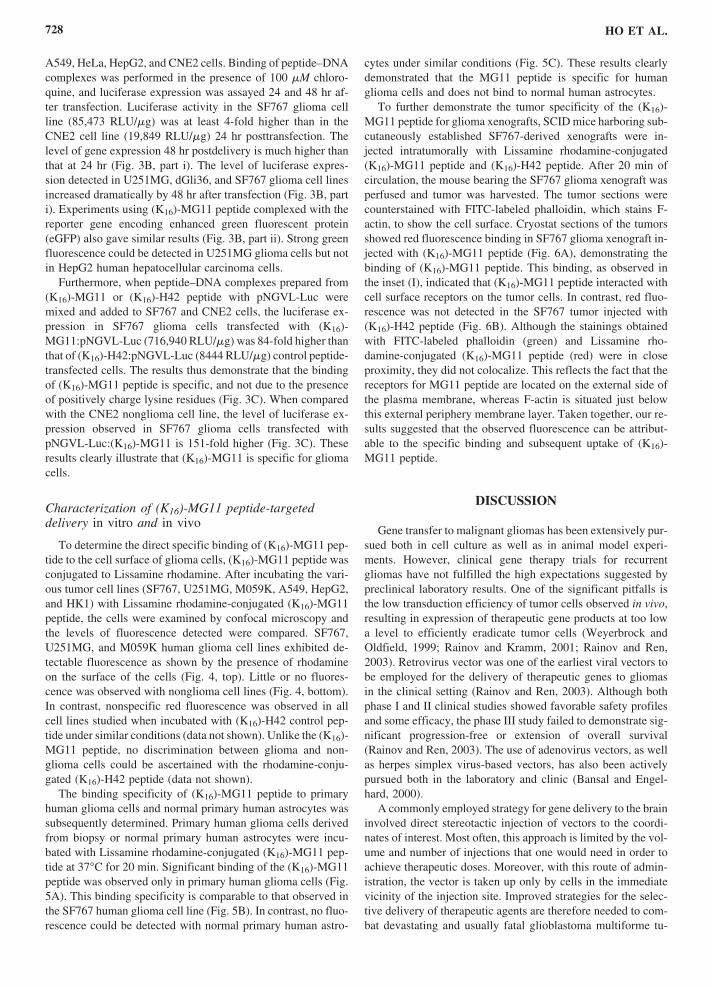