Development of a novel mouse glioma model using … of a novel mouse glioma model using lentiviral...
7
Development of a novel mouse glioma model using lentiviral vectors Tomotoshi Marumoto 1,2 , Ayumu Tashiro 3 , Dinorah Friedmann-Morvinski 1 , Miriam Scadeng 4 , Yasushi Soda 1 , Fred H Gage 1 & Inder M Verma 1 We report the development of a new method to induce glioblastoma multiforme in adult immunocompetent mice by injecting Cre-loxP–controlled lentiviral vectors expressing oncogenes. Cell type- or region-specific expression of activated forms of the oncoproteins Harvey-Ras and AKT in fewer than 60 glial fibrillary acidic protein–positive cells in the hippocampus, subventricular zone or cortex of mice heterozygous for the gene encoding the tumor suppressor Tp53 were tested. Mice developed glioblastoma multiforme when transduced either in the subventricular zone or the hippocampus. However, tumors were rarely detected when the mice were transduced in the cortex. Transplantation of brain tumor cells into naive recipient mouse brain resulted in the formation of glioblastoma multiforme–like tumors, which contained CD133 + cells, formed tumorspheres and could differentiate into neurons and astrocytes. We suggest that the use of Cre-loxP–controlled lentiviral vectors is a novel way to generate a mouse glioblastoma multiforme model in a region- and cell type-specific manner in adult mice. Mouse models of human cancers have been instructional in under- standing the basic principles of cancer biology. Currently, there are three major types of animal models: xenografts, human tumor tissues or cell lines transplanted in immunodeficient mice; trans- genesis, transgenic mice containing oncogenes with tissue-specific expression; and genetic knockouts, transgenic mice in whom a gene, usually a suppressor gene, is in the heterozygous state or is fully deleted 1 . Additional modifications to these methods, such as condi- tional knock-ins and knockouts, have become useful tools to study initiation, maintenance and progression of a wide variety of neo- plasias. Cancers arise from a single cell or a small number of cells in specific cell types, and the cellular origin of cancers is one of the major determinants of the characteristics of tumor cells. But in most animal models, either the oncogene is expressed in all of the cells in the tissue or a gene is genetically knocked out in the whole tissue. It is, therefore, desirable to develop animal models of human cancers in which one or multiple oncogenes are activated or tumor suppressor genes are inactivated in a single cell or a few cells in an immuno- competent adult mouse. We have established a new method of inducing a mouse cancer model by directly transducing oncogenes into a small number of cells. To test the validity of our technology, we have established a new mouse model of glioblastoma multiforme, a tumor in adult central nervous system neoplasms that is highly malignant and is resistant to existing treatments 2 . Transduction of as few as 60 GFAP + cells into the subventricular zone or hippocampus of adult immunocompetent mice with Cre-loxP–controlled lentiviral vectors expressing activated forms of the oncogenes Harvey-Ras (H-Ras) and AKT led to the formation of human high-grade glioma (World Health Organization grades III–IV)–like tumors. However, when transduction of activated H-Ras- and AKT-expressing lentiviral vec- tors is carried out in these regions of the brains of adult Tp53 heterozygotes, the frequency of formation of human glioblastoma multiforme (World Health Organization grade IV)–like tumors is substantially increased. The resulting tumors faithfully recapitulate the pathophysiology of human glioblastoma multiforme. The tech- nology reported here might be useful to model various human cancers in animals. RESULTS Oncogenic H-Ras and activated AKT induce glioma Brain tumors in adults, including glioblastoma multiforme, are thought to arise from a series of somatic mutations leading to the activation of oncogenes or inactivation of tumor suppressors that occur in a few cells or even a single founder cell in adults 3,4 . Thus, to recapitulate the initiation of glioblastoma multiforme, it is necessary to induce these oncogenic events in a single cell or in a small population of a specific type of cells in an adult mouse brain. To achieve this goal, we performed region-specific injection of lentiviral vectors that can transduce both neural stem and progenitor cells and terminally differentiated astrocytes 5 . To target specific cell types, we chose the Cre-loxP system, which is a reliable method of specifically expressing or deleting a target gene in Cre-expressing cells 6 . We generated Cre-loxP–controlled lentiviral vectors, Tomo lentiviral Received 2 May 2008; accepted 5 August 2008; published online 4 January 2009; doi:10.1038/nm.1863 1 Laboratory of Genetics, The Salk Institute for Biological Studies, 10010 North Torrey Pines Road, La Jolla, California 92037, USA. 2 Department of Neurosurgery, Kobe Medical Center National Hospital Organization, 3-1-1 Nishiochiai, Suma-ku, Kobe 654-0155, Japan. 3 Kavli Institute for Systems Neuroscience and Center for the Biology of Memory Norwegian University of Science and Technology, Medical Technical Research Center, Trondheim NO-7489, Norway. 4 University of California, San Diego Center for Functional Magnetic Resonance Imaging, University of California, San Diego, 9500 Gilman Drive, La Jolla, California 92093, USA. Correspondence should be addressed to I.M.V. ([email protected]). 110 VOLUME 15 [ NUMBER 1 [ JANUARY 2009 NATURE MEDICINE TECHNICAL REPORTS © 2009 Nature America, Inc. All rights reserved.
Transcript of Development of a novel mouse glioma model using … of a novel mouse glioma model using lentiviral...
Development of a novel mouse glioma model usinglentiviral vectorsTomotoshi Marumoto1,2, Ayumu Tashiro3, Dinorah Friedmann-Morvinski1, Miriam Scadeng4, Yasushi Soda1,Fred H Gage1 & Inder M Verma1
We report the development of a new method to induce
glioblastoma multiforme in adult immunocompetent mice
by injecting Cre-loxP–controlled lentiviral vectors expressing
oncogenes. Cell type- or region-specific expression of activated
forms of the oncoproteins Harvey-Ras and AKT in fewer
than 60 glial fibrillary acidic protein–positive cells in the
hippocampus, subventricular zone or cortex of mice
heterozygous for the gene encoding the tumor suppressor
Tp53 were tested. Mice developed glioblastoma multiforme
when transduced either in the subventricular zone or the
hippocampus. However, tumors were rarely detected when the
mice were transduced in the cortex. Transplantation of brain
tumor cells into naive recipient mouse brain resulted in the
formation of glioblastoma multiforme–like tumors, which
contained CD133+ cells, formed tumorspheres and could
differentiate into neurons and astrocytes. We suggest that
the use of Cre-loxP–controlled lentiviral vectors is a novel
way to generate a mouse glioblastoma multiforme model in
a region- and cell type-specific manner in adult mice.
Mouse models of human cancers have been instructional in under-standing the basic principles of cancer biology. Currently, there arethree major types of animal models: xenografts, human tumortissues or cell lines transplanted in immunodeficient mice; trans-genesis, transgenic mice containing oncogenes with tissue-specificexpression; and genetic knockouts, transgenic mice in whom a gene,usually a suppressor gene, is in the heterozygous state or is fullydeleted1. Additional modifications to these methods, such as condi-tional knock-ins and knockouts, have become useful tools to studyinitiation, maintenance and progression of a wide variety of neo-plasias. Cancers arise from a single cell or a small number of cells inspecific cell types, and the cellular origin of cancers is one of themajor determinants of the characteristics of tumor cells. But in mostanimal models, either the oncogene is expressed in all of the cells inthe tissue or a gene is genetically knocked out in the whole tissue. It is,therefore, desirable to develop animal models of human cancers inwhich one or multiple oncogenes are activated or tumor suppressor
genes are inactivated in a single cell or a few cells in an immuno-competent adult mouse. We have established a new method ofinducing a mouse cancer model by directly transducing oncogenesinto a small number of cells.
To test the validity of our technology, we have established anew mouse model of glioblastoma multiforme, a tumor in adultcentral nervous system neoplasms that is highly malignant and isresistant to existing treatments2. Transduction of as few as 60GFAP+ cells into the subventricular zone or hippocampus of adultimmunocompetent mice with Cre-loxP–controlled lentiviral vectorsexpressing activated forms of the oncogenes Harvey-Ras (H-Ras)and AKT led to the formation of human high-grade glioma (WorldHealth Organization grades III–IV)–like tumors. However, whentransduction of activated H-Ras- and AKT-expressing lentiviral vec-tors is carried out in these regions of the brains of adult Tp53heterozygotes, the frequency of formation of human glioblastomamultiforme (World Health Organization grade IV)–like tumors issubstantially increased. The resulting tumors faithfully recapitulatethe pathophysiology of human glioblastoma multiforme. The tech-nology reported here might be useful to model various human cancersin animals.
RESULTS
Oncogenic H-Ras and activated AKT induce glioma
Brain tumors in adults, including glioblastoma multiforme, arethought to arise from a series of somatic mutations leading to theactivation of oncogenes or inactivation of tumor suppressors thatoccur in a few cells or even a single founder cell in adults3,4. Thus, torecapitulate the initiation of glioblastoma multiforme, it is necessaryto induce these oncogenic events in a single cell or in a smallpopulation of a specific type of cells in an adult mouse brain. Toachieve this goal, we performed region-specific injection of lentiviralvectors that can transduce both neural stem and progenitor cells andterminally differentiated astrocytes5. To target specific cell types, wechose the Cre-loxP system, which is a reliable method of specificallyexpressing or deleting a target gene in Cre-expressing cells6. Wegenerated Cre-loxP–controlled lentiviral vectors, Tomo lentiviral
Received 2 May 2008; accepted 5 August 2008; published online 4 January 2009; doi:10.1038/nm.1863
1Laboratory of Genetics, The Salk Institute for Biological Studies, 10010 North Torrey Pines Road, La Jolla, California 92037, USA. 2Department of Neurosurgery,Kobe Medical Center National Hospital Organization, 3-1-1 Nishiochiai, Suma-ku, Kobe 654-0155, Japan. 3Kavli Institute for Systems Neuroscience and Center forthe Biology of Memory Norwegian University of Science and Technology, Medical Technical Research Center, Trondheim NO-7489, Norway. 4University of California,San Diego Center for Functional Magnetic Resonance Imaging, University of California, San Diego, 9500 Gilman Drive, La Jolla, California 92093, USA.Correspondence should be addressed to I.M.V. ([email protected]).
110 VOLUME 15 [ NUMBER 1 [ JANUARY 2009 NATURE MEDICINE
T ECHNICAL REPORTS©
2009
Nat
ure
Am
eric
a, In
c. A
ll ri
gh
ts r
eser
ved
.
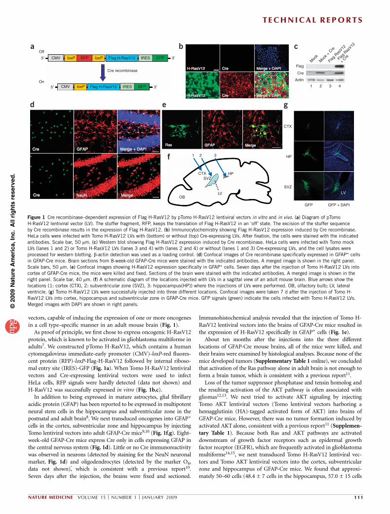
vectors, capable of inducing the expression of one or more oncogenesin a cell type–specific manner in an adult mouse brain (Fig. 1).
As proof of principle, we first chose to express oncogenic H-RasV12protein, which is known to be activated in glioblastoma multiforme inadults7. We constructed pTomo H-RasV12, which contains a humancytomegalovirus immediate-early promoter (CMV)-loxP-red fluores-cent protein (RFP)-loxP-Flag-H-RasV12 followed by internal riboso-mal entry site (IRES)-GFP (Fig. 1a). When Tomo H-RasV12 lentiviralvectors and Cre-expressing lentiviral vectors were used to infectHeLa cells, RFP signals were hardly detected (data not shown) andH-RasV12 was successfully expressed in vitro (Fig. 1b,c).
In addition to being expressed in mature astrocytes, glial fibrillaryacidic protein (GFAP) has been reported to be expressed in multipotentneural stem cells in the hippocampus and subventricular zone in thepostnatal and adult brain8. We next transduced oncogenes into GFAP+
cells in the cortex, subventricular zone and hippocampus by injectingTomo lentiviral vectors into adult GFAP-Cre mice9,10 (Fig. 1f,g). Eight-week-old GFAP-Cre mice express Cre only in cells expressing GFAP inthe central nervous system (Fig. 1d). Little or no Cre immunoreactivitywas observed in neurons (detected by staining for the NeuN neuronalmarker, Fig. 1d) and oligodendrocytes (detected by the marker O4,data not shown), which is consistent with a previous report10.Seven days after the injection, the brains were fixed and sectioned.
Immunohistochemical analysis revealed that the injection of Tomo H-RasV12 lentiviral vectors into the brains of GFAP-Cre mice resulted inthe expression of H-RasV12 specifically in GFAP+ cells (Fig. 1e).
About ten months after the injections into the three differentlocations of GFAP-Cre mouse brains, all of the mice were killed, andtheir brains were examined by histological analyses. Because none of themice developed tumors (Supplementary Table 1 online), we concludedthat activation of the Ras pathway alone in adult brain is not enough toform a brain tumor, which is consistent with a previous report11.
Loss of the tumor suppressor phosphatase and tensin homolog andthe resulting activation of the AKT pathway is often associated withgliomas12,13. We next tried to activate AKT signaling by injectingTomo AKT lentiviral vectors (Tomo lentiviral vectors harboring ahemagglutinin (HA)-tagged activated form of AKT) into brains ofGFAP-Cre mice. However, there was no tumor formation induced byactivated AKT alone, consistent with a previous report11 (Supplemen-tary Table 1). Because both Ras and AKT pathways are activateddownstream of growth factor receptors such as epidermal growthfactor receptor (EGFR), which are frequently activated in glioblastomamultiforme14,15, we next transduced Tomo H-RasV12 lentiviral vec-tors and Tomo AKT lentiviral vectors into the cortex, subventricularzone and hippocampus of GFAP-Cre mice. We found that approxi-mately 50–60 cells (48.4 ± 7 cells in the hippocampus, 57.0 ± 15 cells
Off
5′
5′ 3′
3′
CMV IoxP
IoxPCMV IoxP
Flag H-RasV12
Flag H-RasV12
Cre recombinase
IRES
IRES
GFP
RFP GFP
Flag
Cre
Actin
1 2 3 4
Moc
kM
ock +
Cre
Flag R
asV12
Flag R
asV12
+ Cre
On
a
d e
f
g
b c
GFP
SVZ
SVZ
HP
HP
CTX
CTX
LVOB
1 2 3
GFP + DAPI
Figure 1 Cre recombinase–dependent expression of Flag H-RasV12 by pTomo H-RasV12 lentiviral vectors in vitro and in vivo. (a) Diagram of pTomo
H-RasV12 lentiviral vector (LV). The stuffer fragment, RFP, keeps the translation of Flag H-RasV12 in an ‘off’ state. The excision of the stuffer sequence
by Cre recombinase results in the expression of Flag H-RasV12. (b) Immunocytochemistry showing Flag H-RasV12 expression induced by Cre recombinase.HeLa cells were infected with Tomo H-RasV12 LVs with (bottom) or without (top) Cre-expressing LVs. After fixation, the cells were stained with the indicated
antibodies. Scale bar, 50 mm. (c) Western blot showing Flag H-RasV12 expression induced by Cre recombinase. HeLa cells were infected with Tomo mock
LVs (lanes 1 and 2) or Tomo H-RasV12 LVs (lanes 3 and 4) with (lanes 2 and 4) or without (lanes 1 and 3) Cre-expressing LVs, and the cell lysates were
processed for western blotting. b-actin detection was used as a loading control. (d) Confocal images of Cre recombinase specifically expressed in GFAP+ cells
in GFAP-Cre mice. Brain sections from 8-week-old GFAP-Cre mice were stained with the indicated antibodies. A merged image is shown in the right panel.
Scale bars, 50 mm. (e) Confocal images showing H-RasV12 expression specifically in GFAP+ cells. Seven days after the injection of Tomo H-RasV12 LVs into
cortex of GFAP-Cre mice, the mice were killed and fixed. Sections of the brain were stained with the indicated antibodies. A merged image is shown in the
right panel. Scale bar, 40 mm. (f) A schematic diagram of the locations injected with LVs in a sagittal view of an adult mouse brain. Blue arrows show the
locations (1: cortex (CTX), 2: subventricular zone (SVZ), 3: hippocampus(HP)) where the injections of LVs were performed. OB, olfactory bulb; LV, lateral
ventricle. (g) Tomo H-RasV12 LVs were successfully injected into three different locations. Confocal images were taken 7 d after the injection of Tomo H-
RasV12 LVs into cortex, hippocampus and subventricular zone in GFAP-Cre mice. GFP signals (green) indicate the cells infected with Tomo H-RasV12 LVs.
Merged images with DAPI are shown in right panels.
T ECHNICAL REPORTS
NATURE MEDICINE VOLUME 15 [ NUMBER 1 [ JANUARY 2009 111
©20
09 N
atu
re A
mer
ica,
Inc.
All
rig
hts
res
erve
d.
in the subventricular zone and 56.7 ± 13 cells in the cortex;Supplementary Fig. 1 online) were infected with both Tomo H-RasV12 lentiviral vectors and Tomo AKT lentiviral vectors, and almostall of the cells (498%) showed the expression of both H-RasV12 andAKT proteins (Supplementary Fig. 1).
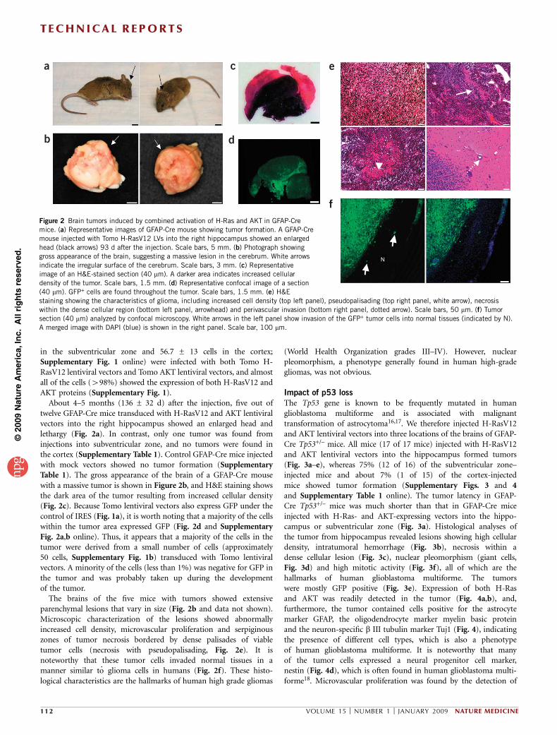
About 4–5 months (136 ± 32 d) after the injection, five out oftwelve GFAP-Cre mice transduced with H-RasV12 and AKT lentiviralvectors into the right hippocampus showed an enlarged head andlethargy (Fig. 2a). In contrast, only one tumor was found frominjections into subventricular zone, and no tumors were found inthe cortex (Supplementary Table 1). Control GFAP-Cre mice injectedwith mock vectors showed no tumor formation (SupplementaryTable 1). The gross appearance of the brain of a GFAP-Cre mousewith a massive tumor is shown in Figure 2b, and H&E staining showsthe dark area of the tumor resulting from increased cellular density(Fig. 2c). Because Tomo lentiviral vectors also express GFP under thecontrol of IRES (Fig. 1a), it is worth noting that a majority of the cellswithin the tumor area expressed GFP (Fig. 2d and SupplementaryFig. 2a,b online). Thus, it appears that a majority of the cells in thetumor were derived from a small number of cells (approximately50 cells, Supplementary Fig. 1b) transduced with Tomo lentiviralvectors. A minority of the cells (less than 1%) was negative for GFP inthe tumor and was probably taken up during the developmentof the tumor.
The brains of the five mice with tumors showed extensiveparenchymal lesions that vary in size (Fig. 2b and data not shown).Microscopic characterization of the lesions showed abnormallyincreased cell density, microvascular proliferation and serpiginouszones of tumor necrosis bordered by dense palisades of viabletumor cells (necrosis with pseudopalisading, Fig. 2e). It isnoteworthy that these tumor cells invaded normal tissues in amanner similar to glioma cells in humans (Fig. 2f). These histo-logical characteristics are the hallmarks of human high grade gliomas
(World Health Organization grades III–IV). However, nuclearpleomorphism, a phenotype generally found in human high-gradegliomas, was not obvious.
Impact of p53 loss
The Tp53 gene is known to be frequently mutated in humanglioblastoma multiforme and is associated with malignanttransformation of astrocytoma16,17. We therefore injected H-RasV12and AKT lentiviral vectors into three locations of the brains of GFAP-Cre Tp53+/– mice. All mice (17 of 17 mice) injected with H-RasV12and AKT lentiviral vectors into the hippocampus formed tumors(Fig. 3a–e), whereas 75% (12 of 16) of the subventricular zone–injected mice and about 7% (1 of 15) of the cortex-injectedmice showed tumor formation (Supplementary Figs. 3 and 4and Supplementary Table 1 online). The tumor latency in GFAP-Cre Tp53+/– mice was much shorter than that in GFAP-Cre miceinjected with H-Ras- and AKT-expressing vectors into the hippo-campus or subventricular zone (Fig. 3a). Histological analyses ofthe tumor from hippocampus revealed lesions showing high cellulardensity, intratumoral hemorrhage (Fig. 3b), necrosis within adense cellular lesion (Fig. 3c), nuclear pleomorphism (giant cells,Fig. 3d) and high mitotic activity (Fig. 3f), all of which are thehallmarks of human glioblastoma multiforme. The tumorswere mostly GFP positive (Fig. 3e). Expression of both H-Rasand AKT was readily detected in the tumor (Fig. 4a,b), and,furthermore, the tumor contained cells positive for the astrocytemarker GFAP, the oligodendrocyte marker myelin basic proteinand the neuron-specific b III tubulin marker Tuj1 (Fig. 4), indicatingthe presence of different cell types, which is also a phenotypeof human glioblastoma multiforme. It is noteworthy that manyof the tumor cells expressed a neural progenitor cell marker,nestin (Fig. 4d), which is often found in human glioblastoma multi-forme18. Microvascular proliferation was found by the detection of
a c
d
e
f
b
N
Figure 2 Brain tumors induced by combined activation of H-Ras and AKT in GFAP-Cremice. (a) Representative images of GFAP-Cre mouse showing tumor formation. A GFAP-Cre
mouse injected with Tomo H-RasV12 LVs into the right hippocampus showed an enlarged
head (black arrows) 93 d after the injection. Scale bars, 5 mm. (b) Photograph showing
gross appearance of the brain, suggesting a massive lesion in the cerebrum. White arrows
indicate the irregular surface of the cerebrum. Scale bars, 3 mm. (c) Representative
image of an H&E-stained section (40 mm). A darker area indicates increased cellular
density of the tumor. Scale bars, 1.5 mm. (d) Representative confocal image of a section
(40 mm). GFP+ cells are found throughout the tumor. Scale bars, 1.5 mm. (e) H&E
staining showing the characteristics of glioma, including increased cell density (top left panel), pseudopalisading (top right panel, white arrow), necrosis
within the dense cellular region (bottom left panel, arrowhead) and perivascular invasion (bottom right panel, dotted arrow). Scale bars, 50 mm. (f) Tumor
section (40 mm) analyzed by confocal microscopy. White arrows in the left panel show invasion of the GFP+ tumor cells into normal tissues (indicated by N).
A merged image with DAPI (blue) is shown in the right panel. Scale bar, 100 mm.
T ECHNICAL REPORTS
112 VOLUME 15 [ NUMBER 1 [ JANUARY 2009 NATURE MEDICINE
©20
09 N
atu
re A
mer
ica,
Inc.
All
rig
hts
res
erve
d.

CD31- and von Willebrand factor–positive endothelial cells in thetumor (Fig. 4g and Supplementary Fig. 2f–h). Consistent with thisresult, the overexpression of vascular endothelial growth factor(VEGF) and VEGF receptor-2 (VEGFR2) in the tumor tissues wereobserved (Fig. 4h,i and Supplementary Fig. 5a online). Five tumorsthat arose from the hippocampus, five tumors that arose from thesubventricular zone and one tumor that arose from the cortex wereexamined by histological analyses, and most of the histologicalcharacteristics of the tumors from the hippocampus were also foundin the tumors from the subventricular zone and cortex (Supplemen-tary Figs. 3 and 4 and Supplementary Table 2 online).
P53 has been shown to have a role in inducing senescence inresponse to oncogenic stimuli, resulting in resistance to cellulartransformation, and, therefore, the loss of Tp53 is thought to allowthe tumor cells to bypass the oncogene-induced senescence19. The p53was more abundant in tumors from GFAP-Cre mice than in tumorsfrom GFAP-Cre Tp53+/– mice (Supplementary Fig. 6a,b online). Wenext examined senescence-associated b-galactosidase (SA–b-gal)activity in the tumors derived from GFAP-Cre or GFAP-Cre Tp53+/–
mice. SA–b-gal activity was more promi-nently found in the tumors from GFAP-Cremice than in those from GFAP-Cre Tp53+/–
mice (Supplementary Fig. 6c–f), indicatingthat loss of Tp53 promotes the cells’ escapefrom the oncogene-induced senescence pro-gram activated by H-Ras and AKT, whichmay result in the shorter latency, higherincidence and higher mitotic activity oftumors found in GFAP-Cre Tp53+/– micecompared to those from GFAP-Cre mice(Fig. 3a,f and Supplementary Table 1). Con-sistent with this result, the degree of apopto-sis in the tumors from GFAP-Cre mice wasmuch lower than that in the tumors fromGFAP-Cre Tp53+/– mice (SupplementaryFig. 6g,h).
The expression of EGFR, which is frequently amplified andoverexpressed in human glioblastoma multiforme, was not prominentin this mouse model of glioblastoma multiforme (SupplementaryFig. 5a), probably because H-Ras and AKT, which are known to bedownstream of EGFR, have sufficiently been activated in thismodel14,15. Also, the expression of MDM2, an oncoprotein thatdegrades p53, is often amplified or overexpressed in human glio-blastoma multiforme but is low in this mouse model of glioblastomamultiforme (Supplementary Fig. 5a), probably because p53 expres-sion is low in the tumor (Supplementary Fig. 6b) and MDM2 isknown to be transcriptionally activated by p53 (ref. 14,15,20). Anothermajor tumor suppressor gene, Cdkn2a (encoding p16), which is oftendeleted in human glioblastoma multiforme, was not deleted in thismouse model of the disease (Supplementary Fig. 5b). Finally, amagnetic resonance image (MRI) of the tumor-bearing mouseshowed a gadolinium-enhanced mass lesion, which is commonlyfound in imaging of human glioblastoma multiforme (Supplemen-tary Fig. 7 online).
Brain tumor–initiating stem cells
Normal neural stem cells can be isolated in culture as clonally derivedcolonies termed neurospheres21. We next established primary culturesof the brain tumor induced by the combined activation of H-Rasand AKT in GFAP-Cre Tp53+/– mice and determined that the culturescontained brain tumor–initiating stem cells (BTICs). The tumorcells (005 tumor cells) were taken from a mouse tumor (originatingfrom the hippocampus in a GFAP-Cre Tp53+/– mouse) and culturedin the normal medium often used for cancer cell lines or in the
100
80
60
Sur
viva
l (%
)
40
20
00 40 80
GFAP-Cre mice, SVZ injection, n = 9GFAP-Cre Tp53+/– mice, SVZ injection, n = 16GFAP-Cre mice, HP injection, n = 12GFAP-Cre Tp53+/– mice, HP injection, n = 17
Time (d) after injection120 160 200
a
Ras and AKT–induced tumorin GFAP-Cre miceRas and AKT–induced tumorin GFAP-Cre Tp53+/– mice
0
1
2
3
Per
cent
age
of th
e K
i67-
posi
tive
cells
in th
e tu
mor
s (%
)
4
5
6 *f
b
d e
c
Figure 3 Effects of p53 loss on tumor formation induced by combined
activation of H-Ras and AKT. (a) A Kaplan-Meier curve showing glioma-free
survival. GFAP-Cre or GFAP-Cre Tp53+/– mice were injected with Tomo H-
RasV12 LVs and Tomo AKT LVs into hippocampus or subventricular zone.
(b–d) H&E staining of the tumor induced by combined activation of H-Ras
and AKT in the right hippocampus in a GFAP-Cre Tp53+/– mouse. T and N
in b indicate the tumor area and the normal tissue area, respectively.
Black arrow in b indicates intratumoral hemorrhage. Black arrowhead
in c indicates the area of necrosis. White arrows in d indicate giant cell
formation in the tumor. Scale bars, 50 mm. (e) Confocal micrograph showing
GFP+ cells throughout the tumor area. T indicates tumor area. Scale bar,
1.5 mm. (f) A graph showing the proportion of the cycling cells in tumors.
Tumor tissues originated from the right hippocampus found in GFAP-Cre or
GFAP-Cre Tp53+/– mice were stained with Ki67-specific antibody, at least
700 cells in three different locations were examined in each tumor, and thenumber of cells positive for Ki67 was calculated. *P o 0.05; data represent
means ± s.d.
a c g h
i j
b
d fe
Figure 4 Tumors induced by combined activation of H-Ras and AKT in GFAP-Cre Tp53+/– micecontain the cells expressing various cell markers including astrocytes, oligodendrocytes and neurons.
(a–j) Immunohistochemical analysis of sections from the tumor induced by combined activation of
H-Ras and AKT in the right hippocampus of GFAP-Cre Tp53+/– mice stained with antibodies to Flag (a),
HA (b), GFAP (c), nestin (d), myelin basic protein (MBP, e), Tuj1 (f), CD31 (g), VEGF (h), VEGFR2 (i)
and Ki67 (j). Scale bars, 50 mm.
T ECHNICAL REPORTS
NATURE MEDICINE VOLUME 15 [ NUMBER 1 [ JANUARY 2009 113
©20
09 N
atu
re A
mer
ica,
Inc.
All
rig
hts
res
erve
d.

medium prepared for neural stem cells. 005 tumor cells in thenormal medium survived for 1 week but eventually died within2 weeks, whereas 005 tumor cells in the medium prepared forculturing neural stem cells could survive and continue to proliferatefor more than 30 passages (data not shown). Furthermore, GFP+
tumor cells cultured in the medium prepared for neural stem cellsformed neurosphere-like structures (often called a ‘tumorsphere’,Fig. 5a). We next examined the self-renewal capacity of 005 tumorcells by dissociating primary tumorspheres and replating single-cellsuspensions of these cells at serial dilutions down to one cell per well.Primary tumorsphere–derived GFP+ cells formed secondary sphereswith a range of 76–88 secondary spheres per 100 primary sphere cells(mean 81.3 ± 5.1) over a period of 1–2 weeks. The secondary sphere-forming cells possessed the potential to grow infinitely, and theproportion of the cells positive for nestin, a marker for immatureneural progenitors, remained stable throughout the course of cellculture (at least 2 months), indicating that 005 tumor cells have thepotential to self renew.
A vast majority of tumorsphere-forming cells (499% of 005 tumorcells) were positive for nestin (Fig. 5a) and negative for neuronalmarkers (microtubule-associated protein 2, and Tuj1, data notshown), astrocyte markers (s100b and GFAP, data not shown) andoligodendrocyte markers (RIP and O4, data not shown). We nexttested whether 005 tumor cells can differentiate in response to serumaddition to the medium. We found that 005 tumor cells showedelongated processes (Fig. 5b) and expressed markers for astrocytesand neurons (Fig. 5c–e) upon addition of 10% FBS, although theydid not express oligodendrocyte markers under these conditions (datanot shown).
It has been shown that most xenograft models for brain tumorsusing tumor cell lines require at least 1 � 105 cells to formgliomas22,23, whereas only 100 BTICs are enough to form tumors in
the immunodeficient mouse brain24. Thus, we next examinedwhether 005 tumor cells can form brain tumors when injected intothe brains of nonobese diabetic–severe combined immuno-deficient (NOD-SCID) mice. Injection of only 100 005 tumor cellsinto the hippocampus of NOD-SCID mice resulted in the formationof tumors (seven of nine mice, average latency 40 ± 6 d, Fig. 5f–i),whereas no control mice (zero of eight) injected with 1 � 105
wild-type mouse neural stem cells formed any tumors (data notshown). Moreover, injection of only ten 005 cells into the hippo-campus in NOD-SCID mice resulted in tumor formation (fourof eight mice, average latency: 63.2 ± 6.8 d), indicating thestrong tumor-forming activity of 005 tumor cells. The gross appear-ance of the brains of the injected mice showed a darker area, whichsuggests a massive lesion with hemorrhage (Fig. 5f). The tumorshowed glioma-like histopathology, including increased cellular den-sity, necrosis within the dense cellular lesion and intratumoralhemorrhage (Fig. 5g–i). Notably, the tumor also showed infiltrativecharacteristics (Fig. 5i), which is not observed in xenograft modelsusing glioblastoma multiforme cell lines25. The characteristics ofthe 005 tumor cells described here were also observed in experi-ments using 006 tumor cells, which were independently isolated froma tumor induced by combined activation of H-Ras and AKT inthe hippocampus of a GFAP-Cre Tp53+/– mouse (SupplementaryFig. 8 online).
In addition to being a marker hematopoietic stem and progenitorcells, CD133 has also been found to be a marker for BTICs. FACSanalysis revealed that CD133 was found in 40–50% of 005 tumor cells(Fig. 5j). We observed that the proportion of the cells that expressCD133 gradually decreased during the course of the culture (especiallyafter 3 months of culture), indicating that 005 tumor cells differentiateupon culturing in the dish, even in the medium for neural stem cells.We conclude that tumorsphere-forming cells derived from our mouse
a
b
c
d
e f
GFAP DAPI
Tuj1 DAPI
MAP2
0
10
20
30
Per
cent
age
ofth
e ce
lls (
%) 40
50
TuJ1
NeuronsDay 0 Day 2 Day 5
Astrocytes
S100β
GFAP
g
j
h i
1000
60
120
Cou
nts
180
101 102
CD133103 104
M1
Control005 tumor cells
Figure 5 005 tumor cells show the
characteristics of BTICs. (a) Confocal
microscopic analysis of neurosphere-like
structures formed by 005 tumor cells.
Cells stained with nestin-specific antibody
are shown in the middle panel. A merged
image with DAPI staining is shown in the
right panel. Scale bar, 50 mm. (b) Phase-
contrast images of 005 tumor cells
showing elongated processes in response
to serum addition in the medium. Images were taken on days 0, 2 and 5 after the serum addition. Scale bars, 50 mm. (c–e) 005 tumor cells expressing
neuronal or astrocyte markers. Five days after the addition of FBS to the medium (final 10% FBS), cells were fixed and stained with GFAP-specific antibody
(c) or Tuj1-specific antibody (d) and observed by confocal microscopy. Merged mages with DAPI are shown. Scale bars, 30 mm. The proportions of cells
positive for neuronal or astrocyte marker are shown in e. Data represent means ± s.d. (f) The gross appearance of tumor formation in the brain of a NOD-
SCID mouse after injection of 100 of 005 tumor cells. Scale bar, 3 mm. (g–i) Histological characteristics of the tumor caused by the injection of 005 tumor
cells in NOD-SCID mice, including increased cellular density (g), intratumoral hemorrhage (indicated by H in h), necrosis (indicated by N in h), giant cell
formation (indicated by white arrows in h) and infiltrative characteristics (i). Scale bars, 50 mm. (j) FACS analysis of 005 tumor cells in culture incubated
with CD133-specific antibody. Unstained cells were used as a control.
T ECHNICAL REPORTS
114 VOLUME 15 [ NUMBER 1 [ JANUARY 2009 NATURE MEDICINE
©20
09 N
atu
re A
mer
ica,
Inc.
All
rig
hts
res
erve
d.
model of glioblastoma multiforme, self renew, differentiate in vitroand have strong tumor-initiating activity similar to previouslyreported BTICs4,24. These results indicate that this mouse model ofglioblastoma multiforme is useful not only for the understanding ofthe initiation of glioblastoma multiforme but also for the investigationof BTICs.
DISCUSSION
The use of somatic cell gene transfer using retroviral vectors in to theneonatal mouse brain to model gliomas has been previouslydescribed26,27. In one such case, avian leukosis virus (ALV)-basedreplication-competent ALV-LTR (long terminal repeat) splice acceptor(RCAS) vectors were injected into the brain of neonatal mice expres-sing the RCAS receptor, tv-a (a receptor for the ALV envelopeglycoprotein) from GFAP or the nestin promoter11. The researchersshowed that combined activation of K-Ras and AKT in nestin+ neuralprogenitors caused the formation of glioblastoma multiforme–liketumor11. They did not observer tumor formation when K-Ras andAKT were induced in GFAP+ astrocytes11; thus, nestin+ neuralprogenitors may be the cellular origin of glioblastoma multiforme.In our model, combined activation of H-Ras and AKT and the loss ofp53 in GFAP+ cells in the hippocampus or subventricular zone causedthe formation of glioblastoma multiforme–like disease. BecauseGFAP+ cells in the hippocampus or subventricular zone are likely tocontain both differentiated astrocytes and neural progenitor cells8,28,these cell types could be the cellular origin of the glioblastomamultiforme–like tumors in our model. To induce glioblastoma multi-forme–like disease in mice, the other group used mice with a wild-typeTp53 genetic background, whereas we needed to use Tp53 hetero-zygotes to obtain all of the neoplastic features associated withglioblastoma multiforme (Fig. 3). Some of the discrepancies betweenthe two models might be attributed to the differences between K-Rasand H-Ras in vivo. The other researchers also used neonatal mice,whereas we used adult mice, and thus the discrepancies may haveoriginated from the differences between the cells used in theirproliferative and differentiation characteristics.
One of the advantages of our strategy is that this method can beused to clarify the differences between tumors caused by the sameoncogenic events occurring in different regions of the brain. There waslittle tumor formation from the cortex, whereas more than 75% ofmice showed tumor formation from the neurogenic areas such as thesubventricular zone and hippocampus (Supplementary Table 1). Thisresult is consistent with a previous report showing that neurogenicareas such as the subventricular zone and hippocampus are moresensitive to oncogenic stimuli, such as chemicals, than other areas29.One explanation for this is that there may be unknown growth factorsin neurogenic areas that promote the transformation of GFAP+ cells.Another explanation is that GFAP+ neural progenitor cells, which areabundant in neurogenic areas, may be more susceptible to transfor-mation than GFAP+ differentiated astrocytes, which are abundant inthe cortex.
Because there are growing numbers of transgenic mice that expressCre in a specific cell type, our proposed strategy of introducingoncogenes could become a versatile and useful method to modelvarious tumors in any organ.
METHODSPlasmids. To generate pTomo, we inserted a loxP-mRFP-loxP fragment ampli-
fied from the pSETB mRFP1 vector by PCR between the XbaI and BamHI sites
of the p156RRLsin PPTCMVIRESPRE vector (a third-generation lenti-
viral vector)30. To construct pTomo H-RasV12, we amplified N-terminus
Flag-tagged H-RasV12 from pGEM H-RasV12 by PCR and inserted it into
the BamHI site of the pTomo vector. To construct pTomo AKT, we amplified
the HA-tagged constitutively active AKT (we added the myristoylation signal of
the c-Src oncoprotein, MGSSKSKPKDPSQR, to the N terminus of human
AKT1) from p156RRLsin PPTCMVIRESPRE myr-AKT by PCR and inserted it
into the BamHI site of the pTomo vector. We confirmed all PCR fragments
by sequencing.
Viral vector production. We produced all lentiviral vectors were by the method
described previously31. We determined the biological titer of lentiviral vectors
by infecting HeLa cells (American Type Culture Collection) with various
amounts of the viruses.
Mice. We obtained GFAP-Cre transgenic mice9 from The Jackson Laboratories.
We maintained the colonies of this strain by crossing to wild -C57BL/6 mice.
We crossed Tp53–/– mice with the GFAP-Cre line. All mice in this study were
cared for according to the guidelines that were approved by the Animal Care
and Use Committee of the Salk Institute.
In vivo vector injection. We stereotaxically injected a small amount of viruses
(0.8 ml, 1 � 108 international units) into mice at the age of 8–16 weeks old who
were under anesthesia with ketamine-xylazine solution. To inject a mixture of
Tomo H-RasV12 lentiviral vectors and Tomo AKT lentiviral vectors, we mixed
the two viral preparations (1:1) and injected 0.8 ml.
Western blotting. We lysed cells or tissues in NP-40 lysis buffer (50 mM
Tris-HCl, pH 7.4, 150 mM NaCl, 1 mM EDTA and 0.5% NP-40), and we
subjected equal amounts of the lysates to SDS-PAGE. We transferred the
proteins to a nitrocellulose membrane and probed the membrane with the
antibodies listed in Supplementary Table 3 online.
Southern blotting. We digested genomic DNA (20 mg) with BamHI, separated
the fragments through an 0.8% agarose gel and blotted them on a nylon
membrane before hybridization with a 32P-random-prime–labeled, 737–base
pair SalI-BstXI GFP fragment from the pTomo vectors or a 501–base pair
genomic probe (corresponding to the DNA sequence from exon 1 to exon 2)
for Cdkn2a, which we PCR-amplified from genomic DNA of normal mouse
endothelial cells.
b-galactosidase staining and TUNEL staining. We subjected 40-mm coronal
tumor sections fixed with 4% paraformaldehyde in PBS to SA–b�gal staining
and TUNEL staining as previously described19.
Cell culture in vitro. We cultured tumor cells obtained from mouse glioblas-
toma multiforme–like tumors (005 and 006) in N2-supplemented (Invitrogen)
DME:Ham’s F12 (Omega Scientific) medium containing 20 ng ml–1 fibroblast
growth factor-2 (PeproTech), 20 ng ml–1 epidermal growth factor (Promega)
and 50 mg ml–1 heparin (Sigma), which is prepared for mouse neural stem
cells32. To induce differentiation, we allowed cells to proliferate in DME:Ham’s
F12 medium supplemented with 10% FBS for 5 d.
Immunocytochemistry. We performed immunocytochemistry as previously
described33. The antibodies used are listed in Supplementary Table 3.
Immunohistochemistry and hematoxylin and eosin staining. We perfused
mice with 0.9% NaCl followed by 4% paraformaldehyde in PBS. We then
excised the brains, stored them in fixative overnight and transferred them to
30% sucrose in PBS. For detection with fluorescence, we cut 40-mm coronal
sections on a sliding microtome and processed the sections for standard
immunohistochemical staining as previously described34. We obtained the
images by confocal laser-scanning microscopy (Leica TCS SP2 ABS). For
H&E staining, we embedded the fixed brains in paraffin, cut them into
6-mm sections and stained with H&E. For immunohistochemical analysis of
paraffinized tissues, we deparaffinized the tissues, rehydrated them and stained
them as previously described35. We used diaminobenzidine (Vector) as a
substrate for a peroxidase. After counterstaining with Mayer’s hematoxylin
(Sigma), we mounted the sections with Vectamount AQ (Vector). We obtained
the images with a Retiga digital camera on a Nikon optical microscope and
analyzed them with OpenLab software (Improvision).
T ECHNICAL REPORTS
NATURE MEDICINE VOLUME 15 [ NUMBER 1 [ JANUARY 2009 115
©20
09 N
atu
re A
mer
ica,
Inc.
All
rig
hts
res
erve
d.

Flow cytometric analysis. We stained 005 tumor cells with CD133-specific
antibody and analyzed them on a BD LSR I flow cytometer (Becton Dickinson)
with CELLQuest software.
Magnetic resonance imaging. We imaged the mice in vivo both before and
after contrast. We acquired images with a 1.5-cm custom-built surface MRI
coil, which was manually tuned and placed in a horizontal-bore 7T MR scanner
(General Electric). We acquired images both before and after administration of
the magnetic resonance contrast agent gadolinium dimeglumine (Magnevist)
with a three-dimensional fast spoiled gradient sequence: TR (repitition time) /
TE (echo time) ¼ 7.5/3.2, FA (flip angle) 20, FOV (field of view) 1.4 mm,
matrix 160 � 160 � 300, BW (bandwidth) 15.63; ten averages providing three-
dimensional datasets of the brain at 87 mm in plane resolution. We also
acquired T2-weighted anatomical images before with a two-dimensional fast
spin-echo sequence TR / TE ¼ 2900/35.5, FA 90, BW 15.63. The after-contrast
dataset was semi-manually segmented and volume-rendered with Amira soft-
ware (Template Graphic Software) to produce quantitative three-dimensional
models of the area of contrast enhancement in the brain tissue.
Note: Supplementary information is available on the Nature Medicine website.
ACKNOWLEDGMENTSWe thank R. Shaw for discussions and critical reading of the manuscript; N. Varkiand H. Powell for pathological analyses; S. Yla-Herttuala, K. Suzuki, N. Tanaka,G. Pao, A. Parker and H. Suh for useful discussions; T. Sawai, G. Estepa andM. Lawrence for technical assistance; M. Schmitt and B. Coyne for admini-strative assistance; and S. Withoff (St. Jude Hospital), V. Tergaonkar (BioprocessingTechnology Institute), O. Singer (Salk Institute) and G. Wahl (Salk Institute) forproviding pSETB mRFP1, pGEM H-RasV12, p156RRLsin PPTCMVIRESPRE myr-AKT vectors and Tp53–/– mice, respectively. T.M. is supported by the AmericanBrain Tumor Association. I.M.V. is an American Cancer Society Professor ofMolecular Biology and is supported in part by grants from the US NationalInstitutes of Health and the H.N. and Frances C. Berger Foundation. The projectdescribed was supported in part by the National Institutes of Health. The contentis solely the responsibility of the authors and does not necessarily represent theofficial views of the National Institutes of Health.
AUTHOR CONTRIBUTIONST.M. conducted most of the experiments, prepared figures and wrote themanuscript. A.T., D.F.-M. and Y.S. participated in in vivo studies and histologicalanalysis. M.S. performed MRI imaging. F.H.G. supervised the experiments andthe project. I.M.V. is the principle investigator, supervised the experiments andthe project and wrote the manuscript.
Published online at http://www.nature.com/naturemedicine/
Reprints and permissions information is available online at http://npg.nature.com/
reprintsandpermissions/
1. Hesselager, G. & Holland, E.C. Using mice to decipher the molecular genetics of braintumors. Neurosurgery 53, 685–694; discussion 695 (2003).
2. Holland, E.C. Glioblastoma multiforme: the terminator. Proc. Natl. Acad. Sci. USA 97,6242–6244 (2000).
3. Holland, E.C. Gliomagenesis: genetic alterations and mouse models. Nat. Rev. Genet.2, 120–129 (2001).
4. Vescovi, A.L., Galli, R. & Reynolds, B.A. Brain tumour stem cells. Nat. Rev. Cancer 6,425–436 (2006).
5. Kafri, T., Blomer, U., Peterson, D.A., Gage, F.H. & Verma, I.M. Sustained expression ofgenes delivered directly into liver and muscle by lentiviral vectors. Nat. Genet. 17,314–317 (1997).
6. Abremski, K., Hoess, R. & Sternberg, N. Studies on the properties of P1 site-specificrecombination: evidence for topologically unlinked products following recombination.Cell 32, 1301–1311 (1983).
7. Guha, A., Feldkamp, M.M., Lau, N., Boss, G. & Pawson, A. Proliferation of humanmalignant astrocytomas is dependent on Ras activation. Oncogene 15, 2755–2765(1997).
8. Doetsch, F., Caille, I., Lim, D.A. & Garcia-Verdugo, J.M. & Alvarez-Buylla, A. Subven-tricular zone astrocytes are neural stem cells in the adult mammalian brain. Cell 97,703–716 (1999).
9. Zhuo, L. et al. hGFAP-Cre transgenic mice for manipulation of glial and neuronalfunction in vivo. Genesis 31, 85–94 (2001).
10. Malatesta, P. et al. Neuronal or glial progeny: regional differences in radial glia fate.Neuron 37, 751–764 (2003).
11. Holland, E.C. et al. Combined activation of Ras and Akt in neural progenitors inducesglioblastoma formation in mice. Nat. Genet. 25, 55–57 (2000).
12. Steck, P.A. et al. Identification of a candidate tumour suppressor gene, MMAC1, atchromosome 10q23.3 that is mutated in multiple advanced cancers. Nat. Genet. 15,356–362 (1997).
13. Li, J. et al. PTEN, a putative protein tyrosine phosphatase gene mutated in humanbrain, breast, and prostate cancer. Science 275, 1943–1947 (1997).
14. Ekstrand, A.J. et al. Functional characterization of an EGF receptor with a truncatedextracellular domain expressed in glioblastomas with EGFR gene amplification.Oncogene 9, 2313–2320 (1994).
15. Maher, E.A. et al. Malignant glioma: genetics and biology of a grave matter. Genes Dev.15, 1311–1333 (2001).
16. Lang, F.F., Miller, D.C., Koslow, M. & Newcomb, E.W. Pathways leading to glioblastomamultiforme: a molecular analysis of genetic alterations in 65 astrocytic tumors.J. Neurosurg. 81, 427–436 (1994).
17. Watanabe, K. et al. Overexpression of the EGF receptor and p53 mutations aremutually exclusive in the evolution of primary and secondary glioblastomas. BrainPathol 6, 217–223; discussion 223–214 (1996).
18. Dahlstrand, J., Collins, V.P. & Lendahl, U. Expression of the class VI intermediatefilament nestin in human central nervous system tumors. Cancer Res. 52, 5334–5341(1992).
19. Xue, W. et al. Senescence and tumour clearance is triggered by p53 restoration inmurine liver carcinomas. Nature 445, 656–660 (2007).
20. Barak, Y., Juven, T., Haffner, R. & Oren, M. Mdm2 expression is induced by wild typep53 activity. EMBO J. 12, 461–468 (1993).
21. Reynolds, B.A. & Weiss, S. Generation of neurons and astrocytes from isolated cells ofthe adult mammalian central nervous system. Science 255, 1707–1710 (1992).
22. Houchens, D.P., Ovejera, A.A., Riblet, S.M. & Slagel, D.E. Human brain tumorxenografts in nude mice as a chemotherapy model. Eur. J. Cancer Clin. Oncol. 19,799–805 (1983).
23. Hu, B. et al. Angiopoietin-2 induces human glioma invasion through the activation ofmatrix metalloprotease-2. Proc. Natl. Acad. Sci. USA 100, 8904–8909 (2003).
24. Singh, S.K. et al. Identification of human brain tumour initiating cells. Nature 432,396–401 (2004).
25. Finkelstein, S.D. et al. Histological characteristics and expression of acidic and basicfibroblast growth factor genes in intracerebral xenogeneic transplants of human gliomacells. Neurosurgery 34, 136–143 (1994).
26. Uhrbom, L., Hesselager, G., Nister, M. & Westermark, B. Induction of brain tumors inmice using a recombinant platelet-derived growth factor B-chain retrovirus. CancerRes. 58, 5275–5279 (1998).
27. Holland, E.C. & Varmus, H.E. Basic fibroblast growth factor induces cell migration andproliferation after glia-specific gene transfer in mice. Proc. Natl. Acad. Sci. USA 95,1218–1223 (1998).
28. Garcia, A.D., Doan, N.B., Imura, T., Bush, T.G. & Sofroniew, M.V. GFAP-expressingprogenitors are the principal source of constitutive neurogenesis in adult mouseforebrain. Nat. Neurosci. 7, 1233–1241 (2004).
29. Lantos, P.L. & Cox, D.J. The origin of experimental brain tumours: a sequential study.Experientia 32, 1467–1468 (1976).
30. Dull, T. et al. A third-generation lentivirus vector with a conditional packaging system.J. Virol. 72, 8463–8471 (1998).
31. Ikawa, M., Tanaka, N., Kao, W.W. & Verma, I.M. Generation of transgenic mice usinglentiviral vectors: a novel preclinical assessment of lentiviral vectors for gene therapy.Mol. Ther. 8, 666–673 (2003).
32. Ray, J. & Gage, F.H. Differential properties of adult rat and mouse brain-derived neuralstem/progenitor cells. Mol. Cell. Neurosci. 31, 560–573 (2006).
33. Marumoto, T. et al. Aurora-A kinase maintains the fidelity of early and late mitoticevents in HeLa cells. J. Biol. Chem. 278, 51786–51795 (2003).
34. Gage, F.H. et al. Survival and differentiation of adult neuronal progenitor cells trans-planted to the adult brain. Proc. Natl. Acad. Sci. USA 92, 11879–11883 (1995).
35. Gagneux, P. et al. Human-specific regulation of a 2–6-linked sialic acids. J. Biol.Chem. 278, 48245–48250 (2003).
T ECHNICAL REPORTS
116 VOLUME 15 [ NUMBER 1 [ JANUARY 2009 NATURE MEDICINE
©20
09 N
atu
re A
mer
ica,
Inc.
All
rig
hts
res
erve
d.