HUMAN BODY
56
-
Upload
diamond-blue -
Category
Education
-
view
250 -
download
7
Transcript of HUMAN BODY


About the pagination of this eBook
Due to the unique page numbering scheme of this book, the
electronic pagination of the eBook does not match the pagination
of the printed version. To navigate the text, please use the
electronic Table of Contents that appears alongside the eBook or
the Search function.
For citation purposes, use the page numbers that appear in the text.

Encyclopædia Britannica, Inc.
Chicago ■ London ■ New Delhi ■ Paris ■ Seoul ■ Sydney ■ Taipei ■ Tokyo
Britannica Illustrated Science LibraryBritannica Illustrated Science Library
HUMAN BODY IHUMAN BODY I

© 2008 Editorial Sol 90All rights reserved.
Idea and Concept of This Work: Editorial Sol 90
Project Management: Fabián Cassan
Photo Credits: Corbis, ESA, Getty Images, Graphic News,NASA, National Geographic, Science Photo Library
Illustrators: Guido Arroyo, Pablo Aschei, Carlos FranciscoBulzomi, Gustavo J. Caironi, Hernán Cañellas, Leonardo César,José Luis Corsetti, Vanina Farías, Manrique Fernández Buente,Joana Garrido, Celina Hilbert, Inkspot, Jorge Ivanovich, IvánLonguini, Isidro López, Diego Martín, Jorge Martínez, MarcoMenco, Marcelo Morán, Ala de Mosca, Diego Mourelos, LauraMourelos, Pablo Palastro, Eduardo Pérez, Javier Pérez, ArielPiroyansky, Fernando Ramallo, Ariel Roldán, Marcel Socías,Néstor Taylor, Trebol Animation, Juan Venegas, ConstanzaVicco, Coralia Vignau, Gustavo Yamin, 3DN, 3DOM studio
Composition and Pre-press Services: Editorial Sol 90Translation Services and Index: Publication Services, Inc.
Portions © 2008 Encyclopædia Britannica, Inc.Encyclopædia Britannica, Britannica, and the thistle logo areregistered trademarks of Encyclopædia Britannica, Inc.
Britannica Illustrated Science Library Staff
EditorialMichael Levy, Executive Editor, Core EditorialJohn Rafferty, Associate Editor, Earth SciencesWilliam L. Hosch, Associate Editor, Mathematics and
ComputersKara Rogers, Associate Editor, Life SciencesRob Curley, Senior Editor, Science and TechnologyDavid Hayes, Special Projects Editor
Art and CompositionSteven N. Kapusta, DirectorCarol A. Gaines, Composition SupervisorChristine McCabe, Senior Illustrator
Media AcquisitionKathy Nakamura, Manager
Copy DepartmentSylvia Wallace, DirectorJulian Ronning, Supervisor
Information Management and RetrievalSheila Vasich, Information Architect
Production ControlMarilyn L. Barton
ManufacturingKim Gerber, Director
Britannica IllustratedScience LibraryBritannica IllustratedScience Library
Encyclopædia Britannica, Inc.
Jacob E. Safra, Chairman of the Board
Jorge Aguilar-Cauz, President
Michael Ross, Senior Vice President, Corporate Development
Dale H. Hoiberg, Senior Vice President and Editor
Marsha Mackenzie, Director of Production
International Standard Book Number (set): 978-1-59339-797-5
International Standard Book Number (volume): 978-1-59339-809-5
Britannica Illustrated Science Library: Human Body I 2008
Printed in China
www.britannica.com

Human Body I
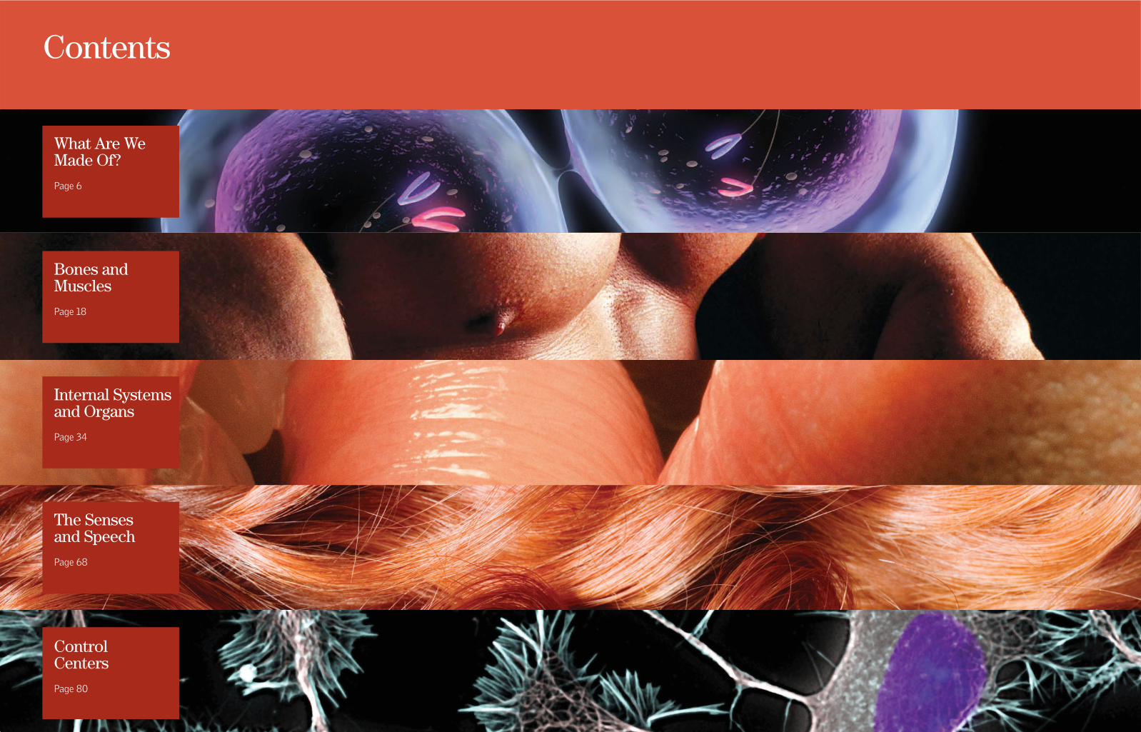
Contents
What Are WeMade Of?Page 6
Bones andMusclesPage 18
Internal Systemsand OrgansPage 34
The Sensesand SpeechPage 68
ControlCentersPage 80

What are cells like, and how do they formtissue? What is blood, and why are proteinsso important? The heart, usually thought of asthe wellspring of love and the emotions, isactually the engine of the circulatory system.It is because of the heart that all the cells ofthe body receive a constant supply ofnutrients, oxygen, and other essentialsubstances. The heart is so powerful that itpumps about 10 pints (4.7 l) of blood perminute. The nervous system is the mostintricate of all the body's systems. It works
A LIVING STRUCTUREThe skeleton consists of206 separate bones, which differ in form, size,and name. It supports and shapes the body,protects the internalorgans, and—in the bone marrow of certainbones—manufacturesvarious types of blood cells.
every second of every day, gatheringinformation about the organism and itssurroundings and issuing instructions so thatthe organism can react. It is this computerthat permits us to think and remember andthat makes us who we are.
The nervous system is a complex networkof sensory cells, originating in the brainand spinal cord, that transmits signals
throughout the body, employing a caravan ofchemical messengers to make sense of thismarvelous complex that we catalogue astouch, taste, smell, hearing, and vision. In fact,at this precise moment, because of anextraordinary relationship between our eyesand our brain, we are able to see andunderstand what we are reading. Moderncameras are designed on the same basicprinciples as our eye, but they have never beenable to equal the visual power of the eye. Thefocus and the automatic aperture of the humaneye are perfect. Our ears share a similarcomplexity and allow us to have excellenthearing. The external ear operates by receivingsound waves in the air. Sound waves travelthrough the auditory canal and are transmittedby the bones of the intermediate ear towardthe cochlea, which contains liquid and isspiraled like the shell of a small sea snail. Thecochlea converts waves of air into vibrations ofliquid, which are detected by special filamentsin the ear that are of many lengths and thatdetect sound waves of different lengths. Thesefilaments then transmit nerve impulses to thebrain and provide us with our ability tointerpret what we hear. This book will alsotell you about the function of our skin, thelargest organ of the body, which serves as anelastic barrier covering and protectingeverything inside our bodies. Captivatingimages will show you how each of ourextraordinary body systems function, andincredible facts will help you understand whythe human body is so amazing.
How can we understand what we are?What are we made of? Are we awarethat all that we do—including reading
this book—is the work of a marvelousmachine? We know very little about how weare able to be conscious of our own actions;nevertheless, even though we are usually notvery aware of it, this community of organsthat is the body—an integrated system thatincludes the brain, heart, lungs, liver, kidneys,muscles, bones, skin, and endocrine glands—acts together in exquisitely regulatedharmony. It is interesting that variousmechanisms work together to keep thetemperature of the body at 98.6° F (37° C);
thanks to the dynamic structure of bonesand cartilage, the body is maintained inperfect balance. The body also has afantastic ability to transform the food it
ingests into living tissues, bones, andteeth, all of which contribute to its growth.By this same process, we obtain the energyfor working and playing. It is hard toimagine that not long ago the cells of thebody of the person reading this book were
autonomous and were duplicatingthemselves freely within the walls of amother's uterus. Certainly no onereading this book could recognize herself
or himself in those cells. Nevertheless,each cell carried within it the informationnecessary for the development of thatperson. Everything that happens inside us istruly fascinating. Therefore, we invite you toenjoy this book. It is full of incredible factsand illustrations that will show you thecomplex ways each part of the body works.
A PerfectMachine

What Are We Made Of?
To understand the truest andmost elementary characteristicsof life, we must begin with thecell-the tiny organizingstructure of life in all its forms.
Most cells are too small to beobserved with the naked eye, but theycan be distinguished easily through anordinary microscope. Human bodytissues are groups of cells whose size
and shape depend on the specifictissue to which they belong. Did youknow that an embryo is a mass ofrapidly dividing cells that continue todevelop during infancy? We invite you
to turn the page and discover manysurprising things in this fascinatingand complex world.
UNDIVIDED ATTENTION 8-9
WATER AND LIQUIDS 10-11
THE CELL 12-13
MITOSIS 14-15
SYSTEMS OF THE BODY 16-17
MITOSISAn enlarged view that showsthe process of mitosis, themost common form ofcellular division

HUMAN BODY I 98 WHAT ARE WE MADE OF?
UndividedAttentionFrom birth the infant's braincells develop rapidly,making connections that canshape all of life'sexperiences. The first threeyears are crucial. Whenneurons receive visual,auditory, or gustatory stimuli,they send messages thatgenerate new physicalconnections withneighboring cells. The signalsare sent through a gap calleda synapse by means of acomplex electrochemicalprocess. What determinesthe formation of a person'ssynapses and neuralnetworks? One key factor isbelieved to be the undividedattention and mental effortexerted by the person.
THE SENSE OF TOUCHIt is predominant in the fingers and hands. Theinformation is transmitted through neurotransmitters,nerves that carry these impulses to the brain andthat serve to detect sensations such as cold, heat,pressure, and pain.
SKINThe skin is one of themost important organs of thebody. It contains approximatelyfive million tiny nerve endingsthat transmit sensations.
LearningEach child has his or her own intellectual filter; thequality of the filter depends on undivided attention andon how the child responds to a broad variety of stimuli.
BrainAt birth the infant brain contains 100billion neurons. That is about as manynerve cells as there are stars in theentire Milky Way Galaxy! Then as theinfant receives messages from thesenses, the cerebral cortex begins itsdynamic development.
RespirationRespiration is usually an involuntary,automatic action that allows us to take inthe oxygen we need from the air and exhalecarbon dioxide. These gases are exchangedin the pulmonary alveoli.
NeuronsEach neuron in the brain can beconnected with several thousand otherneurons and is capable of receiving100,000 signals per second. The signalstravel through the nervous
system at a speed of 225 miles per hour(360 km/h). Thanks to this complexcommunication network, the brain iscapable of remembering, calculating,deciding, and thinking.
A WORLD OF SENSATIONSThe tongue recognizes four tastes (sweet,salty, sour, and bitter), and the nasal fossascontain cells that have more than 200 millionfilaments, called cilia, which are capable ofdetecting thousands of odors.
DENDRITESThey are the branchesthrough which a neuronreceives and sends messages.With this system each neuroncan be stimulated bythousands of other neurons,which in turn can stimulateother neurons, and so forth. 3 pounds
(1.4 kg) IS THE WEIGHT OF A HUMAN BRAIN.
225(360 km/h)
THE VELOCITY OF THE NERVOUSSYSTEM'S SIGNALS
milesper hour

HUMAN BODY I 1110 WHAT ARE WE MADE OF?
Water and FluidsW
ater is of such great importance that it makes up almosttwo thirds of the human body by weight. Water is present in allthe tissues of the body. It plays a fundamental role in digestion and
absorption and in the elimination of indigestible metabolic waste. Water alsoserves as the basis of the circulatory system, which uses blood to distributenutrients to the entire body. Moreover, water helps maintain body temperatureby expelling excess heat through the skin via perspiration and evaporation.Perspiration and evaporation of water account for most of the weight a personloses while exercising.
N 3% NITROGENPresent in proteinsand nucleic acids
Water Balance and FoodIn its continuous process of taking in andeliminating water, one of the most
important functions of the body is to maintain acontinuous equilibrium between the water thatenters and the water that leaves the body.Because the body does not have an organ orother place for storing water, quantities that arelost must be continuously replenished. Thehuman body can survive for several weekswithout taking in food, but going without waterfor the same length of time would have tragicconsequences. The human being takes in about2.5 to 3 quarts (2.5-3 l) of water per day. Abouthalf is taken in by drinking, and the rest comesfrom eating solid food. Some foods, such as fruitsand vegetables, consist of 95 percent water.Eggs are 90 percent water, and red meat andfish are 60 to 70 percent water.
HOW THIRST IS CONTROLLEDThirst is the sensation throughwhich the nervous system informsits major organ, the brain, that thebody needs water. The controlcenter is the hypothalamus. If theconcentration of plasma in the bloodincreases, it means the body is losingwater. Dry mouth and a lack ofsaliva are also indications that thebody needs water.
HOW WATER IS ABSORBEDWater for the body is obtainedprimarily by drinking and ingestingfood and through internal chemicalreactions.
HOW WATER ISELIMINATED
Water is expelled not only withurine but also with sweat, throughthe elimination of feces, and throughevaporation from the lungs and skin.
50%of the watercomes from
ingesting fluids.
35%of the water
is obtained from food.
15%comes from
metabolicactivities.
60%is eliminated with urine.
18%is eliminated bysweating and throughevaporation from the skin.
14%is eliminated duringexhalation by thelungs.
8%is eliminatedin excrement.
C 18% CARBONPresent in allorganic molecules
O 65% OXYGENPresent in water and inalmost all organic molecules
H 10% HYDROGENPresent in water,nutrients, andorganic molecules
Chemical Elements The body contains many chemical elements. The most common are oxygen, hydrogen,carbon, and nitrogen, which are found mainly in proteins. Nine chemical elements arepresent in moderate amounts, and the rest (such as zinc) are present only in very smallamounts, so they are called trace elements.
0.004% IRONFluids and tissues, bones,proteins. An iron deficiencycauses anemia, whosesymptoms include fatigueand paleness. Iron isessential for the formationof hemoglobin in the blood.
THE PERCENTAGE OF A PERSON'SWEIGHT THAT IS DUE TO WATER. INGENERAL, A 10 PERCENT LOSS OF WATERLEADS TO SERIOUS DISORDERS, AND ALOSS OF 20 PERCENT RESULTS IN DEATH.
60%
SULFUR 0.3%Contained in numerousproteins, especially in thecontractile proteins
S
POTASSIUM 0.3%Nerves and muscles;inside the cell
K
SODIUM 0.15%Fluids and tissues, inthe form of salt
Na
MAGNESIUM 0.05%Lungs, kidneys, liver,thyroid, brain, muscles,heart
Mg
PHOSPHORUS 1%Urine, bonesP
CHLORINE 0.2%maintains theequilibrium of waterin the blood.
Cl
CALCIUM 1.5% Bones, lungs, kidneys,liver, thyroid, brain,muscles, heart
Ca
0.0004% IODINEUrine, bones. Whenconsumed, iodine passesinto the blood and fromthere into the thyroid gland.Among its other functions,iodine is used by the thyroidto produce growthhormones for most of theorgans and for braindevelopment.
Fe
I
ProteinsProteins are formed throughthe combination of the fourmost common chemicalelements found in the body.Proteins include insulin, whichis secreted by the pancreas toregulate the amount ofsugar in the blood.

12 WHAT ARE WE MADE OF? HUMAN BODY I 13
The CellI
t is the smallest unit of the human body—and of allliving organisms—able to function autonomously. It isso small that it can be seen only with a microscope.
Its essential parts are the nucleus and cytoplasm,which are surrounded by a membrane. Each cellreproduces independently through a process calledmitosis. The animal kingdom does have single-celled organisms, but in a body such as that ofa human being millions of cells are organizedinto tissues and organs. The word “cell”comes from Latin; it is the diminutive ofcella, which means “hollow.” The scienceof studying cells is called cytology.
MATHIAS SCHLEIDEN
NUCLEUS
ROUGH ENDOPLASMIC
RETICULUM
MITOCHONDRIA
THEODOR SCHWANN
Cell TheoryBefore the invention of themicroscope, it was impossible to
see cells. Some biological theories weretherefore based on logical speculationsrather than on observation. People believedin “spontaneous generation” because it wasinconceivable that cells would regenerate.The development of the microscope,including that of an electronic version inthe 20th century, made detailedobservation of the internal structure of thecell possible. Robert Hooke was the first tosee dead cells in 1665. In 1838 MathiasSchleiden observed living cells, and in 1839,in collaboration with Theodor Schwann, hedeveloped the first theory of cells: that allliving organisms consist of cells.
MitochondriaThe mitochondria provide large amounts ofenergy to the cell. They contain a variety of
enzymes that, together with oxygen, degradeproducts derived from glycolysis and carry outcellular respiration. The amount of energyobtained in this process is almost 20times as great as that released byglycolysis in the cytoplasm.Mitochondria are very differentfrom other organelles becausethey have a unique structure: anexternal membrane enclosing aninternal membrane with a greatnumber of folds that delimitthe internal area, ormitochondrial matrix. Inaddition, the mitochondriahave a circular chromosomesimilar to that of bacteriathat allows the mitochondriato replicate. Cells that need arelatively large amount ofenergy have manymitochondria because thecells reproduce frequently.
TRANSPORT MECHANISMSThe cell membrane is a semipermeable barrier. The cellexchanges nutrients and waste between its cytoplasmand the extracellular medium via passive and activetransport mechanisms.
DIFFUSION It is a passivetransport mechanism in whichthe cell does not use energy. Theparticles that cross the cellmembrane do so because of aconcentration gradient. For example,water, oxygen, and carbon dioxidecirculate by diffusion.
FACILITATED DIFFUSIONPassive transport in whichsubstances, typically ions (electricallycharged particles), that becauseof their size could not otherwisepenetrate the cell's bilayer can do sothrough a pore consisting of proteins.Glucose enters the cell in this way.
ACTIVE TRANSPORT It occursby means of proteins and requiresenergy consumption by the cellbecause the direction of ion transportis against the concentration gradient.In some cells, such as neurons, theNa+/K+ pump uses active transportto move ions into or out of the cell.
UNDER THE MICROSCOPEThis cell has been magnified4,000 times with an electronmicroscope. The nucleus isclearly visible, along with sometypical organelles in the green-colored cytoplasm.
CENTRIOLESThey are cylindrical,hollow structures thatare part of thecytoskeleton.
NUCLEUSThe nucleus consistsof chromatin andregulates cellmetabolism, growth,and reproduction.
POREA discontinuity inthe nuclearmembraneformed byproteins
SMOOTH ENDOPLASMICRETICULUMVarious membranes, whosefunctions include transportand synthesis. They aretube-shaped and do nothave ribosomes.
VESICLEA closedcompartment.It transportsor digests cellproducts andresidues.
NUCLEOLEThe nucleole canbe single ormultiple. Thenucleole consistsof ribonucleicacid and proteins.
CELLULAR MEMBRANEThe covering of the cellsurrounding the cytoplasm.It is also known as theplasma membrane.
VACUOLETransports andstores ingestedmaterials, waste,and water
DNAIt is organizedinto chromosomeswithin the nucleus.DNA is genetic materialthat contains informationfor the synthesis andreplication of proteins.
GOLGI APPARATUSThis structure processes
proteins produced by therough endoplasmatic
reticulum and places themin sacs called vesicles.
CYTOPLASMThe region locatedbetween the plasmamembrane and thenucleus. It containsorganelles.
MITOCHONDRIAAn organelle of theeukaryotic cell responsiblefor cellular respiration
LYSOSOMEThis is the “stomach”of the cell because it
breaks down wastemolecules with its
enzymes.
RIBOSOMEThis organelle iswhere the laststages of proteinsynthesis takeplace.
CYTOSKELETONComposed of fibers,the cytoskeleton isresponsible for cell
motion, orcytokinesis.
ROUGHENDOPLASMATIC
RETICULUMA labyrinthine assembly of
canals and membranousspaces that transport
proteins and are involvedin the synthesis of
substances.
PEROXISOMEOrganelles presentin eukaryotes thatfunction to metabolizeand eliminate toxicsubstances from cells
100 billionTHE AVERAGE NUMBER OF CELLS IN THEBODY OF AN ADULT. ONE CELL ALONE CANDIVIDE UP TO 50 TIMES BEFORE DYING.

14 WHAT ARE WE MADE OF? HUMAN BODY I 15
MitosisI
t is the cell-division process that results in the formation ofcells that are genetically identical to the original (or mother)cell and to each other. The copies arise through replication
and division of the chromosomes, or genetic material, in such away that each of the daughter cells receives a similar inheritanceof chromosomes. Mitosis is characteristic of eukaryotic cells. Itensures that the genetic information of the species and theindividual is conserved. It also permits the multiplication of cells,which is necessary for the development, growth, and regeneration of the organism. The word “mitosis” comes from the Greek mitos,which means “thread,” or “weave.”
THE ESTIMATED NUMBER OF CELLSREPLACED EVERY SECOND IN THE HUMANBODY THROUGH CELLULAR DIVISION
50,000
50 MITOSES MARK THELIFETIME OF A CELL ANDARE KNOWN AS THE“HAYFLICK LIMIT.” THISIDEA IS NAMED AFTERLEONARD HAYFLICK, WHO IN1961 DISCOVERED THAT THESECTION OF DNA CALLEDTHE TELOMERE INFLUENCESCELL LIFE SPAN.
Limit
The Ever-Changing SkinMitosis, or cellular division, occurs intensely within theskin, a fundamental organ of the sense of touch. The dead
cells on the surface are continuously being replaced by new cells,which are produced by mitosis in the lowest, or basal, layer. Fromthere the cells move upward until they reach the epidermis, theouter layer of the skin. A person typically sheds 30,000 deadskin cells every minute.
AntioxidantsAntioxidants are various types of substances (vitamins,enzymes, minerals, etc.) that combat the pernicious effects of
free radicals—molecules that are highly reactive and form as a resultof oxidation (when an atom loses an electron), which is often causedby coming into contact with oxygen. A consequence of this oxidativeaction is the aging of the body. One action of antioxidants is theregulation of mitosis. Preventive geriatrics has focused on usingantioxidants to prevent disease and to slow aging, in part becauseproperly regulated mitosis is fundamental to these processes.
CENTROMERE
SUPERFICIALCELLS
GRANULARCELLS
SPINOUS CELLS
BASAL CELLS
SPINDLEFILAMENT
CELLULARMEMBRANE
CENTRIOLE
CYTOPLASM
CHROMATIN
NUCLEUS
NUCLEUS
ORGANELLES
SISTERCHROMOSOMES
CHROMATIDCHROMOSOME
SHEDDING SUPERFICIAL CELLS LAYERS OF THE SKIN
1. INTERPHASEAn independent stagethat precedes mitosis.The chromatinconsists of DNA.
2. PROPHASEIn prophase the chromatincondenses to form chromosomes.The karyotheca (nuclearenvelope) begins to disappear.Chromosomes are formed by twochromatids that are joinedtogether by a centromere.
3. METAPHASEIt is characterized by theappearance of the spindle. Thecentromere—the “center” ofeach chromosome—and thechromatids are joined togetherand align at the center of thespindle complex. The nuclearmembrane disappears.
4. ANAPHASEIn this crucial stage the copies ofgenetic information separate: thechromatids move apart and formsister chromosomes that migrate toopposite poles of the cell.
5. TELOPHASEThe spindle disappears, and anew nuclear membrane beginsto form around each new set ofchromosomes. The membranedivides, resulting in two newcells that are identicaldaughters of the original cell.
NUCLEUS

16 WHAT ARE WE MADE OF? HUMAN BODY I 17
Systems of the BodyT
he body has various systems with different functions. Thesefunctions range from reproducing a cell to developing a newhuman being, from circulating the blood to capturing
oxygen from the air, and from processing food throughgrinding and chemical transformations to absorbing nutrientsand discarding waste. These functions act in harmony, andtheir interaction is surprisingly efficient.
MuscularSystem
Its function is to define the shape of the organism andprotect it. The muscular system is essential forproducing movement. It consists of muscles, organsmade of fleshy tissue, and contractile cells. There aretwo types of muscles: striated and smooth. Striatedmuscles are attached to the bones and governvoluntary movement. Smooth muscles also obey thebrain, but their movement is not under voluntarycontrol. The myocardium, the muscle tissue of theheart, is unique and is in a class by itself. See page 30.
RespiratorySystemAir from the external world enters the bodythrough the upper airways. The centralorgans, the lungs, absorb oxygen and expelcarbon dioxide. The lungs send oxygenatedblood to all the cells via the circulatorysystem and in turn receive blood that requirespurification. See page 46.
EndocrineSystemThe endocrine system is formed by glands thatare distributed throughout the body. Itsprimary function is to produce approximately50 hormones, the body's chemical messengers.The endocrine system secretes the hormonesinto the bloodstream so that they can reachthe organs they are designed to influence,excite, or stimulate for such activities asgrowth and metabolism. See page 62.
MALE
The various male organs contributeone of the two cells needed tocreate a new human being. Twotesticles (or gonads) and a penis arethe principal organs of the system.The system is continuously active,producing millions of tiny cells calledspermatozoa. See page 64.
ReproductiveSystemFEMALE
A woman's internal organs are the vagina, theuterus, the ovaries, and the fallopian tubes.The basic functions of these organs are theproduction of ova and the facilitation offertilization of an ovum by a spermatozoon (amature male sperm cell). When fertilizationoccurs, it sets a group of processes in motionthat result in pregnancy. See page 66.
Skeletal SystemThe skeleton, or skeletal system, is a solid structureconsisting of bones that are supported by ligamentsand cartilage. The main functions of the systemare to give the body form and to support it, tocover and protect the internal organs, and toallow motion to occur. The skeleton alsogenerates red blood cells (callederythrocytes). See page 20.
Circulatory SystemThis system carries blood to and from the heart and reaches theorgans and cells in every part of the body. The supreme pump—theheart—drives the vital fluid—blood—through the arteries andcollects it by means of the veins, with a continuous driving impulsethat makes the heart the central engine of the body. See page 36.
NervousSystemThe central nervous system consists of thebrain, which is the principal organ of thebody, along with the spinal cord. Theperipheral nervous system consists of thecranial and spinal nerves. Together theysend external and internal sensations to thebrain, where the sensations are processedand responded to whether the person isasleep or awake. See page 82.
Lymphatic SystemIts basic functions are twofold. One is to defend the bodyagainst foreign organisms, such as bacteria or viruses. Theother is to transport interstitial fluid and substances from thedigestive system into the bloodstream via the lymphaticdrainage system. See page 42.
Digestive SystemThis system is a large tract that changes form andfunction as it goes from the mouth to the rectum andanus, passing through the pharynx, the esophagus, thestomach, and the small and large intestines. The liver andpancreas help process ingested food to extract itschemical components. Some of these components arewelcome nutrients that are absorbed by the system, butothers are useless substances that are discarded andeliminated. See page 50.
Urinary SystemThis system is a key system for homeostasis—that is,the equilibrium of the body's internal conditions. Itsspecific function is to regulate the amount of waterand other substances in the body, discarding any thatare toxic or that form an unnecessary surplus. Thekidneys and the bladder are the urinary system'sprincipal organs. The ureters transport the urine fromthe kidneys to the bladder, and the urethra carriesthe urine out of the body. See page 58.

JOINTS 28-29
MUSCULAR SYSTEM 30-31
MUSCLE FIBER 32-33
Bones and Muscles
The musculoskeletal systemconsists of the skeletal systemof bones, attached to each otherby ligaments to form joints, andthe skeletal muscles, which use
tendons to attach muscles to bone. Theskeleton gives resistance and stabilityto the body and serves as a supportstructure for the muscles to work andproduce movement. The bones also
serve as a shield to protect the internalorgans. In this chapter you will see indetail—even down to the inside of amuscle fiber—how each part works.Did you know that bones are constantly
being regenerated and that, besidessupporting the body, they are chargedwith producing red blood cells? In thischapter you will find incredible images,curiosities, and other information.
SKELETON 20-21
BONE TISSUE 22-23
CRANIUM AND FACE 24-25
THE GREAT AXIS OF THE BODY 26-27
MUSCLES OF THE THORAXThey play an important role inbreathing by facilitating thecontraction and expansion ofthe thoracic cavity.
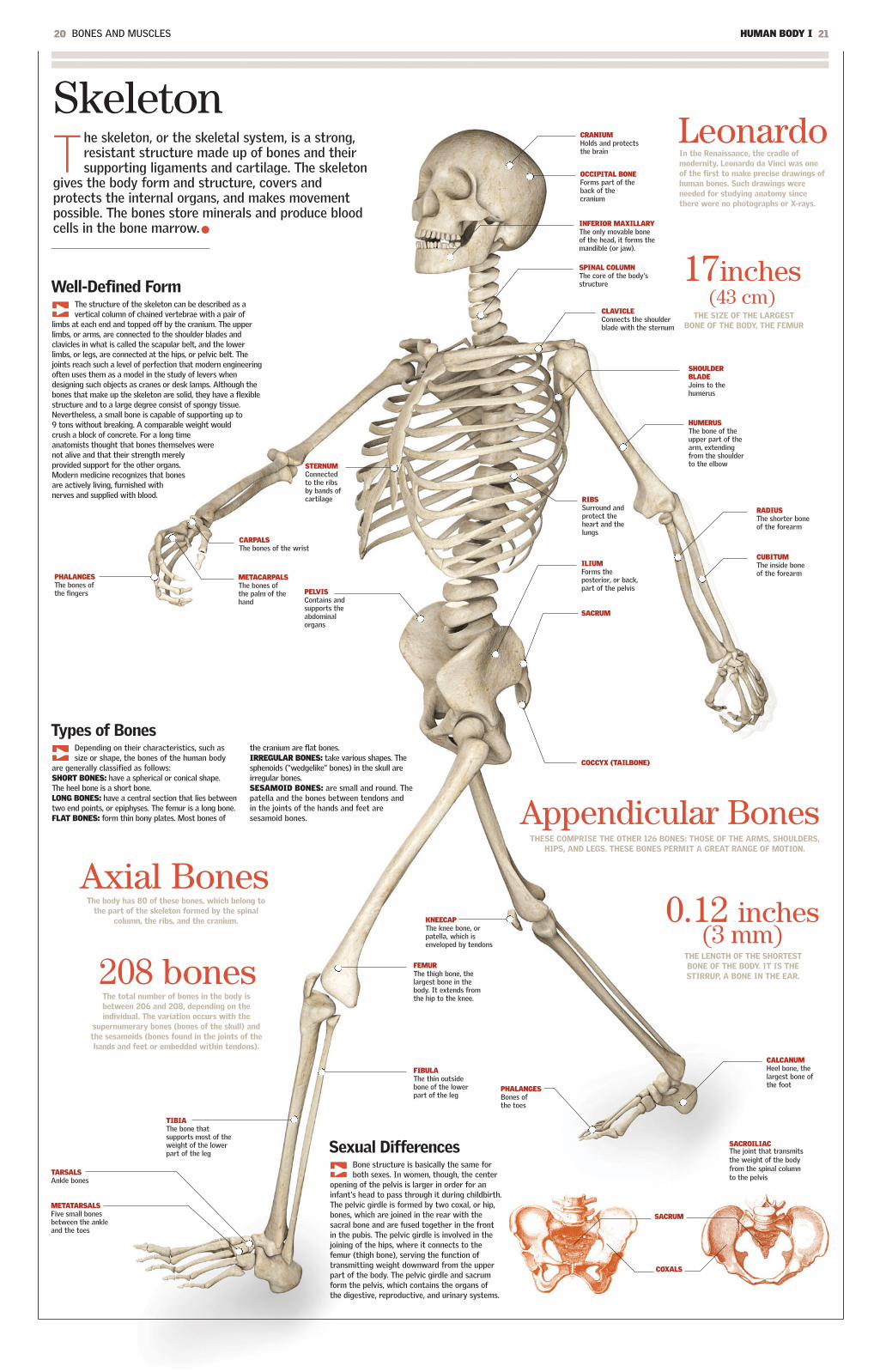
Skeleton
20 BONES AND MUSCLES HUMAN BODY I 21
The skeleton, or the skeletal system, is a strong,resistant structure made up of bones and theirsupporting ligaments and cartilage. The skeleton
gives the body form and structure, covers andprotects the internal organs, and makes movementpossible. The bones store minerals and produce bloodcells in the bone marrow.
CRANIUM Holds and protectsthe brain
INFERIOR MAXILLARYThe only movable boneof the head, it forms themandible (or jaw).
SHOULDERBLADEJoins to thehumerus
Sexual DifferencesBone structure is basically the same forboth sexes. In women, though, the center
opening of the pelvis is larger in order for aninfant's head to pass through it during childbirth.The pelvic girdle is formed by two coxal, or hip,bones, which are joined in the rear with thesacral bone and are fused together in the frontin the pubis. The pelvic girdle is involved in thejoining of the hips, where it connects to thefemur (thigh bone), serving the function oftransmitting weight downward from the upperpart of the body. The pelvic girdle and sacrumform the pelvis, which contains the organs ofthe digestive, reproductive, and urinary systems.
Types of BonesDepending on their characteristics, such assize or shape, the bones of the human body
are generally classified as follows:SHORT BONES: have a spherical or conical shape.The heel bone is a short bone.LONG BONES: have a central section that lies betweentwo end points, or epiphyses. The femur is a long bone.FLAT BONES: form thin bony plates. Most bones of
the cranium are flat bones.IRREGULAR BONES: take various shapes. Thesphenoids (“wedgelike” bones) in the skull areirregular bones.SESAMOID BONES: are small and round. Thepatella and the bones between tendons andin the joints of the hands and feet aresesamoid bones.
Well-Defined FormThe structure of the skeleton can be described as avertical column of chained vertebrae with a pair of
limbs at each end and topped off by the cranium. The upperlimbs, or arms, are connected to the shoulder blades andclavicles in what is called the scapular belt, and the lowerlimbs, or legs, are connected at the hips, or pelvic belt. Thejoints reach such a level of perfection that modern engineeringoften uses them as a model in the study of levers whendesigning such objects as cranes or desk lamps. Although thebones that make up the skeleton are solid, they have a flexiblestructure and to a large degree consist of spongy tissue.Nevertheless, a small bone is capable of supporting up to9 tons without breaking. A comparable weight wouldcrush a block of concrete. For a long timeanatomists thought that bones themselves werenot alive and that their strength merelyprovided support for the other organs.Modern medicine recognizes that bonesare actively living, furnished withnerves and supplied with blood.
The body has 80 of these bones, which belong tothe part of the skeleton formed by the spinal
column, the ribs, and the cranium.
Axial BonesTHESE COMPRISE THE OTHER 126 BONES: THOSE OF THE ARMS, SHOULDERS,
HIPS, AND LEGS. THESE BONES PERMIT A GREAT RANGE OF MOTION.
Appendicular Bones
THE LENGTH OF THE SHORTESTBONE OF THE BODY. IT IS THESTIRRUP, A BONE IN THE EAR.
0.12 inches(3 mm)
THE SIZE OF THE LARGESTBONE OF THE BODY, THE FEMUR
17inches(43 cm)
In the Renaissance, the cradle ofmodernity, Leonardo da Vinci was oneof the first to make precise drawings ofhuman bones. Such drawings wereneeded for studying anatomy sincethere were no photographs or X-rays.
Leonardo
The total number of bones in the body isbetween 206 and 208, depending on theindividual. The variation occurs with the
supernumerary bones (bones of the skull) andthe sesamoids (bones found in the joints of thehands and feet or embedded within tendons).
208 bones
OCCIPITAL BONEForms part of theback of thecranium
CARPALSThe bones of the wrist
METACARPALSThe bones ofthe palm of thehand
COCCYX (TAILBONE)
CLAVICLEConnects the shoulderblade with the sternum
SPINAL COLUMNThe core of the body'sstructure
STERNUMConnectedto the ribsby bands ofcartilage
PELVISContains andsupports theabdominalorgans
HUMERUSThe bone of theupper part of thearm, extendingfrom the shoulderto the elbow
SACRUM
ILIUMForms theposterior, or back,part of the pelvis
RIBSSurround andprotect theheart and thelungs
CUBITUMThe inside boneof the forearm
RADIUSThe shorter boneof the forearm
CALCANUMHeel bone, thelargest bone ofthe foot
PHALANGESThe bones ofthe fingers
KNEECAPThe knee bone, orpatella, which isenveloped by tendons
FIBULAThe thin outsidebone of the lowerpart of the leg
FEMURThe thigh bone, thelargest bone in thebody. It extends fromthe hip to the knee.
TIBIAThe bone thatsupports most of theweight of the lowerpart of the leg
TARSALSAnkle bones
METATARSALSFive small bonesbetween the ankleand the toes
PHALANGESBones of the toes
SACRUM
COXALS
SACROILIAC The joint that transmitsthe weight of the bodyfrom the spinal columnto the pelvis
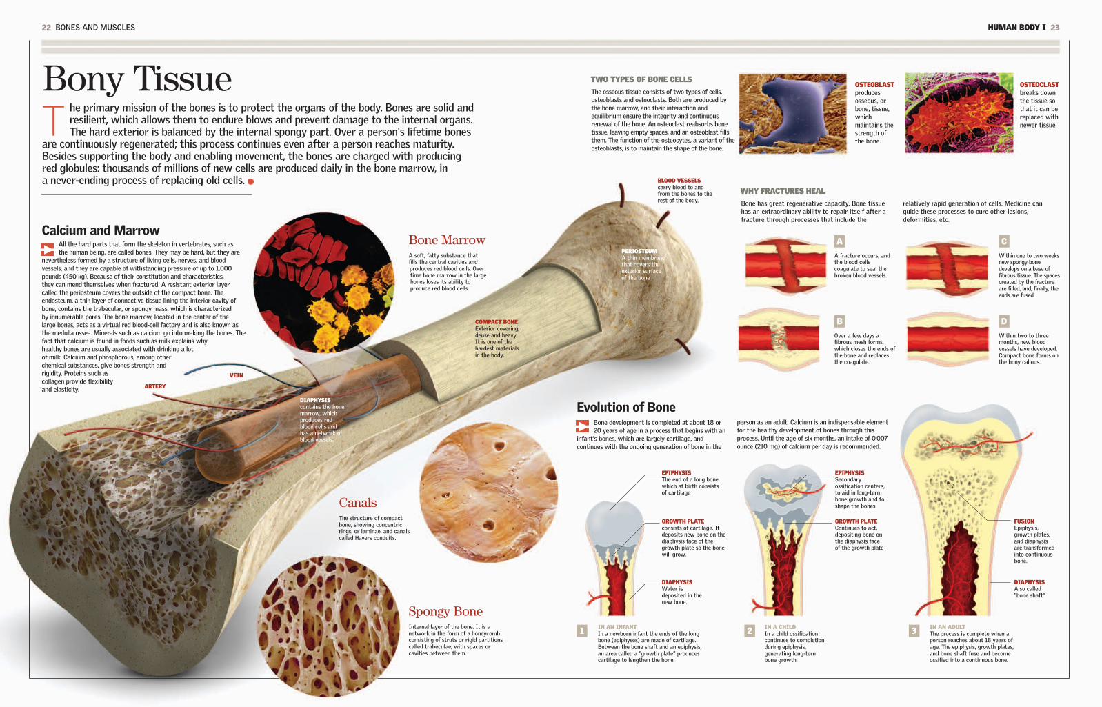
TWO TYPES OF BONE CELLS
22 BONES AND MUSCLES HUMAN BODY I 23
Bony TissueT
he primary mission of the bones is to protect the organs of the body. Bones are solid andresilient, which allows them to endure blows and prevent damage to the internal organs.The hard exterior is balanced by the internal spongy part. Over a person's lifetime bones
are continuously regenerated; this process continues even after a person reaches maturity.Besides supporting the body and enabling movement, the bones are charged with producingred globules: thousands of millions of new cells are produced daily in the bone marrow, ina never-ending process of replacing old cells. BLOOD VESSELS
carry blood to andfrom the bones to therest of the body.
PERIOSTEUM A thin membranethat covers theexterior surfaceof the bone
OSTEOBLASTproducesosseous, orbone, tissue,whichmaintains thestrength ofthe bone.
Bone MarrowA soft, fatty substance thatfills the central cavities andproduces red blood cells. Overtime bone marrow in the largebones loses its ability toproduce red blood cells.
OSTEOCLASTbreaks downthe tissue sothat it can bereplaced withnewer tissue.
IN AN INFANTIn a newborn infant the ends of the longbone (epiphyses) are made of cartilage.Between the bone shaft and an epiphysis,an area called a “growth plate” producescartilage to lengthen the bone.
EPIPHYSISSecondaryossification centers,to aid in long-termbone growth and toshape the bones
GROWTH PLATEContinues to act,depositing bone onthe diaphysis faceof the growth plate
GROWTH PLATEconsists of cartilage. Itdeposits new bone on thediaphysis face of thegrowth plate so the bonewill grow.
EPIPHYSISThe end of a long bone,which at birth consistsof cartilage
CanalsThe structure of compactbone, showing concentricrings, or laminae, and canalscalled Havers conduits.
COMPACT BONEExterior covering,dense and heavy.It is one of thehardest materialsin the body.
FUSIONEpiphysis,growth plates,and diaphysisare transformedinto continuousbone.
DIAPHYSISWater isdeposited in thenew bone.
DIAPHYSISAlso called“bone shaft”
Calcium and MarrowAll the hard parts that form the skeleton in vertebrates, such asthe human being, are called bones. They may be hard, but they are
nevertheless formed by a structure of living cells, nerves, and bloodvessels, and they are capable of withstanding pressure of up to 1,000pounds (450 kg). Because of their constitution and characteristics,they can mend themselves when fractured. A resistant exterior layercalled the periosteum covers the outside of the compact bone. Theendosteum, a thin layer of connective tissue lining the interior cavity ofbone, contains the trabecular, or spongy mass, which is characterizedby innumerable pores. The bone marrow, located in the center of thelarge bones, acts as a virtual red blood-cell factory and is also known asthe medulla ossea. Minerals such as calcium go into making the bones. Thefact that calcium is found in foods such as milk explains whyhealthy bones are usually associated with drinking a lotof milk. Calcium and phosphorous, among otherchemical substances, give bones strength andrigidity. Proteins such ascollagen provide flexibilityand elasticity.
Evolution of BoneBone development is completed at about 18 or20 years of age in a process that begins with an
infant's bones, which are largely cartilage, andcontinues with the ongoing generation of bone in the
person as an adult. Calcium is an indispensable elementfor the healthy development of bones through thisprocess. Until the age of six months, an intake of 0.007ounce (210 mg) of calcium per day is recommended.
Spongy BoneInternal layer of the bone. It is anetwork in the form of a honeycombconsisting of struts or rigid partitionscalled trabeculae, with spaces orcavities between them.
The osseous tissue consists of two types of cells,osteoblasts and osteoclasts. Both are produced bythe bone marrow, and their interaction andequilibrium ensure the integrity and continuousrenewal of the bone. An osteoclast reabsorbs bonetissue, leaving empty spaces, and an osteoblast fillsthem. The function of the osteocytes, a variant of theosteoblasts, is to maintain the shape of the bone.
WHY FRACTURES HEAL
Bone has great regenerative capacity. Bone tissuehas an extraordinary ability to repair itself after afracture through processes that include the
relatively rapid generation of cells. Medicine canguide these processes to cure other lesions,deformities, etc.
A
A fracture occurs, andthe blood cellscoagulate to seal thebroken blood vessels.
1 IN A CHILDIn a child ossificationcontinues to completionduring epiphysis,generating long-termbone growth.
2 IN AN ADULTThe process is complete when aperson reaches about 18 years ofage. The epiphysis, growth plates,and bone shaft fuse and becomeossified into a continuous bone.
3
B
Over a few days afibrous mesh forms,which closes the ends ofthe bone and replacesthe coagulate.
C
Within one to two weeksnew spongy bonedevelops on a base offibrous tissue. The spacescreated by the fractureare filled, and, finally, theends are fused.
D
Within two to threemonths, new bloodvessels have developed.Compact bone forms onthe bony callous.
VEIN
ARTERY
DIAPHYSIScontains the bonemarrow, whichproduces redblood cells andhas a network ofblood vessels.

24 BONES AND MUSCLES HUMAN BODY I 25
Cranium and FaceT
he cranium surrounds and protects the brain, cerebellum, and cerebral trunk (sometimescalled the encephalus). In an adult the cranium consists of eight bones that form the skull andthe base of the cranium. The face is the anterior part of the skull. It consists of 14 bones, all
of which are fixed except the lower maxillary, which makes up the mandible. The total number ofbones in the head as a whole exceeds the total of the face and cranium (22) because it includes thelittle bones of the middle ear.
Cranial SinusesThe sinuses are air-filled cavities whose principal knownfunction is to humidify and heat the air that enters therespiratory tract via the nose. The sinuses reduce theweight of the head, and they also act as resonancecavities, giving the voice its timbre. The sinuses arecovered by a moist membrane and are connected viasmall openings with the interior of the nasal cavity.When the sinuses become inflamed or filled with mucus,there is a risk of infection.
VibrationWhen a person speaks, the bonesof the cranium vibrate. In Japana technology was developedbased on this vibration. In 2006the firefighters of the Madridmunicipality in Spain adoptedthis technology. A helmet,furnished with a cranial contactmicrophone, amplifies thevibrations produced in the bonesof the cranium during speechand sends them to radioequipment.
FRONTAL SINUS
ETHMOID SINUS
SPHENOID SINUS
MAXILLARY SINUS
Foramen MagnumIn Latin this term means “big hole.” It is a circularopening, also called the occipital orifice, which islocated at the base of the cranium. The foramenmagnum allows for the passage of the spinalcolumn, the medulla oblongata, the vertebralarteries, and the spinal nerve. The placement of theforamen magnum toward the bottom of the skull isassociated with more highly evolved species.
The cranium can be comparedto a sphere, which consists of
separate bones at birth and closescompletely at maturity. Thenarrow separations between thebones, which appear as lines inthe fetus for the first months ofits life, are called sutures.Spaces called fontanels formwhere the sutures meet. Theirseparation has the functionalpurpose of allowing the brainto grow. Therefore, when braingrowth is complete, the spherecloses tightly, because itsfunction is to protect the brain.
Cranial Bones (8)
PARIETAL (2)The superior and lateral parts of the cranium
OCCIPITAL (1)Together with the temporals,
it forms the base of the cranium.
FRONTAL (1)It makes up the forehead.
TEMPORAL (2)The lateral part of the cranium
SPHENOID (1)The front part of the base of the cranium and part of the orbital bone (eye socket)
ETHMOID (1)Upper part of the nasal cavity
Facial Bones (14)
ZYGOMATIC (2)The cheekbones
PALATINES (2)Internal bones that form
the roof of the mouth
LACHRYMAL BONES (2)form the eye socket.
SUPERIOR MAXILLARIES (2)The upper mandible
NASAL CONCHAS (2)Independent of
the ethmoid conchas
VOMER (1)divides the nasal cavity
into two halves.
NASAL BONE (2)forms the bridge of the nose
(the rest of the nose is cartilage).
INFERIOR MAXILLARY (1)constitutes the mandible
and is the only facial bone that can move freely.
FORAMENMAGNUM
22THE TOTAL NUMBER OF BONESIN THE CRANIUM
83(1,360 cu cm)THE TYPICAL VOLUMEOF THE CRANIUM
Suturesand Fontanels
cubicinches
9THE WEIGHT OF ANADULT HUMAN HEAD
pounds(4 kg)

26 BONES AND MUSCLES HUMAN BODY I 27
The vertebral, or spinal, column is the flexible axis that lends supportto the body. It consists of a series of bones jointed together in aline, or chain, called the vertebrae. The spinal column forms a
protective inner channel through which the spinal cord runs. The ribsperform a similar function, wrapping and shielding the vital internalorgans, which include the heart and lungs.
DownwardsAll the vertebrae except the cervicalaxis and atlas have a cylindrical body,which gives them a particularcharacteristic: as they approach thepelvis they tend to be longer andstronger.
CARPALS (8)1. LUNATE2. PISIFORM3. TRIQUETRUM4. TRAPEZIUM5. TRAPEZOID6. CAPITATE7. SCAPHOID8. HAMATE
TARSUS (7)1. MEDIAL CUNEIFORM2. INTERMEDIATECUNEIFORM3. LATERAL CUNEIFORM4. TALUS5. TARSAL SCAPHOIDS6. CALCANEUS7. CUBOIDS
METACARPALS (5)
CARPALS (8)
METATARSALS (5)
PHALANGES (14)
PHALANGES (14)
Bones of the Hands and FeetEach hand (see the drawing below) has 27 bones, and eachfoot (see above) has 26. The hand has great mobility, and eachof its fingers (five in all) has three phalanges (distal, medial,and proximal), except for the thumb, which has two. Thecomplex of carpal bones makes up the wrist and is connectedto the forearm. The metacarpal bone sustains the medial part.The feet function in a similar manner; the toes have first,second, and third phalanges, except for the big toe.
Stability and MotionThe vertebrae have a centrum that allowsthem to support the body's weight, each
vertebra upon the next, as well as the weight of therest of the body. The vertebrae also have extensionsthat allow them to articulate with other vertebrae oract as supports for the ligaments and the muscles.This system gives the axis of the body both strengthand flexibility. In addition, most of the nerves of theperipheral system (that is, those responsible for
voluntary movement, for pain, and for the sense oftouch) are connected to the spinal cord inside thespinal column. In the centrum the vertebrae areseparated from each other by intervertebraldisks that are made of cartilage and have agelatinous interior. When an intervertebraldisk is damaged, some of this material canescape and pinch a nerve. This condition,called a herniated disk, can be very painful.
1
1
2
3
47
8
5
6
2
3
76
45
SACRUMThis bone isformed by fivefused vertebrae.
COCCYX This bone iscomposed of fourfused vertebrae.
The Ribs and the Rib CageThe 12 pairs of ribs, which also extend from thespinal column, protect the heart, lungs, majorarteries, and liver. These bones are flat andcurved. The seven upper pairs are called “trueribs,” and they are connected to the sternum (aflat bone consisting of fused segments) by
cartilage. The next two or three pairs (called“false ribs”) are connected indirectly. Theremaining pairs (“floating ribs”) are notattached to the sternum. The rib cage,formed by the ribs and its muscles, is flexible:it expands and contracts during breathing.
SACRALCANALNerves passthrough thesacral canal.
BLADE
LUMBAR VERTEBRAEThere are five of them, andthey bear the weight of theupper part of the body.
The Three CurvesThe three types of naturalcurvature in the spinalcolumn include cervicallordosis (forward, or inward,bending in the cervicalregion of the spine),kyphosis (outward bendingof the thoracic region of thespine), and lumbar lordosis(forward bending of thelower back). Shown hereis the right side of thespinal column.
AXISThe second cervicalvertebra. Together withthe atlas, it permits themovement of the head.
CERVICALThese sevenvertebrae (includingthe atlas and theaxis) support thehead and the neck.
THORACIC, OR DORSAL,VERTEBRAEThere are 12, and they arejoined to the ribs.
ATLASThis bone is the first ofthe seven cervical bones;it unites the spinalcolumn with the head.
PARTS OF THE VERTEBRAE
1. SPINAL APOPHYSIS2. TRANSVERSE
APOPHYSIS (2)3. ARTICULAR
APOPHYSIS (4) (2 SUPERIOR AND 2 INFERIOR)
4. LAMINAE (2)5. PEDICULAE (2)6. FORAMEN MAGNUM7. BODY
1
2
3
76
4
5
STERNUM
LUNG
LIVER
DIAPHRAGM
HEART
SPLEEN
STOMACH
RIBCARTILAGE
33 bonesOR VERTEBRAE, MAKE UP THESPINAL COLUMN. DEPENDING ON THEINDIVIDUAL, SOMETIMESTHERE ARE 34. THEY ARECONNECTED BY DISKS OFCARTILAGE THAT ACT ASSHOCK ABSORBERS. THESACRUM AND THE COCCYX AREA RUDIMENTARY TAIL LOSTDURING EVOLUTION.
The Great Axis of the Body

HUMAN BODY I 2928 BONES AND MUSCLES
The KneeThe knee is the biggestjoint of the body. It
maintains its stability because it isconstrained by four ligaments: theanterior and posterior cruciate andthe internal and external lateral. Theligaments link the femur (the thighbone) with the tibia (a bone of theleg). The knee is protected by thekneecap, a bony disk covered withcartilage that encases the anteriorand superior part of the kneejoint. Like the majority of thejoints, it is synovial.
FEMURThe thigh bone,which is theupper region ofthe lower limb
MUSCLE
MUSCLE
TIBIAThe larger ofthe two bonesof the lower leg
KNEECAPProtectivebony diskcovered withcartilage
SYNOVIALMEMBRANEproduces thesynovial liquid.
PATELLARLIGAMENTThis ligamentcrosses overthe kneecapand encases it.
MENISCUSFibrouscartilage thathelps theweight-supportingbones toabsorb a blow
EXTERNALLIGAMENTSStabilize the jointduring movement.The knee also hasinternal ligaments.
ARTERYThe femoral artery(artery of the femur)changes into thepopliteal artery at theposterior face of theknee. Like all arteriesit carries oxygenatedblood from the heart.
JointsT
hey are the structures where two or more bones come together, either directly or bymeans of strong fibrous cords called ligaments. The skeleton has movement thanks toits joints. Most joints, like the knee, are synovial joints. They are characterized by
mobility, versatility, and lubrication. The muscles that surround them contract to causemovement. When they work as a whole, the bones, muscles, and joints—together with thetendons, ligaments, and cartilage—constitute a grand system that governs the motoractivity of the body and allows us to carry out our daily physical activities.
Flexion
Extension
Circumduction
MOVEMENTSThe complex of joints,together with the musclesand bones, allows thebody to performnumerous actions, withmovements that includeturns and twists.
HypermobilityThe versatility of the joints refersto their characteristic range of
motion. Just as there are mobile,semimobile, and fixed joints, there is alsoa group of joints that are hypermobile.Such joints are less common but areeasily recognizable, especially in childrenand adults who have not lost theflexibility of their joints. The elbows,wrists, fingers, and knees can at an earlyage and in certain individuals have agreater-than-normal range of motion.For people with hypermobile joints thisextra range of motion can beaccomplished without difficulty or riskof dislocation.
Rotation
Abduction
DorsiflexionPlantarFlexion
Adduction
FIBULAThe smallest boneof the lower leg
A CHARACTERISTIC OF THE JOINTSIS THAT THEY CAN MAKE A SOUND,SUCH AS THAT MADE WHENSOMEONE CRACKS HER OR HISKNUCKLES. THIS IS BECAUSE THEREIS AN EXPLOSIVE RELEASE OF GASTHAT PERMITS A SHOCK-ABSORBINGFLUID TO FLOW IN THE JOINT.
Noise
IN THIS YEAR PROFESSORKENJI TAKAGI OF JAPANUSED A CYSTOSCOPE FOR THE FIRST INTERNAL OBSERVATION OF THE KNEE. Technological advances nowpermit arthroscopy to makeprecise observations for diagnosis.
1918
IN THE FORM OF A PIVOTThe joint of the upper bones of the neck.One bone is nested within the other andturns within it. This is the case of the atlasand the axis, in the upper part of the neck,which allow the head to turn from side toside. This is a limited movement.
SPHEROIDArticulation of the shoulder.A bone that has a spherical endthat can be inserted into anotherbone. The motion is extremelyvaried, such as that of theshoulders.
HINGEArticulation of the knee. One bonewith a cylindrical end is inserted intothe patellar groove of the other. Thereis flexion and extension, as in the knee.
PLANEArticulation of the foot. Twosurfaces that slide, one on top of theother, forward, backward, and sideways,as in some joints of the foot and wrist.
ELLIPSOIDThe joint betweenthe humerus and theradius. A bone with anoval end is inserted intothe cavity of anotherbone. The motion isvaried, but there isminimal rotation, as isthe case for the wrists.
BASAL JOINTThe joint at the baseof the thumb. The endsof the two bones cometogether at a rightangle. This allows themto turn, and they movebackward and forward,as occurs with thethumbs.
MobileThese are also called diarthroses; theyare the joints with the greatest range ofmotion. The ends of the bones linkedtogether are structured in various waysthat facilitate their movement relative toeach other, while ensuring the stability ofthe joint. Most joints in the body are ofthis type.
SemimobileAlso known as amphiarthroses. Thesurfaces of the bone that make contacthave cartilaginous tissue. One exampleis the vertebral joints: they have littleindividual movement, but as a wholethey have ample flexion, extension, androtation.
FixedAlso known as synarthroses. Most fixedjoints are found in the cranium and haveno need for motion because their primaryfunction is to protect internal organs.They are connected by bone growth orfibrous cartilage and are extremely rigidand very tough.
Where thepatellar tendon
connects to the bone

Muscular System
30 BONES AND MUSCLES HUMAN BODY I 31
The muscles are organs formed by fleshy tissue consisting of contractilecells. They are divided into striated, smooth, and, in a unique case,cardiac (the myocardium is the muscular tissue of the heart). Muscles
shape and protect the organism. The muscles of the skeleton are attachedto the bones to permit voluntary movement, which is consciously directedby the brain. The smooth muscles are also directed by the brain, but theirmotion is not voluntary, as in the case of digestion. These muscles getmost of their energy from alimentary carbohydrates, which can be storedin the liver and muscles in the form of glycogen and can later pass into theblood and be used as glucose. When a person makes a physical effort,there is an increased demand for both oxygen and glucose, as well as anincrease in blood circulation. A lack of glucose leads to fatigue.
FRONTAL MUSCLE wrinkles the forehead.
ORBICULAR MUSCLEallows blinking.
STERNOCLEIDOMASTOIDallows the head to turn and move forward.
PECTORALIS MAJORstretches the arm forward. It turns it and brings it close to the body.
BRACHIAL BICEPbends the arm at the elbow.
EXTERNAL OBLIQUEturns the trunk and bends it to both sides.
RECTUS ABDOMINISbends the trunk forward.
SPLENIUSkeeps the head erect.
TRAPEZIUMturns the head and theshoulders forward. Itstabilizes the shoulders.
OCCIPITALpulls the scalp backward.
ANTERIOR TIBIAlifts the foot and isconnected to the metatarsalbones of the foot.
EXTENSOR DIGITORUMLONGUSCalled the “pedis,” itconnects to the dorsal partof the foot.
FEMORAL QUADRICEPSA powerful muscular complexthat stretches the knee whena person runs and kicks. Thequadriceps include fourmuscles, with their upperextremes connected to thefemur and the pelvis and theirlower extremes anchored inthe tibia. When the musclescontract, the lower part ofthe leg is thrust forward.
STRIATEDThey are also called “skeletal” (because theycover the skeleton) and “voluntary.” Theyare composed of cells and fibers thatcontract rapidly.
CARDIACComposed of small interconnected fibers,which maintain the rhythmic andcontinuous pumping of the heart.
SMOOTHPerform unconscious actions such asdigestion. Their fibers contract slowly overan extended period of time.
ACHILLES TENDONconnects the gastrocnemius tothe calcaneus bone (talus bone).
GASTROCNEMIUSAlso called “twins.”There are two, and theyextend from the femurto the calcaneus. Theybend the leg.
DELTOIDA triangular muscle surroundingthe shoulder. It lifts the arm tothe side and causes it to swingwhen walking.
FEMORAL BICEPbends the leg atthe knee.
Clearly, a lot fewermuscles are needed tosmile than to frown.
FOREHEAD
WRINKLE THEEYEBROWS
UPPER LIPELEVATOR
ZYGOMATICMINOR
MUSCLES FORFROWNING
MUSCLES FOR SMILING
GLUTEUS MAXIMUSextends from the hipto the thigh.
BRACHIAL TRICEPstretches the arm at the elbow.
When the Skeleton MovesThe great number of muscles ofvoluntary action available to the human
body makes possible thousands of distinctmovements. Actions from the simple blink ofan eyelid to the twisting of a belt areaccomplished by muscular action. The eyemuscles involve the most activity becausethey carry out 100,000 movements per day.Some 30 muscles control all the movementsof the face and define an infinite possiblecombination of facial expressions. It iscalculated that to pronounce one word, theorgans for speech and respiration move some70 muscles. The stirrup muscle, whichcontrols the stirrup of the ear, is one of thesmallest in the body. It measuresapproximately 0.05 inch (1.2 mm). There areother muscles that are very large, includingthe latissimus dorsi of the shoulder. The foothas 40 muscles and more than 200ligaments. Because the muscles areconnected by a great number of nerves, alesion or blow causes the brain to react,
producing pain. Approximately 40 percent ofthe total weight of the body consists of themuscular system. When the organismreduces the quantity of calories it normallyingests (for example, when a person goes ona diet), the first thing the body loses iswater, which is reflected in a rapid weightloss. Then the metabolism adapts to the diet,and the body resorts to using up muscletissue before drawing on the fats stored forburning calories. For this reason, when thediet begins this second phase, theconsequences can be lack of vigor and loss ofmuscle tone, which is recovered when thediet returns to normal.
OR VOLUNTARY MUSCLES ARE IN THETYPICAL HUMAN BODY.
650 skeletalmuscles
RISORIUS
ZYGOMATICMAJOR
OCULAR ORBIT
NASAL
LOWER LIPDEPRESSOR
MENTALISMUSCLE
PLATYSMA
THE THREE TYPES OF MUSCLES

HUMAN BODY I 3332 BONES AND MUSCLES
Muscular FiberA
fiber is the long, thin cell that, when organizedby the hundreds into groups called fascicles,constitutes the muscles. It is shaped like an
elongated cylinder. The amount of fiber presentvaries according to the function accomplished byeach muscle. Fibers are classified as white, whichcontract readily for actions that require force andpower, and red, which perform slow contractions inmovements of force and sustained traction. Eachmuscle fiber contains in its structure numerousfilaments called myofibers. Myofibers, inturn, have two classes of proteinfilaments: myosin, also called thickfilaments, and actin, or thin filaments.Both kinds of fibers are arranged intiny matrices called sarcomeres.
MUSCLEComposed ofhundreds of fiberbundles
MUSCLEFIBER
MYOFIBRILA filament that usually has asticklike form and that is foundinside a muscle fiber
PERINEURIUMThe sheath of connectivetissue that surroundseach fascicle
AXONThe extension of thenerve cell, whose endmakes contact with themuscle and other cells
SARCOMEREEach smallinternal cylinderof the myofibril,consisting ofactin and myosin
CONNECTEDFILAMENTSActin and myosin arelinked through thesefilaments.
MYOSIN AND ACTIN FILAMENTSThe actin and myosinfilaments overlapeach other to causemuscular contraction.
Z BANDmarks theboundarybetweensarcomeres.
THE HEAD OF A MOLECULEThe head of a myosin moleculeextends. It makes contactwith the actin, and the myocinand actin overlap each other,producing a muscularcontraction.
THICK MYOFILAMENT (MYOSIN)The principal protein in thethick muscles, whichenables the reaction thatleads to contraction
THIN MYOFILAMENT(ACTIN)determines muscularcontraction whenlinked with myosin.
The order to contract given by thenervous system ceases, and themuscle fibers return to a positionof rest. This happens to all muscles,regardless of the duration ofcontraction.
Relaxation
The nervous system orders the musclefibers, no matter which type, toshorten. In order to create musclecontraction, calcium is released withinthe muscle cell, which allows the actinand the myosin to come together andoverlap each other.
Contraction
THE LENGTH AMUSCLE FIBER CAN
REACH
12 inches(30 cm)
THE POTENTIALCONTRACTION OF AMUSCLE FIBER IN TERMSOF THE FIBER'S LENGTH
70%
Marathon runners may haveas much as 90 percent red,or slow, fibers in their twinmuscles. Champions in the100-meter dash have only20 to 25 percent.
Running
SpecializationThe quantity of muscle fiber varies according to the sizeand function of the muscle. Also, the same muscle can
combine white fibers (rapid contracters) and red fibers (slowcontracters). Even though their percentages differ from oneperson to the next, the composition of the muscles of the upperlimbs tends to be the same as that of the lower in the sameperson. In other words, the relation between motor neurons andmuscle fibers is inscribed in a person's genes. Depending on thetype of neuron that stimulates them, the fibers are differentiatedinto slow fibers (when the neuron or motor neuron innervatesbetween five and 180 fibers) and rapid fibers (when the neuroninnervates between 200 and 800 fibers). The neurons and thefiber constitute what is called a motor unit.
A Bone LeverIn a lever system a force is applied to one end of abar that is placed on a fixed point of support (thefulcrum) to move a weight at the other end. In the
body the bones are the bars, and the joints actlike a fulcrum. The force is proportional to themuscular contraction.
FASCICLEEach of the hundreds of fiber bundles thatmake up one muscle
CAPILLARIESThese bring blood tothe muscle fibers.
FIRST CLASS LEVERThe joint is located between themuscular contraction and the bodypart that is moved. Examples arethe muscles that pull the craniumto move the head backward.
1 SECOND CLASS LEVERThe body part that is moved islocated between the joint and themuscular contraction. Examplesare the muscles of the calf that liftthe heel.
2 THIRD CLASS LEVERThe most common type in the body,where the muscular contraction isapplied between the joint and thebody part moved. Examples are themuscles that bend the elbow.
3OppositesThe muscles contract or relax according to the movementto be accomplished. To make the brain's directive takeeffect, the muscles involved carry out opposing actions.
EXTENDEDARM
FLEXED ARM
RelaxedBiceps
ContractedTriceps
ContractedBiceps
RelaxedTriceps
Force
Fulcrum
Weight
Force
Fulcrum
WeightForce
Fulcrum
Weight
KIDNEYS 60-61
ENDOCRINE SYSTEM 62-63
MALE REPRODUCTIVE SYSTEM 64-65
FEMALE REPRODUCTIVE SYSTEM 66-67
Internal Systems and Organs
It is difficult to explain that thesexual attraction between a man andwoman—something that appears tobe so natural and intimate—is achemical phenomenon. What is
certain is that when a couple feels theyare in love, it is because hormones havegone into action. Without them, amorousthoughts and sexual fantasies would bedrab and dull. We invite you to find out to
what extent hormones determine manyof our actions and also to investigate indetail, one by one, how the body'ssystems function. You will learn tounderstand how various organs of the
body work as a team. Although eachorgan accomplishes specific tasks onits own, they all communicate witheach other, and together they form acomplete human being.
LUNGS 48-49
DIGESTIVE SYSTEM 50-51
STOMACH 52-53
LIVER, PANCREAS, BILE 54-55
LARGE AND SMALL INTESTINE 56-57
URINARY SYSTEM 58-59
CIRCULATORY SYSTEM 36-37
ALL ABOUT THE HEART 38-39
COMPONENTS OF THE BLOOD 40-41
LYMPHATIC SYSTEM 42-43
GANGLIA 44-45
RESPIRATORY SYSTEM 46-47
THE CHEMISTRY OF LOVEEven a light kiss results inthe release of adrenaline,causing a sensation ofeuphoria and joy.

Circulatory System
36 INTERNAL SYSTEMS AND ORGANS HUMAN BODY I 37
Its function is to carry blood to and from all the organs of the body. Todrive the constant movement of the blood, the system uses the pumpingof the heart, the organ that acts as the system's engine. The arteries
bring oxygen-rich blood to all the cells, and the veins retrieve the blood sothat it can be oxygenated once again and so that wastes can be removed.
VeinsThe veins are the conduits that transportdeoxygenated blood back toward the heart afterit has traveled to different parts of the body. Theveins have thin walls with less muscular fiber andless elasticity than the arteries. The principalveins have valves to prevent the reflux of blood,forcing it to travel in only one direction.
CapillariesThese are branchings of the arterioles, smallvessels into which the arteries are subdivided.The capillaries are tiny, and they come togetherto form small veins, which combine to form largerveins. The capillaries are crucial in the exchangeof oxygen, nutrients, and waste, and they form anetwork to carry out this activity. Ten capillariestogether are as thick as a human hair.
THE EXTERNAL DIAMETER OF THE AORTA (THELARGEST ARTERY) ANDTHE VENA CAVA (THELARGEST VEIN)
1 inch(2.5 cm)
TEMPORAL ARTERYruns along the side ofthe head.
SUPERIOR VENA CAVAbrings the blood fromthe upper part of thebody for purification.
The superior vena cavaand the inferior vena
cava together form thelargest vein.
INFERIOR VENA CAVAtakes blood arrivingfrom the area below
the diaphragm andbrings it up to the
heart.
LEFT PRIMITIVEILIAC VEINThis is the primaryvein of the hip area.
JUGULAR VEINSThere are two oneach side of the neck:the internal and theexternal.
LEFT CAROTID ARTERYruns along the neck andsupplies blood to thehead.
AORTIC ARTERY (AORTA)The body's principal artery
HEARTThe greatengine
HUMERAL ARTERY(Axillary) The right one arisesfrom the brachiocephalictrunk and the left from theaortic arch.
TUNICAADVENTITIA
ELASTICMEMBRANET
TUNICAMEDIA
OUTSIDE OF TUNICAINTIMA
INSIDE OF TUNICAINTIMA
PULMONARY ARTERYcarries blood to the lungs.
PALMAR VENOUS ARCHchannels the hand'svenal blood flow.
FEMORAL ARTERYcarries oxygenatedblood along the thigh.
TIBIAL ARTERYirrigates the leg.
BLOODDISTRIBUTIONDURINGCIRCULATION
SUBCLAVIAN VEINconnects the axillary withthe superior vena cava.
A System That Goes Around The center of the system is the heart, which, together with anetwork of vessels, forms the cardiovascular machinery. This vital
engine beats more than 30 million times a year-approximately 2 billiontimes in a person's lifetime. With each beat it pumps about 5 cubicinches (82 ml) of blood. This means that an adult heart could fill a2,000-gallon (8,000-l) tank in just one day. Beginning at the heart, thecirculatory system completes two circuits: the main, or systemic,circulation via the aortic artery and the minor, or pulmonary,circulation. The main circulation brings oxygenated blood to thecapillary system, where the veins are formed; the minor circulationbrings oxygen-poor blood through the pulmonary artery to be enrichedwith oxygen and to have carbon dioxide removed from it, a processcalled hematosis. Other secondary circuits are the hepatic portalsystem and the hypophyseal portal system.
TRUNCUS OF THEPORTAL VEIN
It terminates in thesinusoids of the liver.
RENAL VEINBlood exits the kidneys
through this vein.
TEMPORAL VEINruns along the sideof the head.
RADIAL ARTERYruns along the radialside of the forearm.
FEMORAL VEINruns along the thigh,channeling thedeoxygenated bloodtoward the heart.
TIBIAL VEIN
LEFT PRIMITIVEILIAC ARTERYprovides blood to thepelvis and the legs.
THE TOTAL LENGTH OF THEBLOOD VESSELS. NINETY-EIGHT PERCENT OF THEMARE CAPILLARIES.
60,000 miles(100,000 km)
THE RANGE IN DIAMETER OFCAPILLARIES THE AVERAGELENGTH IS 0.04 INCH (1 MM).
0.00001to 0.1 inch(0.001 to 0.2 mm)
67% VEINS
17% ARTERIES
11% HEART
5% CAPILLARIES
EXTERNAL MEMBRANE
INTERNAL COVERING
VALVE
MUSCULAR MEMBRANE
CAPILLARY WALL
NUCLEUS
ArteriesMuscular elastic blood vessels. Theirfunction is to bring oxygenated bloodfrom the heart (from the primaryartery, the aorta) to all the cells of thebody. Arteries have thick walls,allowing them to withstand the highpressure of the blood.

38 INTERNAL SYSTEMS AND ORGANS HUMAN BODY I 39
The heart is the engine of the circulatory apparatus: it supplies 10 pints (4.7 l) ofblood per minute. Its rhythmic pumping ensures that blood arrives in every partof the body. The heart beats between 60 and 100 times per minute in a person
at rest and up to 200 times per minute during activity. The heart is a hollow organ,the size of a fist; it is enclosed in the thoracic cavity in the center of the chest abovethe diaphragm. The name of the stomach's entrance, or cardias, comes from the Greekword for heart, kardia. Histologically, one can distinguish three layers of tissue in theheart, starting from the inside out: the endocardium, the myocardium, and the pericardium.
DIASTOLICThe atria and the ventriclesare relaxed. The blood,supercharged with carbondioxide, flows from all thecorners of the body andenters the right atrium, whilethe blood that wasoxygenated through thework of the lungs returns tothe left part of the heart.
THE SEQUENCE OF THE HEARTBEAT
SUPERIORVENA CAVAbrings theblood to beoxygenatedfrom the lowerpart of thebody.
RIGHT ATRIUMIt sends theblood throughthe tricuspidvalve to theright ventricle.
LEFTVENTRICLEreceives theoxygenatedblood via themitral valve.
LEFT ATRIUMreceives theoxygenated bloodfrom the lungs
TRICUSPIDVALVEopens so thatblood can passfrom the atrium tothe ventricle andthen closes toprevent it fromgoing back.
PAPILLARYMUSCLES
MITRAL VALVEThis valve, also knownas the bicuspid valve,opens the path for theblood from the leftauricle toward theventricle and thenprevents it fromreturning.
SEPTUMThe interventricularwall that separatesthe two inferiorcavities
PULMONARYVALVEThrough this valveblood to beoxygenated passesfrom the rightventricle toward thepulmonary artery.
AORTAThe principalartery of thebody.Oxygenatedblood exitsthrough thisartery.
AORTIC VALVEregulates the passage ofthe oxygenated bloodtoward the aorta.
VALVESThe valves control the bloodflow between the atria and theventricles. In the graphic above(right) the pressure of theblood pumped by the heartforces the valve open. Thegraphic below shows that oncethe blood has entered, its ownweight leads to a pressurereversal that causes the valveto close.
IS THE AVERAGE WEIGHT OFAN ADULT HEART (RANGE: 7 TO14 OUNCES [200 TO 400 G]).
The Return Flow of BloodThese cells are phantom cells,because all they contain is a large
amount of hemoglobin, a protein thathas a great affinity for combining withoxygen. The red blood cells, whichcirculate in the blood, bring oxygen tothe cells that need it, and they alsoremove a small part of the carbondioxide that the cells are discardingas waste. Because they cannotreproduce themselves, they mustbe replaced by new red bloodcells that areproduced by thebone marrow.
AORTA
PULMONARYVEIN
PORTALVEIN
PULMONARYARTERY
Network ofvessels in theupper part of
the body
Network ofvessels in thelower part of
the body
Network of vesselsin the liver
Network ofvessels in theright lung
Network ofvessels in
the left lung
Network ofvessels in the
digestiveapparatus
SUPERIORVENA CAVA
INFERIORVENA CAVA
IS THE APPROXIMATE NUMBER OFTIMES THAT THE HEART BEATS PERMINUTE. IT PUMPS 2,000 GALLONS(8,000 L) OF BLOOD PER DAY.
70
A RED BLOOD CELLTRAVERSES THE BODY IN 20 SECONDS.THEREFORE, THEDISTANCE THAT ITTRAVELS AMOUNTSTO 12,000 MILES(19,000 KM).
1
ATRIAL SYSTOLEThe atria contract to pushthe blood down toward theventricles. The rightventricle receives the bloodthat will have to be sent tothe lungs to be oxygenated.The left ventricle receivesblood coming from thelungs, which is alreadyoxygenated and must bepumped toward the aorta.
2
VENTRICULAR SYSTOLEThe ventricles contractafter a brief pause. Thesystole, or contraction, ofthe right ventricle sendsimpure blood to the lungs.The contraction of the leftventricle pumps thealready oxygenated bloodtoward the aorta; it isready for distributionthroughout the body.
3RIGHTVENTRICLEreceives theblood from itsatrium andpumps it to thepulmonaryvalve.
TENDINOUS CORDSThese are the smallfibrous threadswhosefunction is to fastenthe ends of the tricuspidvalve to the heart wall.
LEFT
RIGHT
VALVE
TENDINOUSCORDS
All About the Heart
10ounces(300 g)
20 seconds

40 INTERNAL SYSTEMS AND ORGANS
Components of the BloodT
he blood is a liquid tissue composed of water, dissolved substances, and blood cells. Theblood circulates inside the blood vessels thanks to the impulse it receives from thecontraction of the heart. A principal function of the blood is to distribute nutrients to all the
cells of the body. For example, the red blood cells (erythrocytes) carry oxygen, which associateswith the hemoglobin, a substance in the cell responsible for the blood's red color. The blood alsocontains white blood cells and platelets that protect the body in various ways.
THE APPROXIMATE VOLUME OFBLOOD PRESENT IN A HUMAN ADULT
5 quarts (4.7 l)
Blood ComponentsThe blood is a tissue, and assuch it is characterized by the
same type of cells and intercellularsubstance as tissue. It isdistinguished from the rest of thetissues in the human body by anabundance of intercellular material,
which consists primarily of water.The intercellular material, calledplasma, is yellow, and it containsabundant nutrients and othersubstances, such as hormones andantibodies, that take part in variousphysiological processes.
White Blood Cells,or LeukocytesThis is what a leukocyte, or whiteblood cell, looks like swimming inblood plasma. They are called whitebecause that is their color whenviewed under a microscope.
Plateletsare cell fragments thathave separated from themegakaryocytes, cellslocated in the bonemarrow. They have a rolein blood coagulation. Nextto the red blood cells, theplatelets are the mostabundant component ofthe blood.
Red Blood CellsThese cells are phantom cells, because all they containis a large amount of hemoglobin, a protein that has agreat affinity for combining with oxygen. The redblood cells, which circulate in the blood, bring oxygento the cells that need it, and they also remove a smallpart of the carbon dioxide that the cells are discardingas waste. Because they cannot reproduce themselves,they must be replaced by new red blood cells that areproduced by the bone marrow.
PlasmaRed and white blood cells and platelets(which contribute to coagulation)make up 45 percent of the blood. Theremaining 55 percent is plasma, a fluidthat is 90 percent water and the restvarious nutrients.
Red Blood Cells 4 to 6 million
White Blood Cells 4,500 to 11,000
Platelets 150,000 to 400,000
Normal pH 7.40
COMPONENTS OF THE BLOOD PER 0.00006 cubicinch (1 cu ml)
DAILY PRODUCTION IN MILLIONS
200,000
10,000
400,000
Red Blood CellsWhite Blood Cells
Platelets
COMPOSITIONGRANULOCYTES Neutrophils
Eosinophils
Basophils
AGRANULOCYTES Lymphocytes
Monocytes
0.0003 INCH (0.008 MM)
90% Water8% Protein2% other(salts, nutrients,glucose, amino acidfats, and waste)
0.0003 INCH (0.008 MM)
0.0003 INCH (0.008 MM)
HUMAN BODY I 41
Each person belongs to a bloodgroup. Within the ABO system thegroups are A, B, AB, and O. Eachgroup is also identified with anantigen, or Rh factor, that ispresent in the red blood cells of 85percent of the population. It is of
vital importance to know whatblood group a person belongs to soas to give only the right type duringa blood transfusion. The immunesystem, via antibodies and antigens,will accept the body's own bloodtype but will reject the wrong type.
GROUP AAn individual with red blood cells with antigen A in its membranesbelongs to blood group A, and thatperson's plasma has antibodies against type B. These antibodiesrecognize red blood cells with antigenB in their membranes as foreign.
FLEXIBILITYRed blood cells areflexible and take on abell shape in order topass through thethinnest blood vessels.
COMPATIBILITYDonors of group O can give blood to any group,but group AB donors can give only to otherswith AB blood. The possibility of blood donationdepends on the antibodies of the recipient.
A B AB0
A B AB0
ANTI-B ANTIBODY
ANTIGEN A
ANTIGEN B
ANTI-AANTIBODY
BICONCAVE FORM BELL-SHAPED
GROUP BMembers of this group haveantigen B in the membrane oftheir red blood cells and anti-Aantibodies in their blood plasma.
GROUP ABMembers of this group haveantigen A and B in themembrane of their red bloodcells and no antibodies in theirblood plasma.
GROUP OMembers of this group have noantigens in the membranes of theirerythrocytes and anti-A and anti-Bantibodies in their blood plasma
1
2
3
4
The Blood Groups
ANTIGEN A
ANTIGEN B
ANTI-BANTIBODY
ANTI-AANTIBODY
THE BLOOD MAINTAINSTHE BODY AT THISAVERAGE TEMPERATURE.
98.6º F(37º C)
IS THE PORTIONOF BODY WEIGHTREPRESENTED BYTHE BLOOD.
7%

Lymphatic System
42 INTERNAL SYSTEMS AND ORGANS HUMAN BODY I 43
It accomplishes two basic functions: defense against foreign organisms (suchas bacteria) and aid with transport of liquid and matter via the circulation ofthe lymph from the interstices of the tissue and from the digestive apparatus
to the blood. About 3 to 4 quarts (2.8-3.7 l) of the liquid circulating in the systemdo not return. This liquid is known as lymph, and it is reabsorbed into the plasmaonly through the lymphatic vessels. The lymph contains cells called lymphocytesand macrophages, which are part of the immune system.
One part of the liquid that exits fromblood flow and distributes itself in thebody returns only through the action of
the lymphatic tissue, which reabsorbs itvia the lymphatic capillaries and returnsit to the blood via the lymphatic vessels.
SPLEENThe largest lymphatic organ, itperforms specific tasks, suchas filtering the blood,producing white blood cells,and eliminating old blood cells.It also stores blood. Thespleen can weigh between 3and 9 ounces (100 and 250g). It is about 5 inches (12 cm)long and 3 inches (7 cm) wide.
THYMUSA gland consisting of twolobes, located in the uppersection of the sternum. Itdevelops during puberty andthen begins to decline,transforming itself into amass of connective tissue.The thymus transforms bloodcells produced in the bonemarrow into specialized Tlymphocytes.
PEYER'S PATCHLymphatictissuelocated inthe lowerregion ofthe smallintestine
BONE MARROWThe bonemarrowgenerates white bloodcells, orlymphocytes,within thebones.
LEFT SUBCLAVIAN VEINHas the same function asthe right subclavian vein.The name derives from its
location beneath the clavicle.
TONSILSSimilar to the ganglia,their tissue detectsinvading organisms.
SPLEENThe mainlymphorgan forthe entirebody
LYMPHATIC VESSELS
receive the lymphfrom the lymphatic
capillaries.
POPLITEAL LYMPH NODES
are located behind theknees, and they filter
the lymph from thelower extremities.
THYMUStransforms the
white blood cellsin the bone
marrow into Tlymphocytes.
THORACIC DUCTsends the lymph
to the leftsubclavian vein.
INGUINALLYMPH NODESfilter the lymphfrom the lowerregions of the
body.
1 The lymphatic systemgenerates lymphocytes (alsofound in the blood and in othertissue) and macrophages.Together they constitute theimmune system. Here invadingbacteria are devoured by amacrophage, and the Blymphocytes take informationfrom the surface of thebacteria that they need to“recognize” other similarbacteria.
2 The B lymphocytes are activatedand upon recognizing a pathogendivide themselves into plasmaticcells and memory cells. Theplasmatic cells secrete thousandsof antibody molecules persecond, which are carried by theblood to the site of the infection.The memory cells retain theantigen information, and, whenfaced with a new invasion, willonce again divide rapidly in orderto deal with it.
3 The antibodies, also called“immunoglobin,” are proteinmolecules in the form of a “Y,”with arms unique to eachspecific type of antibody. It isthis feature that attaches themto a specific antigen. Theirfunction is to “mark” invaders,which can then be destroyed bythe macrophages.
Immune Response
Lymphatic Tissue
RIGHT SUBCLAVIAN VEIN
brings the lymph from theupper part of the body to
the lymphatic duct.
AXILLARYLYMPHATICGANGLIAThe lymph fromthe chest andthe arms isfiltered justabove thearmpits.
Lymphatic NetworkThis network contains vesselsthat extend throughout the body
and that filter the liquid that comesfrom the area surrounding the cells. Thelymph circulates in only one directionand returns to the blood through thewalls of small blood vessels. There arevalves that prevent the lymph from
flowing in the opposite direction. Thelymph nodes filter harmfulmicroorganisms from the lymph, whichreturns via blood vessels to maintainthe equilibrium of the body's fluids.Together with the white blood cells, thelymph nodes are in charge ofmaintaining the immune system.
ARTERIOLE
BLOOD CAPILLARY
LYMPHATIC CELL
LYMPHATIC CAPILLARY
VENULE
DIRECTION OF BLOOD FLOW
LYMPHATIC CELL
INTERSTITIALLIQUIDpenetratesthrough theultra-fine spacesin the tissues.
CAPILLARYCELLSlie along, butdo not impede,the passage of fluid.
VALVEopens whenthe liquid haspassed.
LATERALAORTIC NODES
Together with the thymusand the spleen, bonemarrow constitutes thelymphatic system tissues,whose function is tomature the lymphocytes.
BONEMARROW
THE AMOUNT OF LIQUID THATLEAVES THE BLOOD ANDPASSES THROUGH THE SYSTEMDAILY, MOVING THROUGH THETISSUES AND RETURNING TOTHE BLOODSTREAM
6 gallons (24 l)

44 INTERNAL SYSTEMS AND ORGANS HUMAN BODY I 45
Lymph NodeA
lso called a lymph gland, this node has a round shape and is about0.4 inch (1 cm) in diameter. Lymph nodes are distributed throughoutthe body—in the neck, armpits, groin, and popliteal bone (behind the
knees), as well as in the thorax and abdomen. The lymphatic vessels are theducts for the lymph and the pathways for communication among thelymph nodes. The battle of the immune system against invading germstakes place within the nodes, which then enlarge because of inflammation.
THE AREA OF THE SKINCOVERED BY SWEATGLANDS, A PART OF THENATURAL DEFENSES THATCOMPLEMENT THE WORKOF THE GANGLIA IN THEIMMUNE SYSTEM
100(600 sq cm)
THE COLOR OF INFLAMED SKINWHEN BACTERIAL ACTION IN A WOUND CAUSESVASODILATION. THIS OCCURSBECAUSE THE BLOOD VESSELSEXPAND TO INCREASE BLOODFLOW AS A MEANS OF DEFENSE.
Red
InvadersDisequilibrium can be causedin the homeostatic
mechanisms of the human body,causing disease that may or may notbe infectious. Noninfectious diseaseis usually produced by heredity,
external factors, or lifestyle.Infections are brought on byparasitic organisms, such asbacteria, viruses, fungi, and protozoa(single-celled organisms belonging tothe protist kingdom).
Natural DefensesBesides the immune system,composed in part by the lymphatic
system, the body has another group ofresources called natural defenses, whichpeople possess from birth. The body's firstdefensive barrier is the skin. If pathogenicagents succeed in passing through itsfilters, however, both the blood and thelymph possess specialized antimicrobialcells and chemical substances.
A Defensive FilterThe glands are covered with asheath of connective tissue, which
in turn forms an interior network thatconsists of clusters filled withlymphocytes. Their immunologicalfunctions are to filter the fluid that arrivesvia both the sanguine and lymphaticafferent veins, which then goes toward theheart to be returned to circulation via theefferent vessels and to produce immunecells for attacking and removing bacteriaand carcinogenic cells.
RESISTANTCAPSULEHas the functionof envelopingand protectingthe ganglia
B LYMPHOCYTESacquire their
immunologicalcapacity in the
bone marrow andin the liver of
the fetus.
RETICULARFIBERSThe networks thatsupport the lymphnodes
VEIN
ARTERY
T CELLSSpecializedlymphocytescreated in thethymus to helpdetect antigens
EFFERENTLYMPHATIC VESSELThe conduit for thelymph that exits theganglia and returns tothe bloodstream
VALVEregulates the passageof the lymph andprevents its reflux.
LYMPHOCYTESWhite blood cellsthat, together withthe macrophages,are the basis ofthe cellularcomponent of theimmune system.
GERMINAL CENTER The area that contains B lymphocytes. There aretwo types: B cells, whichproduce antibodies, andT cells.
MACROPHAGESTogether with thelymphocytes, they arethe basis of the immunesystem. They devour theinvading bodies that aredetected.
AFFERENT LYMPHATIC VESSELThe afferent vesselscarry the lymphaticliquid from the bloodto the ganglia, orlymphatic nodes.
A BACTERIAare found by the billions in anymedium. Not all of them are harmful.Bacteria known as germs arepathogenic and release poisonoussubstances called toxins.
B VIRUSESare not really living beings butchemical packages. They consist ofgenetic material. When they enterthe body, they invade a cell, wherethey reproduce and then spread.
C PROTOZOAare organisms that typically live inwater and in soil. There are about 30pathogenic species, which canproduce a range of diseases fromsleeping sickness and severe diarrheato malaria.
SWEAT GLANDsecretes sweat, which helps tocontrol body temperature, toeliminate toxins, and to protectthe skin immunologically.
MUCOUS SECRETIONSThese secretions, called mucus,form in the upper and lowerrespiratory tracts, where theycapture bacteria and carry themto the throat to be spit out.
SALIVARY GLANDproduces saliva, which containsbactericidal lysozymes.
INTESTINAL MUCOSAThe goblet cells in thismembrane produce adefensive mucus.
VAGINAL BACTERIAUnder normal conditions, theseare inoffensive, and they occupyareas that could be invaded bypathogenic bacteria.
SEBACEOUS GLAND Located on the surface of theskin, this gland secretes a fattysubstance called sebo.
LACHRYMAL GLANDSecretes tears that protect theeyes. Tears, like saliva andperspiration, kill bacteria.
squareinches

46 INTERNAL SYSTEMS AND ORGANS HUMAN BODY I 47
RespiratorySystemT
he respiratory system organizes and activatesrespiration, a process by which the human bodytakes in air from the atmosphere, extracts the
oxygen that the circulation will bring to all the cells, andreturns to the air products it does not need, such ascarbon dioxide. The basic steps are inhalation, throughwhich air enters the nose and mouth, and exhalation,through which air is expelled. Both actions are usuallyinvoluntary and automatic. Respiration involves theairway that begins in the nose and continues throughthe pharynx, larynx, trachea, bronchi, bronchioles, andalveoli; however, respiration occurs primarily in the twolungs, which are essentially bellows whose job it is tocollect oxygen from the air. The oxygen is thendistributed to the entire body via the blood.
Component Percentage in Percentage of Inhaled Air Exhaled Air
Nitrogen 78.6 78.6
Oxygen 20.8 15.6
Carbon Dioxide 0.04 4
Water Vapor 0.56 1.8
Total 100 100
The air enters the nasal cavity, where it is heated,cleaned, and humidified (it also enters through themouth).
The air passes through the pharynx, where thetonsils intercept and destroy harmful organisms.
The air passes through the larynx, whose upper part,the epiglottis, a cartilaginous section, prevents foodfrom passing into the larynx when swallowing. Fromthe larynx the air goes into the esophagus.
The air passes through the trachea, a tube lined withcilia and consisting of rings of cartilage that preventits deformation. The trachea transports air to andfrom the lungs.
In the thoracic region the trachea branches into twobronchi, which are subdivided into smaller branches,the bronchioles, which in turn carry the air to thepulmonary alveoli, elastic structures shaped like sacswhere gas exchange occurs.
From the alveoli the oxygen passes into the bloodand then from the blood to the tissues of the body.The carbon dioxide exits the bloodstream and travelstoward the alveoli to be subsequently exhaled.Exhaled air contains more carbon dioxide and lessoxygen than inhaled air.
1
2
3
4
5
6
HAIRS The interior of the trachea is covered with hairs(cilia), which, like the hairs in the nose, capturedust or impurities carried by the air.
TRACHEAThe great
respiratorypathway between
the larynx andthe bronchi
LARYNXA pharynx and trachea.It participates inphonation.
PHARYNXThe muscular tract inthe neck. Food and airpass through it.
LUNGSTwo organs that
take oxygenfrom the air
EPIGLOTTIS
THYROID CARTILAGE(ADAM'S APPLE)
VOCAL CORDSThe larynx alsoparticipates inphonation, or theemission of the voice.It does this with thetwo lower of the foursmall elastic muscles,called vocal cords.
RINGCartilaginous ring ofthe trachea
BRONCHITwo fibrouscartilaginous tubes,which begin in thetrachea and terminatein the lungs
DIAPHRAGMMembrane primarilyconsisting of muscularfiber that separates thethoracic cavity from theabdominal cavity
LegendOXYGENATEDBLOODDEOXYGENATEDBLOOD
THE APPROXIMATE VOLUME OF AIRTHAT ENTERS AND EXITS THE LUNGSDURING ONE MINUTE OF BREATHING
6 quarts (5.5 l)WE NORMALLY BREATHE BETWEEN15 AND 16 TIMES A MINUTE.
15
WHAT ENTERS AND WHAT EXITS
Route
LarynxThe resonance box that houses thevocal cords; it consists of various
components of cartilaginous tissue. Oneof these components can be identifiedexternally: it is the Adam's apple, orthyroid cartilage, located in the middleof the throat. The larynx is important
for respiration because it links thepharynx with the trachea and ensuresthe free passage of air entering andleaving the lungs. It closes the epiglottislike a door when the organism isingesting food in order to prevent foodfrom entering the airway.
1
2
3
4
5
6
TRACHEAThe great pathway forincoming air, whichdivides into the twosmaller bronchial tubesgoing to the lungs
RESPIRATORYPROCESS
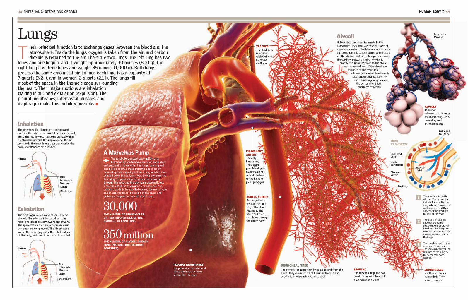
HUMAN BODY I 4948 INTERNAL SYSTEMS AND ORGANS
LungsT
heir principal function is to exchange gases between the blood and theatmosphere. Inside the lungs, oxygen is taken from the air, and carbondioxide is returned to the air. There are two lungs. The left lung has two
lobes and one lingula, and it weighs approximately 30 ounces (800 g); theright lung has three lobes and weighs 35 ounces (1,000 g). Both lungsprocess the same amount of air. In men each lung has a capacity of3 quarts (3.2 l), and in women, 2 quarts (2.1 l). The lungs fillmost of the space in the thoracic cage surroundingthe heart. Their major motions are inhalation(taking in air) and exhalation (expulsion). Thepleural membranes, intercostal muscles, anddiaphragm make this mobility possible.
InhalationThe air enters. The diaphragm contracts andflattens. The external intercostal muscles contract,lifting the ribs upward. A space is created withinthe thorax into which the lungs expand. The airpressure in the lungs is less than that outside thebody, and therefore air is inhaled.
ExhalationThe diaphragm relaxes and becomes dome-shaped. The external intercostal musclesrelax. The ribs move downward and inward.The space within the thorax decreases, andthe lungs are compressed. The air pressurewithin the lungs is greater than that outsideof the body, and therefore the air is exhaled.
AlveoliHollow structures that terminate in thebronchioles. They store air, have the form of a globe or cluster of bubbles, and are active ingas exchange. The oxygen comes to the bloodvia the alveolar walls and then passes towardthe capillary network. Carbon dioxide is
transferred from the blood to the alveoliand is then exhaled. If the alveoli are
damaged as the result of apulmonary disorder, then there is
less surface area available forthe interchange of gases, and
the person might feelshortness of breath.
A Marvelous PumpThe respiratory system accomplishes itsfunctions by combining a series of involuntary
and automatic movements. The lungs, opening andclosing like bellows, make inhalation possible byincreasing their capacity to take in air, which is thenexhaled when the bellows close. Inside the lungs thefirst stage of processing the gases that came inthrough the nose and the trachea is accomplished.Once the exchange of oxygen to be absorbed andcarbon dioxide to be expelled occurs, the next stagescan be accomplished: transport of the gases anddelivery of oxygen to the cells and tissues.
30,000THE NUMBER OF BRONCHIOLES, OR TINY BRANCHINGS OF THEBRONCHI, IN EACH LUNG
350 millionTHE NUMBER OF ALVEOLI IN EACHLUNG (700 MILLION FOR BOTHTOGETHER)
BRONCHIAL TREEThe complex of tubes that bring air to and from thelungs. They diminish in size from the trachea andsubdivide into bronchioles and alveoli.
BRONCHIOLESare thinner than ahuman hair. Theysecrete mucus.
BRONCHIOne for each lung; the twogreat pathways into whichthe trachea is divided
TRACHEAThe trachea isreinforcedwith C-shapedpieces ofcartilage.
PULMONARYARTERYThe onlyblue artery.The oxygen-poor blood goesfrom the rightside of the heartto the lungs topick up oxygen.
AORTAL ARTERYRecharged withoxygen from thelungs, the bloodreturns to theheart and thencirculates throughthe entire body.
ALVEOLIIf dust ormicroorganisms enter,the macrophage cellsdefend againstthem.defienden.
PLEURAL MEMBRANESare primarily muscular andallow the lungs to movewithin the rib cage.
3
2
1 The alveolar cavity fillswith air. The red arrowsindicate the direction theoxygen travels toward thered blood cells and thenon toward the heart andthe rest of the body.
The blue indicates thedirection the carbondioxide travels to the redblood cells and the plasmafrom the heart so that thealveolar can return it tothe lungs.
The complete operation ofexchange is hematosis.The carbon dioxide will bereturned to the lungs bythe venae cavae andexhaled.
HOW IT WORKS
Airflow
IntercostalMuscles
Ribs
Lungs
Diaphragm
Airflow
IntercostalMuscles
Alveolar Wall
Entry andExit of Air
Red BloodCells
LiquidSurfactant
AlveolarCavity
Capillary
IntercostalMuscles
Ribs
Lungs
Diaphragm

50 INTERNAL SYSTEMS AND ORGANS HUMAN BODY I 51
Digestive SystemT
he digestive system is the protagonist of a phenomenaloperation that transforms food into fuel for the entirebody. The process begins with ingestion through the
mouth and esophagus and continues with digestion in thestomach, the small intestine, and the large intestine, fromwhich the feces are evacuated by the rectum and anus. Bythen the task will have involved important chemicalcomponents, such as bile, produced by the liver, and otherenzymes, produced by the pancreas, by which the food isconverted into nutrients. Separating the useful from theuseless requires the filtering of the kidneys, whichdiscard the waste in urine. 00:00:10
00:00:00
03:00:00
08:00:00
Between 20 and 44 hours afterhaving entered the mouth asfood, the residue that wasconverted into semisolid fecesin the previous stage arrivesat the rectum. The waste willbe evacuated through theanus as excrement.
24:00
The First Step: IngestionThe digestive process begins with themouth, the entry point to the large tract
that changes in form and function and ends atthe rectum and anus. The tongue and teeth arethe first specialists in the task. The tongue is incharge of tasting and positioning the food,
which is cut and ground by the teeth. Thissynchronized activity includes the maxillarybones, which are controlled by theircorresponding muscles. The palate, in the upperpart of the mouth, prevents food from passinginto the nose. The natural route of the food isdown the esophagus to the stomach.
TeethThere are 32 teeth, and they are extremely hard, a conditionnecessary for chewing food. There are eight incisors, fourcanines, eight premolars, and 12 molars. Humans developtwo sets of teeth, a provisional or temporary set (the babyteeth) and a permanent set (adult teeth). The firsttemporary teeth appear between six and 12 months of age.At 20 years of age the process of replacement that beganat about age five or six is complete.
Digestion ChronologyThe process that converts food into nutrients begins a fewseconds after the food is raised to the mouth and chewingbegins. The average digestion time is about 32 hours,though digestion can range from 20 to 44 hours.
Enzymes and HormonesThe complex chemical processes that transform food areessentially accomplished by enzymes and hormones. Bothtypes of substances are secreted by various glands of thedigestive system, such as the salivary glands. Enzymes aresubstances that act as catalysts. Hormones are substancesthat regulate processes such as growth metabolism,reproduction, and organ function.
ESOPHAGUSIts muscles force thebolus toward thestomach. Theesophagus and stomachare separated by asphincter.
PHARYNXThe muscles in thewalls of the pharynxcontract, forcing thebolus of chewed foodinto the esophagus.
ROOTEntirely
buried in themaxillary
bone
PULPSoft tissue that
is the core ofthe tooth
THE INSIDE OF A TOOTHTHE MOUTH
A SET OF TEETH
ENAMELhas no feelingand is thehardestsubstance inthe body.
DENTINSensitive tocold and heat,it is strongerthan bonetissue.
NERVESpassinformationabout thetooth to thenervoussystem.
CEMENTThe thin layer
that covers theroots and
anchors them inthe jawbone
CANINES
PREMOLARS
MOLARS
INCISORS
06:00:00
32A PERSON
NORMALLY HASTHIS NUMBER
OF TEETH.
TractThe muscular
movement calledperistalsis pushes
the food along.That is why it ispossible to eatupside down or
duringweightlessness, as
astronauts do.
TONGUEIts notable flexibilitymakes eatingpossible. It alsotastes the food.
THE HARD PALATEThe “roof” of the oralcavity. It is made ofbone.
THE SOFT PALATEAlso called the velarpalate, the palate keepsthe food from goinginto the nose.
1
3
4
5
6
2
2
1
3
4
5
6
About 10 seconds after chewing hasbegun, the food is transformed into amoist alimentary bolus that makes itsway through the pharynx to theesophagus and then to the stomach,where other changes will take place.
Three hours after its arrival, thefood leaves the stomach, which hasaccomplished its function. The firstphase of digestion is over. The bolusnow has a liquid and creamyconsistency.
Three hours later, the food that hasbeen digested in the stomach arrivesat the midpoint of the smallintestine. At this point it is ready tobe absorbed.
Two hours later, the non-digested,watery residue arrives at thejunction of the small and largeintestines. The useless materialrejected by the body's chemicalselectors continues its course, and itis now prepared to be expelled fromthe organism in the form of feces.
The process begins when the food reaches the mouth.The entire organism is involved in the decision, but it isthe digestive system that plays the main role. The firststeps are taken by the teeth and the tongue, aided bythe salivary glands, which provide saliva to moisten thealimentary bolus. The morsels are chewed so that theycan pass through the esophagus.
20:00:00The alimentary residueremains in the large intestinebetween 12 and 28 hours. Inthis part of the process theresidue is converted intosemisolid feces.
10 inches (25 cm)IS THE LENGTH OFTHE ESOPHAGUS.

HUMAN BODY I 5352 INTERNAL SYSTEMS AND ORGANS
StomachT
he part of the digestive tract that is a continuation of the esophagus. It issometimes thought of as an expansion of the esophagus. It is the first sectionof the digestive system that is located in the abdomen. It has the shape of
an empty bag that is curved somewhat like a bagpipe, thehandle of an umbrella, or the letter “J.” In the stomach,gastric juices and enzymes subject the swallowed food tointense chemical reactions while mixing it completely.The stomach connects with the duodenum throughthe pylorus. Peristalsis, or the muscular contractionsof the alimentary canal, moves the food from thestomach to the duodenum, the next station in theprogress of the alimentary bolus.
THE STOMACH INCREASES UP TO 20 TIMES ITS ORIGINAL SIZEAFTER A PERSON EATS.
20 times
DUODENUMThe initial
section of thesmall intestine
STOMACH WALLA covering of three muscular
layers that contract indifferent directions to mash
the food. It contains millionsof microscopic glands that
secrete gastric juices. WRINKLES OR FOLDSare formed when the stomach isempty, but they stretch out as thestomach fills and increases its size.
PYLORUSA muscular ring that opens
and closes the pyloricsphincter to allow (or
prevent) passage of liquefiedfood from the stomach on its
way to the intestine
ESOPHAGUScarries chewed food tothe stomach.
Peristalsis: Muscles in ActionPeristalsis is the group of muscularactions that moves the food toward thestomach and, once the digestive stagehas been completed, moves it on to the
small intestine. The sphincters arestationary, ring-shaped muscularstructures whose opening and closingregulates the passage of the bolus.
How We SwallowAlthough swallowing is a simple act, it does require thecoordination of multiple parts. The soft palate moves backwardwhen the alimentary bolus passes through the esophagus. Theepiglottis moves downward to close the trachea and prevent thefood from entering the respiratory pathways. The alimentarybolus is advanced by the muscular motions of peristalsis.
Full stomach. Food enters. The pyloricsphincter remains closed. The gastric juiceskill bacteria and are mixed with the foodthrough muscular motions.
Food is sent toward the stomach, pumpedby the muscular contractions of theesophageal walls. Gravity helps accomplishthis downward journey.
The stomach in full digestive action. Theperistaltic muscles mix the food until itbecomes a creamy, viscous liquid(chyme).
The stomach is being emptied. The pyloricsphincter relaxes, the muscles move thefood, and small quantities of food exittoward the duodenum.
INFERIOR ESOPHAGEAL SPHINCTERcloses the junction between theesophagus and the stomach to preventreflux of the stomach contents.
THREE LAYERS OF MUSCLEThey are thecircular, the
longitudinal, andthe oblique.
SUBMUCOSATissue that connects
the mucosa to thelayers of muscle
MUSCULAR LAYERS OFTHE MUCOSA
Two fine layers ofmuscular fibers extend
under the mucosa.
GASTRIC MUCOSAcontains the gastric glands, which produce3 quarts (2.8 l) of gastric juice per day.
GASTRIC WELLSFrom three to seven
glands open to form agroove.
GLÁNDULASGÁSTRICAS
SEROSALayer that covers the
outer surface
SUBSEROSALayer that connects the
serosa to the muscles
SOFT PALATE
Stomach WallThe structure of the wall accounts for the twoimportant functions of the stomach: the muscularlayers and the activity of the gastric glandsguarantee that digestion will run its course.
EPIGLOTTIS IN OPEN POSITION
TRACHEA
ESOPHAGUS
LARYNX
FOOD
TONGUE
SOFT PALATE
EPIGLOTTISCLOSED
TRACHEA
ESOPHAGUS
LARYNX
FOOD
TONGUE
The Esophagus
AlimentaryBolus
Duodenum
PyloricSphincter
Stomach
Food
Stomach
Food
Stomach
Food
PyloricSphincterClosed
PyloricSphincterOpen
X-ray of the StomachThe stomach is the best known of the internal bodyorgans, but it is also the most misunderstood. This J-
shaped sac stretches to fill up with food, but it does not absorbany of the nutrients. Its work consists of starting the digestionprocess, storing semi-digested food, and releasing the foodslowly and continuously. Internal gastric juices make it possiblefor the enzymes to decompose the proteins, while muscularcontractions mix the food.

HUMAN BODY I 5554 INTERNAL SYSTEMS AND ORGANS
Liver, Pancreas, BileT
he liver is the largest gland of the human body and the second largest organ (theskin is the largest). It has numerous functions, and a large part of the body's generalequilibrium depends on it. The liver produces bile, a yellowish-green fluid
that helps in the digestion of fats. The liver is the great regulator of the glucose level of the blood, which it stores in the form ofglycogen. Glycogen can be released when the organismrequires more sugar for activity. The liver regulates themetabolism of proteins. Proteins are the essential chemicalcompounds that make up the cells of animals and plants.The liver is also a large blood filter and a storage site forvitamins A, D, E, and K. The pancreas is a gland thatassists in digestion, secreting pancreatic juice.
PancreasThe pancreas is a gland thataccomplishes various functions. Its
exocrine component secretes pancreatic juiceinto the duodenum to aid in digestion. This juicecontains enzymes that break down fats,proteins, and carbohydrates. It contains sodiumbicarbonate, which neutralizes the strongstomach acid. The pancreas also performs afunction in the endocrine system: itsecretes the hormone insulin into theblood, where it regulates glucose levels.
ESOPHAGUSbrings food to thestomach.
PANCREATICDUCT
PANCREASreleases pancreaticjuice, whichcontains digestiveenzymes.
DUODENUMThe initial part of thesmall intestine
SINUSOIDIts function is totransport blood tothe lobule, where itis processed.
HEPATICARTERIALBRANCHtransportsoxygenatedblood to thelobule.
GALLBLADDERstores bileproduced bythe liver.
BRANCH OF THE BILIARYDUCTCarries bile tothe commonbiliary duct
Vesicle and BileThe biliary system stores bile thatis produced by the hepatocytes in
a specialized pouch called thegallbladder. The path the bile takes fromthe liver to the gallbladder leads throughlittle canals, biliary ducts, and hepatic
ducts, whose diameter increases as thebile moves along. When the body ingestsfat, the bile is sent from the gallbladderto the small intestine to accomplish itsmain function: emulsifying fats to helppromote their later absorption.
LobulesAmong its other functions, the liverprocesses nutrients to maintain anadequate level of glucose in the blood.This task requires hundreds of chemicalprocesses that are carried out by thehepatocytes, or liver cells. These are
arranged in columns, formingstructures called lobules. They producebile and a sterol (a solid steroid alcohol)called cholesterol. They also eliminatetoxins that might be present in food.
LiverAmong its numerous functions, the liverrids the blood of potentially harmful
chemical substances, such as drugs and germs.It filters out toxins, starting in the smallintestine, and it is involved in maintaining theequilibrium of proteins, glucose, fats,cholesterol, hormones, and vitamins. The liveralso participates in coagulation.
THE CONNECTIONThe esophagus, stomach,gallbladder, spleen, andsmall intestine are linkedfunctionally and by theirposition in the body. Theyconstitute the greatcrossroads of digestion.
SPLEENThe spleen has adouble function. It ispart of the immunedefense system, andit destroys defectivered blood cells.
ENERGYThe body's cells basically obtaintheir energy from the breakdown ofglucose stored in the liver. When noglucose is available, the body turnsto fatty acids for energy.
ADIPOSE CELLSare cells in which theorganism storesexcess fatty acids inthe form of fat.
MUSCULAR FIBERMuscle cells in theliver together withthe hepatic cellsstore glycogen.
HEPATIC TISSUEExcess glucose in theorganism is stored asglycogen in the cellsof the liver.
CELLULAR GROWTH AND REPAIRAmino acids are convertedinto proteins by a processcalled anabolism. Proteinsare fundamental formitosis, cellularregeneration, and enzymeproduction.
The complex of chemical reactions thatoccur in the cells of living beings,transforming simple substancesinto complex substancesand vice versa. When thenutrients are absorbedinto the bloodstream andpassed to the liver, theliver breaks downproteins into aminoacids, fats into fattyacids and glycerol, andcarbohydrates intosmaller components. Anormal diet includescarbohydrates, proteins,fats, vitamins, andminerals.
Metabolism
COMMONBILIARYDUCT
PANCREAS
COMMON HEPATIC DUCT
THE AMOUNT OF BILE THE LIVERCAN PRODUCE IN A DAY. THE LIVERIS THE HEAVIEST INTERNAL ORGANOF THE BODY.
1 quart (0.9 l)
CYSTIC DUCT
PANCREATICDUCTcarriespancreatic juiceto theduodenum.

HUMAN BODY I 5756 INTERNAL SYSTEMS AND ORGANS
The longest part of the digestive tract. It is about 26to 30 feet (8 to 9 m) long and runs from the stomachto the anus. The small intestine receives the food
from the stomach. Digestion continues through enzymeactivity, which completes the chemical breakdown of the food.Then the definitive process of selection begins: the walls ofthe small intestine absorb the nutrients derived from thechemical transformation of the food. The nutrients thenpass into the bloodstream. Waste substances, on theother hand, will go to the large intestine. There thefinal stage of the digestive process will occur: theformation of the feces to be excreted.
RECTUMThe final point of theaccumulation of the feces.Its storage capacity is small.
SIGMOID COLONcontains a structurethat permits thegases to passwithout pushing the feces.
DESCENDINGCOLONThe feces aresolidified andaccumulatebefore beingexpelled.
TRANSVERSECOLONThe undigestedremains beginto betransformedinto feces.
SMALLINTESTINE
LARGEINTESTINE
CAECUMInitial section
of the largeintestine
DUODENUMThe initial
section of thesmall intestine,
to which thesecretions ofthe pancreasand the liverare directed
WATER THAT ENTERS THE ALIMENTARY CANAL
Saliva 34 (1 l) Water from Drinking 77 (2.3 l)Bile 34 (1 l) Pancreatic Juice 68 (2 l)Gastric Juice 68 (2 l)Intestinal Juice 34 (1 l)
WATER REABSORBED BY THEALIMENTARY CANAL
Small Intestine 280 Large Intestine 30 Subtotal 310 Water Lost in the Feces 3 Total 313
(8.3 l)(0.9 l)(9.2 l)
(0.1 l)(9.3 l)
ILEUMFinal section of the
small intestine, linkedwith the large intestine
ANUSOpening in the large
intestine throughwhich the feces exit
JEJUNUMThe intermediatepart of the smallintestine, whichlinks theduodenum withthe ileum
VillaThe internal wall of the smallintestine is covered with millions of
hairlike structures called villi. Each one hasa lymphatic vessel and a network ofvessels that deliver nutrients to it. Each
villus is covered by a cellular layer thatabsorbs nutrients. Together with epithelialcells, the villi function to increase thesurface area of the intestine and optimizethe absorption of nutrients.
The Union of BothThe small and large intestines join at the section called theileum (which is the final section of the small intestine; the
duodenum and jejunum come before the ileum). The iliac valveacts as a door between the small intestine and large intestine, orcolon. The ileum terminates in the caecum (of the large intestine).The ileum measures approximately 13 feet (4 m) in length. Itsprimary function is the absorption of vitamin B12 and biliarysalts. The primary function of the large intestine is the absorptionof water and electrolytes that arrive from the ileum.
SEROSAThe external protectivemembrane in both
SUBMUCOSAIn both, the loose coveringwith vessels and nerves
MUCOSAIt is thin and absorbsnutrients via projections or hairs.Absorbent fat thatexcretes mucus
MUSCULARThin muscle fibers that arelongitudinal externally andcircular internally. Thefibers are also coveredwith hairs, maximizing thearea of the mucosa.Fatty rigid layer that mixesand pushes the feces
Large and Small Intestine
Opening of theappendix
APPENDIX
ILEUM
CAECUM Initialsection ofthe largeintestine
In fluid ounces In fluid ounces
HAUSTRUMOF THECOLON
ILEOCECALVALVE relaxesbetween meals,allowing theflow toaccelerate.
TAENIA MUSCLE
ASCENDINGCOLON
The water andmineral saltsare absorbed
along thelength of the
large intestinein a process
that removeswater from the
digestive waste.
Differences and SimilaritiesThe small intestine is longer than the largeintestine. The length of the small intestine isbetween 20 and 23 feet (between 6 and 7 m),
and the large intestine averages 5 feet (1.5 m).Their respective composition and functionsare complementary.
ARTERIALCAPILLARY
VENOUSCAPILLARY
VILLA LYMPHATICCAPILLARY
TUNICAMUSCULARIS
SUBMUCOSA
SEROSA
MUCOSA
313 (9.3 1)Total
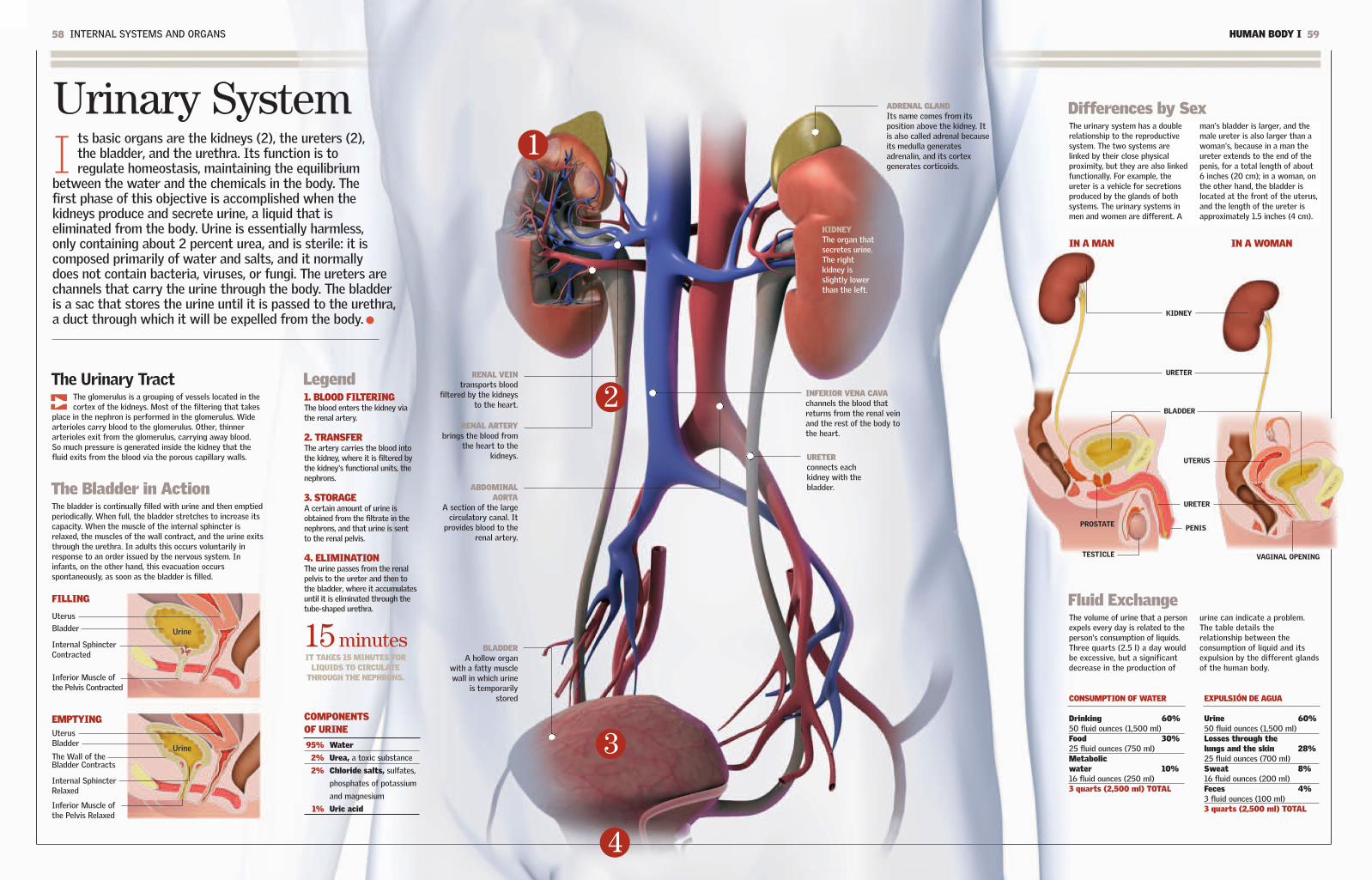
HUMAN BODY I 5958 INTERNAL SYSTEMS AND ORGANS
Urinary SystemI
ts basic organs are the kidneys (2), the ureters (2),the bladder, and the urethra. Its function is toregulate homeostasis, maintaining the equilibrium
between the water and the chemicals in the body. Thefirst phase of this objective is accomplished when thekidneys produce and secrete urine, a liquid that iseliminated from the body. Urine is essentially harmless,only containing about 2 percent urea, and is sterile: it iscomposed primarily of water and salts, and it normallydoes not contain bacteria, viruses, or fungi. The ureters arechannels that carry the urine through the body. The bladderis a sac that stores the urine until it is passed to the urethra,a duct through which it will be expelled from the body.
KIDNEYThe organ thatsecretes urine.The rightkidney isslightly lowerthan the left.
RENAL VEINtransports blood
filtered by the kidneysto the heart.
RENAL ARTERYbrings the blood from
the heart to thekidneys.
ADRENAL GLANDIts name comes from itsposition above the kidney. Itis also called adrenal becauseits medulla generatesadrenalin, and its cortexgenerates corticoids.
ABDOMINAL AORTA
A section of the largecirculatory canal. It
provides blood to therenal artery.
BLADDERA hollow organ
with a fatty musclewall in which urine
is temporarilystored
LegendThe Urinary TractThe glomerulus is a grouping of vessels located in thecortex of the kidneys. Most of the filtering that takes
place in the nephron is performed in the glomerulus. Widearterioles carry blood to the glomerulus. Other, thinnerarterioles exit from the glomerulus, carrying away blood.So much pressure is generated inside the kidney that thefluid exits from the blood via the porous capillary walls.
1. BLOOD FILTERINGThe blood enters the kidney viathe renal artery.
2. TRANSFERThe artery carries the blood intothe kidney, where it is filtered bythe kidney's functional units, thenephrons.
3. STORAGEA certain amount of urine isobtained from the filtrate in thenephrons, and that urine is sentto the renal pelvis.
4. ELIMINATIONThe urine passes from the renalpelvis to the ureter and then tothe bladder, where it accumulatesuntil it is eliminated through thetube-shaped urethra.
IN A MAN IN A WOMAN
2
3
4
INFERIOR VENA CAVAchannels the blood thatreturns from the renal veinand the rest of the body tothe heart.
95% Water
2% Urea, a toxic substance
2% Chloride salts, sulfates,
phosphates of potassium
and magnesium
1% Uric acid
COMPONENTS OF URINE
1
15 minutesIT TAKES 15 MINUTES FOR
LIQUIDS TO CIRCULATETHROUGH THE NEPHRONS.
UrineThe Wall of theBladder Contracts
Internal SphincterRelaxed
Inferior Muscle ofthe Pelvis Relaxed
The Bladder in ActionThe bladder is continually filled with urine and then emptiedperiodically. When full, the bladder stretches to increase itscapacity. When the muscle of the internal sphincter isrelaxed, the muscles of the wall contract, and the urine exitsthrough the urethra. In adults this occurs voluntarily inresponse to an order issued by the nervous system. Ininfants, on the other hand, this evacuation occursspontaneously, as soon as the bladder is filled.
EMPTYING
Bladder
Uterus
Urine
Internal SphincterContracted
Inferior Muscle of the Pelvis Contracted
FILLING
BladderUterus
CONSUMPTION OF WATER
Drinking 60%50 fluid ounces (1,500 ml)Food 30%25 fluid ounces (750 ml)Metabolic water 10%16 fluid ounces (250 ml)3 quarts (2,500 ml) TOTAL
EXPULSIÓN DE AGUA
Urine 60%50 fluid ounces (1,500 ml)Losses through the lungs and the skin 28%25 fluid ounces (700 ml)Sweat 8%16 fluid ounces (200 ml)Feces 4%3 fluid ounces (100 ml)3 quarts (2,500 ml) TOTAL
Differences by SexThe urinary system has a doublerelationship to the reproductivesystem. The two systems arelinked by their close physicalproximity, but they are also linkedfunctionally. For example, theureter is a vehicle for secretionsproduced by the glands of bothsystems. The urinary systems inmen and women are different. A
man's bladder is larger, and themale ureter is also larger than awoman's, because in a man theureter extends to the end of thepenis, for a total length of about6 inches (20 cm); in a woman, onthe other hand, the bladder islocated at the front of the uterus,and the length of the ureter isapproximately 1.5 inches (4 cm).
Fluid ExchangeThe volume of urine that a personexpels every day is related to theperson's consumption of liquids.Three quarts (2.5 l) a day wouldbe excessive, but a significantdecrease in the production of
urine can indicate a problem.The table details therelationship between theconsumption of liquid and itsexpulsion by the different glandsof the human body.
KIDNEY
URETER
BLADDER
URETER
TESTICLE
PROSTATE PENIS
UTERUS
VAGINAL OPENING
URETERconnects eachkidney with thebladder.

60 INTERNAL SYSTEMS AND ORGANS HUMAN BODY I 61
KidneysL
ocated on either side of the spinal column, the kidneys are thefundamental organs of the urinary system. They regulate theamount of water and minerals in the blood by producing urine that
carries away the waste the kidneys discard. They keep the composition ofthe bodily fluids constant, regulate the pressure of the arteries, andproduce important substances such as the precursor of vitamin D anderythropoietin. Every day they process 500 gallons (1,750 l) ofblood and produce 2 quarts (1.5 l) of urine. The kidneys measureapproximately 5 inches (12 cm) long and 3 inches (6 cm)wide. Their weight is only 1 percent of the total bodyweight, but they consume 25 percent of its energy. Ifone kidney ceases to function, the body is able tosurvive with the activity of the other.
ONE KIDNEY HAS ABOUT ONEMILLION NEPHRONS.
1 million
CO
RT
EXM
EDU
LLA
IS THE AMOUNT OF URINEELIMINATED EACH DAY BY AN ADULT.
41 to 51fluid ounces (1,200 to 1,500 cc)
GlomerulusA grouping of vessels and capillaries in the kidney'scortex, or sheath. Most of the filtering done by thenephrons takes place there. The wide, afferentarterioles bring blood to the glomerulus. Other,narrower, efferent arterioles lead out of theglomerulus, transporting blood. Inside theglomerulus so much pressure is generated thatsubstances in the blood pass out through the porouscapillary walls.
45 minutesTHE FRENCH PHYSIOLOGIST CLAUDEBERNARD (1813-78) WAS THE FIRST TONOTE THE IMPORTANCE OF THE KIDNEYS.
At that time it was not known that thekidneys filter all the water content of theblood in the body every 45 minutes and that,even so, it is possible to survive with onlyone kidney (or none, in the case of dialysis).
The Renal CircuitUrine is produced in the nephrons in each kidney;there are thought to be a million nephrons in
each kidney. From the nephrons the urine flows intothe proximal convoluted tubule, where all thenutrients, such as glucose, amino acids, and most ofthe water and salts, are reabsorbed into the blood.After passing through the nephron the urine is filtered,and it arrives at the common collecting duct whereonly the residues and excess water are retained.
NephronsThe functional units of the kidney that filter theblood and produce urine. The basic structure of thenephron consists of two parts: (1) the renal orMalpighian corpuscle, where filtration occurs,including the glomerulus and Bowman's capsule thatenvelops it; and (2) the renal tubule, a tube thatcollects the filtered liquid (urine) that is to beeliminated from the body.
GLOMERULUSSecond stage, orultrafiltrate
EFFERENT ARTERIOLE
Glomerulus salt
Direction of Filtering
INTERLOBULARARTERY
Section of the cortex
INTERLOBULARARTERY
Section of themedulla
LOOP OF HENLEThe shape of the
nephron curve
BOWMAN'S CAPSULE
First stage of thefiltrate. It surrounds a
fluid consisting ofwater, potassium,
bicarbonate, sodium,glucose, amino acids,
urea, and uric acid.
NEPHRON
PROXIMALCONVOLUTEDTUBULE First section of thefiltrate's exit route
PERITUBULARCAPILLARIESThe thinnestconduits
INTERLOBULARVEINSection in the cortex
COLLECTINGTUBULETransports andconcentrates thefiltered liquid fromthe nephrons.
1
2 3
4
5
1. ENTRY OF BLOODThe blood enters the kidney via the renal artery.
2. FILTRATIONThe blood is filtered in the nephrons, the functionalunits of the kidneys.
3. URINE IS OBTAINEDA certain amount of urine is obtained from the filtratein the nephrons, and it is sent to the renal pelvis. Thefiltered blood, free from waste, is sent to the renal veinand reenters the bloodstream.
4. URINEThe urine passes through the renal pelvis to the ureterand from there to the bladder, where it accumulatesuntil it is eliminated through the tube-shaped urethra.
5. CLEAN BLOODThe clean blood exits the kidney via the renal vein,which is connected to the vena cava. The blood thenreturns to the heart.
URETERThe tube that transportsthe urine to the bladder
RENAL VEINThe blood flows out of thekidney through the renalvein toward the vena cava,one of the principal veinsof the body.
RENAL ARTERYA branch of the aorticartery, which providesthe kidney with blood
RENAL PYRAMIDA fluted structure in theform of a pyramid, locatedin the renal medulla
RENAL PELVIStransports theurine to theureter.
ARCUATE ARTERYfeeds the afferent
arterioles leading tothe glomerulus.
ARCUATE VEINTakes blood from theefferent arterioles ofthe glomerulus
INTERLOBULARVEINSection in themedulla
RENALCAPSULE
Protective layerthat covers each
kidney. Itconsists of white
fibrous tissue.

The Hormonal MessageThe endocrine system is made up of the so-calledendocrine glands. This complex, controlled by the
pituitary (hypophysis), or master, gland, includes thethyroid, parathyroid, pancreas, ovaries, testicles,adrenals, pineal, and hypothalamus. The role of theseglands is to secrete the many hormones needed for bodyfunctions. The word “hormone” comes from the Greekhormon, which means to excite or incite. The term wassuggested in 1905 by the British physiologist ErnestStarling, who in 1902 assisted in the isolation of the firsthormone, secretin, which stimulates intestinal activity.Hormones control such functions as reproduction,metabolism (digestion and elimination of food), and thebody's growth and development. However, by controllingan organism's energy and nutritional levels, they alsoaffect its responses to the environment.
HUMAN BODY I 6362 INTERNAL SYSTEMS AND ORGANS
Endocrine SystemC
onsists of the glands inside the body that secrete approximately 50 specific substancescalled hormones into the blood. The hormones activate or stimulate various organs andcontrol reproduction, development, and metabolism. These chemicals control many of the
body's processes and even meddle in our love lives.
AKiss
THYROID
PARATHYROID
PINEAL
PHEROMONESare chemical substancesreleased by the glandsdistributed in the skinthat are related tosexual attraction. Theyact like hormones(whether or not they areactually hormones is amatter of dispute). Theytransmit sensations ofattraction, excitation,and rejection.
The LH hormone excitesthe production of
estrogen hormones, whichregulate female sexuality;
the activity of themammary glands; and the
menstrual cycle. Pubertyis marked by an increase
of estrogen production.
MAMMARY GLANDS
ADRENAL GLANDS
The hormone adrenaline“awakens” the body
before a risk—or before akiss. It increases thecardiac rhythm, the
arterial pressure, thelevel of glucose in theblood, and the flow of
blood to muscles.
PANCREASBefore a kiss, it increases the
glucose level in the blood.The pancreas produces thetwo hormones that control
the blood sugar level: insulinand glycogen.
SEXUAL GLANDSThe reproductive system respondsto the same pituitary hormones in
men and women: luteinizinghormone (LH) and follicle-
stimulating hormone (FSH). (Bothare released and activated in
anticipation of a kiss.)
PITUITARY GLAND ORHYPOPHYSIS: The pituitary gland is locatedat the base of the brain, andit is the most importantcontrol center of theendocrine system. It releasesoxytocin in anticipation of akiss; it is the hormone thatstimulates orgasm, birth, andbreast-feeding; and it is alsoassociated withpsychological behaviors suchas affection and tenderness.
ARTERYVEIN
IN THE SKINMSH stimulatesthe production
of melanin.
IN THE URINARYSYSTEMAntidiuretic.Equilibrium of thefluids in the body.
IN THE UTERUS ANDTHE BREASTSStimulates thesecretion of mother'smilk and contractionsduring birth.
MSH
ADH
OXYTOCIN
ACTH
TSH
GH
FSH, LH
THE ADRENALGLAND
ACTH stimulatesthe adrenal glands
to produce theantistress hormone
cortisol.
THYROIDGLAND
TSH acts on thethyroid and
influencesmetabolism.
IN THE BONES AND MUSCLES
GH stimulatesgrowth in an infantand influences thehealth of an adult.
IN THE TESTICLES AND THE OVARIES
FSH stimulates the productionof spermatozoa and the release
of ovules. LH also generatestestosterone.
ANTERIOR LOBEProduces sixhormones, includingprolactin
POSTERIOR LOBULEThe hormones of thehypothalamus arestored here.
The Master GlandThe pituitary gland, or hypophysis, is also calledthe master gland because it controls the rest ofthe endocrine glands. It is divided into twoparts, the anterior lobe and the posteriorlobe. The pituitary hormonesstimulate the other glands togenerate specific hormonesneeded by the organism.
Oxytocin, the hormone that influencesbasic functions, such as being in love,orgasm, birth, and breast-feeding, is
associated with affection and tenderness.It is a hormone that stimulates theformation of bonds of affection.
The Confidence Hormone
PituitaryHormonesACTH Adrenocorticotropin hormone.It goes to the adrenal gland.TSH A hormone that stimulates thethyroid to produce the thyroidhormones, which influence metabolism,energy, and the nervous system.GH Growth hormoneFSH Follicle-stimulating hormoneLH Luteinizing hormone;testosterone and estrogenMSH Hormone that stimulates themelanocyte of the skin.ADH Antidiuretic hormonePRL Prolactin; stimulates milkproduction by the mother.OXYTOCIN Stimulates the release ofmilk by the mother, as well as thecontractions needed during labor.
Kissing is considered to be healthybecause, among other things, itstimulates the production of numeroushormones and chemical substances.
2
3
1
4
NEUROSECRETORYCELLS This type of cellproduces thehormones ADH andoxytocin in thehypothalamus.

HUMAN BODY I 6564 INTERNAL SYSTEMS AND ORGANS
Male Reproductive SystemT
he male reproductive system is the complex of organs that leads to a man's production of one oftwo types of cells necessary for the creation of a new being. The principal organs are the twotesticles, or male gonads, and the penis. The testicles serve as a factory for the production of
millions of cells called spermatozoa, which are minute messengers of conception bearing the geneticinformation for the fertilization of the ovum. The penis is linked to the urinary apparatus, but forreproduction it is the organ that functions as a vehicle for semen, a liquid through which thespermatozoa can reach their destination. The word “semen” comes from Greek and means “seed.”
THE NUMBER OF SPERMATOZOA THAT EACH 0.06CUBIC INCH (1 ML) OF SEMEN CAN CONTAIN
150 million
PREPUCECovers andprotects thehead of thepenis
BLADDERReceptacle of theurinary systemthat temporarilystores urine
Internal Structure of the PenisThe most characteristic organof a man's body, the penis has a
cylindrical form with a doublefunction for the urinary system andthe reproductive system. In itsnormal, or relaxed, state the peniscarries urine from the body via theurethra during urination. In its erectstate its rigidity permits it to beintroduced into the female vagina andto release sperm through ejaculation.The penis consists of spongy tissue
supplied with blood vessels. Thecirculatory system supplies abundantblood to these vessels during sexualarousal so that the spongy tissuebecomes swollen because of the filledblood vessels. This produces anerection, which makes copulationpossible. The body of the penissurrounds the urethra and is connectedto the pubic bone. The prepuce coversthe head (glans) of the penis, which islocated above the scrotum.
Testicles and SpermatozoaThe seminiferous tubes in thetesticles are covered with
spermatogenic cells. By a process ofsuccessive cellular divisions calledmeiosis, the spermatogenic cells aretransformed into spermatozoa, theterm for the gametes, or male sexualcells, the bearers of half of the geneticinformation of a new individual. Thespermatozoa fertilize the ovum, or
female gamete, which contains theother half of the genetic information.The number of chromosomes is keptconstant because the spermatozoaand the ovum are both haploid cells(cells that possess half of the geneticinformation of other cells). When thetwo haploid cells unite, the fertilizedegg, or zygote, is a diploid cell (whichcontains a total of 46 chromosomes).
SPERMATIC CORDConnects the testicles to the body
DEFERENT DUCT(DUCTUS DEFERENS)Connects the epididymiswith the seminal vesicle
BLOOD VESSELSare numerous, and theyconnect to the vas deferens.
EPIDIDYMISThe tube where thesemen matures andenters the deferent duct
Prostate and EpididymisThe prostate is a gland located in front of the rectum and belowthe bladder. It is the size of a walnut, and it surrounds the
urethra, a tube that carries urine from the bladder. The prostateproduces the liquid for the semen, which carries the spermatozoa.During orgasm, muscular contractions occur that send the liquid fromthe prostate out through the urethra. The epididymis is a duct that,when stretched out to its full length, is approximately 20 feet (5 m)long. In the male body it is extremely coiled and lies on the backsurface of the testicles, where it is connected with the correspondingvas deferens. The vas deferens stores spermatozoa and provides themwith an exit route. The seminal vesicles are two membranousreceptacles that connect to both sides of the vas deferens and formthe ejaculatory duct.
ARTERYIts dilation causes theerection.
CAVERNOUS BODIESLike the spongy tissue,these also fill with blood.`
SPONGY TISSUESwells when it fills withblood, sustaining theerection
URETHRAExtends through thespongy tissue
EXTERNAL SKINCovers thewhole organ
SEMINIFEROUS TUBESemen is produced here.Each testicle hasthousands of them.
GLANSExtremity of the penis
SCROTUMSac of skinthat containsthe testicles
EPIDIDYMISSpiral tubewhere thesperm matures
TESTICLEGland thatproducessperm
EJACULATORYDUCTA short tube thatcarries the spermto the urethra
SEMINALVESICLESecretes fluidand assortednutrients intothe spermduringejaculation
PROSTATEGland thatsecretes a creamyliquid (semen)along with theejaculated sperm
IS THE IDEAL APPROXIMATETEMPERATURE REQUIRED BY THE TESTICLES TOPRODUCE SEMEN. It is lower than the normalbody temperature of 98.6° F(37° C) because thattemperature would be toowarm for this function. Thisexplains why the testicles areoutside of the body. Dependingon the ambient temperature,they extend or retract.
93º F(34º C)
HEADContains genetic
information (DNA)
SEMINIFEROUSTUBULEWhere spermatozoaare produced
SEMINIFEROUS TUBULE
THE GLANDS
TESTICLES
PITUITARY GLAND
FHS LH
TESTOSTERONE
SPERMATOZOON Male reproductive cell
THE PENIStransfers the spermto the woman.
THE TESTICLESThe sexual organs that produce sperm
TAILEngine for thespermatozoon'spropulsion
SPERMATOCYTESare formed byrepeatedreproduction ofthe spermatogonia.
MATURE SPERMThe division of thespermatocytesformsspermatozoa.
SPERMATOZOAmove from theseminiferous tubules tothe epididymis, wherethey are stored.
Production ofSpermatozoa
maintains male reproductivestructures and secondarysexual characteristics.
InterstitialCells
SustentacularCells
INTERMEDIATE PARTContains mitochondria thatrelease energy to move the tail
POINTor acrosome. Containsenzymes that help thespermatozoonpenetrate the externalmembrane of the ovum.

66 INTERNAL SYSTEMS AND ORGANS HUMAN BODY I 67
Female Reproductive SystemI
ts primary function is the production of ova, and its organs are arranged so as to allow thefertilization of the ovum by a spermatozoon of the male reproductive system and from that momentto facilitate a series of processes known collectively as pregnancy for the creation of a new being.
The internal organs of the female reproductive system are the vagina, the uterus, the ovaries, and thefallopian tubes. The external genitalia, generally referred to as the vulva, are relatively hidden andinclude the labia majora and minora, the clitoris, the urinary meatus, Bartholin's glands, and thevaginal orifice that leads to the vagina. The menstrual cycle governs the system's function.
CERVIXThe neck of the uterusthrough which the menstrualfluid and other secretionspass. It allows the sperm toenter and the fluid from themenstrual cycle to exit. Itgreatly expands during birth.
OVARYContains follicles ofthe ova, one ofwhich maturesduring eachmenstrual cycle
FALLOPIAN TUBEA tube close to each ovarythat receives the matureovum and transports it tothe uterus. It measures 4inches (10 cm) long and0.1 inch (0.3 cm) indiameter.
FIMBRIAEFilamentaryformations thatguide the releasedovum toward thefallopian tubeduring ovulation
UTERUSThe muscular wallsstretch to accommodatethe fetus during itsdevelopment.
VAGINAAn elastic muscular tube thatstretches during sexual relationsand birth; it has an internalmucous membrane that provideslubrication and an acid mediumthat acts as a defense againstinfection. It serves as the pathwayof the uterus to the exterior.
IS THE APPROXIMATE NUMBER OF OVATHAT AN INFANT GIRL HAS IN HER BODY AT BIRTH. BETWEEN THE AGES OF 10 AND 14, ABOUT 300,000 TO 400,000 OVAREMAIN, OF WHICH ONLY 400 WILLMATURE COMPLETELY OVER HER LIFETIME.
2 million
EMPTY FOLLICLECORPUS LUTEUMOVULATIONMATURE FOLLICLEThe egg is completelyready.
IMMATUREFOLLICLEIt encases the ovum,which has only justbegun to grow.
DAY 0 DAY 4 DAY 14 DAY 28
At the beginning of the cyclethe uterus eliminates itsinternal lining. Theunfertilized ovum exits theuterus during menstruation.
As the estrogen level increases, theblood vessels expand.The glands produce nutrients.
The yellow body (from the empty follicle) produces hormones(estrogens and progesterones) that stimulate the formation oftissues that are ready for pregnancy. The tissues shrink if there isno fertilization. The tissues are expelled from the body,together with the unfertilized egg, with themenstrual fluid.
In t
he E
ndom
etri
um
The ovum begins to grow,stimulated by FSH.
The follicle (which includes the ovum)develops and matures until it ruptures andreleases the mature ovum.
The released ovumgoes to the uterus.
The empty follicle is convertedinto a corpus luteum and secretesprogesterone, preparing the bodyfor a possible pregnancy.
The corpus luteum (in this stageit is no longer called a follicle)breaks down and dies, thusconcluding the menstrual cycle.
Wit
hin
the
Ova
ry
The follicle-stimulatinghormone (FSH) induces thedevelopment of follicles thatenvelop the ova.
The ova produce estrogen duringdevelopment.
The luteinizinghormone (LH)triggers ovulation onday 14 of the cycle.
The empty follicle (the ovum has already been expelled)is converted into a corpus luteum, which producesprogesterone to thicken the endometrium (uterine lining)and receive the ovum if fertilized.
At
the
Hor
mon
al L
evel
The 28 Days of the Menstrual Cycle
FSH ESTROGEN PROGESTERONE
Menstruation: The Key to Female ReproductionThe female reproductive system is more protected than that of the malebecause the bony structure of the pelvis houses and shields it. Its
development begins around the age of 10, when the female hormones begin athree- to four-year process during which the genital organs, the breasts, thepubic hair, and the general shape of the body change. Toward the age of 13,sometimes earlier or later, the first menstruation, called the menarche, occurs,signaling the beginning of a woman's fertility. She will normally remain fertilefor several decades. During menopause, when fertilization is no longer possible,a woman's sexual life is usually not affected and can continue normally.
CLITORISA sensitive protuberanceof tissue that responds tosexual stimulation
Cilia, tiny hairlikestructures, movethe ova verysmoothly.
LH
THE GLANDS
OVARY
PITUITARY GLAND
FHS LH
ESTROGEN
Development of theCorpus Luteum
PROGESTERONE
maintains thereproductive structuresand secondary sexualcharacteristics.
prepares for theimplementation and controlof the menstrual cycle.
Ovulation
Ovum
Developmentof the Follicle
Everything we know about theworld comes to us through thesenses. Traditionally it wasthought that we had only five:vision, hearing, touch, smell,
and taste. However, for some time nowwe have known that we have manyadditional classes of sensations—suchas pain, pressure, temperature,muscular sensation, and a sense of
motion—that are generally included inthe sense of touch. The areas of thebrain involved are calledsomatosensory areas. Although weoften take our senses for granted, each
one of them is delicate and irreplaceable.Without them it is nearly impossible tounderstand our surroundings. They area bridge between us and everythingalive on the Earth.
SMELL AND TASTE 70-71
TOUCH AND THE SKIN 72-73
ANATOMY OF THE EYE 74-75
THE MECHANICS OF HEARING 76-77
SPEECH AND NONVERBAL LANGUAGE 78-79
The Senses and Speech HEALTHY AND SHINY SKINThe health of the skindepends upon a diet thatprovides the organism with asufficient amount of proteinsand minerals.

70 THE SENSES AND SPEECH
Smell and TasteT
hese two senses of the body function as powerful allies of the digestive system. Tasteinvolves the perception of dissolved chemical substances arriving, for example, in the form offood. Taste sensation is principally seated on the upper surface of the tongue, and saliva is a
fundamental ingredient for dissolving and tasting. Smell involves the perception of these chemicalswhen they take the form of dispersed aromas. The sense of smell operates at a greater distancethan that of taste and can capture substances floating in the environment. It is thought that smellis some 10,000 times more sensitive than any of our other senses.
THE SURFACE OF THETONGUE CAN
DISTINGUISH:SWEET, SALTY,
SOUR, AND BITTER.
4 Flavors
SWEETCompoundsthat containsugar
SALTYContainsmore saltthannecessary
SOURProducesacidity
Olfactory CellsThese are located deep in the nasal cavity, extended overthe so-called olfactory epithelium. It is calculated that
some 25 million cells are located there. Their useful life is, onaverage, 30 days, after which they are replaced by new cells.They have a dual function. One end of each olfactory receptoris connected to the olfactory bulb and transmits thesensations it records, so that the bulb is able to send the nerveimpulses to the brain with the necessary information. Theother end terminates in a group of cilia, ormicroscopic hairs, which serve a protectivefunction within the mucosa.
Gustatory PapillaeThe tongue is the principal seat of the sense of taste. It hasgreat mobility at the bottom of the mouth and contains
between 5,000 and 12,000 gustatory papillae. Each of thesepapillae has approximately 50 sensory cells, which have anaverage life span of 10 days. The salivary glands are activated bythe ingestion of food or just before ingestion. They generate analkaline liquid called saliva, a chemical solvent that, together withthe tongue, breaks down the substances of which food iscomposed and makes it possible to differentiate between them bytaste. The tongue takes charge of perceiving these tastes via thefungiform papillae, which give the tongue its rough appearance.
CELLRECEPTOR
TASTE HAIRS
TASTE PORE
SURFACE OF THE TONGUE
GUSTATORY PAPILLA
OLFACTORYNERVE CELL
OLFACTORY BULB
ETHMOID BONE OF THE CRANIUM
SUPPORT CELL
RECEPTOR CELL CILIA
RECEPTOR CELL
SMELL MOLECULE
NERVE FIBER
SUPPORT CELL
Taste Center
The area of the brain thatreceives information from
the tongue
OLFACTORY BULBLocated behind thenose, it receivesinformation directlyfrom the nasal fossae.
TRIGEMINALNERVEReceives sensoryinformation fromthe entire face, butespecially from thenasal fossae andthe mouth
GLOSSOPHARYNGEALNERVECollects the sensoryimpressions of taste fromthe posterior one third ofthe tongue
OLFACTORY NERVE FIBERSThe upper section ofthe nasal fossae isthe seat of theolfactory nerve andthe sense of smell.The complex, as awhole, is called the
“yellow spot.”
TRIGEMINALNERVEIMPULSES
IMPULSES FROM THEGLOSSOPHARYNGEALNERVE
BITTERA disagreeableand enduringsensation
TONGUEThe principal seatof the sense oftaste, with itsthousands ofgustatory papillae
THE NUMBER OF ODORS THE SENSEOF SMELL CAN DISTINGUISH
10,000

72 THE SENSES AND SPEECH HUMAN BODY I 73
Touch and the SkinT
ouch is one of the five senses. Its function is to perceive sensations of contact, pressure, andtemperature and to send them to the brain. It is located in the skin (the integument), the organthat covers the entire outside of the body for protection. The cellular renewal of the skin is
continuous, and when recording external changes (of temperature, for example), it activates reflexivemechanisms to open or close the pores and, thus, to maintain the required body temperature. Secretions,such as those of the sweat glands, also contribute to this process by reducing heat. Like the sebaceousglands, they are important for hydration and hygiene in the areas where they are located.
NailsThey are hard and hornlike.Their principal component is
keratin, a protein that is alsopresent in the skin and the hair.Their function is to cover andprotect the ends of the fingers
and toes. Their cells arise fromthe proliferative matrix andadvance longitudinally. Onceoutside the body, they die. That iswhy there is no pain when youcut them.
A MAN'S SKIN PRODUCES AGREATER QUANTITY OFSEBUM, OR OILY SECRETION,THAN THAT OF A WOMAN.THEREFORE, A MAN'S SKINIS TOUGHER AND GREASIERTHAN A WOMAN'S.
Skin
EPIDERMISImpermeable towater. It isexternal and isthe thinnestlayer. It iswear-resistant.
DERMISThe middlelayer, which isbelow theepidermis andis thicker
SUBCUTANEOUS FATAlso called thehypodermis. It is anenergy reservoir thatacts as a thermalinsulator and cushion.
Responding to TemperatureWhen the skin perceives thesensation of cold, the blood
vessels and the muscles contract.The purpose of this is to preventthe escape of heat; as aconsequence, the hairs stand onend, resulting in what is commonlycalled goose bumps. The oppositehappens in response to heat: the
blood vessels dilate because theskin has received instructions fromthe brain to dissipate heat, and thevessels emit heat as if they wereradiators. The sudoriferous glandsexude sweat onto the surface ofthe skin. The evaporation of sweatremoves heat from the skin.
The Thinnest and the ThickestThe thinnest skin on the body is that of the eyelids.The thickest is that of the sole of the foot. Both
provide, like all the skin of the body, a protective functionfor muscles, bones, nerves, blood vessels, and interiororgans. It is thought that hair and fingernails aremodified types of skin. Hair grows over the whole body,except for the palms of the hands, the soles of the feet,the eyelids, and the lips.
GOOSEBUMPSThe epidermissurroundingthe hair alsoforms aprotuberance,or bump.
NAILThe cells calledcorneocytes are fullof keratin.
ROOTThe keratinizationprocess pushesthe cells outward,toward the nail.
CONTRACTEDVESSEL
DILATEDVESSEL
SUDORIFEROUS GLANDsecretes sweat, which rises to thesurface of the epidermis.
HAIRSTANDINGON ENDThis is causedby muscularcontractionand guided bythe open pore.
PERSPIRATIONcomes to the surface,
taking up heat.
HAIRFOLLICLE The sheaththat coversa hair
BULBUS PILI (HAIR BULB)The lower extremity of the hair. It isthick and surrounds the nerve papilla.
SEBACEOUSGLANDA holocrine glandnear the surface ofthe skin, it secretesan oily substancethat coats the skinand keeps it softand flexible.
BASAL CELLLAYERThe deepest layerof the epidermis
UPPERSQUAMOUSLAYERor hornlike layer.It is superficial,granulated, andtransparent.
HAIR SHAFTThe part ofthe hair bulbthat extendsabove the skin
SUDORIFEROUSCONDUITSweat, a liquidsecreted by thesudoriferous glandand composed ofwater, salts, andtoxins, passesthrough this conduit.
VENULAESmall blood vessels. Whenthey break, because of ablow for example,hematomas appear.
RUFFINI CORPUSCLECapsules deep in theskin and the ligaments;stretch receptors
A BONE OF THE FINGER
NAIL MATRIX
PACINIAN CORPUSCLESOval-shaped bodies, these receptors aresensitive to pressure and vibration. They
are visible to the naked eye, measuring0.02 inch (0.5 mm) in length. They are
located deep in the hypodermis.
MEISSNER CORPUSCLESTheir task is to detect fine touch. They are
in the fingers, breasts, genitals, and lips.
MERKEL DISKor Merkel cell. It isspecialized to detectpressure. They arelocated in the palms ofthe hand and the solesof the feet.
SUDORIFEROUS GLANDSregulate the temperature of the body.The eccrine glands are tubular andcover the entire surface of the body.The apocrine glands are specialized;they are located only in the armpitsand the genital area. They are largeand do not empty directly onto theskin but into the pilous follicle.
CONTRACTEDMUSCLE
A SHIELD FOR THE FINGERS AND TOESThe fingernail can be seen withthe unaided eye, but theprotective structure of the fingers
and toes also includes their matrixand bone structure.
A COLDAs with fear, cold puts aperson's hair on end—literally!The contraction of both theblood vessels and the musclescauses the hair on the skin tostand on end.
B HEATcauses the secretion of sweat,which increases as thetemperature rises. Cooling iscaused by the evaporation of thesweat, which carries heat awayfrom the body.

74 THE SENSES AND SPEECH HUMAN BODY I 75
Anatomy of the EyeA
lmost all the information that comes from the world into thebrain depends on vision. The eye, one of the most complexorgans of the body, allows us to judge the size and texture
of an object even before we touch it or to know how far away itis. More than 100 million cells are activated instantaneously in thepresence of light, converting the image perceived into nerveimpulses that are transmitted to the brain. For this reason 70percent of all the body's sensory receptors are concentrated in the eyes. It is vital that the brain receive information in a correctform: otherwise, things would appear to be distorted.
THE EYELIDS PROTECT THE EYES FROMBRIGHT LIGHT AND DUST. THE EYELASHESREDUCE EXCESS LIGHT. THE EYEBROWSKEEP SWEAT OUT OF THE EYES. THENASOLACHRYMAL DUCT TAKES THE TEARSFROM THE NASAL CAVITY TO THELACHRYMAL DUCTS—THE OPENINGS ATTHE EXTREMITIES OF THE EYES—WHERETHEY ARE SECRETED.
Protection
How Does the Eye See?An object reflects light in alldirections. The light is partially
focused by the cornea, which refractsthe entering rays. The lens focusesthe rays of light, changing its shapeto give the light the focus it needs.The rays cross the inside of the eye.The light arrives at the retina, and
the rays perceived produce aninverted image of the object. Theretina sends this information to thebrain, which processes it andconstructs a correct image of theobject. Thanks to the fovea the eyecan perceive details such as theshape and color of objects.
Seeing in Three DimensionsWhen the eyes look ahead, thefield of vision is binocular
because both eyes see at the sametime, each one from a differentperspective. The images aresuperimposed at an angle of
approximately 120°. This allowsstereoscopic vision (two images ofthe same object from differentangles, without deformation). Thebrain perceives the image in threedimensions.
Rods and ConesThere are two types ofphotosensitive cells that
transform light into electricalimpulses. The rods function to “see”only in black and white. The cones arefound in the fovea (the part of theretina where the light is focused withthe most precision) and allow us tosee colors in detail. The impulses ofboth types of cells pass through thenerve-cell connectors and arrive atthe optic nerve.
OPTIC DISKThe junction of the nervefibers that are grouped toform the optic nerve
CILIARY BODYContains the musclesthat change the shapeof the lens as required
SCLERAA hard, opaque, and whitishmembrane. It covers the eye almostcompletely. It has two apertures.The rear opening allows the opticnerve to pass. The cornea is mountedin the anterior aperture.
EYELIDThe movable membranes that open and shutthe eyes. They have a cartilaginous frame toprotect the eyes.
IRIS
OPTIC NERVETransmits impulsesfrom the retina tothe brain
LENSIts function is to focusand construct the image.
CORNEAIt refracts the rays oflight passing through it.
LIGHTThe rays crossinside the eye.
IMAGE The object is perceived upsidedown.
IMAGE 1The left eye perceives anobject at an angle of 45°.
IMAGE 2The images from both eyescome together, and thebrain reflects the object ata right angle.
IMAGE 3The perception of the right eyecompletes the binocular arc of 120°.
RETINAInner lining that converts lightinto nerve impulses
EYELASHA row of hairsgrowing from theedges of the eyelids.They protect the eyes.
CORNEAHard andtransparentmembrane. Itrefracts the lightas it enters. Theiris can be seenthrough thecornea.
PUPILThe opening in the iris thatallows light to enter
IrisA colored membranous disk,with a pupil in the center. It hastwo types of muscular fibers:circular and radial. In responseto bright light the circularfibers contract and the radialfibers relax: the pupil diminishesin size to reduce the amount oflight that enters. When there is
less light, the circular muscles relax, and the radialones contract. The pupil thendilates so that more light willenter to facilitate vision.
LENSA disk that focuseslight in order to seethings that are closeor far away
The most common problems involve seeingthings out of focus. These are hypermetropiaand myopia. Both can be corrected by the useof lenses. A hereditary condition called colorblindness, or Daltonism, is less frequent.
VISION PROBLEMSEYE MUSCLEOne of the six muscles thatenvelops the eye and makesit turn in all directions
FOVEAA part of theretina that makesit possible todistinguishshapes and colors
EYEBROWSblock theperspiration ofthe forehead.
EYELASHThey protect against excess light.
LACHRYMAL GLANDThere is one at theinner extremity of
each eye.
Retina
Retina
A HYPEROPIA (FARSIGHTEDNESS)This condition makes it difficult to see objectsthat are close to us. It happens when the imageis focused behind the retina. It can be correctedby convex (converging) lenses, which make therays of light strike the retina properly.
B MYOPIA (NEARSIGHTEDNESS)Here the image is formed in front of theretina. This usually occurs when the ocularsphere is longer than normal. The myopicperson has difficulty seeing distant objects.Myopia is corrected with concave (diverging)lenses or by an operation using a laser.
C COLOR BLINDNESSPersons who are color blind have problemsdistinguishing between certain colors. It is ahereditary illness caused by the absence of thetypes of cone cells that are sensitive to yellow,green, or blue.
VITREOUS HUMORThe material behindthe lens. It has agelatinousappearance.

76 THE SENSES AND SPEECH HUMAN BODY I 77
Mechanics of HearingT
he ear is the sense responsible for hearing and maintaining equilibrium. When the earperceives sounds, it registers its characteristics—volume, tone, and timbre—as well asthe direction from which it comes. A group of nerve terminals receives information about
the body's motion and transmits this to the brain in order to maintain dynamic and staticequilibrium. The ear is important for communication by means of speech or other means, suchas music. The ear is capable of distinguishing a great range of volumes, from the buzzing of amosquito to the roar of an airplane. The ear contains the smallest bones of the body.
AURICULAR PAVILIONor pavilion of the ear. Theonly visible part of the ear. Itconsists of cartilage and skin.It captures the soundvibrations and redirects theminto the ear, preventing echo.
COCHLEAR NERVEBrings the nerveimpulses of the innerear to the brain
VESTIBULARAPPARATUS
VESTIBULARNERVE
EquilibriumDynamic and static equilibrium are maintained by theinner ear. Above the cochlea there are three
semicircular canals, which are spiral-shaped conduits. Insidethe canals are a gelatinous membrane and thousands of cilia,or hairlike structures, traversed by a cranial nerve thatconnects them to the brain. When the head moves, this
gelatinous membrane is displaced, and the tiny cilia send thebrain information about the velocity and the direction of thisdisplacement. On that basis the body can move as requiredto maintain equilibrium. Excessive motion producesseasickness, because the cilia continue to move even whenthe motion stops.
The frequency of a sound is the speed at which thesound makes the air vibrate. It is measured in units
called hertz (Hz): one hertz corresponds to one vibration persecond. High frequencies correspond to high sounds, and lowfrequencies to low sounds. The human ear can hear soundsbetween 20 and 20,000 vibrations per second.
FREQUENCIES AUDIBLE TO HUMANS AND ANIMALSSUBJECT MINIMUM MAXIMUM
Person 10 years old 20 Hz 20,000 Hz
Person 60 years old 20 Hz 12,000 Hz
Dog 60 Hz 45,000 Hz
Frog 100 Hz 3,000 Hz
Bat 1,000 Hz 120,000 Hz
Cat 60 Hz 65,000 Hz
Frequencies
HAMMERTransmits the eardrum'svibrations. It is 0.3 inch
(8 mm) long.
LIGAMENTMaintains the hammer
in its position.
ANVILReceives the
hammer's vibrationsVESTIBULEOval window or labyrinth.Encased in the temporalbone, one conduit goes to thecochlea (for hearing), andtwo go to the semicircularcanals (for equilibrium).
COCHLEAA tubular, spiralstructure filled withfluid that receivesvibrations, which arethen transmitted tothe brain by theorgan of Corti. Thesevibrations producewaves in the fluid,which stimulate thecilia of the organ ofCorti. The cochleaallows differences involume to beidentified.
EARDRUMIt vibrates, and its vibrations
are perceived by the threebones of the inner ear
(hammer, anvil, and stirrup).
Organ of CortiContains ciliary cells that collectvibrations and transform mechanicalenergy into energy of the nervoussystem. Next the impulsesarrive at the brain via thecochlear nerve. The nervecells do not have aregenerative capacity,so if they are losthearing will be lostalong with them.
THE PROCESSING OF SOUND
EUSTACHIAN TUBEConnects the middle earwith the back of the noseand the pharynx. It controlsthe air pressure in the ear,at times through yawning.
LINEAR MOTIONThe displacement of the gelatinous membrane, caused bya difference in height, changes the structure of theauditory cilia.
ROTATIONAL MOTIONThe gelatinous membrane takes on the shape of a domeso that lateral motion will also disturb its equilibrium.
EXTERNALAUDITORYCANALIt is on average 1inch (2.5 cm) long.
1 ENTRANCEThe sound wave iscaptured by the earand enters via theauditory canal.
2 VIBRATIONThe tympanumregisters the intensityof the wave.
3 TRANSMISSIONThe vibration of theeardrum is transmitted tothe hammer, from thehammer to the anvil, fromthe anvil to the stirrup,
from the stirrup to the ovalwindow, from there to thecochlea, and from there tothe cochlear nerve, whoseelectrical impulses aretransmitted to the brain.
AuditoryCanal
Eardrum(Tympanum)
EustachianTube
Organof Corti
CiliaryCells
TectorialMembrane
Hammer Anvil Stirrup OvalWindow
Cochlea CochlearNerve
TO THEBRAIN
FROM THEOUTSIDE
GelatinousMembrane
Fluid
Dome
SensoryFilaments
Sense ofPressure
DisplacedDome
InclinedFilaments
SensoryFilaments
CiliaryCells
Gravity
InclinedFilaments
EXTERNAL EAR INNER EAR
STIRRUPTransmits vibrations to
the oval window. It is0.15 inch (4 mm) long.
MIDDLE EAR

HUMAN BODY I 7978 THE SENSES AND SPEECH
Speech and Nonverbal LanguageS
peaking is the verbal expression of a language and includes articulation, which is the mannerin which words are formed. However, one can make oneself understood by means other thanthe spoken word, such as with signs, facial
expressions, or gestures. These areexamples of what is callednonverbal communication,whereby even silence canbe expressive.
Language and Speech
Linguists explain that the organsof speech necessary to express
language in sounds, which constitutethe fundamental elements of speech,are just as independent of language asa telegraph apparatus is of the Morsecode it transmits. Linguists alsocompare language (the verbal systemof communication that is almostalways written) with a symphonywhose score exists independently ofthe musicians who play it.The vocal cords behave likeinstruments. They are folds of musclethat open and close to produce sounds.When they are not producing vocalsounds, normal breathing occurs.Under the control of the brain, thevocal cords produce sounds that aremodified by the lips and the tongue tocreate speech.
NASAL CAVITYAdds resonance tospeech
LIPSmodify soundsby changingtheir shape.
TONGUEBy changing itsshape andposition, thetongue varies thesounds produced.
ORAL CAVITYActs like aresonancechamber
LARYNXContains thevocal cords
ESOPHAGUSIn its respiratoryfunction it brings inair, which is pushedby the diaphragm.
TRACHEAInfluences speechbecause the airpasses through it
Language of GestureThe expressivity of the human face is theresult of more than 30 muscles that tense
small areas of the skin when they contract.Most of them operate in pairs. Their use isreflexive in most cases, as in the gestures, facial
expressions, and grimaces that often accompanythe spoken word and are silent expressions incertain situations. In other cases, however, suchas the art of acting, their use and mastery canbe studied and practiced. The usual example of
this is the art of mimes, who can stagecomplete dramas that are transmitted veryeffectively with no recourse to the spoken wordor use of the voice.
BrocaControls the articulation
of speech.
VisualReceives and analyzes
the nerve impulsesfrom the eye.
WernickeControls the comprehension
of language.
FROWNINGAction of thecorrugator muscleson the eyebrows
SURPRISEThe muscles of theforehead arecontracted.
SMILEAction of the smilemuscles and thezygomaticus major
FACIALEXPRESSIONSThe muscles of the facealso serve tocommunicate feelings.
A Passage of AirThe vocal cords relaxand open to allow air topass to and from thelungs. No sound isproduced because thevocal cords do notvibrate, which is thebasis for sound.
B Sound Is Produced.The vocal cordsstretch horizontallyabove the larynx.They tighten whenair flows past them.Sound is the vibrationof the vocal cords.

Control Centers
Brain tissue consists ofthousands of millions ofneurons that continually sendeach other signals throughconnections called synapses.
Thanks to this network the brain canremember, calculate, decide, think,dream, create, and express emotions. Weinvite you to understand the secretsabout how these activities of the brain
are accomplished. What determines theformation of synapses and neuronalnetworks? Where are intelligence andmemory located? Is it possible tostimulate brain cells? What happens
during a dream? What are nerves, andhow are they formed? What functions arecarried out by each region of the brain?You will find all this and much more inthis chapter, including incredible images.
NERVOUS SYSTEM 82-83
NEURONS 84-85
THE BRAIN 86-87
THE PERIPHERAL NERVES 88-89
DREAM AND MEMORY 90-91
NERVE CELLSMicroscopephotograph of agroup of neurons

Nervous System
82 CONTROL CENTERS HUMAN BODY I 83
The body's most complex system, many of whosecharacteristics and potentialities are still unknown. Togetherwith the endocrine system, the brain has the job of controlling
the organism. Its specific functions are rapid and intellectual
activities, such as memory, emotions, and will. The brain is dividedinto three portions: the central (the brain and the spinal cord), theperipheral (nerves of the spinal cord and cranium), and thevegetative (or autonomic function).
Consists of the brain (cerebrum, cerebellum, andspinal bulb) and the spinal column. It receivesinformation from the sense organs and sendsinstructions to the muscles and other organs. It alsoprocesses and coordinates the nervous signalstransmitted by the peripheral system.
Central
Its functions are to provide information to the centralnervous system and to coordinate movements. It isdivided into sensory, somatic, and autonomicdivisions. The sensory division informs the centralnervous system about external changes detected bythe senses (such as pain) or internal changes (such asa full bladder). The somatic division sendsinstructions for the conscious movement of differentmuscles, such as for shaking hands or kicking a ball.The autonomic division (vegetative nervous system)automatically controls the functioning of the internalorgans, such as the heart.
Peripheral
PAIN AND THE NERVOUS RESPONSE
Structure of a NerveThe reflex action of withdrawingthe hand or another part of the
body from an object that may causepain (for example, by being pricked orsubjected to heat) is an automaticresponse. Thus the pain receptors in theskin of the fingers detect the heatstimulus from a flame and send nerveimpulses via the sensory nerves to thespinal cord. The impulses move at highspeed through the medulla alongpathways of associated neurons.
Within thousandths of a secondafter detecting the pain stimulus,
the nerve impulses reach the motorneurons. These neurons transmit theimpulses to the flexor muscles in theupper part of the arm. Once theimpulses have been received, themuscles contract, the arm bends, andthe fingers move away from the flamebefore any pain is consciously felt.
The pain is felt when the nervefibers in the spinal cord bring the
nerve impulses to the sensory areas ofthe brain. The sensation of pain is feltonly after the hand has been withdrawnfrom the fire by reflex action.
NERVEFIBERtransmitsnerveimpulses tothe entirebody.
NERVE FASCICLEConsists of a bundle of
nerve fibers
BLOOD VESSELS
PERINEURALenvelops the fascicles.
EPINEURALenvelops the nerve.
GANGLIAGroup of neuronalcells
The Great CoordinatorThe nervous system acts as the great coordinatorof the functions of all the parts and organs of
the body. In simpler organisms, such as unicellularorganisms, the same cell receives sensations andresponds to them without requiring intermediation orspecialized coordination. However, in more complexorganisms such as the human body, the cells of thedifferent parts of the body are differentiated, as arethe functions of the organs that these cells make up.Thus there are receptor cells, which receive stimuli (suchas the cells of the organs linked to the eye or the senses).There are also effector cells (such as those of themuscles or the glands), which are involved in theorganism's responses. The nervous system linksthese functions together through its threeprincipal parts: the brain, the spinal cord, andthe nerves in general. The nerves consist ofnumerous axons and dendrites, envelopedby a sheath of conjunctive tissue. Thesegroups of neurons are called gangliawhen they are outside the brain and thespinal cord, and they are called nucleiwhen they are inside. SPINAL CORD
A bundle of nerves thatstarts at the base of thebrain and extends alongtwo thirds of thevertebral column
TIBIAL NERVEInnervates themuscles of the leg
COMMON NERVEOF THE FIBULAControls themovements of themuscles that liftthe leg
EXTERNALPLANTAR NERVEPermits flexion of the toes
LUMBAR PLEXUSControls the lowerregion of the shoulderand part of the hip andthe legs. It receivesthe nerves that arise inthe lumbar region ofthe spine.
SCIATIC NERVEInnervates the
joints andmuscles of the
hip tendon
CUBITAL NERVE Muscles of the
forearm and hand
COMMON PALMARDIGITAL NERVE Controls the muscles of the palm of the hand
MEDIAL NERVE Controls themuscles that
cover the wristand surround the
forearm
VAGUS NERVE Branches out towardvarious organs andparticipates in thecontrol of cardiacrhythm
FACIAL NERVEPermits the movementof facial muscles
BRAINThe greatcenter ofactivity
THE SPEED AT WHICH ITIS CALCULATED THAT ANERVE IMPULSE TRAVELSIN A NERVE WITH AMYELINATED SHEATH
300feet(90 m) per second
WHEN A FIBER TRANSMITS ANERVE IMPULSE, A CERTAINAMOUNT OF TIME IS NEEDED
BEFORE IT CAN TRANSMIT THENEXT IMPULSE. THIS “REST,”
WHICH IS KNOWN AS THEREFRACTORY PERIOD, LASTS
BETWEEN 0.001 AND 0.005 SECOND.
0.001
1 2
3
SIGNALS The nerve endingsreceive pain signalsthat arrive at thebrain through thespinal column.
REFLEXThe retransmissionof nerve impulsesprovokes thereflex ofwithdrawing thehand.
PAINThe signal arrives atthe brain, and theperson perceives andexperiences pain.
CEREBELLUMControlsequilibriumand thecoordinationof movements

84 CONTROL CENTERS HUMAN BODY I 85
NeuronsN
eurons are cells that make up the nervous system. Their function is to transmitimpulses in the form of electrical signals carrying information to the brain andfrom there to the periphery. The neurons provide the basis for the system's
activities and form a highly complex communication network. They are surroundedand protected by other nerve cells that are not excitable, called glial cells, whichconstitute more than half of all an organism's nerve cells.
SCHWANN CELLA glial cell thatsurrounds an axon
CELL BODYGenerates the vital processesof the neuron cell
NUCLEUSContains theneuron's genetic material
MITOCHONDRIAprovide energy tothe cell.
DENDRITEProtuberance that captures signals
from other neurons. A neuron canhave about 200 dendrites; the number
of dendrites varies from cell to cell.
AXONNerve fiber thattransmits impulses
MYELIN SHEATHA fatty layer that insulates the axons ofsome neurons in order to acceleratenerve impulse transmission. In theperipheral nervous system, this sheathconsists of Schwann cells.
RANVIER'S NODEAn opening in the myelinsheath that aids in thetransmission of nerve impulses
SYNAPTIC NODEThe terminal pointof the axon branch,it containschemicals thattransmit nerveimpulses.
Transmission and SynapsesThe synapse is the point of communication betweenneurons. It comprises a synaptic cleft, a synapticknob, and a target to which the nerve signal isdirected. In order for a neuron to be activated, theremust be a stimulus that converts the electricalcharge inside the membrane of the cell from
negative to positive. The nerve impulse travels viathe axon toward the synaptic knob and brings aboutthe release of chemical substances calledneurotransmitters. These in turn can elicit aresponse from the target to which the stimulus isdirected.
500 millionIS THE NUMBER OF SYNAPSES(CONNECTIONS AMONG NEURONS)FORMED IN 0.06 CUBIC INCH (1 CUMM) OF A BRAIN'S NERVE TISSUE.OVERALL, THE BRAIN HAS 1
QUADRILLION SYNAPSES.
Neuromuscular UnionThis is a special kind of synapse between theneurons and the skeletal muscle fibers thatcauses voluntary contraction of the muscles.
SYNAPTIC VESICLESSacs that contain neurotransmittermolecules brought to the synaptic cleftvia calcium ions
NEUROTRANSMITTERSChemical molecules released by thesynaptic vesicles toward the synapticcleft. From there they influence thetransmission of the impulse.
POINT OF RECEPTIONThe neurotransmitter combines withprotein receptors at the point ofneuronal communication.
CELL MEMBRANEThe charge inside the cell membrane isnegative.
MICROTUBULESStructures that help transportneurotransmitter molecules to thesynaptic membrane.
FIRST IMPULSEReceived by the dendritesthat transmit it to the axon
SECOND IMPULSEThe sequence repeats,maintaining its shape.
Myelin Dendrites
SYNAPSE
Axon
1st CELL 2nd CELL
TYPES OF NEURONS ACCORDING TO THEIR COMPLEXITY
COMPONENTS OF THE SYNAPSE
TRANSMISSION OF NERVE IMPULSES
UNIPOLAR. Two branches of the same axonextend from one cell body.
BIPOLAR. Two separate axons extend fromeach end of a cell body.
MULTIPOLAR.: One axon and a number ofdendrites extend from a cell body.
ASTROCYTES are cells located in cerebraltissue, where they exceed neurons in number.Astrocytes have some delicate protuberancesthat are linked to the blood vessels and thatregulate the flow of nutrients and wastebetween neurons and blood.
The axon of a neuron links itself with a musclefiber. At the point of contact a chemicalsynapse is produced between the neuron andan effector, a muscle with electricallyexcitable tissue, and movement results.
OLIGODENDROCYTES are the cells thatform the myelin sheath around the nervefibers of the brain and the spinal column.Their function is similar to that of Schwanncells in the peripheral nervous system
Plasticity Each neuron is essentially made upof a body, an axon, and many
dendrites. The communication that isestablished among neurons resembles aconversation, or a continuous ongoingexchange of information. Until recently it wasthought that neurons, unlike other tissue,could not be regenerated once lost. Today notonly is it known that this is not so, but it isalso known that the capabilities of the brainand the nervous system are more a functionof the circuits and connections that areestablished among the neurons than of thenumber of neurons per se. Theseconnections are activated, deactivated,and modified by very diverse factors(such as learning, food, habits,exercise, the effects of drugs andaccidents). Some neurons canregenerate if they have beendamaged.
1 Without InformationWhen the neuron is at rest, thesodium ions inside it areuniformly distributed so thatthe electrical charge inside thecell membrane is permanentlynegative.
1 2 3
2 The Impulse ArrivesThe arrival of the neurotransmissionsat the dendrites causes a reversal ofthe charge, which becomes positivein this area, giving it a tendency tomove in the direction of thenegatively charged part of the cell.
3 Transmission of InformationThe positive charge travelstoward the negatively chargedaxon until it reaches thesynapse and thus the other cell.The areas it has left return totheir stable (negative) state.
100 billionTHE NUMBER OF INTERCONNECTED
NEURONS IN A HUMAN BEING

86 CONTROL CENTERS HUMAN BODY I 87
The BrainT
he brain is the body's control center. Underneathits folds more than 100 billion neuronsorganize and examine incoming information
and act as a guide for the organism. In spite ofamounting to only 2 percent of the totalweight of a human body, the brain aloneuses one fifth of the oxygen inhaled. It isone of the most fragile parts of the bodyand, therefore, one of the mostprotected. Along with the spinal cord,the brain forms the central nervoussystem, which gives instructions to theperipheral nervous system.
Spinal MedullaThe spinal medulla is thespinal cord, which goes from
the cephalic trunk to the lumbarregion. Together with the brain itforms the central nervous system.It can reach a length of 18 inches(45 cm). It is composed of gray andwhite matter. The gray matter islocated in its core, in tissueconsisting essentially of neurons.
Surrounding the gray matter iswhite matter that contains thenerve fibers that transmit signals toand from the brain. The spinalnerves extend outward from themedulla to the body and itsextremities. Paralysis in one ormore parts of the body can result ifthe spinal cord is damaged.
GRAYMATTER
WHITEMATTER
MENINGES
ROOT OFTHE MOTORNERVE
PRE-FRONTAL CORTEXPromotes the developmentof reasoning and planning
(area of association andanalysis of information)
ASSOCIATIVE AUDITORY CORTEXArea for association
and analysis of sounds
PRIMARY AUDITORYCORTEX
A sensory area. It receivesinformation from the sensory
receptors of the eyes.
PRE-MOTOR CORTEXCoordinates complex movements
of the muscle motor area
MOTOR CORTEXSends instructions to themuscles telling them tocontract
PRIMARYSENSORY CORTEXReceives signalsfrom the sensoryreceptors in the skin
SENSORYASSOCIATION CORTEXAreas of the cortex thatdo not process sensoryor motor information
WERNICKE'S AREALinguistic area forauditory decoding
VISUALASSOCIATIONCORTEXForms images byassociation andanalysis of information
PRIMARY VISUALCORTEXReceives sensoryinformation sentby the eyes
BROCA'S AREASpeech production. It is a
motor area that commandsthe phonation muscles.
ARACHNOIDS
CRANIALBONE
FrontalLobeContains neurons thatgovern the production ofspeech, the elaborationof thought and emotion,and the performance ofcomplex movements
TemporalLobeWhere sound and itspitch and volume arerecognized. Thetemporal lobe playsan important role inthe storage ofmemories.
CEREBRAL CORTEXGray matter. It isbetween 0.08 and0.24 inch (2 and 6mm) thick. The whitematter is underneath.
OccipitalLobeDetects andinterprets visualimages
MeningesThere are three membranes,called meninges, that cover the
brain. The outermost one covers theinside of the cranium, and it containsveins and arteries that feed blood tothe cranial bones. It is called duramater. The middle membrane is knownas the arachnoid and consists of netlikeelastic connective tissue. The piamater,the thinnest of the three, is the closestto the surface of the cerebral cortex.Its functions are primarily protective.
On one hand it acts as a filter toprevent the entry of harmfulsubstances and microorganisms into thenervous system. On the other hand, asthe covering of the most importantorgan of the body, it acts like an elastichelmet (remember that death takesplace when the brain ceases tofunction). The cephalo-spinal liquid, atransparent fluid that acts like a shockabsorber, circulates within themeninges.
Gray and White MatterThe so-called gray matter, located inthe cerebral cortex and in the spinal
column, consists of groups of neuronal cells.White matter, on the other hand, consistsprimarily of myelin-sheathed axons or nervesthat extend from the neuron cell bodies. Thefatty layers of myelin allow for an increase inthe transmission speed of nerve impulses.
HYPOTHALAMUSControls the
endocrine system(produces hormones)
CALLUS BODYA bundle of nerve fibersthat connect the twocerebral hemispheres
CerebellumAssociated with controllingthe body's equilibrium
THALAMUSRetransmits nerve
signals to thecerebral cortex
CEPHALO-SPINALLIQUID
BLOODVESSEL
BRAIN
AVERAGE WEIGHT OF AN ADULT BRAIN.AT BIRTH THE BRAIN WEIGHS BETWEEN12 AND 14 OUNCES (350 AND 400 G).
3 pounds (1.4 kg)
Parietal LobeIn Latin parietal means “wall.”Located on the sides, this areareceives sensory information andinfluences spatial orientation.
DURA MATER
SENSORYROOT OFTHE NERVE
MENINGESProtective membranes covering the brain
MAP OF THE BRAIN
PIAMATER

III
FRONTALLOBE
TEMPORALLOBE
CEREBELLUM
IXXII
IV
VI
HUMAN BODY I 8988 CONTROL CENTERS
Peripheral NervesT
he peripheral nerves have the task of bringing information toand from the brain and spinal column. Depending on theirlocation, they may be cranial or spinal nerves. The sensory
fibers in the peripheral nerves receive information from the outsideworld, the skin, and the internal organs and transmit it to the centralnervous system; the motor fibers begin to contract the skeletalmuscles and transmit signals in the opposite direction from thesensors. The nerves are located deep in the body, with someexceptions, such as the cubital nerve in the elbow.
Cranial NervesThe 12 pairs of cranial nerves extend fromthe lower part of the brain, as can be seen in
the main illustration. Except for the vagus nerve,the cranial nerves control the muscles of the headin the neck region or bring nerve impulses fromsense organs, such as the eyes, to the brain. In thecase of nerve impulses that come from the eyes, itis the pair of optical nerves that record thesensations from the retina of the eye. Theolfactory nerve works the same way for the nose.
THORACIC SPINAL NERVESTwelve pairs. The anteriorbranch forms the intercostals.
LUMBAR SPINAL NERVES Five pairs. The last onesform the “horse's tail.”
SACRAL SPINAL NERVES Five pairs. They are locatedin the lowest segment ofthe spinal cord.
Spinal NervesThere are 31 pairs of spinalnerves that begin at the
spinal cord and extend through thespaces between the vertebrae.Each nerve is divided intonumerous branches. These nervescontrol most of the body's skeletalmuscles, as well as the smoothmuscles and the glands. Thecervical nerves serve the musclesof the chest and shoulders. Thelumbar nerves serve the abdomenand part of the legs, and the sacralnerves control the rest of the legsand the feet.
SPINALCERVICALNERVESEight pairs.Theyinnervatethe neck.
PAIR IOlfactory nerve.Innervates theinternal and upperregion of the noseand transmitssignals from theolfactory cells thatare perceived as thesense of smell.
PAIR XVagus nerve. Also called the10th cranial nerve. Amongits other functions, itcontrols the muscles andglands of various internalorgans, such as the heart,the lungs, and the stomach.
PAIR IIIOculomotor nerve. Controlsthe movements of the eyeand the eyelid. It changesthe shape of the pupil andthe lens.
PAIR IVTrochlear nerve. Controls theoblique muscle above the eye.
PAIR VIAbducens nerve. The nervethat moves the external lateralrectus muscle of the eye.
PAIR VTrigeminal nerve. Controls themuscles involved in chewing andtransmits sensory information fromthe eyes, the teeth, and the side ofthe face.
PAIR VIIFacial nerve. Controls the muscles offacial expressions and the salivary andtear glands. Transmits sensoryinformation from the taste buds.
PAIR VIIIThe cochlear vestibularnerve. Transmits sensorysignals from the inner ear,which are perceived assound; enables equilibrium.
PAIR IXGlossopharyngeal nerve.Controls the salivaryglands and transmitssensory signals from thetongue and the pharynx.
PAIR XIIHypoglossal nerve.Controls the movementsof the tongue.
PAIR XIAccessory nerve.Its function is tocontrol themuscles involvedin swallowing andmoving the head.
PAIR IIOptic nerve. Supplies the retina.Transmits signals, from the photoreceptors, perceived as vision.
Brain Stem
CerebralCortex
Cerebellum
SympatheticNerve Impulses
SensoryNerveImpulses
SensoryNerveImpulse
MotorNerveImpulse
SensoryNerveImpulse
MotorNerveImpulse
ParasympatheticNerve Impulses
SpinalCord
SpinalCord
COCCYGEALSPINALNERVEThe onlyunpaired spinalnerve, it islocated in thetailbone, orcoccyx.
THE THREE RESPONSESThe nerve receptors gather informationthat goes to the cerebral cortex and tothe spinal cord. The response can beautomatic, ordering dilation orcontraction. Voluntary response implies
a complex nerve path. Reflex responsesare simpler; some of them areprocessed in the brain, but most ofthem are processed in the spinal cord.
A AUTOMATIC RESPONSEThe impulses, or sympathetic(dilation) or parasympathetic(contraction) response signals,travel over separate pathways.
B VOLUNTARY RESPONSEThe sensory impulses thatactivate voluntary responsesoccur in various areas of thebrain. The nerve path is complex.
C REFLEXESSome are processed in the brain,but most of them are processed inthe spinal cord, where the impulseis processed and the reply is sent.
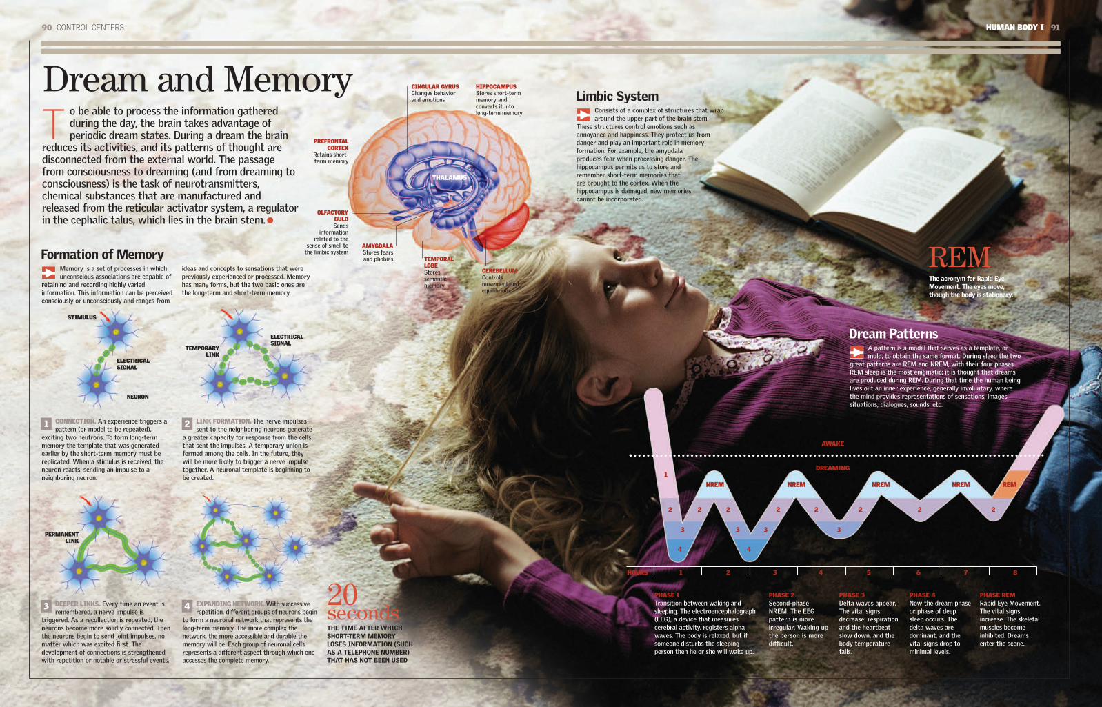
90 CONTROL CENTERS HUMAN BODY I 91
Dream and MemoryT
o be able to process the information gatheredduring the day, the brain takes advantage ofperiodic dream states. During a dream the brain
reduces its activities, and its patterns of thought aredisconnected from the external world. The passagefrom consciousness to dreaming (and from dreaming toconsciousness) is the task of neurotransmitters,chemical substances that are manufactured andreleased from the reticular activator system, a regulatorin the cephalic talus, which lies in the brain stem.
PHASE 2Second-phaseNREM. The EEGpattern is moreirregular. Waking upthe person is moredifficult.
PHASE 3Delta waves appear.The vital signsdecrease: respirationand the heartbeatslow down, and thebody temperaturefalls.
PHASE 4Now the dream phaseor phase of deepsleep occurs. Thedelta waves aredominant, and thevital signs drop tominimal levels.
PHASE REMRapid Eye Movement.The vital signsincrease. The skeletalmuscles becomeinhibited. Dreamsenter the scene.
A pattern is a model that serves as a template, ormold, to obtain the same format. During sleep the two
great patterns are REM and NREM, with their four phases.REM sleep is the most enigmatic; it is thought that dreamsare produced during REM. During that time the human beinglives out an inner experience, generally involuntary, wherethe mind provides representations of sensations, images,situations, dialogues, sounds, etc.
Limbic SystemConsists of a complex of structures that wraparound the upper part of the brain stem.
These structures control emotions such asannoyance and happiness. They protect us fromdanger and play an important role in memoryformation. For example, the amygdalaproduces fear when processing danger. Thehippocampus permits us to store andremember short-term memories thatare brought to the cortex. When thehippocampus is damaged, new memoriescannot be incorporated.
Formation of MemoryMemory is a set of processes in whichunconscious associations are capable of
retaining and recording highly variedinformation. This information can be perceivedconsciously or unconsciously and ranges from
ideas and concepts to sensations that werepreviously experienced or processed. Memoryhas many forms, but the two basic ones arethe long-term and short-term memory.
AWAKE
DREAMING
NREM NREM
1
2 2 2 2 2 2 2 2
3 3 3 3
4 4
NREM NREM REM
HOURS 1 2 3 4 5 6 7 8
CONNECTION. An experience triggers apattern (or model to be repeated),
exciting two neutrons. To form long-termmemory the template that was generatedearlier by the short-term memory must bereplicated. When a stimulus is received, theneuron reacts, sending an impulse to aneighboring neuron.
LINK FORMATION. The nerve impulsessent to the neighboring neurons generate
a greater capacity for response from the cellsthat sent the impulses. A temporary union isformed among the cells. In the future, theywill be more likely to trigger a nerve impulsetogether. A neuronal template is beginning tobe created.
DEEPER LINKS. Every time an event isremembered, a nerve impulse is
triggered. As a recollection is repeated, theneurons become more solidly connected. Thenthe neurons begin to send joint impulses, nomatter which was excited first. Thedevelopment of connections is strengthenedwith repetition or notable or stressful events.
EXPANDING NETWORK. With successiverepetition, different groups of neurons begin
to form a neuronal network that represents thelong-term memory. The more complex thenetwork, the more accessible and durable thememory will be. Each group of neuronal cellsrepresents a different aspect through which oneaccesses the complete memory.
THALAMUS
HIPPOCAMPUSStores short-termmemory andconverts it intolong-term memory
PREFRONTALCORTEX
Retains short-term memory
OLFACTORYBULBSends
informationrelated to the
sense of smell tothe limbic system
STIMULUS
NEURON
ELECTRICALSIGNAL
AMYGDALAStores fearsand phobias TEMPORAL
LOBEStoressemanticmemory
CEREBELLUMControlsmovement andequilibrium
CINGULAR GYRUSChanges behaviorand emotions
TEMPORARYLINK
ELECTRICALSIGNAL
PERMANENTLINK
21
43
THE TIME AFTER WHICHSHORT-TERM MEMORYLOSES INFORMATION (SUCHAS A TELEPHONE NUMBER)THAT HAS NOT BEEN USED
20seconds
The acronym for Rapid EyeMovement. The eyes move,though the body is stationary.
REM
Dream Patterns
PHASE 1Transition between waking andsleeping. The electroencephalograph(EEG), a device that measurescerebral activity, registers alphawaves. The body is relaxed, but ifsomeone disturbs the sleepingperson then he or she will wake up.

92 GLOSSARY HUMAN BODY I 93
Glossary
AcidSubstance that, in solution, increases theconcentration of hydrogen ions and combineswith bases to form salts.
AdrenalineHormone secreted primarily by the adrenalmedulla of the adrenal glands. It constrictsblood vessels and is used as a medicine.
AlleleGene variant that encodes a trait. One diploidcell contains one allele of each parent for eachcharacteristic.
Amino AcidOrganic chemical whose molecular compositionincludes an amino group (derived fromammonia) and a carboxyl group (a radical thatcharacterizes organic acids).
AntigenSubstance that causes an immune response,such as the production of antibodies, whenintroduced into the body.
AortaLargest artery in the body, originating in the leftventricle of the heart. Down to the diaphragm itis called the thoracic aorta and then theabdominal or ventral aorta to the point where itbranches.
Aortic ArchCurve in the aortic artery near its origin at theheart. The arch has the shape of a shepherd'scrook.
ApparatusComplex of organs that fulfills one function. Inthe physiology of the human body it is also usedas a synonym for system. For example, thedigestive apparatus, reproductive apparatus, orrespiratory apparatus.
ArteryBlood vessel that brings blood from the heart tothe entire body.
ArthroscopySurgical procedure used by orthopedic surgeonsto inspect, diagnose, and treat problems in thejoints. It consists of making a small incision andinserting an arthroscope, an instrument the sizeof a pencil that contains a small lens and alighting system to magnify and illuminate theinterior. The light is transmitted via fiber opticsto the end of the arthroscope, and the interior ofthe joint can be observed via a miniaturetelevision camera.
ArticulationJoint between two bones of the body.
ATPAdenosine triphosphate. A molecule producedprimarily by mitochondria that functions as theprimary energy source for the cells.
AtriumThe name for each of the two chambers of theheart that receive blood from the veins.
Basal MetabolismActivity level of the body functions during restor while fasting.
BonesRigid structures, rich in calcium, that make upthe skeleton.
CarpalThe structure of the wrist, composed of eightconnected bones arranged in two rows. On theside toward the arm it joins with the cubital andradial bones, and on the side toward the hand itjoins with the metacarpal bones.
Coronary ArteriesA pair of arteries, originating in the aorticartery, that branch out and supply blood to the heart.
CortexThe gray material present in most areas of thebrain. It is the largest part of the centralnervous system. The majority of the mostadvanced functions occur in the cortex.
CorticoidsHormonal steroids produced by the adrenalgland cortex. Corticoids can be producedartificially. They have a therapeutic applicationas anti-inflammatory drugs.
CystoscopeApparatus used to explore the inner surface ofthe bladder.
CytoplasmA compartment of eukaryotic cells, bounded bya cellular membrane and the membranes of thecell's organelles.
DiaphragmRespiratory muscle between the thorax and theabdomen.
DigestionThe set of processes through which thedigestive system converts food into substancesthat can be assimilated by the organism.
DiploidA cell with two complete sets of chromosomes.It is denoted by the symbol 2n.
DislocationThe displacement of any bone from its normalposition in a joint.
DNADeoxyribonucleic acid. A double helix moleculecontaining encoded genetic information.
EjaculationThe action of expelling semen.
EmbryoThe result of the fertilization of an ovum by asperm cell. It can develop to become a matureorganism.
Emulgent ArteriesArteries that bring blood from the heart to thekidneys, also called renal arteries.
EndocardiumMembrane that lines the walls of the heart. Itconsists of two layers: an exterior, consistingof connective tissue, and an interior, ofendothelial tissue.
EndometriumMucous membrane covering the inner walls ofthe uterus.
Endoplasmatic ReticulumNetwork of membranes in the cell that areinterconnected through the cytoplasm andwhose function is the synthesis and assemblyof proteins.
EndothelialOrganic tissue that lines wall-like structureswithin the body, such as those of the pleura orof blood vessels.
EnzymeProtein that helps regulate the chemicalprocesses within a cell.
ErythropoiesisThe creation of red blood cells, stimulated bythe action of a protein called erythropoietin.
FollicleInward fold of the epidermis in the form of asac, which usually surrounds the base of a hair.
GeneUnit of information of a chromosome; it is asequence of nucleotides in a DNA molecule thatfulfills a specific function.
GlandOrgan that has the function of producingsecretions that can be expelled through the skinor mucous membranes (salivary glands or sweatglands, for example) or into the bloodstream(the thyroid, for example).
HaploidFrom the Greek haplous, meaning single. Ahaploid cell has a single set of chromosomes,unlike the diploid cells. Gametes are haploid.
HemostaticSubstance or agent that halts hemorrhaging.
HippocampusPart of the brain that governs the memory.
HolocrineGland with an exclusively secretory function orwhose secretion consists of disintegrated cellsof the gland itself, such as the sebaceous glands.
HomeostasisComplex of self-regulatory phenomena that keepthe composition and the properties of the body'sinternal environment constant. It is said thathomeostasis is reached when the body's internalenvironment contains the optimum concentra-tions of gases, nutrients, ions, and water; when
CartilageFlexible skeletal tissue consisting of isolatedgroups of cells within a collagenous matrix.
Celiac ArteryArtery that brings blood from the heart to thestomach and the other organs of the abdomen.
Cellular MembraneThe flexible covering of all living cells, whichcontains the cytoplasm. It regulates theexchange of water and gases between the celland its exterior.
ChromatinComplex substance in the cell nucleus composedof nucleic acid and proteins.
CiliumTiny hairlike protuberance on a cell with alocomotive function in a liquid medium.
CoagulationOrganic process in which the blood turns from aliquid to a solid state and whose normal purposeis to stop bleeding.
CoccyxBone formed by the fusion of the last vertebrae.At its base it articulates with the sacral bone. Inhuman beings and other vertebrates that do nothave a tail, it is an actual bone.
CoronalA name given to the frontal bone, located at the anterior and superior part of thecranium. At birth the frontal bone or coronalis divided into two halves, which fuse overtime. In medicine this can also refer to asuture that joins the frontal bone with the twoparietal bones.

94 GLOSSARY HUMAN BODY I 95
its temperature is optimum; and when the vol-ume of fluids is optimum for the life of the cells.
HormoneThe product of the glandular secretion whosefunction is to stimulate, inhibit, or regulate theaction of other glands, systems, or organs ofthe body.
Innominate BonesA pair of bones, one in each hip, which join thesacrum and the coccyx to form the pelvis. Theyconsist of the fusion of the iliac, the ischium, andthe pubic bones.
LobesRounded protuberances of organs, such as theliver, the lungs, or the brain.
LysosomeProtein that can break down the constituentsubstances of the walls of certain bacteria andis, hence, a potent antibacterial.
MeiosisType of cell division in which two successivedivisions of the nucleus of a diploid cell createfour haploid nuclei. As a result of thismechanism, gametes or spores are produced.
MeristemTissue with cells that produce other cells bycellular division.
MetabolismComplex of chemical reactions that take placecontinuously within cells to synthesize complexsubstances from simpler substances or todegrade a substance into simpler substances.An example is the digestive process.
MetacarpalMiddle part of the skeletal structure of thehand, between the wrist (carpal bones) and thephalanges. It consists of five bones, which arethe largest bones of the hand.
MetatarsalPart of the skeletal structure of the foot,between the tarsus (posterior part of the foot)and the phalanges (toes). It consists of fivebones and is usually called the sole of the foot.
MicturitionAct of urinating, or expelling urine.
MitochondriaOrganelle that has a double membrane. Thefinal stage of the aerobic respiration processtakes place in mitochondria, where ATP isobtained by breaking down sugars and othersubstances.
MitosisNuclear division in a cell that forms daughternuclei identical to the parent.
Mucous MembraneCovering of body cavities that communicatewith the exterior (such as the nose). A mucousmembrane contains numerous single-celledglands that secrete mucus.
MusclesOrgans composed of fibers capable ofcontracting.
MyocardiumMuscular part of the heart, between thepericardium and the endocardium.
Nucleic AcidMolecule that carries genetic information aboutthe cell. There are two types: DNA and RNA.
NucleusThe part of the cell that contains the DNA withits genetic information.
OrganAny part of the body that accomplishes afunction.
amino acids, fundamental for the constitutionand functioning of living material, such asenzymes, hormones, and antibodies.
Ranine ArteryArtery that branches out toward the front ofthe tongue.
RespirationThe act and effect of inhaling air, primarilythrough the nose, to take in the substances thatthe body requires, such as oxygen, and afterprocessing them exhaling unneeded substances,such as carbon dioxide.
RibosomeOrganelle located in the cytoplasm that governsthe formation of proteins based on informationprovided by the nucleic acids.
RibsLong and curved bones. They originate at theback of the body at the spinal column and curveforward. They are called “true” if they end at thesternum and “false” if they remain floatingwithout completely enclosing the rib cage.
Schwann CellsCells that produce myelin, a fatty insulatingsubstance that prevents electrical signals fromlosing strength as they move away from thebody of the neuron.
SemenThe spermatozoa and fluids produced in themale genital organs. It is often called sperm.
SensationPhysiological process of receiving andrecognizing stimuli produced by vision, hearing,smell, taste, touch, or the body's spatialorientation.
SleepState of repose characterized by inactivity orsuspension of the senses and voluntary motion.
The cerebral activity called dreaming takesplace during sleep.
SpinalRelating to the spine.
Spinal BulbarPart of the cerebral trunk that goes from theannular protuberance to the cranium's occipitalforamen.
SpineThe neuroskeletal axis that runs along themedial dorsal of the body and consists of aseries of short bones called vertebrae, whichare arranged in a column and jointed witheach other.
SternumBone of the anterior thorax, which joins thefront of the ribs.
Striated MuscleMuscle used for voluntary motion. Its musclefibers show striations, or grooves.
Subclavian ArteriesPair of arteries, one of which branches off fromthe brachiocephalic trunk (on the right side ofthe body) and the other from the aortic arc (onthe left). They run toward the shoulder on eachside and, after passing below the clavicle,become the axillary artery.
SystemComplex of organs that participates in any ofthe principal functions of the body. A synonymof “apparatus.”
TarsalThe skeletal structure of the leg between thefoot and the metatarsal. It consists of sevenbones that constitute the posterior part of the foot.
TissueGroup of identical cells that togetheraccomplish a function.
UterusHollow viscera of the female reproductivesystem. It is located inside a woman's pelvis. Inthe uterus, or womb, either menstrual fluid isproduced or a fetus develops until it is born.
VeinsBlood vessels that bring blood from the entirebody toward the heart.
VentriclesCavities of the heart that receive blood fromtheir respective atrium (right or left) and pumpit through the arteries.
VisceraOrgans located in the principal cavities of thebody (such as the stomach or the liver withinthe abdominal cavity).
VitaminsOrganic substances present in food. The bodyingests them to ensure the balance of variousvital functions. There are different kinds ofvitamins, designated with the letters A, B, C, etc.
OsmosisMovement of a liquid through a selectivelypermeable membrane.
PapillaeConical protuberances, usually sensory, formedon the skin or mucous membranes (especiallythe tongue) by the branching of nerves andblood vessels.
PericardiumPair of membranes that surround the heart.
PhagocytesCells found in blood and tissue. They capturebacteria or any other kind of noxious particlesand “phagocytize,” or “eat,” them, absorbingthem into their cytoplasm and later digesting them.
PhalangesBones of the fingers and toes. They extend tothe metacarpal bones in the hand and themetatarsals in the foot. Starting from themetacarpals and the metatarsals, they aresequentially numbered: first, second, and thirdphalanges (of each finger or toe). The word“phalanges” commonly designates the firstphalanges, or each of the jointed parts of thefingers or toes.
PhysiologyStudy of the functions of the organism.
PolymerMacromolecule consisting of repeated structuralunits, called monomers.
PopliteusSection of the leg opposed to, or behind, the knee.
ProteinSubstance that makes up the cells. It is abiopolymer consisting of one or several chains of

96 INDEX HUMAN BODY I 97
Index
Aabducens nerve, 89ABO blood system, 40accessory nerve, 89Achilles tendon, 31actin filament, muscle fibers, 33active transport, 13Adam's apple, 46adipose cell, 55adrenal gland, 59, 61, 63adrenocorticotropin hormone (ACTH), 62afferent lymphatic vessel, 44agranulocyte, 41alveoli, 46, 48, 49amino acid, protein synthesis, 55amphiarthrose joint, 28amygdala, 90, 91anabolism, 55anaphase (cell division), 15anterior tibia, 31anti-diuretic hormone (ADH), 62antibody, 43antioxidant, 14aortic artery (aorta), 36, 39aortic valve, 39arm
bones, 20circulatory system, 36-37joints, 28movement, 32, 33muscles, 30
artery, 36-37, 38kidneys, 58, 61knees, 29lungs, 49
astrocyte, 85atlas bone, 26, 28axial bone, 21axis bone, 26, 28axon, 32, 84
Bbacteria, 44, 45basal joint, 28Bernard, Claude, 60bicep muscle, 30, 31bile, 54bladder, 58, 65blood
circulation, 16, 36-37components, 40-41glucose level regulation, 54, 55groups, 40oxygenation, 38, 40purification in kidneys, 60
bonecell types, 23cervical, 27development, 23fracture repair, 23function, 22lever function, 33skeleton, 20-21structure, 22-23types, 21See also joint
bone marrow, 22, 40, 43bone shaft, 22, 23Bowman capsule, 61brain, 86-87
astrocyte, 85communication, 79cranial nerves, 88-89cranium, 20dream states, 90, 91infant, 9limbic system, 91memory formation, 90, 91neurons, 8-9, 84-85olfactory receptors, 71taste center, 71weight, 9
brain cell, 8-9Broca's area, 79, 87
communication, 78, 79compact bone, 22connective tissue, perineurium, 32cornea, 74, 75cortisol, 62cranial nerve, 88-89cranium, 20
muscles, 33sinuses, 25
cubital nerve, 83cubitum, 20cytology, 12cytoplasm, 12, 13cytoskeleton, 12
DDa Vinci, Leonardo, 20deltoid muscle, 30dendrite, 8, 84diaphragm, 47, 48diaphysis (bone), 22, 23diarthrose joint, 28diastolic: See heartbeatdieting, muscle loss, 30diffusion (cell), 13digestive system, 17
digestive process, 51intestine: See intestineliver, 54-55overview, 50pancreas, 54, 55peristalsis, 53spleen, 42, 55stomach, 51, 52-53
disease, 45DNA, 13dorsal vertebra, 26dreaming, 90, 91
Eear, 76, 77
cranial nerves, 89efferent lymphatic vessel, 45ellipsoid joint, 28endocrine system, 17, 62-63
hypothalamus, 10, 86pancreas, 55See also hormone
endoplasmic reticulum, 12, 13enzyme
digestive process, 50pancreatic juice, 55
epididymis, 65epiglottis, 46epiphysis (bone), 23equilibrium, 77
cerebellum, 86esophagus, 50, 52estrogen, 62, 63
menstrual cycle, 66ethmoid bone, 24-25excrement, 10, 51eye, 74-75
brain, 79, 87cranial nerves, 88, 89muscles, 30sleep, 91
Fface
bones, 24-25cranial nerves, 88, 89muscles, 30, 31nonverbal communication, 79
facial nerve, 88facilitated diffusion (cell), 13fallopian tube, 66, 67farsightedness (hyperopia), 75fascicle, muscle fibers, 32
fat, storage, 55female
menopause, 67menstrual cycle, 66milk production, 62pelvis, 21reproductive system, 16, 67sexuality, 63skin, 73urinary system, 59
femur (thigh bone), 20, 21artery, 29vein, 37
fiber: See muscular fiberfibula, 21, 29finger, 20
See also handflat bone, 21follicle-stimulating hormone (FSH), 62, 66fontanel, 24food, 17
digestive process, 50-51, 52, 56-57source of water, 10
footarticulation, 28bones, 27movement, 33muscles, 31nerves, 83toenails, 73
foramen magnum, cranium, 24fracture, repair, 23free radical, 14frontal bone, 24-25frontal lobe (brain), 87, 89frontal muscle, 30frowning, 31, 79fusion, bones, 23
Ggallbladder, 54gastrocnemius, 31
bronchi, 47, 49bronchial tree, 48
Ccalcanum (calcaneus), 21, 27calcium, 11, 22, 23callus body, 87capillary, 36, 37, 49carbon, 11cardiac muscle, 30, 31
See also heartcarotid artery, 36carpal bone, 20cell
division: See mitosissize, 6-7structure, 12-13transport mechanisms, 13
cell theory, 12cellular membrane, 13central nervous system, 82, 87
brain: See brainspinal cord: See spinal cord
centriole, 13, 15cerebellum (brain), 82, 86, 89, 90cerebral cortex (brain) 9, 86-87, 88cervical vertebra, 26cervix (uterine), 67cheekbone (zygomatic bone), 24-25chemical element, contents of human body, 11chlorine, 11cholesterol, formation, 54chromosome, 14circulatory system, 16, 36-37
See also artery; heart; veinclavicle, 20clitoris, 67coccyx (tailbone), 21, 26, 27cochlea, 76, 77cochlear vestibular nerve, 89colon, 57color blindness, 75

98 INDEX HUMAN BODY I 99
glomerulus, 58, 61glosso-pharyngeal nerve, 89gluteus maximus, 31glycogen, storage, 54, 55Golgi apparatus, 12goose bump, 73granulocyte, 41gray matter (brain), 87growth hormone (GH), 62growth plate, 23gustatory papilla, 70
Hhair, temperature regulation, 73hand
bones, 20, 27fingernails, 73joints, 28nerves, 83touch, 9
Havers conduit, bony tissue, 22Hayflick, Leonard, 15Hayflick limit (cell longevity), 15head
bone structure, 24-25circulatory system, 36movement, 33muscles, 30
hearing, 76, 77heart, 36, 38-39
cardiac muscle, 31valves, 39
heartbeat, 38hematosis, 49herniated disc, 26hippocampus, 90, 91homeostasis, 17, 58Hooke, Robert, 12hormone, 17, 62
digestive process, 50menstrual cycle, 66See also endocrine system; pheromone
humerus, 20, 28hydrogen, 11hypermobility, 28hyperopia (farsightedness), 75hypoglossal nerve, 89hypophysis: See pituitary glandhypothalamus, 10, 86
Iilium, 20immune system, 43, 44
spleen, 55See also lymphatic system; white blood cell
infantbones, 23brain development, 8-9cranium, 24
inferior maxillary, 20, 25inferior vena cava, 36, 59insulin, 11, 55interphase (cell division), 14intestinal mucosa, 44intestine, 51, 56-57
duodenum, 52, 54iodine, 11iris, 74iron, 11irregular bone, 21
Jjaw bone, 20joint, 28-29
lever function, 33noise, 29
jugular vein, 36
Mmacrophage, 45-46magnesium, 11male
hormones, 62pelvis, 21reproductive system, 17, 64-65skin, 73urinary system, 59
master gland: See pituitary glandmelanocyte-stimulating hormone (MSH), 62memory formation, 90, 91men: See malemenarche, 67meninges, 86, 87meniscus, 29menopause, 67menstrual cycle, 66metabolism, 55metacarpal bone, 20metaphase (cell division), 14metatarsal bone, 21milk, production, 62mitochondria, 13mitosis, 6-7, 12, 14-15mitral valve, 39mouth
digestive function, 50, 51sound production, 78swallowing, 52
mucous secretion, 44muscle
function: See muscular fibermovement, 30, 87types, 31
muscular fiber, 32-33glycogen storage, 55
muscular system, 17, 30-31musculoskeletal system, 18-19, 30
See also muscular system; skeletal systemmyelin sheath, 84
oligodendrocytes, 85myofibril, muscle fibers, 33
myopia (nearsightedness), 75myosin filament, muscle fibers, 33
Nnails, 73nasal concha, 24-25nasal fossa,
olfactory nerve, 71sensations, 9
nearsightedness (myopia), 75neck, bones, 28nephron, 60, 61nervous system, 16, 82-83
brain: See brainneuron: See neuronpain signals, 83spinal column, 20, 26-27spinal cord, 87
neuromuscular union, 85neuron, 8-9, 84-85, 86
dendrites, 8, 84microscope photograph, 80-81
neurotransmitter, 9, 85nitrogen, 11nonverbal communication, 78, 79nose
bones, 24-25cranial nerves, 88, 89nasal fossa, 9, 71olfactory cells, 70sound production, 78
NREM (non-rapid eye movement) sleep, 91nucleole, 13nucleus, 12, 13
Ooblique muscle, 30occipital bone, 20, 24-25occipital lobe (brain), 86
occipital muscle, 30oculomotor nerve, 89olfactory cell, 70olfactory nerve, 71, 89oligodendrocyte, 85optic nerve, 88orbicular muscle, 30organ of Corti, 76osteoblast, 23osteoclast, 23ovary, 66, 67ovulation, 66ovum, 66, 67oxygen, 11, 40oxytocin, 62, 63
Ppain signal, 83palatine, 24-25pancreas, 11, 54, 55, 63parietal bone, 24patella (kneecap), 21, 29pathogen, types, 45pectoralis major, 30pedis, 31pelvis, 20, 67
joint, 21penis, 59, 64, 65periosteum, 22, 23peripheral nervous system, 82, 88-89peristalsis, digestive system, 51, 53peroxisome (organelle), 13perspiration: See sweat; sweat glandPeyer's patch, 42phalange, 20, 21pharynx, 47, 50pheromone, 63phosphorus, 11photosensitive cell, 75pituitary gland, hormones, 62, 63plane, 28plasma, 41
Kkidney, 58, 59, 60-61
Bowman capsule, 61nephrons, 61renal vein, 36
kissing, hormone stimulation, 63knee
articulation, 28joint, 29
kneecap (patella), 21, 29
Llachrymal bone, 24-25lachrymal gland, 44larynx, 46, 47leg
bones, 21circulatory system, 36-37knee: See kneemuscles, 31nerves, 83
lens (eye), 74Leonardo da Vinci, 20leukocyte, 41ligament, knee, 29limbic system, 91liver, 54-55long bone, 21lumbar vertebra, 27lung, 47, 48-49
circulatory system, 36, 38luteinizing hormone (LH), 62, 63, 66lymphatic system, 16, 42-43
lymph nodes, 44-45lymphocytes, 45
lysosome, 12

100 INDEX
plasma membrane: See cellular membraneplatelet, 41popliteal artery, 29pore (cell), 13potassium (K), 11pregnancy, 66, 67progesterone, menstrual cycle, 66prolactin, 62prophase (cell division), 14prostate gland, 65protein, 11
metabolism, 12, 54synthesis, 55
protozoa, pathogens, 45pulmonary artery, 48pulmonary valve, 39pylorus, 52
Q-Rquadriceps, 31radius, 20, 28Ranvier's node, 84rapid eye movement (REM) sleep, 91rectus abdominis, 30red blood cell, 40REM sleep, 91renal vein, 36reproductive system
female, 16, 66-67hormones, 62male, 17, 64-65
respiration, 9, 46process, 46-47, 48
respiratory system, 17, 46-47See also lung
retina, 74, 75Rh factor, 40rib cage, 20, 26ribosome, 12rough endoplasmic reticulum, 12
Ssacroiliac joint, 21sacrum, 20, 26, 27salivary gland, 44, 70salt: See sodiumsarcomere, muscle fibers, 33Schleiden, Mathias, 12Schwann, Theodor, 12Schwann cell, 84sciatic nerve, 83sclera, 75sebaceous gland, 44, 73sensation: See hearing; smell; taste; touch;
visionseptum, 39sesamoid bone, 21sexual attraction, 63short bone, 21shoulder, articulation, 28sight, 74-75sinus cavity, 25skeletal system (skeleton), 16
structure, 20-21See also joint; musculoskeletal system
skin, 9, 44, 72-73, 87cellular division, 14melanocyte production, 62wound healing, 45
sleep, 91smell, 70, 71, 90smooth endoplasmic reticulum, 13smooth muscle, 30, 31sodium, 11speech, 78, 87speech recognition technology, 24spermatozoa, 64sphenoid bone, 24-25spheroid, 28spinal column, 20, 26-27spinal cord, 26, 82, 87, 88spinal medulla, 87spinal nerve, 88spleen, 42, 43, 55
splenius muscle, 30spongy bone, 22Starling, Ernest, 62sternocleidomastoid muscle, 30sternum, 20, 26stomach, 51, 52-53striated muscle, 17, 30, 31subclavian vein, 42sugar, regulation in blood, 11sulfur, 11superior maxillary, 24-25superior vena cava, 36, 39swallowing, 52sweat, 10, 73sweat gland, 44synapse, 8, 85synaptic node, 84synarthrose joint, 28systole: See heartbeat
TT cell, 45
See also lymphatic systemtailbone (coccyx), 21, 26, 27Takagi, Kenji, 29tarsal bone, 21taste, 70, 71
types, 9technology, speech recognition, 24teeth, structure, 50telophase (cell division), 15temperature regulation, 73temporal artery, 36temporal bone, 24-25temporal lobe (brain), 86, 89, 90temporal vein, 36tendinous cord, 39testicle, 64, 65testosterone, 62, 63, 65thalamus, 86thigh bone: See femurthirst, control, 10
thoracic vertebra, 26thumb, joints, 28thymus, 42, 43thyroid-stimulating hormone (TSH), 62tibia, 21, 29toe, nails, 73tongue
functions, 50, 51gustatory papillae, 70, 71nervous system, 88sensations, 9sound production, 78
tonsils, 42touch, 9, 72-73trachea, 46, 47, 49transport mechanism (cell), 13trapezium muscle, 30tricep muscle, 30tricuspid valve, 39trigeminal nerve, 71, 88trochlear nerve, 89
Uureter, 58, 59urethra, 58urinary system, 17, 58-59
gender differences, 59kidneys, 60-61
urine, 10, 58, 59, 64Bowman capsule, 61production, 60
uterus, 66, 67
Vvacuole, 13vagina, 66, 67
bacteria, 44vagus nerve, 82, 89
valveheart, 39lymphatic system, 45
vein, 36-37inferior vena cava, 36, 59kidneys, 58, 61lymphatic system, 42superior vena cava, 36, 39
vertebral column: See spinal columnvesicle, 13villi (intestine), 57virus, pathogens, 45vision, 74-75, 87vocal cord, 46, 78
See also speechvomer (bone), 24-25vulva, 66
W-Zwater
fluid exchange, 10, 59intake, 10intestines, 56
Wernicke's area (brain), 79white blood cell, 41, 45white matter (brain), 87women: See femalewound healing, 45Z band, muscle fibers, 33zygomatic bone (cheekbone), 24-25
HUMAN BODY I 101

![Human Body Exergy Balance: Numerical Analysis of an Indoor ... · Human body system [1]. A human body energy balance model, or twonode model, - was used for the human body system“](https://static.fdocuments.us/doc/165x107/6000b436c5a9c34ccd5461ab/human-body-exergy-balance-numerical-analysis-of-an-indoor-human-body-system.jpg)