How Does MRI Form A Picture? Application of MRI Field
16
1/10/2014 1 “Lumps and Bumps” of the Lower Extremity/MRI characterization Mark I. Robbins, M.D. American Radiologic Technologies Musculoskeletal Radiology Rye, New Hamshire Utility of MR In Musculoskeletal Imaging • Noninvasive • Multiplanar capabilities • No ionizing radiation • High sensitivity with excellent spatial resolution=early detection How Does MRI Form A Picture? • Fat and Water in the Human body have an abundance of protons • They resonate in a random frequency and orientation Application of MRI Field • When A High Strength Magnetic Field is applied, these protons align with it in proportion to Bo (the field Strength of the Magnet) Energy Released to Create Picture • RF Energy is release as the proton goes from high energy state to low (after our applied pulse stops) • TR is when pulses applied • TE is when you listen for the radio signal A Paradox • "Simplicity, simplicity, simplicity! I say, let your affairs be as two or three, and not a hundred or a thousand; instead of a million count half a dozen, and keep your accounts on your thumbnail." HD Thoreau, Walden, "Where I Lived and What I Lived For" (1854) • “Simplify, but don’t oversimplify” A. Einstein • For today, we’re going with the Concordian
Transcript of How Does MRI Form A Picture? Application of MRI Field

1/10/2014
1
“Lumps and Bumps” of the Lower
Extremity/MRI characterization
Mark I. Robbins, M.D.
American Radiologic Technologies
Musculoskeletal Radiology
Rye, New Hamshire
Utility of MR
In Musculoskeletal Imaging • Noninvasive
• Multiplanar
capabilities
• No ionizing radiation
• High sensitivity with
excellent spatial
resolution=early
detection
How Does MRI Form A Picture?
• Fat and Water in the
Human body have an
abundance of
protons
• They resonate in a
random frequency
and orientation
Application of MRI Field
• When A High
Strength Magnetic
Field is applied, these
protons align with it
in proportion to Bo
(the field Strength of
the Magnet)
Energy Released to Create Picture
• RF Energy is release
as the proton goes
from high energy
state to low (after our
applied pulse stops)
• TR is when pulses
applied
• TE is when you listen
for the radio signal
A Paradox
• "Simplicity, simplicity, simplicity! I say, let
your affairs be as two or three, and not a
hundred or a thousand; instead of a million
count half a dozen, and keep your accounts on
your thumbnail." HD Thoreau, Walden,
"Where I Lived and What I Lived For" (1854)
• “Simplify, but don’t oversimplify” A. Einstein
• For today, we’re going with the Concordian

1/10/2014
2
All Three Planes Illustrative Cases
Gradient Echo Images
• Shows susceptibility
(blooming, foreign
bodies, hemosiderin)
• Demonstrates
hyaline cartilage
• Less imaging time
(no refocusing)
Gradient Echo Images
Identifying Foreign Bodies
6 year-old stepped on a nail Vascular Magnetic Resonance
Angiogram (MRA)

1/10/2014
3
Gradient Echo Flow Related
Enhancement
Gradient Echo Vascular Flow
Related Enhancement
T1 Weighted Images
• Short TE/TR
• Fat is hyperintense (bright)
• Fatty marrow is bright
• Subcutaneous tissue
• Fibrocartilage and cortical bone is dark
• Fluid is dark
Things That Are T1 Hyperintense
• Fat
• Blood
(methemoglobin)
• Proteinaceous fluid
• Melanin
• Paramagnetic
compounds
• 30% calcium
Quadriceps Muscle Traumatic
Hematoma Lipoma
• Follows fat on all
sequences
• May have a capsule,
central fat necrosis,
or calcification
• Pelvis and
retroperitoneal
liposarcomas

1/10/2014
4
72 year-old-male, intermetatarsal
mass, gout, and negative aspirate Liposarcoma Hallux
54 year-old male; first interspace
swelling (companion case) Lipohemarthrosis
Fat Globules within a
Seroma”Degloving injury Morel-Lavallee syndrome
• Tangential trauma to vascularized tissue
• Can be associated with fractures
• Skin and subcutis sheared from facia : fusiform, ovoid or
crescentic hematolymphatic collection, debris
• Closed internal injury resulting in degloving injury, usually
presents acutely, but can be delayed
• Lymphatics are torn, slow to heal
• Development of capsule often requires percutaneous
drainage or evacuation
• Can mimic tumor; MRI useful

1/10/2014
5
Lipoma Arborescens
• Rare synovial
proliferation of
frondlike fatty tissue
• Swelling, effusion
40 year old male with hindfoot pain
while weight-bearing
Intraosseous Lipoma of Calcaneus
• 15% of intraosseous lipomas
• Same location as unicameral bone cysts
• Asymptomatic or variably painful
• Dystrophic calcification
• Differentiation from normal trabecular
rarefaction
• Low attenuation CT or fat signal on MRI
Exuberant Callus Formation
Motion about Fracture Site
Entities that can Have Low Signal
• Fibromas
• Calcified lesions
• Synovitis
• Old blood products like hemosiderin
(PVNS)
• High velocity blood flow (flow voids)
57 year old male with enlarging
anterior mass; rule out a ganglion

1/10/2014
6
Rheumatoid Nodules
2020--30% of RA30% of RA
Long standing Long standing dzdz
RF positiveRF positive
Increase incidence w/ Increase incidence w/ MethtrexateMethtrexate
Usually superficial, Usually superficial, bursa, tendon, joints, bursa, tendon, joints, or ligamentsor ligaments
Often poorly defined Often poorly defined mass low/mass low/isoiso muscle muscle T1, low or bright T2, T1, low or bright T2, variable enhancement.variable enhancement.
Rheumatoid Nodule
• Plantar forefoot
• Can be associated
with RA
Rheumatoid nodule Plantar fibromatosis
• Low signal on T1 and T2
• Enhances
• Conforms to or is exophytic from any cord of the plantar aponeurosis
• Localized or diffuse infiltrative forms
Large Plantar Fibromata Aggressive Fibromatosis
30 year-old female; 5 year history

1/10/2014
7
Calcifying Aponeurotic Fibroma
11 Yr. old Female: enlarging mass Calcifying Aponeurotic Fibroma
• Rare, locally aggressive fibroblastic lesion located in hands and soles of feet in young
• 50% recurrence post resection
• Juvenile aponeurotic fibroma, Keasby 1953
• Male 2:1 female
• Macroscopic: Rubbery gray white mass, adherent to tendons and fascia; gritty (calcium)
Calcifying aponeurotic fibroma
• Spindled fibroblasts bordering chondroid
foci with calcification and cartilage more
prominent in older children
• Differential diagnosis: rheumatoid nodule,
chondroma, fibrous hamartoma of infancy,
schwannoma, fibromatosis
Dermatofibroma Protuberans
Morton’s Neuroma
• Low signal
intermetatarsal mass
• Perineural fibrosis; not a
malignant tumor
• Low T2, enhances
• DDx:Intermetatarsal
bursitis, giant cell tumor
of the tendon sheath,
tophus, callous
Morton’s Neuroma

1/10/2014
8
Morton’s Neuromas
Mixed literature correlating size Mixed literature correlating size
(>5mm transverse) with improved (>5mm transverse) with improved
outcome post op. outcome post op.
High prevalence in asymptomatic feetHigh prevalence in asymptomatic feet
–– Two studies up to 33% asymptomaticTwo studies up to 33% asymptomatic
–– Smaller more frequently asymptomaticSmaller more frequently asymptomatic
Morton’s neuroma/bursitis alcohol
injection
Rheumatoid Nodule Simulating a
Morton’s Neuroma Gout/Tophi
62 year-old male from Georgia with
bilateral swelling
Gout with Erosion; Improved with
Colchicine

1/10/2014
9
Large Tophus with Classic
Erosions of Gout Achilles Xanthoma
Post-operative Achilles Tendon
75 y-old with trauma; intact repair Postoperative Achilles
Low Signal Achilles Masses/Lumpy
Bumpy Disease
• Chronic Achilles Tendinosis without
cavitary changes or large areas of mucoid
degeneration
• Repaired Achilles tendon
• Deposition diseases: gout, amyloid,
xanthomas
Giant Cell Tumor of the Tendon
Sheath
• Extra-articular form
of PVNS
• Mimics Mortons
neuromas, fibromas,
callous from forefoot
overload
• Lack of calcification
may distinguish from
a synovial sarcoma
1/10/2014
10
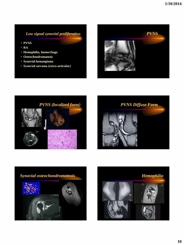
Low signal synovial proliferation
• PVNS
• RA
• Hemophilia, hemorrhage
• Osteochondromatosis
• Synovial hemangioma
• Synovial sarcoma (extra-articular)
PVNS
PVNS (localized form) PVNS Diffuse Form
Synovial osteochondromatosis Hemophilia

1/10/2014
11
Synovial Hemangioma
• Flow voids
• Phleboltihs
• Soft tissue signs
50 year-old female with a mass post
bunionectomy/osteotomy
Anterior tibial pseudo aneurysm
• High velocity flow void (black hole)
producing low signal
• Pulsation “ghosting” (phase encode)
artifact
• Surrounding blood products
• Proximity to an artery
Chronic ATAF Ligament Tear
Meniscoid Lesion
T2 weighted images T2-Weighted Images
• Fluid is hyperintense
• Marrow edema
• Most tumors
• Infiltrative process
• Marrow expansion
• Bursitis, synovial
cysts, ganglia
• CSF

1/10/2014
12
Intermetatarsal Bursitis Anatomic Bursitis First MPJ
Anatomic Bursitis Glomus tumor
• Painful subungual
mass
• Temperature
sensitive digit
• Exhibits signal that
mimics a ganglion or
cyst
Tenosynovitis Ganglia
1/10/2014
13
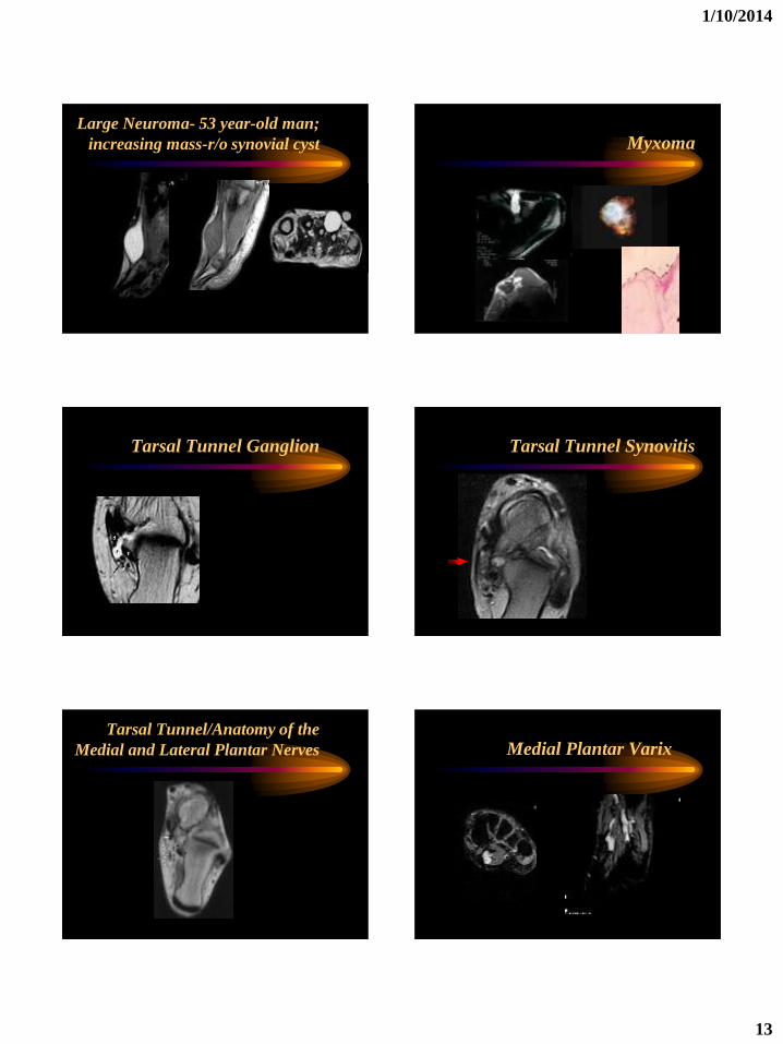
Large Neuroma- 53 year-old man;
increasing mass-r/o synovial cyst Myxoma
Tarsal Tunnel Ganglion Tarsal Tunnel Synovitis
Tarsal Tunnel/Anatomy of the
Medial and Lateral Plantar Nerves Medial Plantar Varix
1/10/2014
14
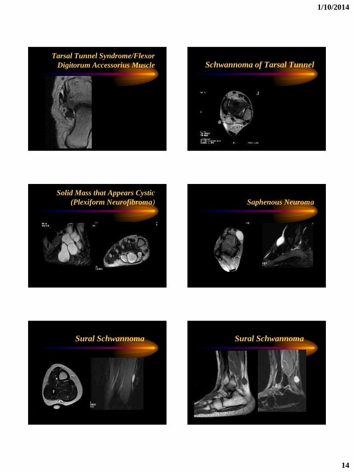
Tarsal Tunnel Syndrome/Flexor
Digitorum Accessorius Muscle Schwannoma of Tarsal Tunnel
Solid Mass that Appears Cystic
(Plexiform Neurofibroma)
Saphenous Neuroma
Sural Schwannoma Sural Schwannoma

1/10/2014
15
Plantar Neuroma 10 year-old with a palpable tibial
mass
• Differential diagnosis: Primary bone
neoplasm such as ABC, exostosis,
unicameral bone cysts, non ossifying
fibroma, Ewings, EG, osteosarcoma
• Soft tissue mass: Myositis ossificans,
granuloma, neurofibroma, synovial cyst
or ganglion
Osteochondroma
• Medullary continuity
• Points away from joint
• T2 bright cartilage cap
• Complications
• Bursitis exostocia,
fracture, pain, malignant
degeneration (cartilage
cap of > 2cm)
20 year-old male with palpable
plantar mass
Soft Tissue Mass/Synovial Sarcoma Rare Intramuscular Metastasis

1/10/2014
16
Gadolinium
• Enhance solid
tumors
• Shortens the T1 of
enhancing tissue
• Rim enhancement of
abscesses, ganglia,
synovial cysts,
myxomas, and other
fluid structures
Schwannoma
Ganglion, myxoma
SUMMARY
• MRI can characterize soft tissue masses based
on properties of the tissue.
• Useful to assess involvement of bone, tendons,
and neurovascular structures
• The exact histology of many lesion cannot be
predicted by imaging features.
• Management of any palpable mass should be
based on the clinical presentation.
THANKS!