MICE VC116 Alain Blondel 18 December 2008 1 MICE NEWS See more details in Macolm’s MICE news!

Hippo-Foxa2 signaling pathway plays a role inperipheral lung maturation and surfactant homeostasisChaeuk Chunga,b,c, Tackhoon Kimb, Miju Kimb, Minchul Kimb, Hoogeun Songb, Tae-Shin Kimb, Eunjeong Seob,Sang-Hee Leea, Hanbyul Kima,b, Sang Kyum Kima,b, Geon Yood, Da-Hye Leeb, Deog-Su Hwangd, Tatsuo Kinashie,Jin-Man Kimf, and Dae-Sik Limb,1
bDepartment of Biological Sciences, National Creative Research Initiatives Center, Biomedical Research Center, aGraduate School of Medical Scienceand Engineering, Korea Advanced Institute of Science and Technology, Daejeon 305-701, Korea; cDepartment of Pulmonary and Critical Care Medicine,and fDepartment of Pathology, Chungnam National University School of Medicine, Daejeon 301-721, Korea; dDepartment of Biological Sciences, SeoulNational University, Seoul 151-742, Korea; and eDepartment of Molecular Genetics, Institute of Biomedical Science, Kansai Medical University, Osaka 570-8506, Japan
Edited by Melanie H. Cobb, University of Texas Southwestern Medical Center, Dallas, TX, and approved April 1, 2013 (received for review November 27, 2012)
Respiratory distress syndrome (RDS), which is induced by insufficientproduction of surfactant, is the leading cause ofmortality in pretermbabies. Although several transcription factors are known to beinvolved in surfactant protein expression, themolecular mechanismsand signaling pathways upstreamof these transcription factors haveremained elusive. Here, using mammalian Hippo kinases (Mst1/2,mammalian sterile 20-like kinase 1/2) conditional knockout mice, wedemonstrate that Mst1/2 kinases are critical for orchestration oftranscription factors involved in surfactant protein homeostasis andprevention of RDS.Mice lackingMst1/2 in the respiratory epitheliumexhibited perinatal mortality with respiratory failure and their lungscontained fewer type I pneumocytes and more immature type IIpneumocytes lacking microvilli, lamellar bodies, and surfactant pro-tein expression, pointing to peripheral lung immaturity and RDS. Incontrast topreviousfindingsofYAP (Yes-associatedprotein)-mediatedcanonical Hippo signaling in the liver and intestine, loss of Mst1/2kinases induced the defects in pneumocyte differentiation in-dependently of YAP hyperactivity. We instead found that Mst1/2kinases stabilized and phosphorylated the transcription factorFoxa2 (forkhead box A2), which regulates pneumocyte maturationand surfactant protein expression. Taken together, our resultssuggest that the mammalian Hippo kinases play crucial roles insurfactant homeostasis and coordination of peripheral lung differ-entiation through regulation of Foxa2 rather than of YAP.
non-canonical Hippo pathway | lung development
Peripheral lung immaturity and deficiency of pulmonary sur-factant cause many intractable pulmonary diseases. Deficit of
surfactant because of preterm birth or genetic disorders of surfac-tant homeostasis induce respiratory distress syndrome (RDS) in thenewborn period (1). Dysregulation of these genes also underlies thepathogenesis of many chronic lung diseases that have been con-sidered idiopathic, such as interstitial lung disease, pulmonary al-veolar proteinosis, and others (2). Elucidating the mechanismunderlying regulation of surfactant would be very helpful for un-derstanding the molecular basis of refractory lung diseases of bothinfants and adults.Alveoli are composed of type I and type II pneumocytes. The type
II pneumocyte is the progenitor cell of the type I pneumocyte andplays a central role in physiological pulmonary functions, especiallysurfactant production (3). Type I pneumocytes contact alveolarcapillaries and participate in gas exchange. Lung morphogenesis inmice is a dynamic process that can be divided into embryonic[embryonic day (E) 9–11.5], pseudoglandular (E11.5–16.5), cana-licular (E16.5–17.5), saccular [E17.5 to postnatal (PN) day 5], andalveolar (PN5–28) stages. During canalicular and saccular stages inlate gestation, peripheral lungs containing type I and II pneumo-cytes fully differentiate in preparation for postnatal respiration (4).Many transcription factors, including Foxa2 (forkhead box A2),
TTF-1 (thyroid transcription factor-1)/Nkx2.1 (NK2 homeobox 1),and C/EBPα (CCAAT/enhancer binding protein-α), are known to
be involved in surfactant production and homeostasis during lategestation (1, 4–7). Mice containing a mutation of TTF-1 or a de-letion of Foxa2 or C/EBPα display immature type II pneumocytesand lack type I pneumocytes without an alteration in branchingmorphogenesis, resulting in RDS (8, 9). Foxa2, TTF-1, and C/EBPαreciprocally interact to regulate transcriptional targets of surfac-tant synthesis and peripheral lung maturation (5). Some upstreamregulators, such as Nfatc3 (nuclear factor of activated T cells,cytoplasmic 3) and protein kinase A were identified (10, 11), butso far the mechanism regulating peripheral lung maturation andsurfactant homeostasis are poorly understood and complex.Mammalian sterile 20-like kinase 1 and 2 (Mst1/2) are upstream
kinases involved in the Hippo pathway, the unique signaling path-way involved in organ development and cancer (12). Interestingly,Mst1/2- and 45kD WW domain protein (WW45)-knockout miceshow a tendency to accumulate undifferentiated progenitor cells(13, 14). For example, liver-specific ablation of Mst1/2 causes anincrease in the number of hepatocytes and facultative progenitorcells (oval cells) (15). Intestine-specific ablation of Mst1/2 alsoresults in intestinal progenitor cell proliferation and subsequentcolonic tumorigenesis (16, 17). The phenotypes of these organsare attributed to hyperactivity of Yes-associated protein (YAP),an oncogenic downstream effector molecule of the Hippo path-way. These studies suggest the interesting possibility that Yap-mediated Hippo signaling pathway, the so-called canonical Hippopathway, works as critical inhibitor of epithelial progenitor cellproliferation in epithelial organs. Whether this paradigm is ap-plicable to other epithelial organs is not yet clear. Recent studieshave also demonstrated the role of Mst1/2 kinases in a YAP-independent mechanism, the so-called noncanonical Hippo path-way. For example, Mst1 suppresses lymphoma development bypromoting faithful chromosome segregation, independently ofYAP (18). Mst1/2 kinases also execute a variety of functions withmultiple partners, such as Foxo1, Foxo3, Akt, and Aurora B, indifferent biological contexts (19–22).We have now generated conditional double-knockout mice
(dKO) in whichMst1 andMst2were deleted in the developing lungepithelium. Our results revealed that Mst1/2 acts through modu-lation of Foxa2 to play a critical role in the regulation of peripherallungmaturation and surfactant homeostasis with use of thesemice.
Author contributions: C.C., T. Kim, Minchul Kim, H.S., T.-S.K., E.S., and D.-S.L. designedresearch; C.C., Miju Kim, H.S., T.-S.K., H.K., and D.-H.L. performed research; D.S.H.,T. Kinashi, and J.-M.K. contributed new reagents/analytic tools; C.C., T. Kim, MinchulKim, E.S., S.-H.L., S.K.K., and J.-M.K. analyzed data; and C.C., T. Kim, G.Y., and D.-S.L.wrote the paper.
The authors declare no conflict of interest.
This article is a PNAS Direct Submission.
Freely available online through the PNAS open access option.1To whom correspondence should be addressed. E-mail: [email protected].
This article contains supporting information online at www.pnas.org/lookup/suppl/doi:10.1073/pnas.1220603110/-/DCSupplemental.
7732–7737 | PNAS | May 7, 2013 | vol. 110 | no. 19 www.pnas.org/cgi/doi/10.1073/pnas.1220603110

ResultsLung-Specific Mst1/2-dKO Mice Succumb to RDS and Die in thePerinatal Period. Mst1/2-null mice lacking both Mst1 and Mst2genes begin dying in utero at embryonic day 8.5 because of defectsin placental development and yolk sac/embryonic vascular pat-terning (21). To investigate the role ofMst1/2 in lung development,we crossedMst1fl/fl;Mst2−/−mice withNkx2.1-Cre mice to generatethe lung-specific Mst1/2-knockout mouse, Mst1fl/fl;Mst2−/−;Nkx2.1-Cre, hereafter termedMst1/2-dKO.Mst2−/−mice did not show anynoticeable developmental or immunological defects (15). Weconfirmed the efficient deletion of Mst1 in the Mst2-null lung tis-sues (Fig. S1 A and B). LacZ staining showed that Nkx2.1 Cre wasexpressed in lung epithelial cells (Fig. S1C).Mst1/2-dKOmicewereborn at normal Mendelian ratio. Lung size and dry lung weightwere comparable between Mst1/2 dKO mice and control mice atE15.5 and E18.5 (Fig. 1A and Fig. S2). However,Mst1/2-dKOmiceexhibited a markedly sick appearance at birth and almost all micedied within the first 15 d after birth (Fig. S1D). At PN10, Mst1/2-dKO lung showed severe accumulation of inflammatory cellsin the alveolar space and bronchiole with multiple atelectasis and
emphysematous lesions (Fig. 1C and Fig. S3A), suggesting theMst1/2-dKO mice died because of respiratory failure.We then determined the onset of aberrant phenotype fromE15.5
to E18.5. Until E17.5, there were no significant differences betweenthe lungs of Mst1/2-dKO mice and control mice (Fig. 1B). At thattime, embryonic markers such as Sox9 (sex-determining region Ybox 9, a marker for distal lung progenitor cells) and Sox2 (markerfor proximal lung progenitor cells) were also normally expressed inMst1/2 dKO lungs (23) (Fig.1D, and Fig. S4 A and B). However,alveoli of Mst1/2-dKO lungs at E18.5 and PN1 showed a re-markably reduced aerated space compactedwith immature-lookingcuboidal cells (Fig. 1 B and C). At around PN5,Mst1/2-dKO lungsdisplayed spontaneous infiltration of inflammatory cells and hya-line membrane formation, which are characteristics typical of RDSin preterm infants (1), at which point the destruction of lung be-came aggravated (Fig. 1C). No bacteria and fungi were detected inlung sections or in cytospins of bronchoalveolar lavage fluid, rulingout the possibility of infection (Fig. 1C and Fig. S3B).We determined whether Mst1 and Mst2 affect cell proliferation
and apoptosis by performing BrdU labeling and TUNEL assays,
Fig. 1. Mst1fl/fl;Mst2−/−;Nkx2.1-Cre mice exhibit perinatal mortality with RDS. (A) Representative lungs of E15.5, E18.5, PN1, and PN10 of control and Mst1/2-dKO mice. (Scale bar, 0.5 cm.) (B) Lung section from E15.5, E16.5, E17.5, and E18.5 of control and Mst1/2-dKO mice were stained with H&E, revealing thatbranching morphogenesis is normal in Mst1/2-dKO mice. (C) Lung sections from PN1, PN5, and PN10 mice were fixed and stained with H&E. (D) Evaluation ofcell proliferation and apoptosis by immunodetection of Brdu and TUNEL assay, respectively, in sections of control and Mst1/2-dKO mice at E17.5. Distalprogenitor marker (Sox9) and proximal progenitor marker (Sox2) were costained. (E) Ki67 and epithelial cell marker (E-cadherin) were costained to reveal theproliferation rates of epithelial cell and nonepithelial cell. TUNEL staining was performed to evaluate the apoptosis at E18.5. Mean ± SD was determined fromthree embryos per group. *P < 0.05. (Scale bars in B–E, 50 μm.)
Chung et al. PNAS | May 7, 2013 | vol. 110 | no. 19 | 7733
DEV
ELOPM
ENTA
LBIOLO
GY

respectively, at E17.5. Proliferation and cell death in the Mst1/2-dKO mice lungs did not appeared significantly altered until E17.5(Fig. 1D and Fig. S4C). The percentages of proliferating pro-genitor cells expressing Sox2 or Sox9 were similar between controland Mst1/2-dKO mice at E17.5 (Fig. 1D and Fig. S4D). However,E18.5Mst1/2-dKO lungs showed a markedly increased number ofproliferative cells and mildly diminished cell death (Fig. 1E andFig. S4E). To confirm the increase of epithelial cell specific pro-liferation at E18.5, we costained the lung sections with Ki67 and E-cadherin (E-Cad). The percentage of Ki67+ cells among E-Cad+
epithelial cells was higher in Mst1/2 dKO versus control lungs atE18.5. In contrast, the percentage of Ki67+ cells among E-Cad−
mesenchymal cells was similar in Mst1/2 dKO and control lungs(Fig. 1E). These observations suggest that the lung phenotype ofMst1/2 dKO mice is a result of the cell-autonomous effects ofMst1/2 deletion.
Deletion of Mst1/2 in Lung Epithelial Cells Causes Abnormal LungMaturation: Impaired Surfactant Homeostasis and Delayed Differen-tiation of Type I and II Pneumocytes. Given that deregulated matu-ration of the lung gives rise to perinatal lethality andRDS (1), whichwere observed inMst1/2-dKO mice (Fig. 1 B and C), we examineddifferentiation markers to assess the degree of lung maturation.Normal maturation was observed in control lungs, which showedwell-differentiated type II cells expressing the markers surfactantprotein-B (SP-B) and prosurfactant protein-C (proSP-C), and typeI cells, which were stained by T1α protein (Fig. 2A). In contrast,alveolar sacs in Mst1/2-dKO lungs at E18.5 were compacted withcuboidal cells lacking SP-B and proSP-C expression (Fig. 2A).These Mst1/2-dKO cuboidal cells seemed to be immature type IIpneumocytes that did not fully mature to produce surfactant
proteins normally. Staining for T1α demonstrated decreasednumbers of squamous type I cells in Mst1/2-dKO lungs (Fig. 2A).However, the distribution and proliferation of CC10-expressingClara cells and acetylated-tubulin expressing ciliated cells weresimilar between Mst1/2-dKO and control mice (Fig. 2A and Fig.S4F). These data suggested that proliferating E-Cad+ epithelialcells were mainly immature type II pneumocytes lacking SP-B andproSP-C expressions rather than airway epithelial cells. We did notsee any abnormal proliferation of mesenchymal cells expressingsmooth muscle actin (SMA) inMst1/2-dKO lungs. Vascularizationwas normal inMst1/2-dKOmice (Fig. S4G). We also examined theexpression of mesenchyme-originated signaling factors, such asFgf10 (fibroblast growth factor 10) and its targets, Spry2 (Sproutyhomolog 2) and Bmp4 (Bone morphogenetic protein 4), and othercardinal factors of morphogenesis, includingWnt7b and Shh (SonicHedgehog) at E15.5 and E18.5 (23). The unaltered expressions ofFgf10, spry2, Bmp4, and Shh reflect that the effects of Mst1/2 de-letion are cell autonomous, and not secondary result of a change infundamental developmental pathway (Fig. S5).Transmission electronic microscopy (TEM) confirmed that
Mst1/2-dKO mice have immature type II pneumocytes and fewsquamous type I pneumocytes. These immature type II pneumo-cytes lacked apical microvilli and lamellar bodies (Fig. 2B). Scan-ning electronic microscopy (SEM) also indicated much more fillingof alveoli surfaces with cuboidal type II pneumocytes in Mst1/2-dKO mice compared with control mice (Fig. 2B). To further con-firm these phenotypes of Mst1/2-dKO lungs, we measured themRNA levels of several lung differentiation markers. A quantita-tive RT-PCR (qRT-PCR) analysis revealed a significant reductionin the levels of SP-A, SP-B, SP-C, SP-D, ABCA3 (lamellar bodymarker), and T1α mRNA in Mst1/2-dKO mice, but no change in
Fig. 2. Mst1/2-dKO mice exhibit defects in pulmonary surfactant protein expression and peripheral lung maturation. (A) SP-B, proSP-C, and T1α staining weredecreased in lungs of E18.5 Mst1/2-dKO mice, whereas CC10 protein was unaltered. (Scale bar, 200 μm.) (B) Ultrastructure of lung epithelial cells in E18.5control andMst1/2-dKO lungs. TEM (Upper andMiddle) showed squamous type I cells (arrowhead, Upper) and cuboidal type II cells (arrow, Upper) containinglamellar bodies (arrow, Middle) and microvilli (arrowhead, Middle) in the lungs of control mice. Type I cells were rarely observed in the lungs of Mst1/2-dKOmice. SEM (Lower) showed alveolar space. Arrowhead indicates type II cells. (Scale bars, 5 μm.) (C) MLE-12 mouse lung epithelial cells were infected withlentivirus encoding scrambled shRNA (control) or Mst1/Mst2 shRNA. Mst1/2-knockdown cells and control cells were immunostained for SP-B (red) and proSP-C(red) and counterstained with DAPI (blue, Upper andMiddle) (Scale bar, 20 μm). (Lower) TEM of MLE-12 cells. (Scale bar, 5 μm.) (D and E) qRT-PCR of total RNAfrom E18.5 Mst1/2-dKO lungs. *P < 0.05. (F) Quantification of mRNA levels in control and Mst1/2-knockdown MLE-12 cells. *P < 0.05.
7734 | www.pnas.org/cgi/doi/10.1073/pnas.1220603110 Chung et al.
the expression of CC10 (Fig. 2 D and E). Taken together, thesefindings demonstrate that the absence of Mst1 and Mst2 results indefective peripheral lung differentiation, characterized by impair-ments in alveolarization and delayed differentiation of type I andtype II pneumocyte, finally causes RDS-like phenotype.
Mst1/2 Knockdown Decreases the Expression of SP-B and SP-C in MLE-12 Cells. To confirm the hypothesis that Mst1 and Mst2 are indeedrequired for peripheral lung maturation and intrinsic surfactantprotein production, we performed in vitro studies using MLE-12(murine lung epithelial) cells expressing surfactant proteins (24).Consistently, depletion of Mst1/2 with small-interfering (hairpin)RNA (shRNA) suppressed the expression of SP-B and proSP-C(Fig. 2C). TEM revealed that the numbers of lamellar bodies, mi-crovilli, and internal cellular organelles weremarkedly decreased inMst1/2 knockdown MLE-12 (Fig. 2C), suggesting that these cellshad become defective type II pneumocytes that cannot producesurfactant normally. Consistently, there was a significant reductionin the levels of SP-B, SP-C, and ABCA3 mRNA in Mst1/2knockdown MLE-12 cells (Fig. 2F).
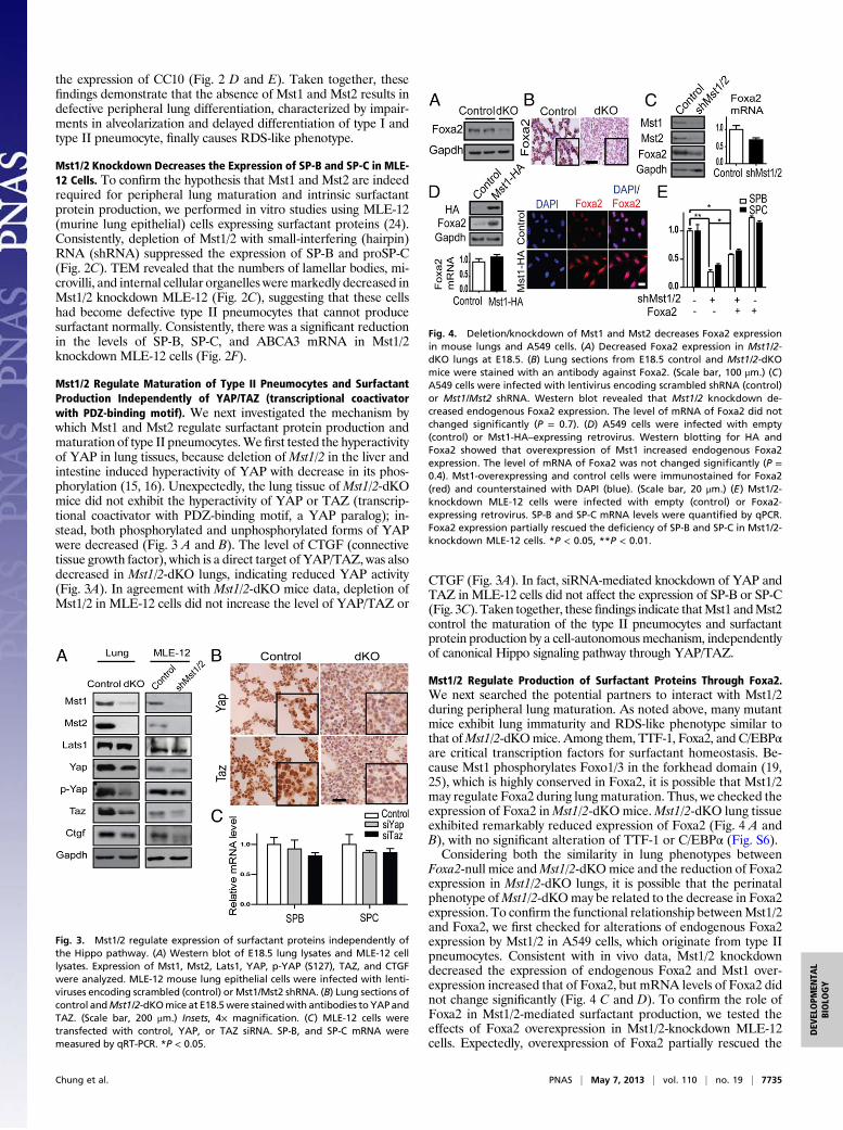
Mst1/2 Regulate Maturation of Type II Pneumocytes and SurfactantProduction Independently of YAP/TAZ (transcriptional coactivatorwith PDZ-binding motif). We next investigated the mechanism bywhich Mst1 and Mst2 regulate surfactant protein production andmaturation of type II pneumocytes.Wefirst tested the hyperactivityof YAP in lung tissues, because deletion ofMst1/2 in the liver andintestine induced hyperactivity of YAP with decrease in its phos-phorylation (15, 16). Unexpectedly, the lung tissue of Mst1/2-dKOmice did not exhibit the hyperactivity of YAP or TAZ (transcrip-tional coactivator with PDZ-binding motif, a YAP paralog); in-stead, both phosphorylated and unphosphorylated forms of YAPwere decreased (Fig. 3 A and B). The level of CTGF (connectivetissue growth factor), which is a direct target of YAP/TAZ, was alsodecreased in Mst1/2-dKO lungs, indicating reduced YAP activity(Fig. 3A). In agreement with Mst1/2-dKO mice data, depletion ofMst1/2 in MLE-12 cells did not increase the level of YAP/TAZ or
CTGF (Fig. 3A). In fact, siRNA-mediated knockdown of YAP andTAZ in MLE-12 cells did not affect the expression of SP-B or SP-C(Fig. 3C). Taken together, thesefindings indicate thatMst1 andMst2control the maturation of the type II pneumocytes and surfactantprotein production by a cell-autonomous mechanism, independentlyof canonical Hippo signaling pathway through YAP/TAZ.
Mst1/2 Regulate Production of Surfactant Proteins Through Foxa2.We next searched the potential partners to interact with Mst1/2during peripheral lung maturation. As noted above, many mutantmice exhibit lung immaturity and RDS-like phenotype similar tothat ofMst1/2-dKOmice. Among them, TTF-1, Foxa2, and C/EBPαare critical transcription factors for surfactant homeostasis. Be-cause Mst1 phosphorylates Foxo1/3 in the forkhead domain (19,25), which is highly conserved in Foxa2, it is possible that Mst1/2may regulate Foxa2 during lungmaturation. Thus, we checked theexpression of Foxa2 inMst1/2-dKO mice.Mst1/2-dKO lung tissueexhibited remarkably reduced expression of Foxa2 (Fig. 4 A andB), with no significant alteration of TTF-1 or C/EBPα (Fig. S6).Considering both the similarity in lung phenotypes between
Foxa2-null mice andMst1/2-dKOmice and the reduction of Foxa2expression in Mst1/2-dKO lungs, it is possible that the perinatalphenotype ofMst1/2-dKOmay be related to the decrease in Foxa2expression. To confirm the functional relationship betweenMst1/2and Foxa2, we first checked for alterations of endogenous Foxa2expression by Mst1/2 in A549 cells, which originate from type IIpneumocytes. Consistent with in vivo data, Mst1/2 knockdowndecreased the expression of endogenous Foxa2 and Mst1 over-expression increased that of Foxa2, but mRNA levels of Foxa2 didnot change significantly (Fig. 4 C and D). To confirm the role ofFoxa2 in Mst1/2-mediated surfactant production, we tested theeffects of Foxa2 overexpression in Mst1/2-knockdown MLE-12cells. Expectedly, overexpression of Foxa2 partially rescued the
Fig. 3. Mst1/2 regulate expression of surfactant proteins independently ofthe Hippo pathway. (A) Western blot of E18.5 lung lysates and MLE-12 celllysates. Expression of Mst1, Mst2, Lats1, YAP, p-YAP (S127), TAZ, and CTGFwere analyzed. MLE-12 mouse lung epithelial cells were infected with lenti-viruses encoding scrambled (control) or Mst1/Mst2 shRNA. (B) Lung sections ofcontrol andMst1/2-dKOmice at E18.5were stainedwith antibodies toYAPandTAZ. (Scale bar, 200 μm.) Insets, 4× magnification. (C) MLE-12 cells weretransfected with control, YAP, or TAZ siRNA. SP-B, and SP-C mRNA weremeasured by qRT-PCR. *P < 0.05.
Fig. 4. Deletion/knockdown of Mst1 and Mst2 decreases Foxa2 expressionin mouse lungs and A549 cells. (A) Decreased Foxa2 expression in Mst1/2-dKO lungs at E18.5. (B) Lung sections from E18.5 control and Mst1/2-dKOmice were stained with an antibody against Foxa2. (Scale bar, 100 μm.) (C)A549 cells were infected with lentivirus encoding scrambled shRNA (control)or Mst1/Mst2 shRNA. Western blot revealed that Mst1/2 knockdown de-creased endogenous Foxa2 expression. The level of mRNA of Foxa2 did notchanged significantly (P = 0.7). (D) A549 cells were infected with empty(control) or Mst1-HA–expressing retrovirus. Western blotting for HA andFoxa2 showed that overexpression of Mst1 increased endogenous Foxa2expression. The level of mRNA of Foxa2 was not changed significantly (P =0.4). Mst1-overexpressing and control cells were immunostained for Foxa2(red) and counterstained with DAPI (blue). (Scale bar, 20 μm.) (E) Mst1/2-knockdown MLE-12 cells were infected with empty (control) or Foxa2-expressing retrovirus. SP-B and SP-C mRNA levels were quantified by qPCR.Foxa2 expression partially rescued the deficiency of SP-B and SP-C in Mst1/2-knockdown MLE-12 cells. *P < 0.05, **P < 0.01.
Chung et al. PNAS | May 7, 2013 | vol. 110 | no. 19 | 7735
DEV
ELOPM
ENTA
LBIOLO
GY
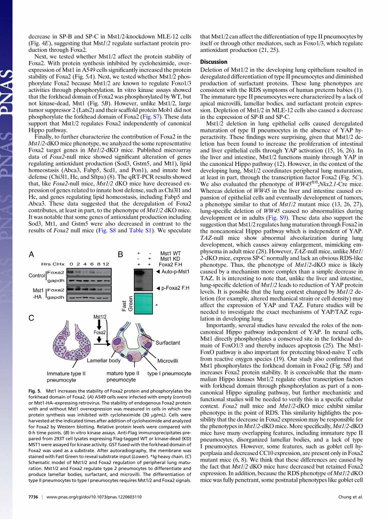
decrease in SP-B and SP-C in Mst1/2-knockdown MLE-12 cells(Fig. 4E), suggesting that Mst1/2 regulate surfactant protein pro-duction through Foxa2.Next, we tested whether Mst1/2 affect the protein stability of
Foxa2. With protein synthesis inhibited by cycloheximide, over-expression ofMst1 in A549 cells significantly increased the proteinstability of Foxa2 (Fig. 5A). Next, we tested whether Mst1/2 phos-phorylate Foxa2 because Mst1/2 are known to regulate Foxo1/3activities through phosphorylation. In vitro kinase assays showedthat the forkhead domain of Foxa2 was phosphorylated byWT, butnot kinase-dead, Mst1 (Fig. 5B). However, unlike Mst1/2, largetumor suppressor 2 (Lats2) and their scaffold proteinMob1 did notphosphorylate the forkhead domain of Foxa2 (Fig. S7). These datasupport that Mst1/2 regulates Foxa2 independently of canonicalHippo pathway.Finally, to further characterize the contribution of Foxa2 in the
Mst1/2-dKOmice phenotype, we analyzed the some representativeFoxa2 target genes in Mst1/2-dKO mice. Published microarraydata of Foxa2-null mice showed significant alteration of genesregulating antioxidant production (Sod3, Gstm5, and Mt1), lipidhomeostasis (Abca3, Fabp5, Scd1, and Pon1), and innate hostdefense (Chi3l1, Hc, and Sftpa) (8). The qRT-PCR results showedthat, like Foxa2-null mice, Mst1/2 dKO mice have decreased ex-pression of genes related to innate host defense, such asChi3l1 andHc, and genes regulating lipid homeostasis, including Fabp5 andAbca3. These data suggested that the deregulation of Foxa2contributes, at least in part, to the phenotype ofMst1/2 dKOmice.It was notable that some genes of antioxidant production includingSod3, Mt1, and Gstm5 were also decreased in contrast to theresults of Foxa2 null mice (Fig. S8 and Table S1). We speculate
thatMst1/2 can affect the differentiation of type II pneumocytes byitself or through other mediators, such as Foxo1/3, which regulateantioxidant production (21, 25).
DiscussionDeletion of Mst1/2 in the developing lung epithelium resulted inderegulated differentiation of type II pneumocytes and diminishedproduction of surfactant proteins. These lung phenotypes areconsistent with the RDS symptoms of human preterm babies (1).The immature type II pneumocytes were characterized by a lack ofapical microvilli, lamellar bodies, and surfactant protein expres-sion. Depletion of Mst1/2 in MLE-12 cells also caused a decreasein the expression of SP-B and SP-C.Mst1/2 deletion in lung epithelial cells caused deregulated
maturation of type II pneumocytes in the absence of YAP hy-peractivity. These findings were surprising, given that Mst1/2 de-letion has been found to increase the proliferation of intestinaland liver epithelial cells through YAP activation (15, 16, 26). Inthe liver and intestine, Mst1/2 functions mainly through YAP inthe canonical Hippo pathway (12). However, in the context of thedeveloping lung, Mst1/2 coordinates peripheral lung maturation,at least in part, through the transcription factor Foxa2 (Fig. 5C).We also evaluated the phenotype of WW45fl/fl;Nkx2.1-Cre mice.Whereas deletion of WW45 in the liver and intestine caused ex-pansion of epithelial cells and eventually development of tumors,a phenotype similar to that of Mst1/2 mutant mice (13, 26, 27),lung-specific deletion of WW45 caused no abnormalities duringdevelopment or in adults (Fig. S9). These data also support thesuggestion thatMst1/2 regulates lungmaturation through Foxa2 inthe noncanonical Hippo pathway which is independent of YAP.TAZ-null mice show abnormal alveolarization during lungdevelopment, which causes airway enlargement, mimicking em-physema in adult mice (28). However,TAZ-null mice, unlikeMst1/2-dKOmice, express SP-C normally and lack an obvious RDS-likephenotype. Thus, the phenotype of Mst1/2-dKO mice is likelycaused by a mechanism more complex than a simple decrease inTAZ. It is interesting to note that, unlike the liver and intestine,lung-specific deletion ofMst1/2 leads to reduction of YAP proteinlevels. It is possible that the lung context changed by Mst1/2 de-letion (for example, altered mechanical strain or cell density) mayaffect the expression of YAP and TAZ. Future studies will beneeded to investigate the exact mechanisms of YAP/TAZ regu-lation in developing lung.Importantly, several studies have revealed the roles of the non-
canonical Hippo pathway independent of YAP. In neural cells,Mst1 directly phosphorylates a conserved site in the forkhead do-main of FoxO1/3 and thereby induces apoptosis (25). The Mst1-FoxO pathway is also important for protecting blood-naïve T cellsfrom reactive oxygen species (19). Our study also confirmed thatMst1 phosphorylates the forkhead domain in Foxa2 (Fig. 5B) andincreases Foxa2 protein stability. It is conceivable that the mam-malian Hippo kinases Mst1/2 regulate other transcription factorswith forkhead domain through phosphorylation as part of a non-canonical Hippo signaling pathway, but further mechanistic andfunctional studies will be needed to verify this in a specific cellularcontext. Foxa2 null mice and Mst1/2-dKO mice exhibit similarphenotype in the point of RDS. This similarity highlights the pos-sibility that the decrease in Foxa2 expressionmay be responsible forthe phenotypes inMst1/2-dKOmice.More specifically,Mst1/2-dKOmice have many overlapping features, including immature type IIpneumocytes, disorganized lamellar bodies, and a lack of typeI pneumocytes. However, some features, such as goblet cell hy-perplasia and decreasedCC10 expression, are present only in Foxa2mutant mice (6, 8). We think that these differences are caused bythe fact that Mst1/2 dKO mice have decreased but retained Foxa2expression. In addition, because theRDS phenotype ofMst1/2 dKOmice was fully penetrant, some postnatal phenotypes like goblet cell
Fig. 5. Mst1 increases the stability of Foxa2 protein and phosphorylates theforkhead domain of Foxa2. (A) A549 cells were infected with empty (control)or Mst1-HA–expressing retrovirus. The stability of endogenous Foxa2 proteinwith and without Mst1 overexpression was measured in cells in which newprotein synthesis was inhibited with cycloheximide (30 μg/mL). Cells wereharvested at the indicated times after addition of cycloheximide and analyzedfor Foxa2 by Western blotting. Relative protein levels were compared with0-h time points. (B) In vitro kinase assays. Anti-Flag immunoprecipitates pre-pared from 293T cell lysates expressing Flag-tagged WT or kinase-dead (KD)MST1were assayed for kinase activity. GST fusedwith the forkhead domain ofFoxa2 was used as a substrate. After autoradiography, the membrane wasstained with Fast Green to reveal substrate input (Lower). *Ig heavy chain. (C)Schematic model of Mst1/2 and Foxa2 regulation of peripheral lung matu-ration. Mst1/2 and Foxa2 regulate type 2 pneumocytes to differentiate andproduce lamellar bodies, surfactant, and microvilli. The differentiation oftype II pneumocytes to type I pneumocytes requires Mst1/2 and Foxa2 signals.
7736 | www.pnas.org/cgi/doi/10.1073/pnas.1220603110 Chung et al.

hyperplasia could not be efficiently observed. Postnatal deletion ofMst1/2 in lung may be required to accurately address these issues.The transcription factor TTF-1 has been shown to be involved
in lung development and is a target of rat Mst2 kinase (29). Anin vitro study reported that Foxa2 regulates TTF-1 expression,but it was also reported that Foxa1−/−;Foxa2Δ/Δ mice exhibit nochange in TTF-1 expression (7). Consistent with this latter study,we found that, although Foxa2 expression was markedly de-creased in the lungs of Mst1/2-dKO mice, TTF-1 expression wasnot changed (Fig. S6).Regulation of type II pneumocyte differentiation is important,
not only in the developmental stage but also during tumorigenesisand lung regeneration after injury (3). However, lung develop-mental problems of mutant mice were ultimately fatal, preventingus from assessing effects ofMst1/2 deletion in adult animals, such asadult interstitial lung disease and tumorigenesis. A recent studyrevealed that K-Ras–induced distal lung adenocarcinomas origi-nate from type II pneumocytes (30). It will thus be interesting toinvestigate the functions of Mst1 and Mst2 at the point of type IIpneumocyte differentiation in K-Ras–induced lung tumorigenesisand lung regeneration after injury.In conclusion, deletion of Mst1/2 from the respiratory epithe-
lium caused deregulation of peripheral lung differentiation, pre-vented maturation of type II pneumocytes, reduced surfactantproteins, and decreased type I pneumocytes, causing RDS at theperinatal stage. The identification of Mst1/2 as unique regulatorsof peripheral lung differentiation may aid in the development ofnew strategies for early diagnosis and effective treatment of manyperipheral lung diseases, such as RDS, interstitial lung disease,and lung cancer.
Materials and MethodsGeneration Mst1fl/fl; Mst2−/−;Nkx2.1-Cre Mice. The generation of Mst1fl/fl;Mst2−/−;Nkx2.1-Cre mice is described in SI Materials and Methods. Mousecare and experiments were performed in accordance with procedures ap-proved by the Korea Advanced Institute of Science and Technology-AnimalCare and Use Committee.
Histological and Electron Microscopy Analysis. Lung tissue was fixed in 10%(vol/vol) formalin for paraffin sections and in 4% (wt/vol) paraformaldehydefor preparation of frozen sections. Sections (4-μm thickness) were stainedwith H&E or were subjected to immunohistochemical analysis using standardprotocols. Detailed protocols and information on antibodies used forstaining are presented in SI Materials and Methods. Electron microscopyanalyses of lung and cell samples were performed according to standardprotocols as previously described (14). Detailed procedures for electron mi-croscopy analysis are described in SI Materials and Methods.
Cell Proliferation and Apoptosis. Cell proliferation was analyzed by stainingfor Ki67 (Novocastra), and apoptosis was evaluated using an In Situ Cell DeathDetection kit (Roche), as described previously (14).
Immunoblot Analysis. Lung tissues were homogenized in Proprep lysis buffer(iNtRON Biotechnology) containing protease and phosphatase inhibitors.Lysates were centrifuged at 15,000 × g for 15 min at 4 °C, and the resultingsupernatants were analyzed by immunoblotting. The antibodies used fordetection are described in SI Materials and Methods.
qRT-PCR Analysis and in Vitro Kinase Assay. Total RNA was isolated from lungtissues using Riboex (Geneall) according to themanufacturer’s protocol. Real-time reactions were performed as described previously (25). The relativeconcentration of mRNA was normalized to the concentration of GAPDHmRNA in each sample. GAPDH mRNA was quantified using the primers 5′-CTT CAC CAC CAT GGA GGA GGC-3′ and 5′-GGC ATG GAC TGT GGT CATGAG-3′ primers. Other primer sequences and in vitro kinase assays are de-scribed in SI Materials and Methods and Tables S2–S4.
Foxa2 Protein Stability. Cycloheximide was added to the cells at a final con-centrationof 30μg/mL. The cellswereharvestedat the indicated times followingcycloheximide treatment, and protein extracts were prepared. The protocolswere performed as previously described (31), with some modifications.
Statistical Analysis. Quantitative data are presented as means ± SDs unlessindicated otherwise. Differences between means were evaluated using Stu-dent’s unpaired test. A P value < 0.05 was considered statistically significant.
ACKNOWLEDGMENTS. We thank Taechang Yang, Eun Soon Lee, and SeungHye Yang for their technical assistance. This study was supported by NationalCreative Research Initiative Program Grant 20120001228.
1. Whitsett JA, Wert SE, Trapnell BC (2004) Genetic disorders influencing lung formationand function at birth. Hum Mol Genet 13(Spec No 2):R207–R215.
2. Whitsett JA, Wert SE, Weaver TE (2010) Alveolar surfactant homeostasis and thepathogenesis of pulmonary disease. Annu Rev Med 61:105–119.
3. Whitsett JA, Weaver TE (2002) Hydrophobic surfactant proteins in lung function anddisease. N Engl J Med 347(26):2141–2148.
4. Maeda Y, Davé V, Whitsett JA (2007) Transcriptional control of lung morphogenesis.Physiol Rev 87(1):219–244.
5. Martis PC, et al. (2006) C/EBPalpha is required for lung maturation at birth. De-velopment 133(6):1155–1164.
6. Chen G, et al. (2010) Foxa2 programs Th2 cell-mediated innate immunity in the de-veloping lung. J Immunol 184(11):6133–6141.
7. Wan H, et al. (2005) Compensatory roles of Foxa1 and Foxa2 during lung morpho-genesis. J Biol Chem 280(14):13809–13816.
8. Wan H, et al. (2004) Foxa2 is required for transition to air breathing at birth. Proc NatlAcad Sci USA 101(40):14449–14454.
9. DeFelice M, et al. (2003) TTF-1 phosphorylation is required for peripheral lungmorphogenesis, perinatal survival, and tissue-specific gene expression. J Biol Chem278(37):35574–35583.
10. Davé V, et al. (2006) Calcineurin/Nfat signaling is required for perinatal lung matu-ration and function. J Clin Invest 116(10):2597–2609.
11. Yan C, Whitsett JA (1997) Protein kinase A activation of the surfactant protein B geneis mediated by phosphorylation of thyroid transcription factor 1. J Biol Chem 272(28):17327–17332.
12. Pan D (2010) The hippo signaling pathway in development and cancer. Dev Cell 19(4):491–505.
13. Lu L, et al. (2010) Hippo signaling is a potent in vivo growth and tumor suppressorpathway in the mammalian liver. Proc Natl Acad Sci USA 107(4):1437–1442.
14. Lee JH, et al. (2008) A crucial role of WW45 in developing epithelial tissues in themouse. EMBO J 27(8):1231–1242.
15. Zhou D, et al. (2009) Mst1 and Mst2 maintain hepatocyte quiescence and suppresshepatocellular carcinoma development through inactivation of the Yap1 oncogene.Cancer Cell 16(5):425–438.
16. Zhou D, et al. (2011) Mst1 and Mst2 protein kinases restrain intestinal stem cellproliferation and colonic tumorigenesis by inhibition of Yes-associated protein (Yap)overabundance. Proc Natl Acad Sci USA 108(49):E1312–E1320.
17. Ren F, et al. (2010) Hippo signaling regulates Drosophila intestine stem cell pro-
liferation through multiple pathways. Proc Natl Acad Sci USA 107(49):21064–21069.18. Kim TS, et al. (2012) Mammalian sterile 20-like kinase 1 suppresses lymphoma
development by promoting faithful chromosome segregation. Cancer Res 72(20):
5386–5395.19. Choi J, et al. (2009) Mst1-FoxO signaling protects naïve T lymphocytes from cellular
oxidative stress in mice. PLoS ONE 4(11):e8011.20. Avruch J, et al. (2012) Protein kinases of the Hippo pathway: Regulation and sub-
strates. Semin Cell Dev Biol 23(7):770–784.21. Oh S, et al. (2009) Crucial role for Mst1 and Mst2 kinases in early embryonic de-
velopment of the mouse. Mol Cell Biol 29(23):6309–6320.22. Oh HJ, et al. (2010) MST1 limits the kinase activity of aurora B to promote stable
kinetochore-microtubule attachment. Curr Biol 20(5):416–422.23. Janson R, Hogan BL (2011) Epithelial progenitor cells in lung development, mainte-
nance, repair, and disease. Annu Rev Cell Dev Biol 27:493–512.24. Sisson TH, et al. (2010) Targeted injury of type II alveolar epithelial cells induces
pulmonary fibrosis. Am J Respir Crit Care Med 181(3):254–263.25. Lehtinen MK, et al. (2006) A conserved MST-FOXO signaling pathway mediates oxi-
dative-stress responses and extends life span. Cell 125(5):987–1001.26. Lee KP, et al. (2010) The Hippo-Salvador pathway restrains hepatic oval cell
proliferation, liver size, and liver tumorigenesis. Proc Natl Acad Sci USA 107(18):
8248–8253.27. Chen L, Qin F, Deng X, Avruch J, Zhou D (2012) Hippo pathway in intestinal ho-
meostasis and tumorigenesis. Protein Cell 3(4):305–310.28. Makita R, et al. (2008) Multiple renal cysts, urinary concentration defects, and pul-
monary emphysematous changes in mice lacking TAZ. Am J Physiol Renal Physiol
294(3):F542–F553.29. Aurisicchio L, Di Lauro R, Zannini M (1998) Identification of the thyroid transcription
factor-1 as a target for rat MST2 kinase. J Biol Chem 273(3):1477–1482.30. Xu X, et al. (2012) Evidence for type II cells as cells of origin of K-Ras-induced distal
lung adenocarcinoma. Proc Natl Acad Sci USA 109(13):4910–4915.31. Rausa FM, 3rd, Hughes DE, Costa RH (2004) Stability of the hepatocyte nuclear factor
6 transcription factor requires acetylation by the CREB-binding protein coactivator.
J Biol Chem 279(41):43070–43076.
Chung et al. PNAS | May 7, 2013 | vol. 110 | no. 19 | 7737
DEV
ELOPM
ENTA
LBIOLO
GY