High-resolution ultrasound (HRUS) evaluation of the ... della spalla cable e... · References: L....
40
Page 1 of 40 High-resolution ultrasound (HRUS) evaluation of the rotator cable (RCa) in young and elderly asymptomatic volunteers Poster No.: C-2299 Congress: ECR 2010 Type: Scientific Exhibit Topic: Musculoskeletal Authors: L. M. Sconfienza 1 , D. Orlandi 2 , E. Fabbro 2 , S. Pucci 2 , E. Silvestri 2 ; 1 Milan/IT, 2 Genoa/IT Keywords: ultrasonography, rotator cuff, rotator cable DOI: 10.1594/ecr2010/C-2299 Any information contained in this pdf file is automatically generated from digital material submitted to EPOS by third parties in the form of scientific presentations. References to any names, marks, products, or services of third parties or hypertext links to third- party sites or information are provided solely as a convenience to you and do not in any way constitute or imply ECR's endorsement, sponsorship or recommendation of the third party, information, product or service. ECR is not responsible for the content of these pages and does not make any representations regarding the content or accuracy of material in this file. As per copyright regulations, any unauthorised use of the material or parts thereof as well as commercial reproduction or multiple distribution by any traditional or electronically based reproduction/publication method ist strictly prohibited. You agree to defend, indemnify, and hold ECR harmless from and against any and all claims, damages, costs, and expenses, including attorneys' fees, arising from or related to your use of these pages. Please note: Links to movies, ppt slideshows and any other multimedia files are not available in the pdf version of presentations. www.myESR.org
Transcript of High-resolution ultrasound (HRUS) evaluation of the ... della spalla cable e... · References: L....

Page 1 of 40
High-resolution ultrasound (HRUS) evaluation of the rotatorcable (RCa) in young and elderly asymptomatic volunteers
Poster No.: C-2299
Congress: ECR 2010
Type: Scientific Exhibit
Topic: Musculoskeletal
Authors: L. M. Sconfienza1, D. Orlandi2, E. Fabbro2, S. Pucci2, E. Silvestri2;1Milan/IT, 2Genoa/IT
Keywords: ultrasonography, rotator cuff, rotator cable
DOI: 10.1594/ecr2010/C-2299
Any information contained in this pdf file is automatically generated from digital materialsubmitted to EPOS by third parties in the form of scientific presentations. Referencesto any names, marks, products, or services of third parties or hypertext links to third-party sites or information are provided solely as a convenience to you and do not inany way constitute or imply ECR's endorsement, sponsorship or recommendation of thethird party, information, product or service. ECR is not responsible for the content ofthese pages and does not make any representations regarding the content or accuracyof material in this file.As per copyright regulations, any unauthorised use of the material or parts thereof aswell as commercial reproduction or multiple distribution by any traditional or electronicallybased reproduction/publication method ist strictly prohibited.You agree to defend, indemnify, and hold ECR harmless from and against any and allclaims, damages, costs, and expenses, including attorneys' fees, arising from or relatedto your use of these pages.Please note: Links to movies, ppt slideshows and any other multimedia files are notavailable in the pdf version of presentations.www.myESR.org
Page 2 of 40
Purpose
INTRODUCTION
The rotator cuff is a functional complex composed of several tendons and muscles thatprovide stability of the shoulder by pressing the humeral head on the glenoid and, at thesame time, to assure a wide range of motion.
Such complex includes a thick bundle of fibers perpendicular to the supraspinatus tendonthat has been described for the first time by Clark in 1990 [1] and called "rotator cable"by Burkhart in 1993 [2]. This structure surrounds the distal zone of the supraspinatustendon, an hypovascular crescentic region that has been called "rotator crescent" whichtears easily [3,4].
The rotator cable plays a central role in shoulder biomechanic and it was described as astructure similar in fashion to the supporting cables of a suspension bridge where stressis trasferred fom the cuff to the cable.
Cable-dominant cuffs are predominant in elderly (>60 years) shoulders [2] in reason ofthe increasing reliance of the stress shielding action that protects the relatively avascularcrescent tissue from tears.
Therefore, according to Morag et al. [5], an accurate characterization of a rotator cufftear and its relationship to the rotator cable may be important.
ROTATOR CUFF ANATOMY
The rotator cuff is a group of four muscles that form a strong complex around the shoulderjoint and help to control the rotation and position of the arm. Each of these muscles hasa tendon at the end that attaches to the humerus. These four muscles are:
• The subscapularis• The supraspinatus• The infraspinatus• The teres minor
The tendons of the rotator cuff are seen to fuse into a single structure near their insertionsinto the tubercles of the humerus. This fusion is apparent when the two surfaces of theintact cuff are exposed by removal of the overlying bursa and the underlying capsule. Thesupraspinatus and infraspinatus tendons join about 15 mm proximal to their insertionson the humerus and cannot be separated by additional blunt dissection (Fig. 1) on page16 .

Page 3 of 40
Fig.: Axial view of the rotator cuff .The function of this complex is to keep the jointcentered on the glenoid rim.References: L. M. Sconfienza; Unit of Radiology, IRCCS Policlinico San Donato,Milan, ITALY
Although there is an interval between the muscular portions of teres minor andinfraspinatus, these muscles merge inseparably just proximal to the musculotendinousjunction. The teres minor and the subscapularis have muscular insertions on the surgicalneck of the humerus, which extends approximately 2 cm downward beyond theirtendinous attachment on the tubercles.The tendons of the cuff are reinforced near theirinsertions on the tubercles of the humerus by fibrous structures that are located bothsuperficial and deep to the tendons. The superficial aspects of the infraspinatus andsupraspinatus tendons are covered by a thick sheet of fibrous tissue that lies directlybeneath the deep layer of the subdeltoid bursa but is not part of the bursa itself.
In a paper published in 2006,Ward et al. [6] report on their examination of the architecturalproperties of the rotator cuff muscles in ten cadaveric specimens,which they performed

Page 4 of 40
in the hope of understanding their functional design. Based on physiological cross-sectional area, the subscapularis have the greatest force-producing capacity, followedin declining order by the infraspinatus, supraspinatus, and teres minor. Based on fibrelength, the supraspinatus operates over the widest range of sarcomere lengths. Thesupraspinatus and infraspinatus have relatively long sarcomere lengths in the anatomicalposition and are under relatively high passive tensions at rest, indicating that they areresponsible for glenohumeral resting stability. However, the subscapularis contributespassive tension at maximum abduction and lateral rotation, indicating that it plays acritical part in glenohumeral stability in the position of apprehension. This informationillustrates the exquisite coupling of muscle architecture and joint mechanics, which allowsthe rotator cuff to produce near-maximal active tensions in the midrange and to producepassive tensions in the various end-range positions (Fig. 2) on page 17 .
Fig.: Coronal view of the rotator cuff .The function of this complex is to keep the jointcentered on the glenoid rim.
Page 5 of 40
References: L. M. Sconfienza; Unit of Radiology, IRCCS Policlinico San Donato,Milan, ITALY
CRESCENT AND CABLE BACKGROUND
• Anatomy
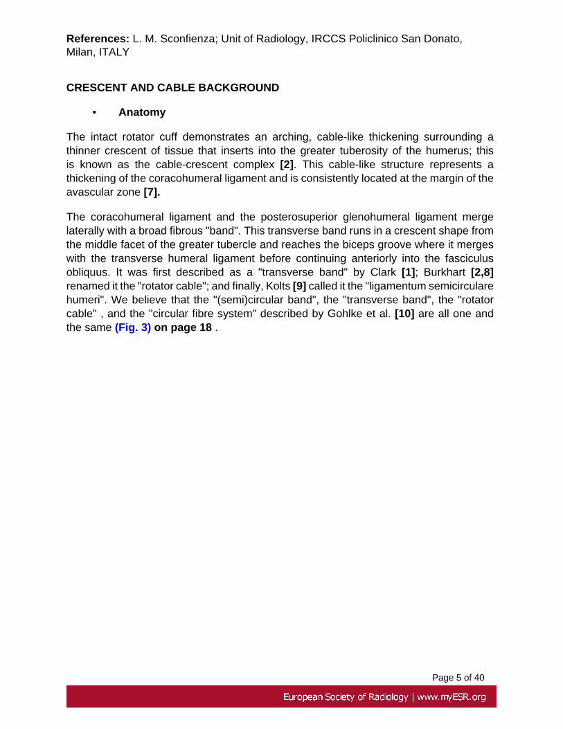
The intact rotator cuff demonstrates an arching, cable-like thickening surrounding athinner crescent of tissue that inserts into the greater tuberosity of the humerus; thisis known as the cable-crescent complex [2]. This cable-like structure represents athickening of the coracohumeral ligament and is consistently located at the margin of theavascular zone [7].
The coracohumeral ligament and the posterosuperior glenohumeral ligament mergelaterally with a broad fibrous "band". This transverse band runs in a crescent shape fromthe middle facet of the greater tubercle and reaches the biceps groove where it mergeswith the transverse humeral ligament before continuing anteriorly into the fasciculusobliquus. It was first described as a "transverse band" by Clark [1]; Burkhart [2,8]renamed it the "rotator cable"; and finally, Kolts [9] called it the "ligamentum semicircularehumeri". We believe that the "(semi)circular band", the "transverse band", the "rotatorcable" , and the "circular fibre system" described by Gohlke et al. [10] are all one andthe same (Fig. 3) on page 18 .

Page 6 of 40

Page 7 of 40
Fig.: Coronal cross-sectional illustration showing the location of rotator cable withsonographic correlation.References: L. M. Sconfienza; Unit of Radiology, IRCCS Policlinico San Donato,Milan, ITALY
The rotator cable may function in a way analogous to the functioning of a load-bearingsuspension bridge [2]. By this model, stress is transferred from the cuff muscles tothe rotator cable as a distributed load, thereby stress-shielding the thinner, avascularcrescent tissue.
Burkhart et al. [2,8] defined the suspension bridge model for the rotator cuff. In 12shoulders with massive rotator cuff tears, they observed that normal kinematics weremaintained when the tears involved only the supraspinatus tendon and a small portion ofthe infraspinatus tendon. In all these shoulders with stable fulcrum kinematics, the freemargin of the rotator cuff tear was thick and rind like (Fig. 4) on page 20 .

Page 8 of 40
Fig.: Axial cross-sectional illustration showing the fibres orientation of rotator cableand rotator crescent.References: L. M. Sconfienza; Unit of Radiology, IRCCS Policlinico San Donato,Milan, ITALY
In a second study, the same authors found a rotator cable-crescent complex in cadavershoulders, corresponding to the free margin of a tear. The rotator crescent was foundto measure an average of 41.35 mm in the anteroposterior direction and of 14.08 mmin the mediolateral direction, with an average thickness of 1.82 mm. The average widthof the rotator cable surrounding the rotator crescent was seen to be 12.05 mm, with anaverage thickness of 4.72 mm.
• Arthroscopy
Pouliart et al. [11,12] observed a distinct rotator cable surrounding a distinct rotatorcrescent in about 50% of cadaveric specimens. "The rotator cable spans from

Page 9 of 40
anterolateral to posterolateral above the intertubercular groove. In about 25% ofshoulders the rotator cable is less obvious but might be identified by adding traction tothe arm or rotating the humerus. In these shoulders, the rotator crescent is not visible.In the rest, the rotator cable and crescent cannot be discerned despite manipulations,and the rotator crescent therefore cannot be marked. In adduction and external rotation,a longitudinal anterosuperior capsular fold with a distinct anterior leading edge developsin all cases (Fig. 5) on page 20 .
Fig.: Arthroscopic view of the right shoulder showing the rotator crescent and therotator cable.References: L. M. Sconfienza; Unit of Radiology, IRCCS Policlinico San Donato,Milan, ITALY

Page 10 of 40
In adduction and internal rotation, the posterosuperior capsule becomes tight enoughto squeeze the arthroscope downwards and out. The longitudinal posterosuperiorfold appears just superior to the posterior arthroscopic portal and runs from theposterosuperior glenoid rim, medial and posterior to the origin of the long tendon of thebiceps and the glenoid labrum, to the posterior part of the greater tubercle. Here it mergeswith the posterior leg of the rotator cable when this is visible. Since both longitudinalsuperior folds are always seen during either external or internal rotation, they may as wellbe assessed with the arthroscopic technique in all cases [11,12]".
• Histologic Correlation
Since the rotator cuff is composed of intimately associated tendons that intersect andinterdigitate in the region of the supraspinatus and infraspinatus tendons, loads fromcontraction are not isolated to a single muscle but are dispersed among neighboring cufftendons. Anatomic dissections of rotator cuffs have demonstrated a thick fibrous sheathcalled the rotator cable, which arises from the coracohumeral ligament and envelopesthe anterior margin of the supraspinatus tendon.
At histologic examination, a fibrillar structure distinct from the rotator cuff was identified,consistent with the rotator cable. This structure is a defined cablelike extension ofthe coracohumeral ligament along the articular surface of the supraspinatus andinfraspinatus tendons that surround the area described by Codman [3] as the "criticalzone," a zone with a propensity for tearing [4]. The orientation and appearance of thecable fibres differed from those of the rotator cuff tendon fibers. Samples from the rotatorcuff midline demonstrated a cablelike structure situated on the articular surface of thecuff tendons. This fibrillar structure could be identified as oriented perpendicular to theorientation of the rotator cuff tendon fibers.
This aspect is confirmed by Kolts's studies [9,13]; "The rotator cable appears as anapproximately 1-cm-wide band of capsular collagen fibres oriented in parallel, runningtransverse to the longitudinal axis of the supraspinatus muscle tendon".
• Biomechanic
Transverse plane force couple is the predominant mechanism resisting superior humeralhead displacement with cuff tears. As long as the force couple between subscapularisand infraspinatus remains balanced the joint remains centred and functional [14]. Theintact rotator cuff demonstrates an arching, cable-like thickening surrounding a thinnercrescent of tissue that inserts into the greater tuberosity of the humerus; this is known asthe cable-crescent complex [2]. This densely packed unidirectional collagen fibres extendfrom the coracohumeral ligament (CH) posteriorly to the infraspinatus, running bothsuperficial and deep to the tendon proper, at the margin of the avascular zone [7]. Thecrescent, comprises supraspinatus and infraspinatus insertions that are contained withinthe avascular zone. On arthroscopic examination, the margin of the crescent is seemed

Page 11 of 40
to have thick bundles of fibres that are perpendicular to the axis of the supraspinatustendon and arch anteriorly and posteriorly to attach on the humerus.
The marked differences in thickness between the rotator cable (4.72 mm) and the rotatorcrescent bordered by the cable
(1.82 mm) is striking. This finding supports the concept of the rotator cable as a functionalcable system in which there is stress transfer from the cuff to the thick cable and stres-shielding of the thin capsular tissue distal to the cable and within the crescent [2] .
The rotator cable work in the same way as the functional cable system of a suspensionbridge. By this model, stress is transferred from the cuff muscles to the rotator cableas a distributed load, thereby stress-shielding the thinner, avascular crescent tissue,particularly in older individuals.
Given their fusion into the rotator cable, the coracoglenohumeral ligament andposterosuperior glenohumeral ligament provide the medial anchorage for the rotatorcable function. This probably allows the superior complex to maintain its depressingand centring effect as long as one of the medial and one of the lateral points of bonyattachment are preserved [11,12].
• Imaging
High resolution ultrasound (HRUS) plays a central role in rotator cuff diagnostic imagingand it could be used to demostrate the clinical implications of the rotator cable [5]. Beingquick, non invasive, and allowing a dynamic evaluation, HRUS is ideal to focus on astructure as implicated in the shoulder biomechanic as the rotator cable.
Moreover, dynamic scans can be acquired during clinical tests in abduction, adduction,extrarotation, thus magnifyng anatomic and pathologic conditions that could be difficultto characterize otherwise.
On HRUS scans the morphology of the rotator cable could vary from a thick cable offibres to thin flattened fibers traversing deep to the rotator cuff. According to Morag etal [5] the ability of US to depict the rotator cable may be greater in older (>60 years)individuals. Larger studies should be undertaken to assess HRUS ability to consistentlyidentify the rotator cable in large groups of symptomatic and asymptomatic patients andto attempt to correlate between rotator cable morphology and shoulder function after arotator cuff tear (Fig. 6) on page 21 .

Page 12 of 40
Fig.: HRUS scans on long (A) and short(B) axis of the supraspinatus tendon showingthe rotator cable (arrow).References: L. M. Sconfienza; Unit of Radiology, IRCCS Policlinico San Donato,Milan, ITALY
• Age Correlation
The marked differences in thickness and width between the rotator cable and the crescentarea supports the concept of a functional cable system where stress is transfered fromthe cuff to the thick cable, acting as a stress-shield of the thin capsular tissue of thecrescent. In younger shoulders with thick rotator crescent, this area is not stress-shieldedby the cable. On the contrary, older shoulders (>60 years of age) with thin crescent tissueare stress-shielded by the cable. These findings suggest that there may be two differentfunctional classes of rotator cuff based on the behaviour of the cable-crescent complexunder load: cable dominant (in which the crescent is stress-shielded by the cable) andcrescent dominant (in which there is no stress-shielding of the crescent). The arthroscopicview of the rotator cable and crescent often shows that the slight crescent tissue has aredundant invagination adjacent to the rotator cable, suggesting that the rotator crescentis not under tension [15] (Fig. 7) on page 22 .

Page 13 of 40
Fig.: HRUS scans on long axis of the supraspinatus tendon showing the two functionalclasses of rotator cuff: cable dominant (A) and crescent dominant (B). Rotator cable(arrow) is significantly thicker in cable dominant.References: L. M. Sconfienza; Unit of Radiology, IRCCS Policlinico San Donato,Milan, ITALY
• Role in rotator cuff tears
The anatomy of the cable-crescent complex and the model of the load-bearingsuspension bridge suggests that the location of a rotator cuff tear is much more importantthan the size of the tear in terms of its effect on shoulder function. That is to say that atear involving the rotator cable may be biomechanically much more significant than a tearthat involves only the rotator crescent (Fig. 8) on page 22.
Page 14 of 40
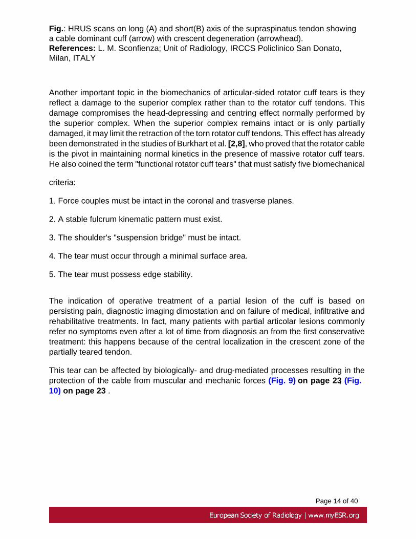
Fig.: HRUS scans on long (A) and short(B) axis of the supraspinatus tendon showinga cable dominant cuff (arrow) with crescent degeneration (arrowhead).References: L. M. Sconfienza; Unit of Radiology, IRCCS Policlinico San Donato,Milan, ITALY
Another important topic in the biomechanics of articular-sided rotator cuff tears is theyreflect a damage to the superior complex rather than to the rotator cuff tendons. Thisdamage compromises the head-depressing and centring effect normally performed bythe superior complex. When the superior complex remains intact or is only partiallydamaged, it may limit the retraction of the torn rotator cuff tendons. This effect has alreadybeen demonstrated in the studies of Burkhart et al. [2,8], who proved that the rotator cableis the pivot in maintaining normal kinetics in the presence of massive rotator cuff tears.He also coined the term "functional rotator cuff tears" that must satisfy five biomechanical
criteria:
1. Force couples must be intact in the coronal and trasverse planes.
2. A stable fulcrum kinematic pattern must exist.
3. The shoulder's "suspension bridge" must be intact.
4. The tear must occur through a minimal surface area.
5. The tear must possess edge stability.
The indication of operative treatment of a partial lesion of the cuff is based onpersisting pain, diagnostic imaging dimostation and on failure of medical, infiltrative andrehabilitative treatments. In fact, many patients with partial articolar lesions commonlyrefer no symptoms even after a lot of time from diagnosis an from the first conservativetreatment: this happens because of the central localization in the crescent zone of thepartially teared tendon.
This tear can be affected by biologically- and drug-mediated processes resulting in theprotection of the cable from muscular and mechanic forces (Fig. 9) on page 23, (Fig.10) on page 23 .

Page 15 of 40
Fig.: US scan of the supraspinatus tendon showing a crescent zone tear (arrowhead)limited by the rotator cable (arrow).References: L. M. Sconfienza; Unit of Radiology, IRCCS Policlinico San Donato,Milan, ITALY

Page 16 of 40
Fig.: US scan of the supraspinatus tendon showing a crescent zone tear (arrowhead)limited by the rotator cable (arrow).References: L. M. Sconfienza; Unit of Radiology, IRCCS Policlinico San Donato,Milan, ITALY
Images for this section:

Page 17 of 40
Fig. 1: Axial view of the rotator cuff .The function of this complex is to keep the jointcentered on the glenoid rim.

Page 18 of 40
Fig. 2: Coronal view of the rotator cuff .The function of this complex is to keep the jointcentered on the glenoid rim.

Page 19 of 40

Page 20 of 40
Fig. 3: Coronal cross-sectional illustration showing the location of rotator cable withsonographic correlation.
Fig. 4: Axial cross-sectional illustration showing the fibres orientation of rotator cable androtator crescent.

Page 21 of 40
Fig. 5: Arthroscopic view of the right shoulder showing the rotator crescent and the rotatorcable.

Page 22 of 40
Fig. 6: HRUS scans on long (A) and short(B) axis of the supraspinatus tendon showingthe rotator cable (arrow).
Fig. 7: HRUS scans on long axis of the supraspinatus tendon showing the two functionalclasses of rotator cuff: cable dominant (A) and crescent dominant (B). Rotator cable(arrow) is significantly thicker in cable dominant.

Page 23 of 40
Fig. 8: HRUS scans on long (A) and short(B) axis of the supraspinatus tendon showinga cable dominant cuff (arrow) with crescent degeneration (arrowhead).
Fig. 9: US scan of the supraspinatus tendon showing a crescent zone tear (arrowhead)limited by the rotator cable (arrow).

Page 24 of 40
Fig. 10: US scan of the supraspinatus tendon showing a crescent zone tear (arrowhead)limited by the rotator cable (arrow).

Page 25 of 40
Methods and Materials
EXPERIMENTAL STUDY• Purpose
In reason of the limited number of publications dealing with the rotator cable and onlya single example about the ultrasonographic (US) appearance of the rotator cable [5],the purpose of our work is to characterize the US appearance of the rotator cable andto compare the HRUS consistency of such structure in young and elderly asymptomaticvolunteers.
• Methods and materials
Twelve young (six males, age range 21-39 years, mean age 33 years) and twelve elderly(six males, age range 62-83 years, mean age 75 years) asymptomatic volunteers wereincluded in our study (twentyfour shoulders for each group).
Volunteers with shoulder pain, limited range of motion, or history of symptomaticshoulders were excluded from the study.
HRUS evaluation of supraspinatus and infraspinatus tendons was performed both onlong and short axis with an ultrasound equipment (Esaote MyLab 70 XVG; EsaoteBiomedica SPA,Italia) provided with a high-resolution 12-5 MHz transducer. Imageswere reviewed for the presence of a hyperechoic bundle of fibers running perpendicularto the supraspinatus or infraspinatus tendons in the expected location of the rotatorcable. Identification of the rotator cable is based on demonstration of cable fibers in bothtransverse and longitudinal scans.
For each shoulder, we noted whether the rotator cable was detectable or not, and - if yes- its thickness (craniocaudal dimensions) and width (mediolateral dimensions). Fisher'sexact and U Mann-Whitney tests were used (Fig. 1) on page 26.

Page 26 of 40
Fig.: HRUS scan on short axis of the supraspinatus tendon showing the rotator cable(arrow).References: L. M. Sconfienza; Unit of Radiology, IRCCS Policlinico San Donato,Milan, ITALY
HRUS of asymptomatic volunteer's and patient's shoulders successfully depicteda hyperechoic fibrillar structure deep to the supraspinatus tendon tracking in aperpendicular fashion relative to the rotator cuff fibers; this hyperechoic and fibrillar rotatorcable demonstrated anisotropy and therefore appeared artifactually hypoechoic whennot imaged perpendicular to the ultrasound beam.
Images for this section:

Page 27 of 40
Fig. 1: HRUS scan on short axis of the supraspinatus tendon showing the rotator cable(arrow).
Page 28 of 40
Results
• Results
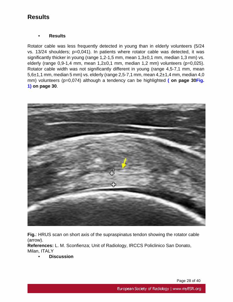
Rotator cable was less frequently detected in young than in elderly volunteers (5/24vs. 13/24 shoulders; p=0,041). In patients where rotator cable was detected, it wassignificantly thicker in young (range 1,2-1,5 mm, mean 1,3±0,1 mm, median 1,3 mm) vs.elderly (range 0,9-1,4 mm, mean 1,2±0,1 mm, median 1,2 mm) volunteers (p=0,025).Rotator cable width was not significantly different in young (range 4,5-7,1 mm, mean5,6±1,1 mm, median 5 mm) vs. elderly (range 2,5-7,1 mm, mean 4,2±1,4 mm, median 4,0mm) volunteers (p=0,074) although a tendency can be highlighted ( on page 30Fig.1) on page 30.
Fig.: HRUS scan on short axis of the supraspinatus tendon showing the rotator cable(arrow).References: L. M. Sconfienza; Unit of Radiology, IRCCS Policlinico San Donato,Milan, ITALY
• Discussion

Page 29 of 40
Rotator cable was more frequently detected in in elderly volunteers (13/24 shoulders).A reason for that can be found in reason of the increased hypoechogenicity of thesurrounding tendon matrix and, according with Burkhart, for the adaptive changes thatlead the crescent to progressive thinning with advancing age [2].
When detected, rotator cable was significantly thicker in young volunteers. We belive thatthis finding can be related to the overall thinning of the tendon during the ageing process.
The prevalence of cable dominant shoulders in elderly volunteers could be explainedby the intrinsic characteristics of resistence, fibres orientation, and strategic positionthat lead the rotator cable to be the last structure involved by degenerative thinningprocesses(Fig. 2) on page 30.
Fig.: HRUS scan on long axis of the supraspinatus tendon showing a cable dominantcuff (arrow) with crescent degeneration (arrowhead).References: L. M. Sconfienza; Unit of Radiology, IRCCS Policlinico San Donato,Milan, ITALY

Page 30 of 40
Images for this section:
Fig. 1: HRUS scan on short axis of the supraspinatus tendon showing the rotator cable(arrow).

Page 31 of 40
Fig. 2: HRUS scan on long axis of the supraspinatus tendon showing a cable dominantcuff (arrow) with crescent degeneration (arrowhead).

Page 32 of 40
Conclusion
• Conclusions
In summary, HRUS is an emergent, rapid an cheap technique able to perform anaccurate, non invasive and dynamic study of the rotator cable, in both young and elderlypatients. The rotator cable is a well-known structure among orthopedic surgeons, oftenvisualized during arthroscopy with proved anatomic correlation.
Our ability in demonstration of cable integrity with a tear isolated to the crescent may haveclinical and surgical implications. It may redirect the terapeutic strategies of cuff tears:clinical and infiltrative strategy for cable dominant patients with central tears localizedin the crescent and a good muscolar trophism (non progressive lesions); arthroscopicor surgical strategy for crescent dominant patient and for tears in eccentic position(progressive and painful lesions).
(Fig. 1) on page 33 (Fig. 2) on page 33
Fig.: US scan of the supraspinatus tendon showing a crescent zone tear (arrowhead)limited by the rotator cable (arrow).References: L. M. Sconfienza; Unit of Radiology, IRCCS Policlinico San Donato,Milan, ITALY

Page 33 of 40
However, further studies are needed to determine if the rotator cable can be uniformlydepicted at US and to determine if this information can potentially alter treatment of rotatorcuff tears.
In conclusion, nevertheless the amazing progress of the diagnostic imaging, we belivethat a wide background above the anatomy and the biomechanic of the shoulder is strictlyreccomended for an appropriate approach at dignostic and treatment of the rotator cuff.Quoting Burkhart SS "As with any breakthrough technology, this rapid progress wasmade possible by the marriage of insight to technology. The great misconception of ourera is that technology alone will advance a discipline; the irony is that technology in avacuum produces no advancement. Technology without understanding produces meregadgets; technology guided by insight produces tools." [16]
Images for this section:
Fig. 1: US scan of the supraspinatus tendon showing a crescent zone tear (arrowhead)limited by the rotator cable (arrow).

Page 34 of 40
Fig. 2: US scan of the supraspinatus tendon showing a crescent zone tear (arrowhead)limited by the rotator cable (arrow).

Page 35 of 40
References
REFERENCES
1. Clark J, Sidles JA,Matsen FA. The relationship of the glenohumeral joint capsule tothe rotator cuff. Clin Orthop (1990); 254:29-34
2. Burkhart SS, Esch JC, Jolson RC. The rotator crescent and rotator cable: an anatomicdescription of the shoulder's "suspension bridge." Arthroscopy (1993); 9: 611-616
3. Codman EA. The shoulder: rupture of the supraspinatus tendon and other lesions inor about the subacromial bursa. Boston, Mass: Thomas Todd, 1934.
4. Codman EA, Akerson IB. The pathology associated with rupture of the supraspinatustendon. Ann Surg (1931); 94:348-359.
5. MoragY, Jacobson JA,LucasD,Miller B, BrigidoMK, Jamadar DA. US appearanceof the rotator cable with histologic correlation: preliminary results. Radiology (2006);241:485-491.
6. Ward SR, Hentzen ER, Smallwood LH et al. Rotator cuff muscle architecture:implications for glenohumeral stability. Clin Orthop Relat Res (2006); 448:157-163
7. Clark JM,Harryman DT. Tendons, ligaments, and capsule of the rotator cuff. J BoneJoint Surg Am (1992); 74:713-725
8. Burkhart SS. Fluoroscopic comparison of kinematic patterns in massive rotator cufftears.A suspension bridge model.Clin Orthop (1992); 284:144-152
9. Kolts I, Busch LC, Tomusk H et al. Macroscopical anatomy of the socalled "rotatorinterval". A cadaver study on 19 shoulder joints. Ann Anat (2002); 184:9-14
10. Gohlke F, Essigkrug B, Schmitz F. The pattern of collagen fiber bundles of the capsuleof the glenohumeral joint. J Shoulder Elbow Surg (1994); 3:111-127

Page 36 of 40
11. Pouliart N. Shoulder instability: Experimental model and related anatomy.Doctoralthesis,Brussels,Vrije Universiteit,Faculty ofMedicine and Pharmacy (2005)
12. Pouliart N, Eid S,Somers K et al. Variations in the superior capsuloligamentouscomplex and description of a new ligament. J Shoulder Elbow Surg (2007) (publishedOnline First)
13. Kolts I,Busch LC,Tomusk H et al. Anatomy of the coracohumeral and thecoracoglenoidal ligaments. Ann Anat (2000); 182:563-566
14. Parsons IM, Apreleva M, Fu FH et al. The effect of rotator cuff tears on reaction forcesat the glenohumeral joint. J Orthop Res(2002) 20:439-446
15. Burkhart SS. Arthroscopic treatment of massive rotator cuff tears: clinical results andbiomechanical rationale.Clin Orthop (1991); 267:45-56
16. Burkhart SS. Arthroscopic Treatment of Massive Rotator Cuff Tears. Clin OrthopRelat Res. (2001); 390:107-18
Personal Information
Luca M Sconfienza1, Davide Orlandi2, Emanuele Fabbro2, Simone Pucci2, Enzo
Silvestri3.
1Unit of Radiology, IRCCS Policlinico San Donato, San Donato Milanese, Italy on page37
2Unit of Radiology, Department of Internal Medicine, University of Genova School ofMedicine, Genova, Italy on page 37
3Unit of Radiology, Ospedale Evangelico Internazionale, Genova, Italy on page 38
Contact: [email protected]

Page 37 of 40
Images for this section:
Fig. 1: Unit of Radiology, IRCCS Policlinico San Donato, San Donato Milanese, Italy

Page 38 of 40
Fig. 2: Unit of Radiology, Department of Internal Medicine, University of Genova Schoolof Medicine, Genova, Italy
Page 39 of 40
Fig. 3: Unit of Radiology, Ospedale Evangelico Internazionale, Genova, Italy

Page 40 of 40