Hervé Delingette Projet Epidaure - Inria · Hervé Delingette Projet Epidaure...
75
-
Upload
truongdung -
Category
Documents
-
view
218 -
download
0
Transcript of Hervé Delingette Projet Epidaure - Inria · Hervé Delingette Projet Epidaure...

Overview• Introduction• Liver Segmentation• Lesion and Vessel Segmentation• Functional Segment Computation• Applications based on Liver Reconstruction• Conclusion
Overview• Introduction• Liver Segmentation
• Context• Segmentation• Additional Work
• Lesion and Vessel Segmentation• Functional Segment Computation• Applications based on Liver Reconstruction• Conclusion

Hepatic SurgeryHepatic Surgery Planning

Research on Liver Segmentation • Context
• Eureka project MASTER 1995-1999
• Subcontracted by IRCAD (J. Marescaux) in Strasbourg (France)
• Two PhD thesis
Johan Montagnat (1996-1999)
Luc Soler (1996-1999)
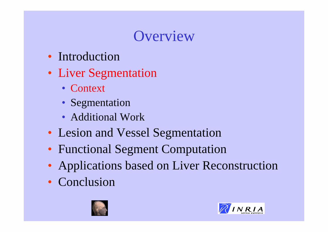
Objectives
Liver Parenchyma Hepatic Lesions Portal Vein
• Objective 1: Delineation of structures

Objectives
• Objective 2: Functional Analysis of the liver
Labeling of the Portal Vein
Couinaud Segmentation
Finding Segments to be resected

Input Images
• Source : Hospital of Strasbourg, Mulhouse
• Contrast Agent Injection :
Better Contrast
More Invasive
Directly in the Portal Vein

Input Images
• Source : Hospital of Strasbourg, Mulhouse
• Contrast Agent Injection :
•
Low Contrast
Less Invasive
Intravenous Injection

Main Difficulties
• Source of Image Contrast
• Image Texture
• Large Inter-Patient Variability

Image constrast
• Parenchyma appearance depends on the delay between injection and acquisition
Acquisition too early
Acquisition OK
Acquisition too late
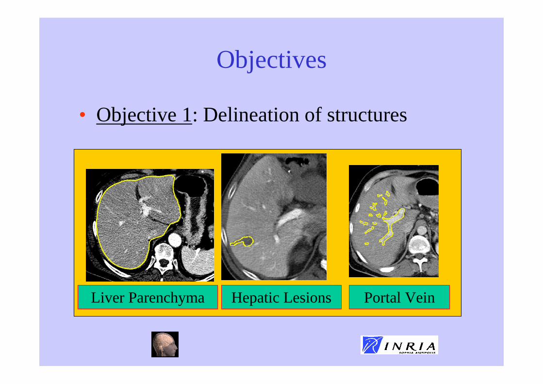
Image Texture
• Textured Aspect

Inter-Patient Variability (1)
• From Textbooks (Couinaud Thesis)
Liver Variability (40 cases)
Vein Branching Variability

Inter-Patient Variability (2)
• From real cases :

Overview• Introduction• Liver Segmentation
• Context• Segmentation• Additional Work
• Lesion and Vessel Segmentation• Functional Segment Computation• Applications based on Liver Reconstruction• Conclusion

Segmentation of the Liver
• Challenges• Texture aspect
• No clear boundary between neighboring structures

Segmentation of the Liver
• Main Approach : combine• 1) Low level image boundary detection
• 2) High Level Shape Constraint
Accurate Localization of Boundaries
Robust Reconstruction
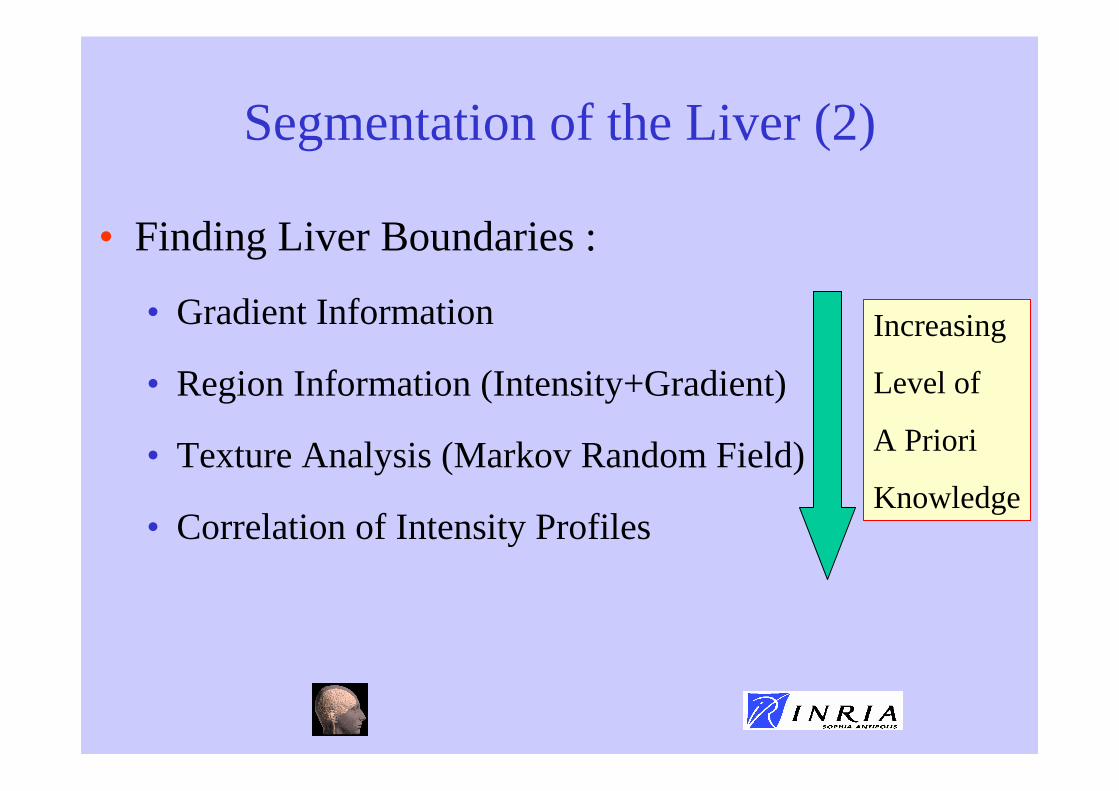
Segmentation of the Liver (2)
• Finding Liver Boundaries :
• Gradient Information
• Region Information (Intensity+Gradient)
• Texture Analysis (Markov Random Field)
• Correlation of Intensity Profiles
Increasing
Level of
A Priori
Knowledge

Segmentation of the Liver (3)
• High Level Shape Constraint :• Use of Deformable Models (Simplex Mesh).
• Minimize the sum of two energies :• Internal Energy : to constraint the shape of the
surface
• External Energy : to fit the apparent boundary from the image

Segmentation of the Liver (4)
• Effect of Internal Energy
Smoothness Constraint Shape Constraint

Segmentation of the Liver (5)
• Combination of Internal and External Energy

Segmentation of the Liver (6)
• Algorithm :• Build a Template Surface
• Perform Low-Level Processing
• Initialize Template Surface in Image
• Do :• Compute Internal and External Forces
• Update Mesh Position
• Until Convergence

Segmentation of the Liver (7)
• Generic Template Surface : Visible Human
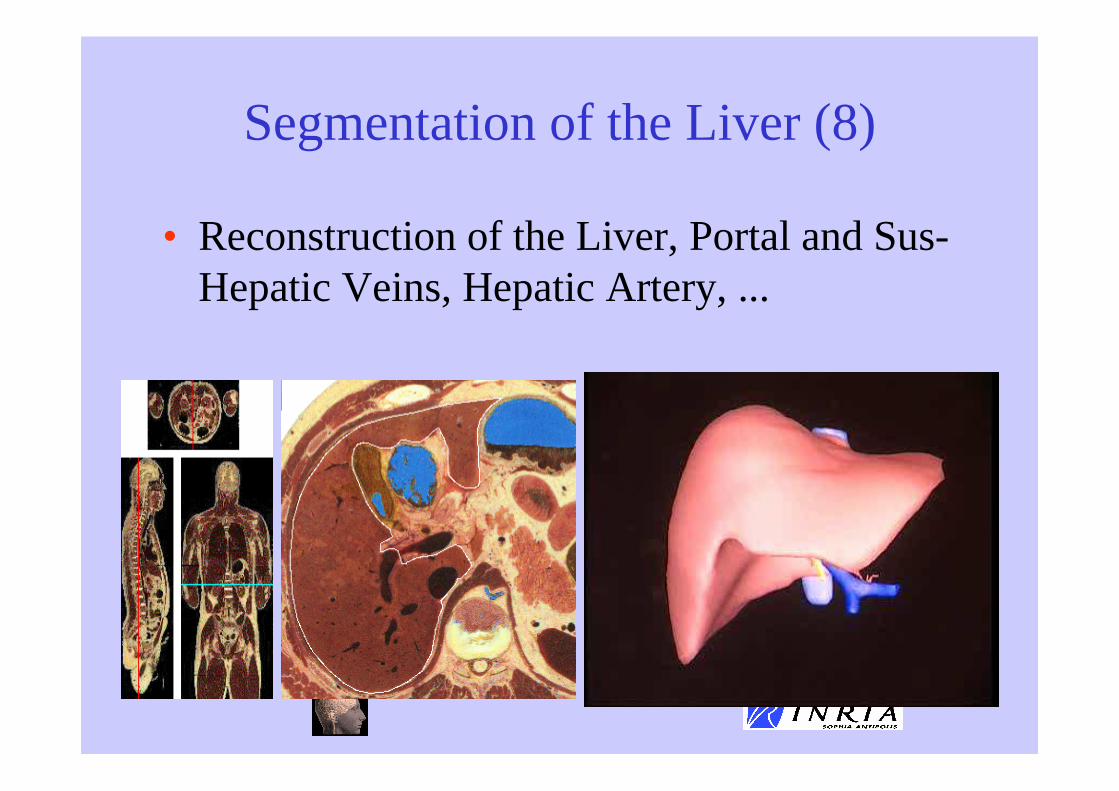
Segmentation of the Liver (8)
• Reconstruction of the Liver, Portal and Sus-Hepatic Veins, Hepatic Artery, ...

Segmentation of the Liver (9)
• Initialization :• Automatic :
• Segmentation of vertebra + ribs by thresholding
• Rough Registration with a template rib cage image
• Detection of the potential location of the liver
• Manual :• ROI drawn by the user

Segmentation of the Liver (10)
• Deformation of the template surface
Computation Time : 2mn 50 s

Segmentation of the Liver (11)
Trace of deformed model

Segmentation of the Liver (11)
• Validation study on 3 images :• Manual Delineation of each image requires 10h for
a radiologist
Image Sensitivity Spécificity Similarity Overlap Correlation1 76% 95% 84% 73% 98%2 90% 95% 93% 87% 99%3 93% 96% 95% 91% 99%
ImageInterslice Distance Mean Distance Standard Deviation Median Distance
1 4 mm 2,5 mm 3,1 mm 2,6 mm
2 1,66 mm 1,3 mm 1,8 mm 1,66 mm
3 2 mm 1,1 mm 1,6 mm 1,3 mm

Segmentation of the Liver (12)
• Robustness of the approach :• Could cope with the segmentation of roughly
70% of the database provided (45 cases) to us by hospitals
• Problems with already resected livers
• Problems often near stomach
• Possibility to interactively modify the segmentation

Overview• Introduction• Liver Segmentation
• Context• Segmentation• Additional Work
• Lesion and Vessel Segmentation• Functional Segment Computation• Applications based on Liver Reconstruction• Conclusion

Additional Work
• Since 1999, segmentation techniques have been much improved :• Initialization based on non-rigid registration
(Brain)
• Better Low-Level Detection (Brain)
• Use of Statistical Shape Model (Liver)
• Cooperation between Deformable Models (Brain)

Initialization
• Use of a brain atlas to initialize deformable model
Low-Level Detection
• Texture classification :• Off-line Computation :
• Gather a set of images representative of inter-patient variability
• Compute a set of texture descriptors for each image (statistical, model-based, signal processing)
• Train classifier (linear, SVM, Neural-Nets)
• On-line Computation :• Compute texture descriptors
• Apply classifier

Low-Level Detection (2)
• Example for Corpus Callosum with SVM

Statistical Shape Model
• Use Statistical Prior to guide the segmentation :• Build a representative set of Liver Surfaces
• Find Correspondences between Points
• Compute the Mean Liver Shape
• Compute the Covariance Matrix
• Keep main modes of variation from the mean shape (Principal Component Analysis andIndependent Component Analysis)

Statistical Shape Model (2)
Mean LiverModel
Training Setof 13 Liver
Models

Statistical Shape Model (3)
• Modes Of Variation
First Mode of Variation

Statistical Shape Model (4)
• Modes Of Variation
Second Mode of Variation

Statistical Shape Model (5)
• Modes Of Variation
Third Mode of Variation

Cooperation Between Models
• Simultaneous segmentation with a family of models• Use a Hierarchy of
Segmentations
• Use Distance constraints to preventintersection or to enforceanatomical knowledge

Cooperation Between Models
Without Distance Constraints With Distance Constraints

Overview• Introduction• Liver Segmentation• Lesion and Vessel Segmentation• Functional Segment Computation• Applications based on Liver Reconstruction• Conclusion

Histogram Analysis
• The liver includes 3 main structures : parenchyma, vessels and lesions
After Anisotropic Diffusion

Lesions and Vessel Segmentation
• Automatic Threshold Computation based on Gaussian Distribution

Lesions and Vessel Segmentation (2)
• This is a crude segmentation :
True Lesions
Loss of connectivity
Post-Processing to improve segmentation based on Prior Knowledge
Portal Vein Hepatic Lesions

Lesion Post-Processing
• Assumes 2 types of Hepatic Lesions :
Shape : NodularLocation : internal or subcapsular
Shape : Flat but min depthLocation : peripheral
Haemangioma cavernous,Carcinoma

Lesion Post-Processing (2)
• Process outcome of rough segmentation :• 1) Detect Distance from Capsule
• 2) Analyze Shape
• Remove False Positive

Lesion Final Segmentation
Nodular Lesions Peripheral Lesions

Portal Vein Segmentation
Connect Isolated Vessels
Compute Vessel Skeleton
Remove False Branches

Connect Isolated Vessels
Input Image Output Image
Combines Thresholding and Topological Closure

Skeleton Computation
• Use algorithm of Bertrand-Malandain• Fuse Junctions,
• Remove Small Branches
• Smooth center line

Remove False Branches
• Makes 3 hypothesis
Tree Structure Next to Arterial Tree
Next to Hepatic Vein
Remove Loops Remove Tangent Network
Remove Crossing Network
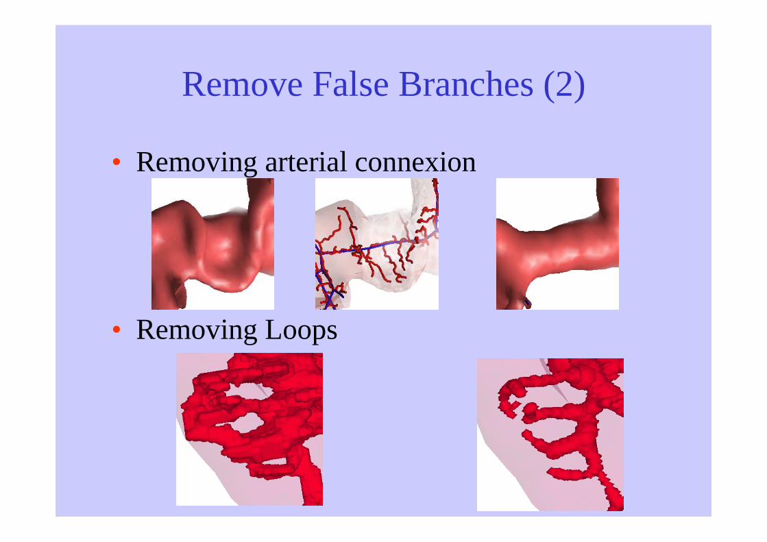
Remove False Branches (2)
• Removing arterial connexion
• Removing Loops

Remove False Branches (3)
• Final Result
Before Processing Branches
After Processing Branches

Remove False Branches (4)
• Final Result
Before Processing Branches
After Processing Branches

Overview• Introduction• Liver Segmentation• Lesion and Vessel Segmentation• Functional Segment Computation• Applications based on Liver Reconstruction• Conclusion
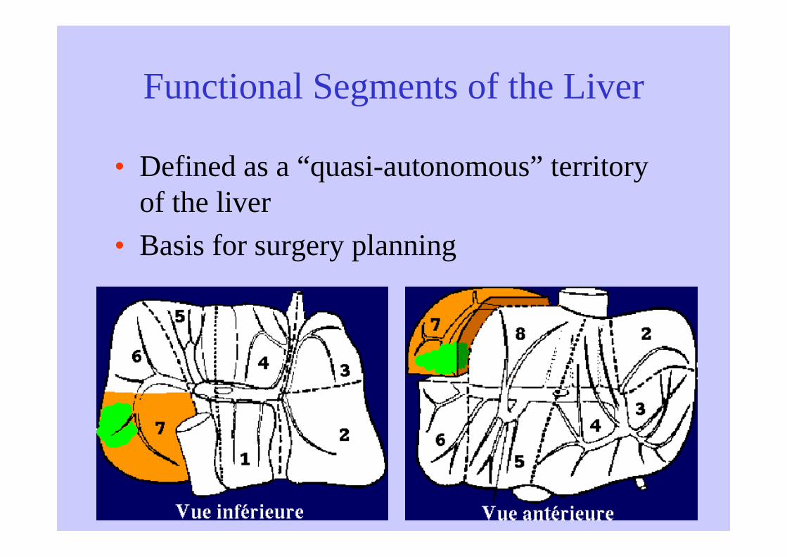
Functional Segments of the Liver
• Defined as a “quasi-autonomous” territory of the liver
• Basis for surgery planning

Functional Segments of the Liver
• Different Definitions of Functional Segments:• Couinaud with 8 segments (portal and suprahepatic
veins)
• North American (arteriobiliary systems)
• Healey and Schroy (arteriobiliary systems)
• Surgical (external landmarks)
• Still Active Debate
Liver Anatomy: Portal (and Suprahepatic) or BiliarySegmentation, C. Couinaud, Digestive Surgery, 1999;No 6, 16:459-467
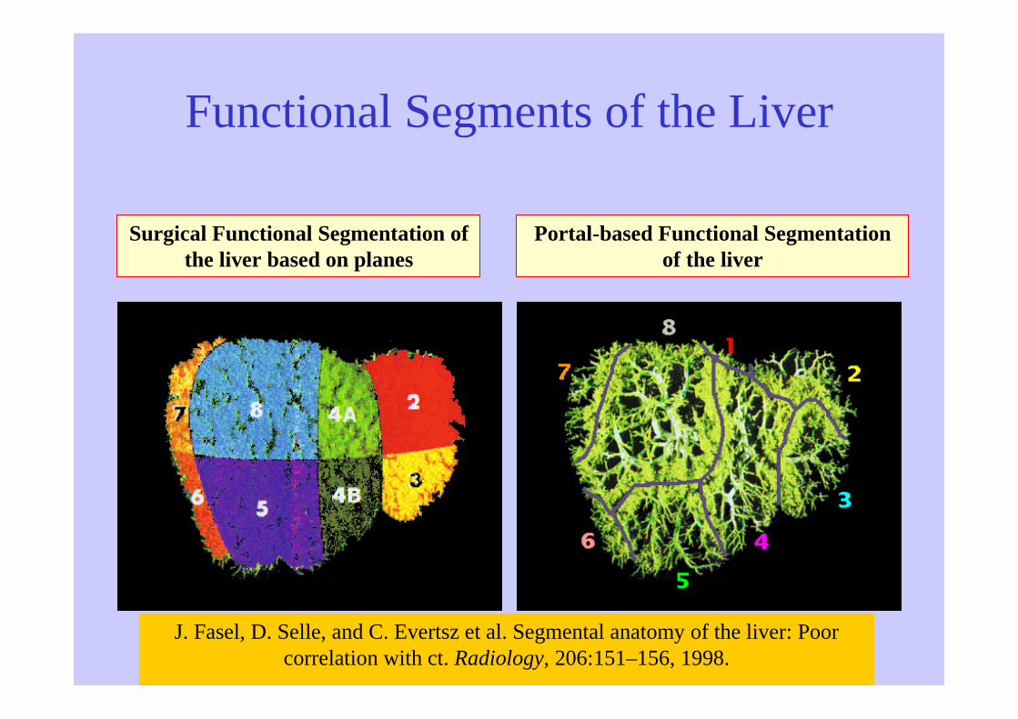
Functional Segments of the Liver
Surgical Functional Segmentation of the liver based on planes
Portal-based Functional Segmentation of the liver
J. Fasel, D. Selle, and C. Evertsz et al. Segmental anatomy of the liver: Poor correlation with ct. Radiology, 206:151–156, 1998.

Automatic Functional Segmentation
• Solely based on the Portal Vein
• Segments are regions surrounding given part of the portal vein
Labeling the portal Vein
Dilation in a non convex region
Building Segment Surfaces

Labeling of the Portal Vein
• Can be done manually
• Automatic labeling
• Use liver segmentation to
define prior knowledge of
segment location
• Use hierarchical approach : coarse to fine

Labeling of the Portal Vein (2)
• Hierarchy:
Right and Left Lobes
Two Sectors for each lobe
Two Segments per sector

Labeling of the Portal Vein (3)
• Start labeling the leaves then progress towards the root
• Solve conflicts by taking into account :• prior knowledge on segment volume
• prior knowledge on segment location
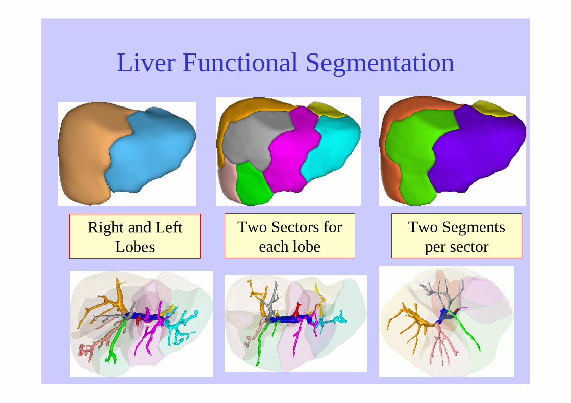
Liver Functional Segmentation
Right and Left Lobes
Two Sectors for each lobe
Two Segments per sector

Liver Functional Segmentation (2)
Overview• Introduction• Liver Segmentation• Lesion and Vessel Segmentation• Functional Segment Computation• Applications based on Liver Reconstruction
• Augmented Reality• Surgery Simulation
• Conclusion

Augmented Reality
• Fusion between pre-operative imaging and video images
Manual Registration
Ongoing Research
Joint Work with IRCAD
@copyright IRCAD

Need for Training
Hand-eyeSynchronisation
Camera being manipulated by an
assistantLong instruments
going through a fixed point in the abdomen

EPIDAURE SIMULATION [Cotin, 1997] [Picinbono, 2001] [Forest 2003]
• Hepatectomy Simulation by laparoscopy
• Include vessels and hepatic parenchyma
Couinaud Segments(source [Soler, 1998])

Modeling basic surgical gesture
Gliding Gripping
Cutting (pliers) Cutting (US)
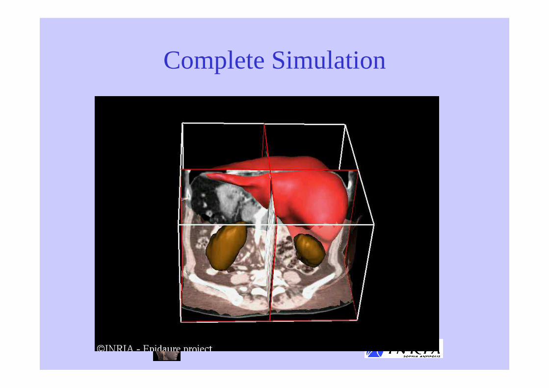
Complete Simulation

Conclusion (1)
• Liver Segmentation :• Use of Deformable Models to make the
segmentation more robust
• Interactive correction of segmentation is possible
• New CT imaging• No breathing artifacts
• Less Texture
• More Slices

Conclusion (2)
• Lesion Segmentation :• Used as Second Reading in a CAD system
• Follow-up of tumor volumetry for oncological studies (registration problems)
• Functional Segmentation :• Still an ill-posed and open problem
• Relies on good segmentation of the portal vein
• Need for more robust algorithms and more validation

Selected Bibliography
• Journals
• Conferences
S. Nicolau, A. Garcia, X. Pennec, L. Soler, and N. Ayache. Augmented reality guided radio-frequency tumor ablation. Computer Animation and Virtual World (previously the Journal of Visualization & Computer Animation), 2004.
L. Soler, G. Malandain, and H. Delingette. Segmentation automatique : application aux angioscanners 3D dufoie. Traitement du signal, 15(5):411-431, 1998
J. Montagnat and H. Delingette. Globally constrained deformable models for 3D object reconstruction. Signal Processing, 71(2):173--186, 1998
J. Montagnat and H. Delingette. A Hybrid Framework for Surface Registration and Deformable Models. In Computer Vision and Pattern Recognition, CVPR'97, San Juan, Puerto Rico, pages 1041--1046, June 1997.
L. Soler, J.-M. Clément, C. Koehl, H. Delingette, G. Malandain, N. Ayache, O. Dourthe, and J. Marescaux. An Automatic Virtual Patient Reconstruction from CT-Scans for Hepatic Surgical Planning. In Medicine Meets Virtual Reality (MMVR'2000), Studies in Health Technology and Informatic, Los Angeles, January 2000

On line references and reportshttp://www-sop.inria.fr/epidaure/