Medical Image Synthesis for Digital Simulation and Image ...

IEEE TRANSACTIONS ON MEDICAL IMAGING, VOL. 22, NO. 10, OCTOBER 2003 1185
Epidaure: A Research Project in Medical ImageAnalysis, Simulation, and Robotics at INRIA
I. INTRODUCTION
E PIDAURE1 (see Fig. 1) is the name of a research projectlaunched in 1989 at INRIA2 Rocquencourt, close to Paris,
France. At that time, after a first experience of research in com-puter vision [1] in the group of O. Faugeras, I was very enthu-siastic about the idea of transposing research results of digitalimage analysis into the medical domain. Visiting hospitals andmedical research centers, I was progressively convinced thatmedical image analysis was an important research domain byitself. In fact I had the impression that a better exploitation ofthe available medical imaging modalities would require moreand more advanced image processing tools in the short andlong-term future, not only to assess the diagnosis on more objec-tive and quantitative measurements, but also to better prepare,control, and evaluate the therapy.
To compare with the domains of computer vision and aerialimagery where research in digital image processing was ex-tremely active already, I had the feeling that medical imageanalysis would become a well-defined scientific domain by it-self. The reasons were multiple, including the fact that new dig-ital representations were available with fully volumetric imagescomposed of voxels instead of pixels, and new measures ofintensity physically linked to each medical imaging modality.Moreover, rigidity or polyhedric constraints typically used incomputer vision or aerial imagery were no longer valid withanatomical shapes. Also, the objectives of speed and full au-tomation, usual requirements in computer vision and aerial im-agery for instance, were partially replaced by robustness andaccuracy, often allowing some degree of interaction with the op-erator. In brief, a new research world was opening, motivatinga small group of scientists at INRIA to embark together in acommon research project named Epidaure.
The project started in 1989 with a young team of researchersincluding I. Herlin, J. Lévy-Véhel, and O. Monga, followedby J. P. Thirion (1990), a number of external collaborators in-cluding L. Cohen, J. M. Rocchisani, and P. Sander, and severalPh.D. students including G. Malandain, I. Cohen, C. Nastar, A.Guéziec, and J. P. Berroir. In October 1992, I decided to movefrom INRIA-Rocquencourt to the rapidly developping center ofINRIA-Sophia Antipolis, close to Nice. It was a major change
Manuscript received November 1, 2002; accepted March 5, 2003.N. Ayache is a research director at INRIA, 06902 Sophia-Antipolis Cedex,
France (e-mail: [email protected]).Digital Object Identifier 10.1109/TMI.2003.812863
1Epidaure is originally the French name of a magnificent site in Greece whichused to be the sanctuary of ancient medicine. For computer scientists, it canalso be interpreted as a recursive acronym (in French:Epidaure: Projet Images,Diagnostic AUtomatique, et RobotiquE).
2INRIA is the French Research Institute in Computer Science and AutomaticControl.
Fig. 1. This image has been the ”Logo” of the Epidaure project for a long time.It was also used as a logo of the first CVRMed conference held in Nice in 1995.(Courtesy of G. Malandain).
in the life of the project, as among permanent members, onlyJ. P. Thirion could follow the move. Hopefully, G. Malandainwas recruited on a permanent position in 1993, followed by H.Delingette in 1994, X. Pennec in 1998 and M. A Gonzalez-Ballester in 2001. J. P. Thirion left the group in 1997 to joinFocus-Imaging, and founded later3 in 2001, a company spe-cialized in quantifying disease evolution through medical imageprocessing.
The research directions of the project were progressivelydefined around the following topics: volumetric image segmen-tation, three-dimensional (3-D) shape modeling, image reg-istration, motion analysis, morphometry, and simulation [2],[3]. I will now describe and illustrate some of the scienticcontributions of the Epidaure team on these different topics.
II. THREE-DIMENSIONAL SEGMENTATION AND
SHAPE MODELING
The main objective was to design new tools to extract quanti-tative information in volumetric images in a hierachical manner[4]. The main contributions were the following ones.
• Three-dimensional edge extraction:some of our ear-liest efforts were devoted to the extraction of edgesin volumetric images. O. Monga, G. Malandain, J. M.Rocchisani, and R. Deriche proposed a generalization
3Web site of QuantifiCare company: www.quantificare.com.
0278-0062/03$17.00 © 2003 IEEE

1186 IEEE TRANSACTIONS ON MEDICAL IMAGING, VOL. 22, NO. 10, OCTOBER 2003
Fig. 2. Segmentation with deformable simplex meshes of the liver surface from a CT image and of a fetus face from a 3-D ultrasound image (courtesy of H.Delingette).
of the Canny–Deriche edge detectors in three dimen-sions [5], [6]. We then realized that some images couldbe processed more efficiently if we had access to theoriginal measurements (raw data). This was the casewith ultrasound (US) images and computed tomography(CT) images. For ultrasound images, we proposed with I.Herlin a new approach called “sonar space filtering” [7]to extract edges in images acquired in polar coordinates.For CT images, J. P. Thirion [8] proposed an originalapproach called “geometric tomography” to extract edgesdirectly from the sinogram. Both approaches showedadvantages over classical methods.
• Discrete Topology of Curves and Surfaces:G. Ma-landain and G. Bertrand designed new local criteria tocharacterize the dimension of a manifold described by aset of points in a voxel grid. These criteria are essentialto refine for instance the representation of a curve or asurface in a volumetric image [9] and they allow a newcharacterization of topologically simple points [10]. Theywere used for the extraction of skeletons [11], [12] whichcan themselves be used to guide registration procedures[13]
• Texture-based approaches: J. Lévy-Véhel andco-workers developed a system called Arthur whichcombined texture modeling and a sophisticated discrim-inant analysis scheme to select texture parameters froma training set of images. The system could computetwo–dimensional (2-D) and 3-D parameters, includingadvanced fractal and multifractal measurements, whichproved to be well adapted to a certain type of medicalimages [14].
• Modeling of tubular structures: Following the pio-neering work of G. Gerig at ETH-Zurich, we proposedwith K. Krissian and G. Malandain an original techniqueto segment vessels from a combined iconic and geometricmodel of vascular structures. The method included a first
stage of anisotropic diffusion controlled by the principaldirections of curvature of the vessel, followed by a mul-tiscale detection of the center line. The method proveditself quite efficient for the quantification of vascularstenoses, and was evaluated through a collaboration withGeneral Electric Medical Systems [15].
• Deformable surface models:Inspired by the work of Ter-zopoulos and his colleagues, we introduced with L. Cohenand I. Cohen new deformable surface models evolving innoisy volumetric images to segment anatomical shapes[16], [17]. These models were used in a variety of volu-metric images [18]. Later, H. Delingette proposed to usedeformable discrete meshes, called Simplex Meshes, quiteefficient to interactively segment anatomical structures involumetric images [19], [20]. An important property ofsimplex meshes stems from the fact that each node hasexactly three neighbors, therefore, allowing a simple ap-proximation of the mean curvature. This property allowedH. Delingette to propose dedicated schemes to preservethe regularity of the deformable surfaces during the seg-mentation process (cf. Fig. 2). Further advances were pro-posed by J. Montagnat and H. Delingette [21] to combineglobal and local deformations in a hierarchical mannerin order to improve robustness. Specific filtering methodsfor model-based segmentation of four-dimensional (4-D)ultrasound images were proposed in [22]. A survey waspublished by Montagnat and Delingette in [23].
• Extraction of surface singularities: with O. Monga andP. Sander we investigated the extraction of differentialproperties of surfaces (like the computation of first andsecond fundamental forms) by filtering the image inten-sity along iso-surfaces [24], [25]. We exploited the implicitfunction theorem and the assumption that anatomical sur-faces often correspond (at least approximately) to someiso-intensity surface. Then, J. P. Thirion and A. Gourdonproposed an efficient algorithm to extract the carefully

IEEE TRANSACTIONS ON MEDICAL IMAGING, VOL. 22, NO. 10, OCTOBER 2003 1187
Fig. 3. Left: sagittal cross section from a 3-D MR images. Middle and right: crest lines automatically computed on the surface of the brain (courtesy ofJ. P. Thirion and G. Subsol; Original MR images courtesy of Prof. R Kikinis, Brigham and Women’s Hospital, Boston).
Fig. 4. Crest lines allow accurate and fully automatic registration of high resolution MR T1 images of the same patient. In this figure, only the 240 matchedcrest lines are displayed. Change of color along a line correspond to the presence of an extremal point on the crest line. One can note that matched crestlinesare found on several anatomical surfaces (skin, skull, brain, etc.) Validation experiments showed that an overall accuracy of 0.1 mm was achieved throughthis registration procedure. (Courtesy of X. Pennec and J. P. Thirion.).
defined crest lines and extremal points in volumetricimages [26], [27]. Crest lines correspond to regions wherethe maximum principal curvature (in absolute value) isextremal in the direction of principal curvature. Intuitivelythese lines correspond to salient lines on smooth surfaces,and could be seen as a generalization of polyhedral edgeson smooth surfaces (cf. Fig. 3). On these lines, extremalpoints are characterized by the extremality of the secondprincipal curvature too. Both crest lines and extremalpoints tend to correspond to known anatomical features,in particular in the skull surface. Because these geometricentities are based on curvature properties, they remaininvariant by rigid transformations, and were extensivelyused for rigid registration as described later [28]. Themultiscale analysis of crest lines was conducted by M.Fidrich and J. P. Thirion [29], [30].
III. REGISTRATION: THE GEOMETRIC APPROACH
Registration of medical images appeared soon as a centralproblem in medical imaging. Influenced by the experience ofimage registration in computer vision, we explored first theso-called “geometric” approach, in which geometric primitivesare extracted in a first stage, and then matched against eachother in a second stage.
• Geometric Hashing:with A. Guéziec [31] we introduceda new method to match crest lines which has the nice prop-erty of exhibiting a sublinear complexity with respect tothe number of points and curves. This approach was ex-ploiting a geometric hashing technique, using five differ-ential invariants computed at each point along each curve:its curvature, torsion, and three angles between the Frenetframe (attached to the curve) and a local frame attached to

1188 IEEE TRANSACTIONS ON MEDICAL IMAGING, VOL. 22, NO. 10, OCTOBER 2003
Fig. 5. Augmented reality combining intraoperative X-ray with preoperative MR angiographies (Courtesy of J. Feldmar.).
the underlying surface (defined by the normal and the di-rections of principal curvatures). This approach was quitesuccessful to achieve a totally automatic registration ofhigh-resolution images of the same patient [typically mag-netic resonance (MR)-MR or CT-CT registration] with anexcellent accuracy. Interestingly enough, this work alsoapplied to the registration of 3-D structures of proteins[32], [33].
• Quantifying registration accuracy: To quantify this ac-curacy, X. Pennec [34]–[36] introduced a new formalismto study the uncertainty attached to the rigid transforma-tions estimated from geometric registration methods. Adifficulty to overcome was the appropriate modeling ofrotations, whose parameters belong to a manifold whichis not a vector space (Lie Group). A similar problem wasarising when modeling the uncertainty on the geometricprimitives used to guide the registration (other thansimple points): this was the case with local frames, ori-ented points, lines, etc. The proposed formalism allowedto rigorously model and propagate the uncertaintiesbetween primitives and geometric transformations andwe showed that submillimeter accuracy was definitelyachievable in the estimation of rigid registration [37] (cf.Fig. 4).
• Iterative Closest Point (ICP) algorithm for Rigidand Deformable registration: with J. Feldmar [38], wemoved from rigid to deformable registration and fromcurves to surfaces. We proposed an extension of theICP algorithm to take into account the local curvaturesof surfaces, and their variation through the applicationof affine transformations. The idea was generalized to3-D volumes in [39]. Another extension was applied tothe case when one image is a projective one, in orderto superimpose video images with medical images, an
important step toward augmented reality [40] (cf. Fig. 5).This work is currently under extension by S. Nicolauand L. Soler at IRCAD (Strasbourg, France) (see, also,Fig. 17 in Section VII).
More recently, S. Granger and X. Pennec revisited theICP algorithm in the framework of the EM algorithm inorder to better control the accuracy of geometric regis-tration in the context of image-guided oral implantology[41], [42].
IV. REGISTRATION: THE ICONIC APPROACH
After these first successes with geometric approaches, we fol-lowed a general orientation toward “iconic” approaches, whereno preliminary image segmentation is required because the in-tensities of superimposed images are directly compared. Theprice to pay is usually the requirement of a good initial solu-tion and more intensive computations.
• The Demons algorithm: revisiting the work of Chris-tensen et al., J. P. Thirion proposed a much more efficientmethod, called the Demon’s algorithm, in order to non-rigidly register monomodal images [43], [44] (cf. Fig. 6).The method was placed in a variationnal framework withP. Cachier and X. Pennec in order to explicit the minimiza-tion of a well-identified energy and applied to the trackingof anatomical structures in temporal sequences of 3-D ul-trasound images [45]. They showed how to compute thenon rigid registration field using convolutions [46]. WithD. Rey, P. Cachier showed how to insure a symmetric reg-istration field using inversion-invariant energy functions[47]. P. Cachier also proposed a new framework for vecto-rial regularization involving isotropic energies, filters andsplines [48]. With P. Cachier, J. F. Mangin, and others, wetried to reconcile the Geometric and Iconic approaches by

IEEE TRANSACTIONS ON MEDICAL IMAGING, VOL. 22, NO. 10, OCTOBER 2003 1189
Fig. 6. Iconic registration of brain images of different subjects with the Demons algorithm; Left: One slice (out of a 128) of the original images of nine differentpatients. Shapes and intensities are very different. Right: The same nine patients after nonrigid matching, re-sampling and intensity correction.The computation isperformed entirely in three dimensions. Note that the morphometrical differences are compensated for, but not the local morphological differences. (Courtesy ofJ. P. Thirion).
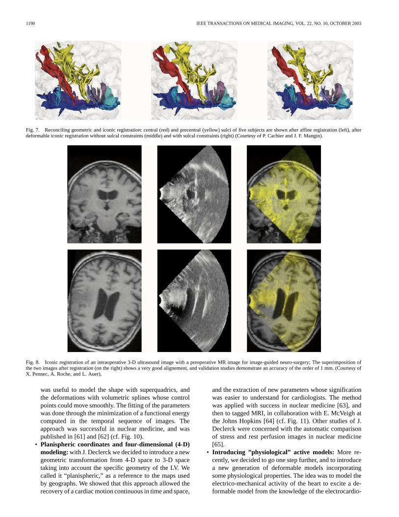
introducing in the previous approach a term related to thegeometric correspondance of sulcal lines. This led to moreaccurate results for the intersubject registration of brainimages (cf. Fig. 7) [49].
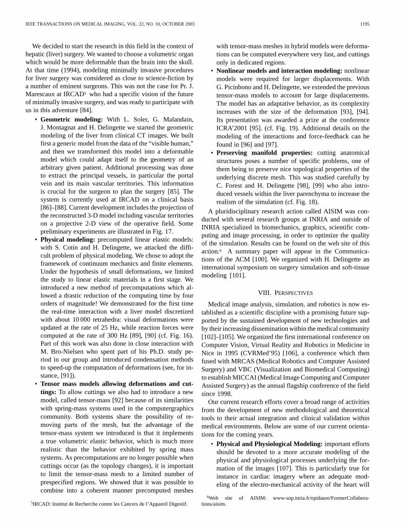
• Unifying and augmenting iconic criterions: withA. Roche and G. Malandain [50], we proposed a max-imum likelihood framework to unify the main criterionsproposed in the litterature to compare multimodal images.A. Roche introduced a new criterion from informationtheory, the correlation ratio [51] which plays an in-termediate role between linear correlation and mutualinformation. More precisely, he showed that the choiceof an optimal criterion depends on the type of expectedrelationship between the intensities of the registeredimages. For instance, an affine relationship between theintensities will lead to the use of a linear correlationcriterion, while a more general functional relationshipbetween the intensities will lead to the correlation ratiocriterion, and finally a general statistical relationship willlead to the mutual information criterion. An extension ofthis work to the difficult problem of multimodal registra-tion of multipatient images was published by A. Guimondet al. [52]. Other extensions related to the problem ofregistration of MR and ultrasound images were exploredwith X. Pennec and P. Cachier with remarkable results inimage-guided neurosurgery [53] (cf. Fig. 8).
• Building histological atlases:S. Ourselin and G. Subsoldevelopped a robust block-matching approach in order tobuild 3-D volumes from 2-D optical cross sections [54].With E. Bardinet and others, the approach was adaptedand applied to several different problems. For instance,the optical cross sections can come from microscopic ormacroscopic histological images (with or without stainingprocess), in order to correlate the detection of abnormalsignals in MRI with postmortem observations [55]. Theoptical cross sections could also come from autoradio-
graphs, and to correlate the detected activity in functionalMRI with ground truth provided by autoradiographs (Eu-ropean project MAPAWAMO). Another project was theconstruction of high resolution atlases of the basal gangliafrom optical cross sections, followed by their fusion withpreoperative MR images, in order to better control the in-troduction of electrodes in the subthalamic nuclei for thetreatment of Parkinson disease [56] (cf. Fig. 9). Extensionsto accelerate the method on parallel architectures were in-vestigated by Stephanescu, Ourselin, and Pennec [57].
V. MODELING AND ANALYZING CARDIAC MOTION
The analysis of cardiac images has been an important re-search topic within the Epidaure project.
• Active contours and differential landmarks: withI. Herlin and I. Cohen we proposed an original modelof active contours to follow the boundary of ventriclesand cardiac valves in temporal sequences of ultrasoundimages [58]. Later, with S. Benayoun, we introduceddifferential criterions to compute deformation fields fromtemporal sequences of volumetric images. The idea wasto detect and use points of high curvature to guide thematching process, and useful results were published in[59].
• Modal analysis: with C. Nastar, we introduced for thefirst time an elementary physical model of the left ven-tricle in order to decompose its periodic motion into a setof principal modes of deformation, the temporal evolutionof each mode being itself compressed through a Fourieranalysis [60]. C. Nastar later founded the company Look-ThatUp (LTU).
• Deformable superquadrics: with E. Bardinet and L.Cohen, we tried to constrain the shape of the left ventricle(LV) with a parametric model deforming itself under theaction of parameterized deformations. We showed that it
1190 IEEE TRANSACTIONS ON MEDICAL IMAGING, VOL. 22, NO. 10, OCTOBER 2003
Fig. 7. Reconciling geometric and iconic registration: central (red) and precentral (yellow) sulci of five subjects are shown after affine registration (left), afterdeformable iconic registration without sulcal constraints (middle) and with sulcal constraints (right) (Courtesy of P. Cachier and J. F. Mangin).
Fig. 8. Iconic registration of an intraoperative 3-D ultrasound image with a preoperative MR image for image-guided neuro-surgery; The superimposition ofthe two images after registration (on the right) shows a very good alignement, and validation studies demonstrate an accuracy of the order of 1 mm. (Courtesy ofX. Pennec, A. Roche, and L. Auer).
was useful to model the shape with superquadrics, andthe deformations with volumetric splines whose controlpoints could move smoothly. The fitting of the parameterswas done through the minimization of a functional energycomputed in the temporal sequence of images. Theapproach was successful in nuclear medicine, and waspublished in [61] and [62] (cf. Fig. 10).
• Planispheric coordinates and four-dimensional (4-D)modeling:with J. Declerck we decided to introduce a newgeometric transformation from 4-D space to 3-D spacetaking into account the specific geometry of the LV. Wecalled it “planispheric,” as a reference to the maps usedby geographs. We showed that this approach allowed therecovery of a cardiac motion continuous in time and space,
and the extraction of new parameters whose significationwas easier to understand for cardiologists. The methodwas applied with success in nuclear medicine [63], andthen to tagged MRI, in collaboration with E. McVeigh atthe Johns Hopkins [64] (cf. Fig. 11). Other studies of J.Declerck were concerned with the automatic comparisonof stress and rest perfusion images in nuclear medicine[65].
• Introducing ”physiological” active models: More re-cently, we decided to go one step further, and to introducea new generation of deformable models incorporatingsome physiological properties. The idea was to model theelectrico-mechanical activity of the heart to excite a de-formable model from the knowledge of the electrocardio-

IEEE TRANSACTIONS ON MEDICAL IMAGING, VOL. 22, NO. 10, OCTOBER 2003 1191
Fig. 9. Automatic 3-D reconstruction of histological atlas from 2-D stained cross sections and fusion with postmortem MR images. Top left: postmortem MRsagittal and coronal cross sections; Top right: automatic superposition of reconstructed 3-D histology; Extraction of deep grey nuclei surfaces from 3-D histology(bottom left)and superposition on postmortem MR (bottom right). This atlas can then be registered with MR images of patients with Parkinson disease in order tobetter locate the subthalamic nuclei in an image-guided stereotactic neurosurgery procedure (Courtesy of E. Bardinet, S. Ourselin, J. Yelnik, and D. Dormont).
Fig. 10. Deformable superquadrics used to model and track the motion of the cardiac left ventricle in nuclear medicine (Courtesy of E. Bardinet and L.Cohen).
gram (ECG). Then, the geometry of the model must beprecisely adjusted to contours measured in a time seriesof cardiac images using standard attraction techniques ofactive contours. The advantage of such an approach is the
potentially improved robustness with respect to sparse ormissing image data, which should allow a better use of 4-Dultrasound images (cf. Fig. 12). This is a quite ambitiousproject, involving several groups at INRIA and outside

1192 IEEE TRANSACTIONS ON MEDICAL IMAGING, VOL. 22, NO. 10, OCTOBER 2003
Fig. 11. A planispheric parametrization of the left ventricle is used to model and track the motion of individual points in the cardiac left ventriclefrom tagged MRI. Left: tracking of the tagging planes; middle and right: measured radial contraction and torsion are shown in false colors (Courtesyof J. Declerck and E. McVeigh).
Fig. 12. A new class of electromechanical models of the heart for the segmentation and analysis of cardiac images. Colors correspond to the values of thesimulated action potential, which triggers the mechanical contraction. These models will also be used to simulate the effects of radiofrequency ablation surgery(Courtesy of M. Sermesant and Y. Coudière).
Fig. 13. Crest lines were used to compare the skulls of a modern man (left) with the skull of a prehistoric man (the man of Tautavel, right). Hundreds of crestlines were automatically registered under the supervision of experts, and a global deformation field was computed in 3-D (illustrated by the center image). Theseresults were presented during one year at the ”Musée de l’Homme” in Paris for the millenium. (Courtesy of G. Subsol, B. Mafart, and M.-A. de Lumley).
INRIA (D. Hill at Guy’s Hospital and E. McVeigh at NIH).More details can be found on the web site of the ICEMAaction4 and early developments are reported in [66]–[68].
4ICEMA2 web site: www-rocq.inria.fr/sosso/icema2/icema2.html.
VI. M ORPHOMETRY
The Epidaure project was involved in the quantitative studyof shapes through several actions. The first one was related tothe automatic averaging and indexing of anatomical structures,while the second and third (measuring brain dissymmetry andmeasuring temportal evolutions in brain images) were part of a

IEEE TRANSACTIONS ON MEDICAL IMAGING, VOL. 22, NO. 10, OCTOBER 2003 1193
European Project called Biomorph, coordinated by Alan Colch-ester (Kent University). The main objective of this project wasthe development of improved techniques for measurement ofsize and shape of biological structures (morphometry).
• Averaging and indexing anatomical structures: thestudy of averaging anatomical structures was the primaryconcern of the Ph.D. dissertation of G. Subsol. He pro-posed a method based on the matching and avering ofhomologous crest lines between subjects which provedto be quite successfull on skull images [69], [70]. Themethod was also applied to compare the evolution of theskull through aging, or even through ages, by comparingfor the skulls of a contemporary and prehistoric men [71](cf. Fig. 13).
However, the method was difficult to apply to humanbrain structures, because of the large variability of crestlines between individuals. Another direction was investi-gated by A. Guimond and J. P. Thirion who proposed ageneral scheme based on the study of dense deformationfields obtained by appropriate iconic registration methods.The idea was to choose an arbitrary volumetric image asa reference image, and to compute all transformations be-tween the other images and this reference image. By av-eraging transformations they showed that it was possibleto compute a new reference image, and the method couldthen be iterated until convergence. The results were quitepromising [72]. A. Guimond also explored the use of non-rigid registration techniques for the exploration of largedatabases of MR images [73].
• Measuring brain dissymmetry: we concentrated first onthe design of statistical measures of brain dissymmetryin volumetric images to compare schizophrenic patientswith normals. Actually, a theory developped by Pr. TimCrow (Oxford) was predicting a significant reduction ofdissymmetry among schizophrenic patients, which had tobe confirmed by quantitative experiments. First, S. Primaand S. Ourselin designed a method to compute the mid-sagittal plane in 3-D brain images in a robust, objectiveand reproducible manner [74].
Then, S. Prima, J. P. Thirion, G. Subsol, and N. Roberts(Liverpool) proposed an original measure of dissymmetry:this measure requires first to symmetrize one of the twohemispheres with respect to the previously defined mid-sagittal plane, and then to compute at each point of agiven hemisphere an elastic registration between a smallregion around this point and the homologous region inthe other and symmetrized hemisphere. In case of perfectsymmetry, a rigid registration is found, whereas in caseof imperfect symmetry, a local deformation is found. Aquantitative measure of dissymmetry can be obtained bymeasuring how far the transformation is from a rigid trans-formation. Prima and Thirion proposed to use the loga-rithm of the Jacobian of the deformation (which is zerofor a rigid transformation, positive for local expansions,and negative for local contractions).
This measure provides an intuitive interpretation ofthe result, as symmetric regions correspond to vanishingvalues of the measure, whereas dissymetric regions show
significantly larger absolute values of the measure, witha sign depending on the hemisphere in which the studiedregion appears larger. Other measures were also proposedin [75], and a new methodology was proposed to comparetwo populations after intensity and spatial normalization[76]. The final result was that, at this stage, no significantstatistical difference could be found between schizo-phrenic patients and normal subjects. Although negative,this result was quite important in showing the importanceof well-controlled quantitative measurements beforedrawing final conclusions on 3-D anatomical shapes (cf.Fig. 14).
• Measuring temporal evolutions in brain images:stillin the Biomorph project, the Epidaure team was also in-volved in the subtle detection of temporal changes in timeseries of MR images of patients with multiple sclerosis.This topic was studied first by J. P. Thirion and G. Calmon[77] who proposed a deformation analysis to detect andquantify active lesions, with criterions similar to the onesabove-described (actually these criterions were introducedby Thirion and Calmon before the studies on brain dissym-metry). The method was then expanded and tested by D.Rey [78] (cf. Fig. 15) who also explored other directions,introducing statistical tests in the normalized temporal (orlongitudinal) series [79], [80].
S. Prima also proposed original statistical tests to ana-lyze longitudinal series in collaboration with L. Collins(Montreal Neurological Institute) [81]. G. Subsol, J. P.Thirion, and N. Roberts (Mariarc, Liverpool, U.K.)studied the deformation of cerebral ventricules [82] or themeasure of the cerebral atrophy [83] through time seriesof MR images.
VII. SURGERY SIMULATION
We started to work on the problem of surgery simulation in1993, initially with S. Cotin, J. Pignon, and H. Delingette. Atthe beginning we concentrated on the cutting and displace-ment of bones and face tissues in cranio-facial surgery, but wesoon decided that it was more adequate to study the simula-tion of laparoscopic surgery. Indeed, the context of minimallyinvasive surgery was appearing as more adequate for sim-ulation, as the surgeon was already working with specificinstruments through a limited number of degrees of freedom,observing the operating field on a video screen. Moreover,a specific training was required, in particular to achieve agood hand-eye synchronization, and was currently availableonly with passive mechanical systems (endotrainers) or withanimals.
A major difference between surgery simulators and flightsimulators stems from the fact that it is not sufficient tomodel the geometry of the structures of interest. Actually,a surgery simulator must provide much more than a simplevisual navigation around these structures. It is also necessaryto model physical properties in order to allow interactions suchas touching organs, gliding instruments, and eventually cuttingand/or suturing tissues and vessels. For this, not only a goodvisual feedback is necessary, but also a realistic force feedback,

1194 IEEE TRANSACTIONS ON MEDICAL IMAGING, VOL. 22, NO. 10, OCTOBER 2003
Fig. 14. Measuring brain dissymmetry with quantitative 3-D tools: after an automatic detection of the mid-sagittal plane (Left), a nonrigid registration is appliedlocally between a small region around each point in one hemisphere, and a symmetrized version of its homologous region in the other hemisphere. The deformationfield (not shown here) is analyzed in order to reveal and quantify local dissymmetries, which are represented (Right) in false colors (white color corresponds tosymmetrical regions, red (respectively blue) corresponds to regions which appear larger (respectively smaller) in the other hemisphere. (Courtesy of S. Prima).
Fig. 15. Automatic detection of evolving lesions in T2 MR images. Left: two images of the same patient acquired two weeks apart; upper middle: zoom of thecomputed apparent deformation field; lower middle: isovalues of the computed logarithm of the Jacobian of the deformation field; right: thresholded Jacobianreveals evolving lesions (all computations done in 3-D after automatic spatial and intensity normalization); (Courtesy of D. Rey. Original images courtesy ofR. Kikinis).
imposing strong constraints on the computing time. Finally,the modeling of physiological properties like for instance the
respiration or the blood circulation is also required to reach thelevel of realism expected by surgeons.
IEEE TRANSACTIONS ON MEDICAL IMAGING, VOL. 22, NO. 10, OCTOBER 2003 1195
We decided to start the research in this field in the context ofhepatic (liver) surgery. We wanted to choose a volumetric organwhich would be more deformable than the brain into the skull.At that time (1994), modeling minimally invasive proceduresfor liver surgery was considered as close to science-fiction bya number of eminent surgeons. This was not the case for Pr. J.Marescaux at IRCAD5 who had a specific vision of the futureof minimally invasive surgery, and was ready to participate withus in this adventure [84].
• Geometric modeling: With L. Soler, G. Malandain,J. Montagnat and H. Delingette we started the geometricmodeling of the liver from clinical CT images. We builtfirst a generic model from the data of the “visible human,”and then we transformed this model into a deformablemodel which could adapt itself to the geometry of anarbitrary given patient. Additional processing was doneto extract the principal vessels, in particular the portalvein and its main vascular territories. This informationis crucial for the surgeon to plan the surgery [85]. Thesystem is currently used at IRCAD on a clinical basis[86]–[88]. Current development includes the projection ofthe reconstructed 3-D model including vascular territorieson a projective 2-D view of the operative field. Somepreliminary experiments are illustrated in Fig. 17.
• Physical modeling: precomputed linear elastic models:with S. Cotin and H. Delingette, we attacked the diffi-cult problem of physical modeling. We chose to adopt theframework of continuum mechanics and finite elements.Under the hypothesis of small deformations, we limitedthe study to linear elastic materials in a first stage. Weintroduced a new method of precomputations which al-lowed a drastic reduction of the computing time by fourorders of magnitude! We demonstrated for the first timethe real-time interaction with a liver model discretizedwith about 10 000 tetrahedra: visual deformations wereupdated at the rate of 25 Hz, while reaction forces werecomputed at the rate of 300 Hz [89], [90] (cf. Fig. 16).Part of this work was also done in close interaction withM. Bro-Nielsen who spent part of his Ph.D. study pe-riod in our group and introduced condensation methodsto speed-up the computation of deformations (see, for in-stance, [91]).
• Tensor mass models allowing deformations and cut-tings: To allow cuttings we also had to introduce a newmodel, called tensor-mass [92] because of its similaritieswith spring-mass systems used in the computergraphicscommunity. Both systems share the possibility of re-moving parts of the mesh, but the advantage of thetensor-mass system we introduced is that it implementsa true volumetric elastic behavior, which is much morerealistic than the behavior exhibited by spring masssystems. As precomputations are no longer possible whencuttings occur (as the topology changes), it is importantto limit the tensor-mass mesh to a limited number ofprespecified regions. We showed that it was possible tocombine into a coherent manner precomputed meshes
5IRCAD: Institut de Recherche contre les Cancers de l’Appareil Digestif.
with tensor-mass meshes in hybrid models were deforma-tions can be computed everywhere very fast, and cuttingsonly in dedicated regions.
• Nonlinear models and interaction modeling:nonlinearmodels were required for larger displacements. WithG. Picinbono and H. Delingette, we extended the previoustensor-mass models to account for large displacements.The model has an adaptative behavior, as its complexityincreases with the size of the deformation [93], [94].Its presentation was awarded a prize at the conferenceICRA’2001 [95]. (cf. Fig. 19). Additional details on themodeling of the interactions and force-feedback can befound in [96] and [97].
• Preserving manifold properties: cutting anatomicalstructures poses a number of specific problems, one ofthem being to preserve nice topological properties of theunderlying discrete mesh. This was studied carefully byC. Forest and H. Delingette [98], [99] who also intro-duced vessels within the liver parenchyma to increase therealism of the simulation (cf. Fig. 18).
A pluridisciplinary research action called AISIM was con-ducted with several research groups at INRIA and outside ofINRIA specialized in biomechanics, graphics, scientific com-puting and image processing, in order to optimize the qualityof the simulation. Results can be found on the web site of thisaction.6 A summary paper will appear in the Communica-tions of the ACM [100]. We organized with H. Delingette aninternational symposium on surgery simulation and soft-tissuemodeling [101].
VIII. PERSPECTIVES
Medical image analysis, simulation, and robotics is now es-tablished as a scientific discipline with a promising future sup-ported by the sustained development of new technologies andby their increasing dissemination within the medical community[102]–[105]. We organized the first international conference onComputer Vision, Virtual Reality and Robotics in Medicine inNice in 1995 (CVRMed’95) [106], a conference which thenfused with MRCAS (Medical Robotics and Computer AssistedSurgery) and VBC (Visualization and Biomedical Computing)to establish MICCAI (Medical Image Computing and ComputerAssisted Surgery) as the annual flagship conference of the fieldsince 1998.
Our current research efforts cover a broad range of activitiesfrom the development of new methodological and theoreticaltools to their actual integration and clinical validation withinmedical environments. Below are some of our current orienta-tions for the coming years.
• Physical and Physiological Modeling:important effortsshould be devoted to a more accurate modeling of thephysical and physiological processes underlying the for-mation of the images [107]. This is particularly true forinstance in cardiac imagery where an adequate mod-eling of the electro-mechanical activity of the heart will
6Web site of AISIM: www-sop.inria.fr/epidaure/FormerCollabora-tions/aisim.

1196 IEEE TRANSACTIONS ON MEDICAL IMAGING, VOL. 22, NO. 10, OCTOBER 2003
Fig. 16. First demonstration of real-time interaction with a deformable model of the liver including visual and haptic feedback (Courtesy of S. CotinandH. Delingette).
Fig. 17. A geometric model of the liver is reconstructed from standard preoperative CT images and includes an automatic parcellization into main vascularterritories; This model can then be combined with an intraoperative image video image of the liver (left) to create an augmented reality visualization (Right) usedto guide the surgery procedure (Courtesy of L. Soler and J. Marescaux).
Fig. 18. Introducing vessels into a deformable model of the liver (Courtesy of C. Forest and H. Delingette).
lead to a better joint exploitation of medical images andelectrophysiological signals [108], and also to a bettersimulation of new forms of intervention like radiofre-
quency ablation for instance. In the same spirit, a betterbiophysical modeling of evolving lesions will also leadto a better detection and measure of their evolution. It

IEEE TRANSACTIONS ON MEDICAL IMAGING, VOL. 22, NO. 10, OCTOBER 2003 1197
Fig. 19. Simulation of nonlinear elastic deformations and cuttings using tensor-mass models (Courtesy G. Picinbono and H. Delingette).
Fig. 20. Activationt-maps computed from functional MRI and after an automatic parcellization of the cortex at various levels of resolution and for (p < 0:05).From left to right:t map computed with 4900, 1700 and 340 parcels. The obtained results show a better sensitivity than a standard voxel-based approach (Courtesyof G. Flandin and J. B. Poline).
Fig. 21. Left and center: 3-D reconstruction of micro-vessels from a mosaic of confocal microscopic images (Courtesy of C. Fouard, G. Malandain, and J. P.Marc-Vergnes). Right:In vivo andin situ acquisition of micro-circulation images (Courtesy of Mauna Kea Technologies and Pr. E. Vicaut).
is quite likely that the quantitative analysis of medicalimages will also play a crucial role in the study of theactual effects of new medicines. The introduction of ac-curate biomecanical and physiological models in surgicalsimulators will provide a dramatic improvement in therealism of a new generation of training systems.
• Building anatomical, histological and functional at-lases: the construction of statistical atlases includinganatomical, histological and functional statistical infor-
mation will play an important role in the field. Some openproblems are related to the statistical analysis of shapesand textures, a very active research area in which we areinvolved, in particular through a collaboration with theLoni group at the University of California at Los Angeles(Prof. Thompson and Prof. Toga) and the Pitié-SalpêtrièreHospital in Paris. The development of new statisticaltools are also central in the analysis of fMRI signals.This is part of our current work in collaboration with the

1198 IEEE TRANSACTIONS ON MEDICAL IMAGING, VOL. 22, NO. 10, OCTOBER 2003
SHFJ department of CEA [109], [110] (cf. Fig. 20) andwith the Odyssée and Vista research projects at INRIA.7
New statistical methods will be required to automaticallydiscriminate “pathological” images from “healthy” ones[111]–[113]. It is likely that the exploitation of largedatabases distributed over the planet will require GridComputing techniques.
• Scalable Microscopic and Macroscopic Models :theintroduction of volumetric microscopic imagery bothinvivo andin situ will provide new opportunities for fusingmicro and macroscopic information in a more systematicmanner (cf. Fig. 21). A potential outcome will be thepossibility to acquire for instance image-guided confocalmicroscopic images providing the same information asclassical biopsies, with the advantage of real-time in-formation and minimally invasive procedures.8 The de-velopment of scalable models going from nanoscopic tomacroscopic scales will certainly play an important rolein the joint analysis of microscopic and macroscopic im-ages, with the huge potential of molecular imaging forgenomics studies. Also, specific data-mining proceduresbased on the image content still have to be invented.
• Image-Guided Therapy and Medical Robotics:Image-guided radiotherapy is an important area of research forwhich we plan to build and evaluate new systems with NiceHospital and Institut Gustave Roussy in Paris. We also planto pursue our work on image-guided liver surgery withIRCAD in Strasbourg, and on image-guided neurosurgerywith the Pitié-Salpêtrière Hospital in Paris and probablysoon with Brigham and Women’s Hospital in Boston.Real-time registration and deformation analysis will cer-tainly require specific software and hardware solutionsinvolving for instance parallelism and Grid Computing.The development of medical robotics also poses a numberof challenging problems, both for image-guided therapy(see for instance the work of the CHIR group and EveCoste-Manière at INRIA)9 and for surgery simulation.The coupling of image analysis, both preoperative andintraoperative with medical robotics opens new avenuesfor applications, including for instance a virtual suppres-sion of cardiac and/or respiratory motions during theintervention.
These directions are by no means exhaustive of all the pos-sible directions of research in the field. The permanent evolu-tion of the current technologies and the regular introduction ofnew ones creates a constant emergence of new problems, main-taining an exceptional level of exciting research activity, whichI feel is not going to decrease during the next decade(s)!
NICHOLAS AYACHE, Guest EditorINRIASophia-Antipolis, F-06902 France
7www-sop.inria.fr/odyssee; www.irisa.fr/vista .8Web site of Mauna Kea Technologies : www.maunakeatech.com9www-sop.inria.fr/chir.
ACKNOWLEDGMENT
The work presented in this invited editorial is the work ofa team, and I wish to express my wholehearted thanks to allthe persons who have contributed to the Epidaure project untiltoday.
In particular I want to express my warmest thanks to the re-searchers currently enrolled in the project H. Delingette, M. A.Gonzalez-Ballester, G. Malandain and X. Pennec, and to thepast members I. Cohen, I. Herlin, J. Lévy-Véhel, O. Monga, andJ. P. Thirion.
I want to thank the research engineers E. Bardinet and G.Subsol in Sophia-Antipolis, and previously P. Sander in Roc-quencourt for their important contributions to the results of theteam. I also wish to thank our research and system engineersJ. Bertot in Sophia-Antipolis, and J. P. Chièze and J. B. Giorgiin Rocquencourt. Special thanks are due to our past and cur-rent development engineers F. Betting, J. D. Lemaréchal, andM. Traina.
I want to thank all the past and current Ph.D. students of theteam for their various contributions to the research project (inchronological order): G. Malandain, I. Cohen, A. Guéziec, J. P.Berroir, C. Nastar, S. Benayoun, H. Delingette, A. Gourdon, E.Bardinet, J. Feldmar, G. Subsol, M. Fidrich, S. Fernández-Vidal,X. Pennec, M. Bro-Nielsen, S. Cotin, J. Declerck, L. Soler, A.Guimond, K. Krissian, J. Montagnat, G. Picinbono, S. Prima, A.Roche, P. Cachier, S. Ourselin, O. Migneco, D. Rey, J. Stoeckel,C. Forest, M. Sermesant, S. Granger, G. Flandin, S. Nicolau,A. Pitiot, C. Fouard, J. Dauguet, V. Moreau, R. Stefanescu, V.Arsigny, G. Dugas-Phocion, O. Clatz, and P. Y Bondiau.
I wish to thank G. Kahn, the Scientific Director of INRIA,who provided an incredibly stimulating support in many formsto the project, and my former Research Director O. Faugeras,from whom I learned (among many other important things) thehigh exigence of scientific research activities. At its creation,the project also received the strong support of A. Schroeder, theformer director of INRIA Rocquencourt in 1989, and then thestrong support of P. Bernhard, former director of INRIA Sophia-Antipolis, when the group moved to Sophia-Antipolis in 1992.
Special thanks are due to the past assistants of the team, N.Gaudechoux, F. Pezé, and E. Lière, and to our current assistantI. Strobant.
Last but not least, I wish to thank all our academic, clin-ical and industrial partners, whose list would be too long to becitedin extensohere. Among them, special thanks are due to M.Brady (Oxford, U.K.) who spent a memorable sabbatical withus in 1994–1995, to L. Cohen, J. F. Mangin, J. B. Poline, andN. Roberts for their longstanding academic collaboration andto the following (very incomplete!) list of medical doctors fortheir precious collaboration: L. Auer, J. Bittoun, P. Cinquin, A.Colchester, J. Darcourt, D. Dormont, R. Kikinis, D. Le Bihan,C. Lebrun-Frenet, J. Marescaux, Y. Marsault, J. M. Rocchisani,and J. Yelnik.
REFERENCES
[1] N. Ayache,Artificial Vision for Mobile Robots – Stereo-Vision and Mul-tisensor Perception. Cambridge, MA: MIT-Press, 1991, 342 pages.

IEEE TRANSACTIONS ON MEDICAL IMAGING, VOL. 22, NO. 10, OCTOBER 2003 1199
[2] N. Ayache, J. Boissonnat, L. Cohen, B. Geiger, J. Lévy-Véhel, O.Monga, and P. Sander, “Steps toward the automatic interpretation of3-D images,” in3D Imaging in Medicine, K. Höhne, H. Fuchs, andS. Pizer, Eds. Berlin, Germany: Springer-Verlag, 1990, vol. 60, ser.NATO ASI, pp. 107–120.
[3] N. Ayache, “Medical computer vision, virtual reality and robotics, apromising research track,”Image Vision Comput., vol. 13, no. 4, pp.295–313, May 1995.
[4] N. Ayache, J. Boissonnat, E. Brunet, L. Cohen, J. Chièze, B. Geiger,O. Monga, J. Rocchisani, and P. Sander, “Building highly structuredvolume representations in 3D medical images,” inComputer AssistedRadiology (CAR’89), vol. 1808, Berlin, Germany, 1989, pp. 765–772.
[5] O. Monga, R. Deriche, G. Malandain, and J.-P. Cocquerez, “Recursivefiltering and edge tracking: Two primary tools for 3-D edge detection,”Image Vision Comput., vol. 9, no. 4, pp. 203–214, Aug. 1991.
[6] J.-M. Rocchisani, O. Monga, R. Deriche, and G. Malandain, “Automaticmultidimensional segmentation of nuclear medicine images using fastrecursive filters,”Eur. J. Nucl. Med., vol. 16, p. 419, 1990.
[7] I. L. Herlin and N. Ayache, “Feature extraction and analysis methods forsequences of ultrasound images,”Image Vision Comput., vol. 10, no. 10,pp. 673–682, 1992.
[8] J.-P. Thirion, “Direct extraction of boundaries from computed tomog-raphy scans,”IEEE Trans. Med. Imag., vol. 13, pp. 322–328, June 1994.
[9] G. Malandain, G. Bertrand, and N. Ayache, “Topological segmentationof discrete surfaces,”Int. J. Comput. Vision, vol. 10, no. 2, pp. 183–197,1993.
[10] G. Bertrand and G. Malandain, “A new characterization of three-dimen-sional simple points,”Pattern Recogn. Lett., vol. 15, no. 2, pp. 169–175,Feb. 1994.
[11] G. Malandain and S. Fernández-Vidal, “Euclidean skeletons,”Image Vi-sion Comput., vol. 16, no. 5, pp. 317–327, Apr. 1998.
[12] G. Bertrand and G. Malandain, “A note on “Building skeleton modelsvia 3D medial surface-axis thinning algorithms”,”Graph. Models ImageProcess., vol. 57, no. 6, pp. 537–538, Nov. 1995.
[13] G. Malandain, S. Fernández-Vidal, and J. Rocchisani, “Mise en corre-spondance d’objets 3D par une approche mécanique: Application auximages médicales multimodales,”Traitement du Signal, vol. 11, no. 6,pp. 541–558, 1994.
[14] J. Levy-Vehel, P. Mignot, and J. Berroir, “Multifractal, texture and imageanalysis,” presented at theIEEE Conf. Computer Vision and PatternRecognition, CVPR’92, Urbana-Champaign, IL.
[15] K. Krissian, G. Malandain, N. Ayache, R. Vaillant, and Y. Trousset,“Model-based detection of tubular structures in 3D images,”Comput.Vision Image Understanding, vol. 80, no. 2, pp. 130–171, 2000.
[16] I. Cohen, L. Cohen, and N. Ayache, “Using deformable surfaces tosegment 3-D images and infer differential structures,”Comput. Vision,Graph. Image Process.: Image Understanding, vol. 56, no. 2, pp.242–263, 1992.
[17] L. Cohen and I. Cohen, “Finite element methods for active contourmodels and balloons for 2-D and 3-D images,”IEEE Trans. PatternAnal. Machine Intell., vol. 15, Nov. 1993.
[18] N. Ayache, P. Cinquin, I. Cohen, L. D. Cohen, I. L. Herlin, F. Leitner,and O. Monga, “Segmentation of complex 3D medical objects: A chal-lenge and a requirement for computer assisted surgery planning andperforming,” inComputer Integrated Surgery. Cambridge, MA: MIT-Press, 1995.
[19] H. Delingette, M. Hébert, and K. Ikeuchi, “Shape representation andimage segmentation using deformable surfaces,”Image Vision Comput.,vol. 10, no. 3, pp. 132–144, Apr. 1992.
[20] H. Delingette, “General object reconstruction based on simplexmeshes,”International Journal of Computer Vision, vol. 32, no. 2, pp.111–146, Sept. 1999.
[21] H. Delingette and J. Montagnat, “Shape and topology constraintson parametric active contours,”Computer Vision and Image Under-standing, vol. 83, no. 2, pp. 140–171, 2001.
[22] J. Montagnat, M. Sermesant, H. Delingette, G. Malandain, and N. Ay-ache, “Anisotropic filtering for model based segmentation of 4D cylin-drical echocardiographic images,”Pattern Recogn. Lett., vol. 24, no.4–5, pp. 815–828, 2003.
[23] J. Montagnat and H. Delingette, “A review of deformable surfaces:Topology, geometry and deformation,”Image Vision Comput., vol. 19,no. 14, pp. 1023–1040, 2001.
[24] O. Monga, P. Sander, and N. Ayache, “From voxel to intrinsic surfacefeatures,”Image Vision Comput., vol. 10, no. 6, pp. 403–417, 1992.
[25] O. Monga, N. Ayache, and P. Sander, “Using uncertainty to link edgedetection and local surface modeling,”Image Vision Comput., vol. 10,no. 6, pp. 673–682, 1992.
[26] J.-P. Thirion and A. Gourdon, “Computing the differential characteris-tics of isointensity surfaces,”J. Comput. Vision Image Understanding,vol. 61, no. 2, pp. 190–202, Mar. 1995.
[27] J.-P. Thirion, “The extremal mesh and the understanding of 3D sur-faces,”Int. J. Comput. Vision, vol. 19, no. 2, pp. 115–128, 1996.
[28] , “New feature points based on geometric invariants for 3D imageregistration,”Int. J. Comput. Vision, vol. 18, no. 2, pp. 121–137, May1996.
[29] M. Fidrich, “Iso-surface extraction in nD applied to tracking featurecurves across scale,”Image Vision Comput., vol. 16, no. 8, pp. 545–556,1998.
[30] M. Fidrich and J.-P. Thirion, “Stability of corner points in scale space:The effect of small nonrigid deformations,”Comput. Vision Image Un-derstanding, vol. 72, no. 1, pp. 72–83, 1998.
[31] A. Guéziec and N. Ayache, “Smoothing and matching of 3-D-spacecurves,”Int. J. Comput. Vision, vol. 12, no. 1, pp. 79–104, Jan. 1994.
[32] X. Pennec and N. Ayache, “A geometric algorithm to find small buthighly similar 3D substructures in proteins,”Bioinformatics, vol. 14, no.6, pp. 516–522, 1998.
[33] A. Guéziec, X. Pennec, and N. Ayache, “Medical image registrationusing geometric hashing,”IEEE Computational Sci. Eng. (Special Issueon Geometric Hashing), vol. 4, no. 4, pp. 29–41, Oct.-Dec. 1997.
[34] X. Pennec and N. Ayache, “Uniform distribution, distance and expecta-tion problems for geometric features processing,”J. Math. Imag. Vision,vol. 9, no. 1, pp. 49–67, July 1998.
[35] X. Pennec and J.-P. Thirion, “A framework for uncertainty and validationof 3D registration methods based on points and frames,”Int. J. Comput.Vision, vol. 25, no. 3, pp. 203–229, 1997.
[36] X. Pennec, “Toward a generic framework for recognition based on un-certain geometric features,”Videre: J. Comput. Vision Res., vol. 1, no.2, pp. 58–87, 1998.
[37] X. Pennec, N. Ayache, and J.-P. Thirion, “Landmark-based registrationusing features identified through differential geometry,” inHandbook ofMedical Imaging, I. Bankman, Ed. New York: Academic, 2000, ch.31, pp. 499–513.
[38] J. Feldmar and N. Ayache, “Rigid, affine and locally affine registrationof free-form surfaces,”Int. J. Comput. Vision, vol. 18, no. 2, May 1996.
[39] J. Feldmar, J. Declerck, G. Malandain, and N. Ayache, “Extension of theICP algorithm to nonrigid intensity-based registration of 3D volumes,”Comput. Vision Image Understanding, vol. 66, no. 2, pp. 193–206, May1997.
[40] J. Feldmar, N. Ayache, and F. Betting, “3D-2D projective registration offree-form curves and surfaces,”J. Comput. Vision Image Understanding,vol. 65, no. 3, pp. 403–424, 1997.
[41] S. Granger and X. Pennec, “Multi-scale EM-ICP: A fast and robust ap-proach for surface registration,” inLecture Notes in Computer Science,A. Heyden, G. Sparr, M. Nielsen, and P. Johansen, Eds. Berlin, Ger-many: Springer-Verlag, 2002, vol. 2353, European Conference on Com-puter Vision (ECCV 2002).
[42] S. Granger, X. Pennec, and A. Roche, “Rigid point-surface registra-tion using an variant of ICP for computer guided oral implantology,”in Lecture Notes in Computer Science, W. Niessen and M. Viergever,Eds. Berlin, Germany: Springer-Verlag, 2001, vol. 2208, Proceedings.4th Int. Conf. on Medical Image Computing and Computer-Assisted In-tervention (MICCAI’01), pp. 752–761.
[43] J.-P. Thirion, “Image matching as a diffusion process: An analogy withMaxwell’s demons,”Med. Image Anal., vol. 2, no. 3, pp. 243–260, 1998.
[44] , “Diffusing models and applications,” inBrain Warping, A. W.Toga, Ed. New York: Academic, 1998, ch. 9, pp. 143–155.
[45] X. Pennec, P. Cachier, and N. Ayache, “Tracking brain deformations intime-sequences of 3D US images,”Pattern Recogn. Lett., vol. 24, no.4–5, pp. 801–813, Feb. 2003.
[46] P. Cachier and X. Pennec, “3-D nonrigid registration by gradient de-scent on a Gaussian-windowed similarity measure using convolutions,”in Proc. IEEE Workshop Mathematical Methods in Biomedical ImageAnalysis, MMBIA’00, 2000, pp. 182–189.
[47] P. Cachier and D. Rey, “Symmetrization of the nonrigid registrationprobem using inversion-invariant energies: Application to multiple scle-rosis,” inLecture Notes in Computer ScienceBerlin, Germany, 2000,vol. 1935, Medical Image Computing and Computer Assisted Interven-tion, MICCAI’00, pp. 472–481.
[48] P. Cachier and N. Ayache, “Isotropic energies, filters and splines forvectorial regularization,” J. Math. Imag. Vision, 2003, to be published.

1200 IEEE TRANSACTIONS ON MEDICAL IMAGING, VOL. 22, NO. 10, OCTOBER 2003
[49] P. Cachier, J.-F. Mangin, X. Pennec, D. Rivière, D. Papadopoulos-Or-fanos, J. Régis, and N. Ayache, “Multisubject nonrigid registration ofbrain MRI using intensity and geometric features,” in , W. Niessenand M. Viergever, Eds. Berlin, Germany: Springer-Verlag, 2001, vol.2208, Proceedings of the 4th International Conference on MedicalImage Computing and Computer-Assisted Intervention (MICCAI’01),pp. 734–742.
[50] A. Roche, G. Malandain, and N. Ayache, “Unifying maximum like-lihood approaches in medical image registration,”Int. J. Imag. Syst.Technol. (Special Issue on 3D Imag.), vol. 11, no. 1, pp. 71–80, 2000.
[51] A. Roche, G. Malandain, X. Pennec, and N. Ayache, “The correlationratio as a new similarity measure for multimodal image registration,” inLecture Notes in Computer Science, vol. 1496, Proceedings of the FirstInternational Conference on Medical Image Computing and Computer-Assisted Intervention (MICCAI’98). Berlin, Germany, Oct. 1998, pp.1115–1124.
[52] A. Guimond, A. Roche, N. Ayache, and J. Meunier, “Multimodal brainwarping using the demons algorithm and adaptative intensity correc-tions,” IEEE Trans. Med. Imag., vol. 20, pp. 58–69, Jan. 2001.
[53] A. Roche, X. Pennec, G. Malandain, and N. Ayache, “Rigid registrationof 3-D ultrasound with MR images: A new approach combining inten-sity and gradient information,”IEEE Trans. Med. Imag., vol. 20, pp.1038–1049, Oct. 2001.
[54] S. Ourselin, A. Roche, G. Subsol, X. Pennec, and N. Ayache, “Recon-structing a 3D structure from serial histological sections,”Image VisionComput., vol. 19, no. 1–2, pp. 25–31, Jan. 2001.
[55] E. Bardinet, A. Colchester, A. Roche, Y. Zhu, Y. He, S. Ourselin, B.Nailon, S. Hojjat, J. Ironside, S. Al-Sarraj, N. Ayache, and J. Wardlaw,“Registration of reconstructed post mortem optical data with MR scansof the same patient,” inLecture Notes in Computer Science, W. Niessenand M. Viergever, Eds. Berlin, Germany: Springer-Verlag, 2001, vol.2208, Proceedings of the Fourth International Conference on MedicalImage Computing and Computer-Assisted Intervention (MICCAI’01),pp. 957–965.
[56] E. Bardinet, S. Ourselin, D. Dormont, G. Malandain, D. Tande, K.Parain, N. Ayache, and J. Yelnik, “Co-registration of histological,optical and MR data of the human brain,” inLecture Notes inComputer Science, T. Dohi and R. Kikinis, Eds. Berlin, Germany:Springer-Verlag, 2002, vol. 2488, Medical Image Computing andComputer-Assisted Intervention (MICCAI’02), pp. 548–555.
[57] S. Ourselin, R. Stefanescu, and X. Pennec, “Robust registration of multi-modal images: Toward real-time clinical applications,” inLecture Notesin Computer Science, T. Dohi and R. Kikinis, Eds. Berlin, Germany:Springer-Verlag, 2002, vol. 2488, Medical Image Computing and Com-puter-Assisted Intervention (MICCAI’02), pp. 140–147.
[58] N. Ayache, I. Cohen, and I. Herlin, “Medical image tracking,” inAc-tive Vision, A. Blake and A. Yuille, Eds. Cambridge, MA: MIT Press,1992, ch. 17, pp. 285–302.
[59] S. Benayoun and N. Ayache, “Dense nonrigid motion estimation insequences of medical images using differential constraints,”Int. J.Comput. Vision, vol. 26, no. 1, pp. 25–40, 1998.
[60] C. Nastar and N. Ayache, “Frequency-based nonrigid motion analysis:Application to four dimensional medical images,”IEEE Trans. PatternAnal. Machine Intell., vol. 18, pp. 1067–1079, Nov. 1996.
[61] E. Bardinet, L. Cohen, and N. Ayache, “Tracking and motion analysisof the left ventricle with deformable superquadrics,”Med. Image Anal.,vol. 1, no. 2, 1996.
[62] , “A parametric deformable model to fit unstructured 3D data,”Comput. Vision Image Understanding, vol. 71, no. 1, pp. 39–54, 1998.
[63] J. Declerck, J. Feldmar, and N. Ayache, “Definition of a four-dimen-sional continuous planispheric transformation for the tracking and theanalysis of left-ventricle motion,”Med. Image Anal., vol. 2, no. 2, pp.197–213, June 1998.
[64] J. Declerck, N. Ayache, and E. McVeigh, “Use of a 4D PlanisphericTransformation for the Tracking and the Analysis of LV Motion withTagged MR Images,” INRIA, Paris, France, Res. Rep. 3535, 1998.
[65] J. Declerck, J. Feldmar, M. Goris, and F. Betting, “Automatic registra-tion and alignment on a template of cardiac stress and rest SPECT re-oriented images,”IEEE Trans. Med. Imag., vol. 16, pp. 727–737, Dec.1997.
[66] M. Sermesant, Y. Coudière, H. Delingette, N. Ayache, and J. Désidéri,“An electro-mechanical model of the heart for cardiac image analysis,”in Lecture Notes in Computer ScienceBerlin, Germany, 2001, vol.2208, 4th Int. Conf. on Medical Image Computing and Computer-As-sisted Intervention (MICCAI’01), pp. 224–231.
[67] N. Ayache, D. Chapelle, F. Clément, Y. Coudière, H. Delingette,J. Désidéri, M. Sermesant, M. Sorine, and J. Urquiza, “Towardmodel-based estimation of the cardiac electro-mechanical activity fromECG signals and ultrasound images,” inLecture Notes in ComputerScience, T. Katila, I. Magnin, P. Clarysse, J. Montagnat, and N. J. ,Eds. Helsinki, Finland, 2001, vol. 2230, Functional Imaging andModeling of the Heart (FIMH’01), pp. 120–127.
[68] M. Sermesant, C. Forest, X. Pennec, H. Delingette, and N. Ayache,“Biomechanical model construction from different modalities: Applica-tion to cardiac images,” inProc. Medical Image Computing and Com-puter-Assisted Intervention (MICCAI’02), vol. 1, T. Dohi and R. Kikinis,Eds., Tokyo, Japan, Sept. 2002, pp. 714–721.
[69] G. Subsol, “Crest lines for curve based warping,” inBrain Warping, A.W. Toga, Ed. New York: Academic, 1998, ch. 13, pp. 225–246.
[70] G. Subsol, J.-P. Thirion, and N. Ayache, “A general scheme for auto-matically building 3D morphometric anatomical atlases: Application toa skull atlas,”Med. Image Anal., vol. 2, no. 1, pp. 37–60, 1998.
[71] G. Subsol, B. Mafart, A. Silvestre, and M. de Lumley, “3D image pro-cessing for the study of the evolution of the shape of the human skull:Presentation of the tools and preliminary results,” inProc. Three-Dimen-sional Imaging in Paleoanthropology and Prehistoric Archaeology, vol.1049, B. Mafart, H. Delingette, and G. Subsol, Eds., Liege, Belgium,2002, pp. 37–45.
[72] A. Guimond, J. Meunier, and J.-P. Thirion, “Average brain models: Aconvergence study,”Comput. Vision Image Understanding, vol. 77, no.2, pp. 192–210, 2000.
[73] A. Guimond, G. Subsol, and J.-P. Thirion, “Automatic MRI databaseexploration and applications,”Int. J. Pattern Recogn. Artif. Intell., vol.11, no. 8, pp. 1345–1366, Dec. 1997.
[74] S. Prima, S. Ourselin, and N. Ayache, “Computation of the mid-sagittalplane in 3-D brain images,”IEEE Trans. Med. Imag., vol. 21, pp.122–138, Feb. 2002.
[75] J.-P. Thirion, S. Prima, G. Subsol, and N. Roberts, “Statistical analysisof normal and abnormal dissymmetry in volumetric medical images,”Med. Image Anal., vol. 4, no. 2, pp. 111–121, 2000.
[76] S. Prima, “Étude de la Symétrie Bilatérale en Imagerie Cérébrale Volu-mique,” Thèse de Sciences, Université Paris XI, Orsay, France, 2001.
[77] J.-P. Thirion and G. Calmon, “Deformation analysis to detect and quan-tify active lesions in three-dimensional medical image sequences,”IEEETrans. Med. Imag., vol. 18, pp. 429–441, May 1999.
[78] D. Rey, G. Subsol, H. Delingette, and N. Ayache, “Automatic detectionand segmentation of evolving processes in 3D medical images: Applica-tion to multiple sclerosis,”Med. Image Anal., vol. 6, no. 2, pp. 163–179,June 2002.
[79] D. Rey, C. Lebrun-Fresnay, G. Malandain, S. Chanalet, N. Ayache, andM. Chatel, “A new method to detect and quantify evolving MS lesions bymathematical operators,”Neurology (Suppl.: Proc. 52nd Annu. MeetingAmer. Acad. Neurol.), vol. 54, p. 123, May 2000.
[80] D. Rey, J. Stoeckel, G. Malandain, and N. Ayache, “A spatio-temporalmodel-based statistical approach to detect evolving multiple sclerosislesions,” presented at theIEEE Workshop Mathematical Methods inBiomedical Image Analysis, MMBIA’01, Kauia, HI, Dec. 2001.
[81] S. Prima, N. Ayache, A. Janke, S. Francis, D. Arnold, and L. Collins,“Statistical analysis of longitudinal MRI data: Application for detectionof disease activity in MS,” inLecture Notes in Computer Science, T.Dohi and R. Kikinis, Eds. Berlin, Germany, 2002, vol. 2488, MedicalImage Computing and Computer-Assisted Intervention (MICCAI’02),pp. 363–371.
[82] N. Roberts, G. Subsol, J.-P. Thirion, M. Puddephat, and G. W. White-house, “Automatic analysis of deformation of the cerebral ventricles,”Magn. Reson. Mater. Phys., Biol., Med., vol. 4, no. 2, pp. 62–63, 1996.
[83] G. Subsol, N. Roberts, M. Doran, and J.-P. Thirion, “Automatic analysisof cerebral atrophy,”Magn. Reson. Imag., vol. 15, no. 8, pp. 917–927,1997.
[84] J. Marescaux, J.-M. Clément, V. Tassetti, C. Koehl, S. Cotin, Y. Russier,D. Mutter, H. Delingette, and N. Ayache, “Virtual reality applied to he-patic surgery simulation : The next revolution,”Ann. Surg., vol. 228, no.5, pp. 627–634, Nov. 1998.
[85] L. Soler, G. Malandain, and H. Delingette, “Segmentation automatique: Application aux angioscanners 3D du foie,”Traitement du Signal, vol.15, no. 5, pp. 411–431, 1998.
[86] L. Soler, J.-M. Clément, C. Koehl, H. Delingette, G. Malandain, N. Ay-ache, O. Dourthe, and J. Marescaux, “An automatic virtual patient re-construction from ct-scans for hepatic surgical planning,” presented atthe Medicine Meets Virtual Reality (MMVR’2000), Los Angeles, CA,Jan. 2000.

IEEE TRANSACTIONS ON MEDICAL IMAGING, VOL. 22, NO. 10, OCTOBER 2003 1201
[87] L. Soler, H. Delingette, G. Malandain, J. Montagnat, N. Ayache, J.-M.Clément, C. Koehl, O. Dourthe, D. Mutter, and J. Marescaux, “A fullyautomatic anatomical, pathological and fonctionnal segmentation fromct-scans for hepatic surgery,”Proc. SPIE (Medical Imaging 2000), pp.246–255, Feb. 2000.
[88] L. Soler, O. Dourthe, G. Malandain, J. Montagnat, H. Delingette, andN. Ayache, “A new 3D segmentation of liver neoplasm, portal venoussystem and parenchymal contours,” presented at theEur. Congr. Radi-ology, Vienne, Austria, Mar. 2000.
[89] N. Ayache, S. Cotin, H. Delingette, J.-M. Clément, J. Marescaux, andM. Nord, “Simulation of endoscopic surgery,”J. Minimally InvasiveTherapy Allied Technologies (MITAT), vol. 7, no. 2, pp. 71–77, July1998.
[90] S. Cotin, H. Delingette, and N. Ayache, “Real-time elastic deformationsof soft tissues for surgery simulation,”IEEE Trans. Visual., Comput.Graphics, vol. 5, pp. 62–73, Jan.-Mar. 1999.
[91] M. Bro-Nielsen and S. Cotin, “Real-time volumetric deformable modelsfor surgery simulation using finite elements and condensation,” in Proc.Computer Graphics Forum, Eurographics’96, vol. 15, pp. 57–66.
[92] S. Cotin, H. Delingette, and N. Ayache, “A hybrid elastic model allowingreal-time cutting, deformations and force-feedback for surgery trainingand simulation,”Visual Comput., vol. 16, no. 8, pp. 437–452, 2000.
[93] G. Picinbono, H. Delingette, and N. Ayache, “Non-linear anisotropicelasticity for real-time surgery simulation,”Graphical Models, vol. 65,no. 5, pp. 305–321, Sept. 2003.
[94] , “Modèle déformableélastique nonlinéaire pour la simulation dechirurgie en temps réel,”Les Comptes Rendus de l’Académie des Sci-ences CRAS, C.R. Biologies, 2002.
[95] , “Non-linear and anisotropic elastic soft tissue models for medicalsimulation,” presented at theIEEE International Conference Roboticsand Automation—ICRA2001, Seoul, Korea.
[96] G. Picinbono, J.-C. Lombardo, H. Delingette, and N. Ayache, “Im-proving realism of a surgery simulator: Linear anisotropic elasticity,complex interactions and force extrapolation,”J. Visual., Comput.Animat., vol. 13, no. 3, pp. 147–167, July 2002.
[97] J.-C. Lombardo, M.-P. Cani, and F. Neyret, “Real-time collision detec-tion for virtual surgery,” inComputer Animation, Geneva, Switzerland,May 1999.
[98] C. Forest, H. Delingette, and N. Ayache, “Cutting simulation of mani-fold volumetric meshes,” in , T. Dohi and R. Kikinis, Eds. Berlin, Ger-many: Springer-Verlag, 2002, vol. 2488, Medical Image Computing andComputer-Assisted Intervention (MICCAI’02), pp. 235–244.
[99] , “Removing tetrahedra from a manifold mesh,” inProc. ComputerAnimation (CA’02), Geneva, Switzerland, pp. 225–229.
[100] H. Delingette and N. Ayache, “Hepatic surgery simulation,”Commun.ACM, 2004, to be published.
[101] N. Ayache and H. Delingette, Eds.,Lecture Notes in ComputerScience. Berlin, Germany: Springer-Verlag, June 2003, vol. 2673,Surgery Simulation and Modeling of Soft Tissues.
[102] N. Ayache, “L’analyse automatique des images médicales,état de l’art etperspectives” (in French),Annales de l’Institut Pasteur, vol. 9, no. 1, pp.13–21, Avril-juin 1998.
[103] H. Delingette, “Toward realistic soft tissue modeling in medical simula-tion,” Proc. IEEE (Special Issue on Surgery Simulation), pp. 512–523,Mar. 1998.
[104] J. Duncan and N. Ayache, “Medical image analysis: Progress over twodecades and the challenges ahead,”IEEE Trans. Pattern Anal. MachineIntell., vol. 22, pp. 85–106, Jan 2000.
[105] N. Ayache, “Medical imaging informatics: From digital anatomyto virtual scapels and image guided therapy,” inIntroduction to the2002 IMIA Yearbook of Medical Informatics. Stuttgart, Germany:Schattauer, 2002.
[106] N. Ayache, Ed.,Lecture Notes in Computer Science. Berlin, France:Springer-Verlag, Proc. Computer Vision, Virtual Reality and Roboticsin Medicine (CVRMed’95), p. 541.
[107] N. Ayache, Ed., “Computational models for the human body,” inHand-book of Numerical Analysis. Amsterdam, The Netherlands: Elsevier,2004, to be published.
[108] M. Sermesant, Y. Coudière, H. Delingette, N. Ayache, J. Sainte-Marie,D. Chapelle, F. Clément, and M. Sorine, “Progress toward model-basedestimation of the cardiac electromechanical activity from ECG signalsand 4D images,” presented at theModeling and Simulation for Com-puter-Aided Medicine and Surgery Conf.(MS4CMS’02), Rocquencourt,France, 2002.
[109] G. Flandin, F. Kherif, X. Pennec, D. Rivière, N. Ayache, and J.-B. Po-line, “A new representation of fMRI data using anatomo-functional con-straints,” presented at theNeuroImage Conf. (HBM’02), Sendai, Japan,2002.
[110] , “Parcellation of brain images with anatomical and functional con-straints for fMRI data analysis,” inProc. IEEE Int. Symp. BiomedicalImaging, 2002, pp. 907–910.
[111] J. Stoeckel, J.-B. Poline, G. Malandain, N. Ayache, and J. Darcourt,“Smoothness and degrees of freedom restrictions when using spm99,”NeuroImage, vol. 13, no. 6, p. 259, June 2001.
[112] J. Stoeckel, G. Malandain, O. Migneco, P. Koulibaly, P. Robert, N. Ay-ache, and J. Darcourt, “Classification of SPECT images of normal sub-jects versus images of alzheimer’s disease patients,” inLecture Notesin Computer Science, vol. 2208, Proceedings of the Fourth InternationalConference on Medical Image Computing and Computer-Assisted In-tervention (MICCAI’01), W. Niessen and M. Viergever, Eds., Utrecht,The Netherlands, pp. 666–674.
[113] O. Migneco, M. Benoit, P. Koulibaly, I. Dygai, C. Bertogliati, P. Desvi-gnes, P. Robert, G. Malandain, F. Bussière, and J. Darcourt, “Perfusionbrain SPECT and statistical parametric mapping analysis indicate thatapathy is a cingulate syndrome: A study in alzheimer’s disease and non-demented patients,”Neuroimage, vol. 13, pp. 896–902, 2001.