
Heart Prelab Questions - State College of Florida, Manatee...
18
Heart Prelab Questions 1. Describe the location of the heart. 2. Briefly describe the heart’s four chambers. 3. Compare atrioventricular and semilunar valves. 4. Briefly describe coronary circulation. 5. Match the blood vessel with the heart chamber ____1. aorta (A) right atrium ____2. superior vena cava (B) right ventricle ____3. pulmonary trunk (C) left atrium ____4. pulmonary veins (D) left ventricle 1
Transcript of Heart Prelab Questions - State College of Florida, Manatee...
Heart Prelab Questions 1. Describe the location of the heart. 2. Briefly describe the heart’s four chambers. 3. Compare atrioventricular and semilunar valves. 4. Briefly describe coronary circulation. 5. Match the blood vessel with the heart chamber ____1. aorta (A) right atrium
____2. superior vena cava (B) right ventricle
____3. pulmonary trunk (C) left atrium
____4. pulmonary veins (D) left ventricle
1
Heart Anatomy The objective of this lab is to learn heart anatomy from: A. A short lecture on heart anatomy B. Illustrations from this manual, your textbook, handouts, lab charts and the
Anatomy and Physiology Coloring Book, if you wish to purchase it. C. Models D. Use of A.D.A.M. Interactive Anatomy software E. Dissection of a sheep heart F. Use of Interactive Physiology software G. Use of a microscope slide of cardiac muscle (optional) H. Demonstration and analysis of electrocardiograms or EKG’s (optional) 1. Position of the heart – figure 20.1 in your text. 2. Heart anatomy Identify the following anatomical features of the heart from
figures 20.3, 20.4, 20.5 and 20.6 in your text. ♦Chambers and associated vessels The right atrium (1) receives deoxygenated blood from the superior
vena cava (2), inferior vena cava (3) and coronary sinus (4). The right ventricle (5) pumps deoxygenated blood into the pulmonary
trunk artery (6), which then divides to form the pulmonary arteries (7). The left atrium (8) receives oxygenated blood from the lungs through pulmonary veins (9).
The left ventricle (10) pumps oxygenated blood into the aorta (11) to all parts (systems) of the body.
♦The anterior interventricular sulcus (12) and the posterior interventricular sulcus (13) are prominent grooves that separate the ventricles externally. The more obvious of the two is the anterior sulcus. It extends at an oblique left to right angle down the front of the heart. Later, you will see some prominent coronary vessels that extend down these sulci.
♦The interventricular septum (14) is a wall that separates the two ventricles internally. ♦Pectinate muscles (35) in the walls of the auricles of the atria. ♦Trabeculae carnae (36) in the walls of the ventricles. ♦Heart valves •Atrioventricular valves (between the atria and ventricles) consist of
flaps or cusps that close to stop the back-flow of blood from the ventricles back into the atria.
▪The tricuspid valve (15) is located between the right atrium and right ventricle. Look for its three flaps (cusps)
Find the chordae tendineae (16) running downward from edges of valve to the papillary muscles (17), which extend from the lining of the right ventricle. What are the functions of the chordae tendineae and papillary muscles?
2
▪The bicuspid (mitral) valve (18) is located between the left atrium and left ventricle. Look for the two stronger cusps
Notice the stronger chordae tendineae and the larger, stronger papillary muscles. Why is the bicuspid valve and associated structures stronger than the tricuspid valve and associated structures?
•Semilunar valves (at base of pulmonary trunk and aorta) consist of three cusps that fill with blood to stop the back-flow of blood from these large vessels into the ventricles.
▪The pulmonary (pulmonic) semilunar valve (19), or just pulmonary valve, is at the base of pulmonary trunk.
▪The aortic semilunar valve (20), or just aortic valve, is at the base of the aorta. 3. Coronary circulation - figure 20.8 in your text. ♦Coronary arteries
•Openings of the right and left coronary arteries just above the aortic semilunar valve
•Right coronary artery (21) ▪marginal branch (22) ▪posterior interventricular branch (23) •Left coronary artery (24) ▪anterior interventricular branch (25) or left anterior
descending coronary artery (LAD) arises from the left coronary artery and passes downward through the anterior interventricular sulcus to supply blood to most of the left ventricle and some of the right ventricle.
▪circumflex branch (26) arises from the left coronary artery and loops around the left atrium to supply blood to the back of the left ventricle.
♦Coronary veins •Coronary sinus (4) receives blood from the coronary veins and
collects it into the right atrium. •Great cardiac vein (28) passes up through the anterior interventricular sulcus, and then loops around the left atrium to the back of the heart where it empties into the coronary sinus.
•Middle cardiac vein (29) passes up through the posterior interventricular sulcus and collects into the coronary sinus on posterior aspect of heart.
•Posterior cardiac vein (30) passes up the posterior wall of the left ventricle to the left of the middle cardiac vein and collects into the coronary sinus.
•Small cardiac vein (31) collects the heart's right inferior margin then collects into the coronary sinus.
3
4. Pericardium and pericardial cavity and the layers of the heart wall - Figure 20.2 in your text. ♦Parietal pericardium is tough and fibrous with a smooth inner lining of simple squamous called mesothelium
♦Pericardial Cavity ♦Visceral pericardium also called the epicardium (32). It is on the surface of
the heart and is the same mesothelium as the inner lining of the parietal ericardium. ♦Myocardium (33) composed mostly of cardiac muscle forms most of the heart wall.
♦Endocardium (34) is a membrane that lines the inner wall (chambers) of the heart. It consists primarily of a simple squamous layer that is continuous with the endothelium of the blood vessels.
5. Flow of blood through the heart. Figure 20.7 in your text. Think about it as
you study heart anatomy.
6. Heart Sounds – Use a stethoscope to listen to heart sounds Heart sounds, called auscultations, can be heard with a stethoscope. Use the provided stethoscope, and information on pages 718 and 719 in your text to listen to your lab partner’s heart sounds. Note that figure 20.15, in your text, shows the various placements of the stethoscope for optimal listening. You may also use a lab computer, and the following website to for proper placement of the stethoscope: http://www.meddean.luc.edu/lumen/MedEd/GrossAnatomy/thorax0/heart/valves.html Be sure to swab the ear pieces of a stethoscope both before and after use. You may use a lab computer, and go to the following websites to listen to normal and abnormal heart sounds: http://www.rain.org/~landon/Heartweb/index.html http://www.bioscience.org/atlases/heart/sound/sound.htm http://members.aol.com/kjbleu/heartsounds.html Be sure the computer speaker is turned on.
4
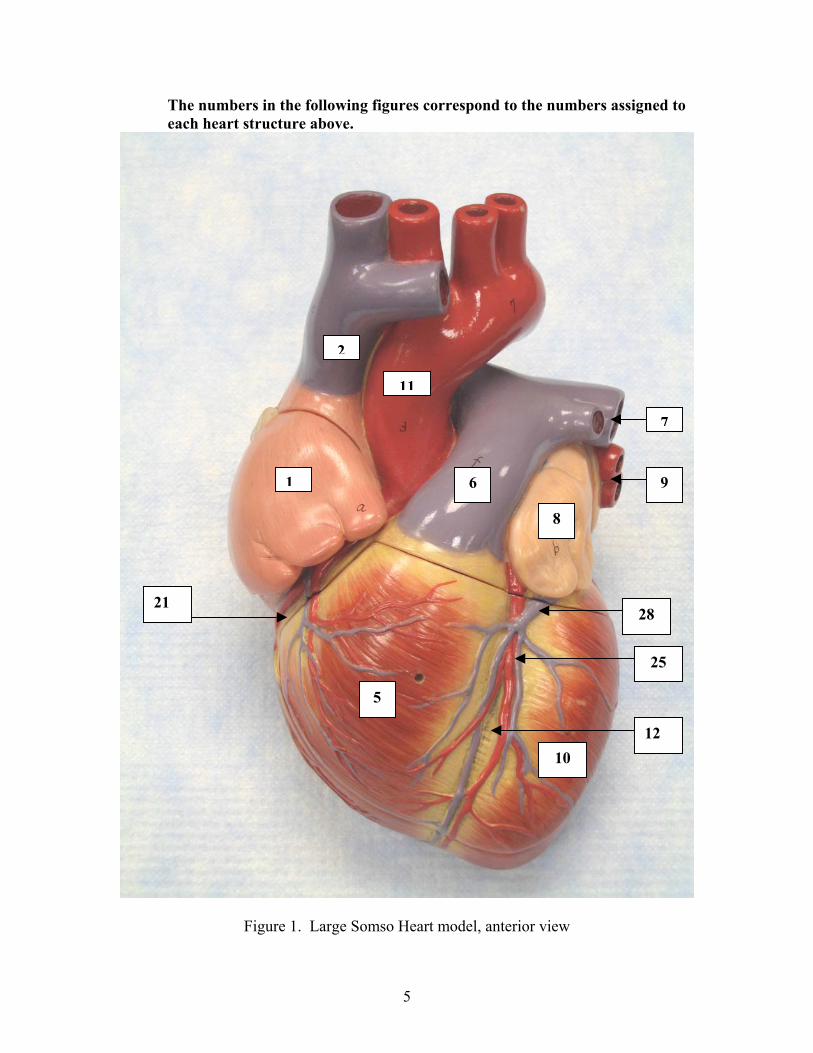
The numbers in the following figures correspond to the numbers assigned to each heart structure above.
1
6 9
21
Figur
1
e 1. Lar
2
8
28
2
5
110
ge Somso Heart model, anterior view
5
7
1
5
2
11
7 7
9 8
9
28
4 1 26
3
30
31 10
29
5
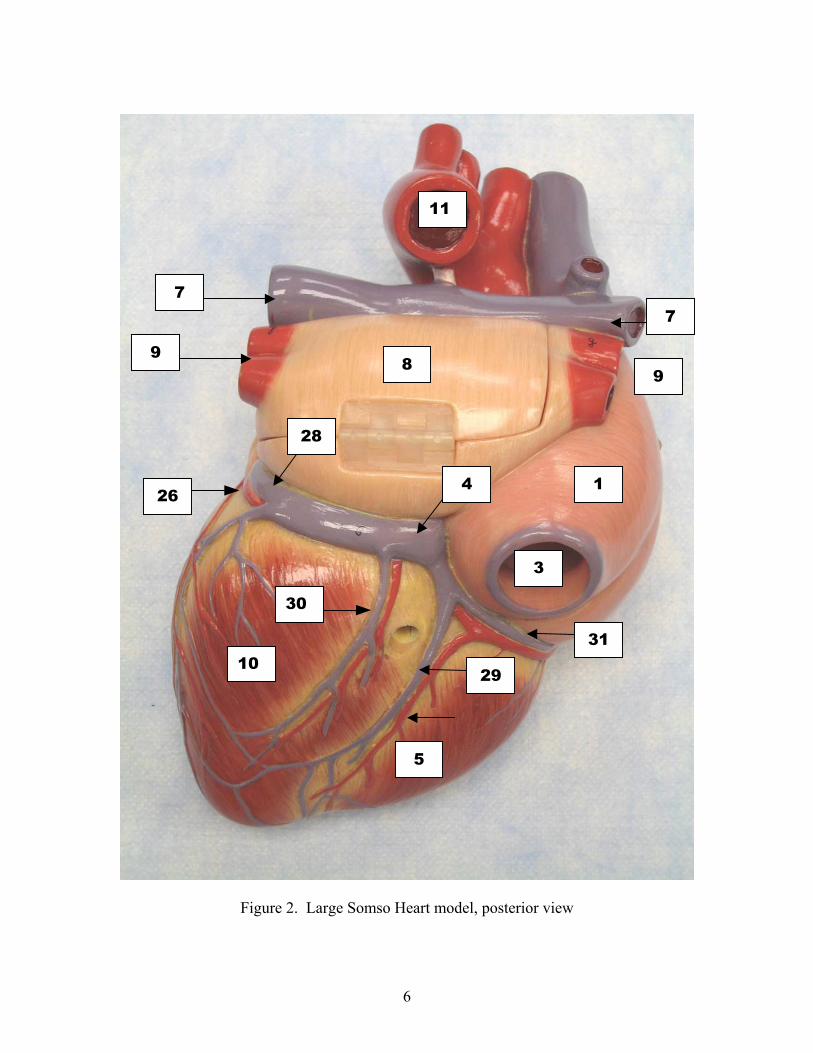
Figure 2. Large Somso Heart model, posterior view
6
1
8
15
16
17
5 10
14
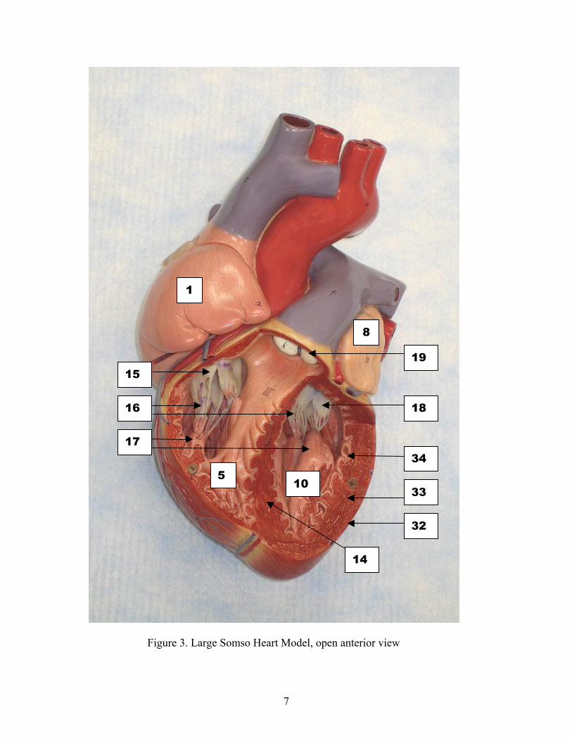
Figure 3. Large Somso Heart Model, open anterior view
7
19
18
34
33
32
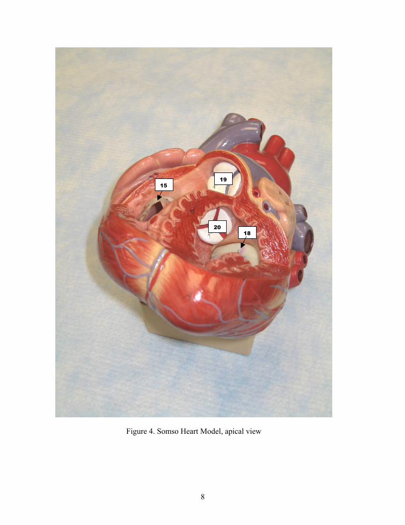
Figure 4. Somso Heart Mode
8
19
20l, apica
18
15
l view
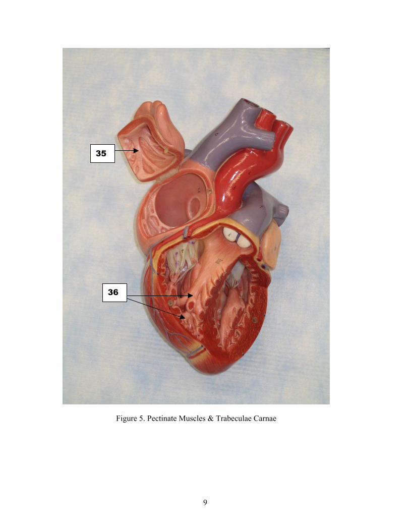
35
36
Figure 5. Pectinate Muscles & Trabeculae Carnae
9

2
11
7 1
6 9
8
21 26
28 22
5 25
12 10
Figure 6. Carolina Heart, anterior view
10
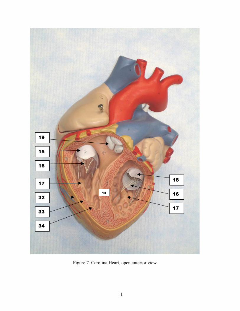
19
15
16
18 17
32
33
34
Figure 7. Caro
14
16
17
lina Heart, open anterior view
11
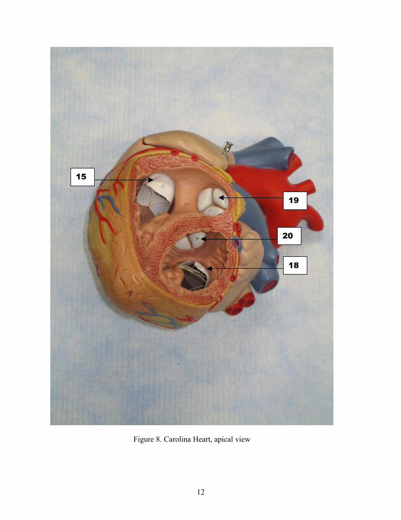
15
19
20
18
Figure 8. Carolina Heart, apical view
12
7. Heart anatomy using A.D.A.M. Interactive anatomy: From now on, every lab will have a part where students will learn anatomy using both models and software called A.D.A.M. Interactive anatomy. Students will alternate using models and A.D.A.M.; half of the students will be on models while the other half will be at the computers studying A.D.A.M.. Instructions on the Use of A.D.A.M. Interactive Anatomy a. To run A.D.A.M. Interactive Anatomy, click on the A.D.A.M. Interactive Anatomy icon on the desktop. b. The next screen has four choices: Dissectible Anatomy, Atlas Anatomy, 3D Anatomy and Slide show. You will start with Dissectible Anatomy, so click on that icon. c. The next screen will include the large A.D.A.M. Interactive Anatomy window with a box marked Open. In the middle of the Open box, you will likely see a smaller box with the message “Please wait loading A.D.A.M. Interactive Anatomy”. This box will not go away unless you click on the black area of the larger AIA window. d. You will see four tabs marked Dissectible Anatomy, Atlas Anatomy, 3D Anatomy, and Pictures. If you are going to use Dissectible Anatomy, click on Open. If you are going to use Atlas Anatomy or 3D Anatomy click on the respective tab and then click Open. You may change content from within Dissectible Anatomy by clicking on the folder icon, at the top left of the window, and selecting Content. e. If you choose Dissectible Anatomy, the next window will show the image of a man. Expand this window by clicking on the square icon ( ) in the upper right of the window. f. You will see a tool bar along the left side of the window. Below is a list of this tool bar’s icons and their functions. 1) Select the arrow icon at the upper left, point to any structure, hold-down the left mouse button and the name of the structure will appear. 2) Select the magnifying glass to change the size of the image. There are only two image sizes, large and small. 3) Selecting the transparency box allows you to see the next layer in certain cases. 4) Below the transparency box, you will see three light bulbs. The upper left bulb selects the normal mode; it displays the image in full color. The upper right bulb extracts and displays a specific, selected structure; you will seldom need to use this function. The lower left bulb allows you to highlight a selected structure. The highlighted structure is in color while everything else is grayed-out. You will find the highlight mode very useful.
13
5) The pull-down menu to the right of the highlight bulb allows you to highlight the parts of a specific system. You might want to do this to get an idea of how the selected system fits in with other systems. 6) Below the highlight bulb, is the gender icon. The male is the default gender. Select the female gender only when specifically studying female anatomy. 7) To the right of the gender icon is the view icon. It allows you to choose six different anatomical views, depending on which one is required. 8) Below the gender and view icons is a long, vertical Depth Bar. You will use this tool most often because it selects different layers of anatomy. Each layers is assigned a number indicated by the number in the bar slider. The quickest way to navigate through the layers is to click on the slider and drag it to the number called for in the lab guide. You may also move one layer at a time, by using the arrows at the top and bottom of the depth bar. The bottom arrow moves deeper one layer at a time, and the top arrow moves out (more superficial) one layer at a time. g. To the extreme right of the dissectible anatomy window is an icon that looks like a small person. If you place the mouse pointer over this icon, a rectangle will appear. Navigate by holding down the mouse button and dragging this rectangle over the part of the anatomy that you need to study. You can also navigate by using the slider bars at he bottom and right side of the window, but this method is slower. Please do not be intimidated by AIA. If you get stuck, someone will help you. A.D.A.M. Exercises using Dissectible Anatomy 1. Select layer #173 and identify the following: a. right atrium b. right ventricle c. auricle of left atrium d. left ventricle e. anterior interventricular sulcus f. superior vena cava g. ascending aorta h. pulmonary trunk i. left pulmonary artery j. right pulmonary artery 2. Select layer #174 and identify the following: a. anterior, septal and posterior cusps of the tricuspid (right AV) valve b. tendinous cords (chordae tendineae) c. anterior and septal papillary muscles d. anterior, right and left cusps of the pulmonary valve e. trabeculae carneae (inside left ventricle) f. pectinate muscle of auricle of right atrium
14
3. Select layer #175 and identify the following: a. right pulmonary artery b. superior vena cava c. arch of aorta d. pulmonary trunk e. right pulmonary artery f. left pulmonary artery g. right superior, right inferior, left superior and left inferior pulmonary veins h. inferior vena cava 4. Select layer #169 and identify the pericardial sac 5. Select layer #170 and identify the epicardium (same as visceral pericardium) 6. Select layer #171 and identify the following: a. interior of pericardial sac b. right coronary artery c. right marginal artery (branch) d. anterior cardiac veins e. anterior interventricular artery (left anterior descending coronary artery) f. great cardiac vein 8. Sheep heart dissection. Each pair of students or each table will dissect a sheep
heart. The procedures for the dissection are outlined below. Read these carefully before attempting the dissection.
a. Remove the parietal pericardium and fat from the heart, if it is present. Be careful not to cut the major vessels too close to the heart. Remove as much fat as possible.
b. Locate the front of the heart (ventral part in a cow or sheep). The anterior
interventricular sulcus extends diagonally across the front. c. Now, locate the three most prominent vessels emerging from the heart. They
are, from front to back, the pulmonary trunk, the aorta and the superior vena cava. Turn the heart so that the top is facing you. The usually prominent pulmonary trunk extends upward, from right to left, between the two atrial auricles (ear-like appendages) at the top of the heart. Just behind the “trunk”, the aorta curves to the left. The “trunk” may run under the aorta if enough length of vessels is present. There is a major branch emerging from the aorta and some students mistake this for the superior vena cava. You can tell it is a branch of the aorta if you use the larger end of a metal (Mall) probe and gently push it from this vessel into the aorta. Posterior to the aorta is the ultra-thin superior vena cava. It is so thin that is usually collapsed and difficult to locate. If you cannot find it, ask your instructor to help you.
15
d. The smaller pulmonary veins connect to the left atrium but are usually not present in your specimen because they may have been cut too close to the heart. You can often see one or more of their openings into the left atrium.
e. The coronary sinus is a bag-like vein that collects blood from the coronary
veins into the right atrium. It runs along a deep groove on the back of the heart between the atria and the ventricles. Its opening into the atrium is located inside the right atrium.
f. You are now ready to open the heart and look at its internal anatomy. The
procedures for this are as follows: 1) Locate the back of the heart. Place the metal probe (not a needle probe)
into the superior vena cava. Gently work the probe straight down through the right atrium into the right ventricle. The probe will be aiming directly at the apex of the heart. Use scissors to cut along the probe toward, but not quite to, the apex of the heart.
2) Probe up anteriorly through the ventricle into the pulmonary trunk. If
your heart is hard, this may not be easy. You might have to cut through the upper end of the pulmonary trunk to get the probe on through. Cut along the probe to open up the ventricle and the “trunk.” Be sure not to cut across the interventricular sulcus; stay to the right of it.
3) Fold back the walls of the right atrium and right ventricle along the first
cut and identify the following: a) inside of right atrium b) opening of the inferior vena cava c) opening of coronary sinus d) pectinate muscles in the wall of the auricle of the right atrium e) tricuspid valve (three flaps) f) chordae tendineae g) papillary muscles h) inside of right ventricle i) epicardium, myocardium and endocardium 4) Fold back the ventricular wall and the pulmonary trunk along the second
cut and identify the following: a) interventricular septum b) pulmonary (pulmonic) semilunar valve at base of pulmonary trunk 5) You are now ready to explore the interior of the left side. Cut through the
left auricle and into the front of the left ventricle. The left ventricle is considerably thicker than the right one, so it will be a little more difficult. Make sure your cut is parallel to and about one-half inch to the left of the interventricular septum. Remember not to cut across the interventricular septum.
16
6) Open the left atrium and the left ventricle and identify the following: a) inside of the left atrium b) bicuspid (mitral) valve (thicker than the tricuspid) c) chordae tendineae (heavier than the ones on the right side) d) papillary muscles (larger than the one on the right side) e) inside of the left ventricle f) interventricular septum g) trabeculae carnae in the wall of the ventricle 7) To see the aortic semilunar valve, you must make a longitudinal cut along
the aorta. Look to the right of the mitral valve, and probe up through the aorta. Cut along the probe to open up the aorta to see the aortic semilunar valve. Notice how smooth the lining of the aorta is. Note the openings of the left and right coronary arteries located just superior to the valve
8) Examine the cut edge of the left ventricular wall. Identify the
epicardium, myocardium and endocardium. Congratulations, you are now board certified sheep heart dissectors. Good luck.
9. Interactive Physiology (Optional)
In this section, you will use software called Interactive Physiology to enhance your
understanding of cardiovascular physiology. Start the program by clicking on the
Interactive Physiology icon on the computer’s desktop. In addition to a module on
cardiovascular anatomy, there are four modules
Heart Focus Questions
1. Name the heart’s receiving chambers. ________________________.
2. Name the heart’s main pumping chambers. _________________________.
3. Name the partition between ventricles. ________________________________
4. Name the specific valve that blood passes through to exit the left ventricle.
________________________
5. Name the specific valve between the right atrium and the right ventricle.
____________________________
6. Name the string-like structures attached to the edges of the atrioventricular valves.
_______________________________________
7. Name the muscles that act as attachment for the strings in number 6.
__________________________________
8. Name the ear-like appendages of the atria. ________________________________
17
9. Name the large artery emerging from the right ventricle.
_________________________________
10. Name the left and right branches of above vessel.
_______________________________________
11. Name the large veins transporting blood into the right atrium.
______________________________ and _________________________________
12. Name the large artery transporting blood out of the left ventricle.
_________________________
13. Name the bag-like vessel that collects blood from the coronary veins, and carries it
into the right atrium. ________________________________
14. Name the vessels that carry oxygenated blood from the lungs to the left atrium.
_____________________________________________________
15. Name the outer-most pericardium. _________________________
16. Name the pericardium on the surface of the heart. (two names)
_____________________________ or _________________________
17. Name the branch of left coronary artery that loops around the left atrium.
____________________________________
18. Name the branch of left coronary artery that runs along the anterior interventricular
sulcus. _____________________________________________
19. Name the coronary vein that also runs along the anterior interventricular sulcus.
______________________________________________
20. Name the branch of right coronary artery that extends down the side of the heart.
_______________________________________________
21. Name the groove along the front of the heart that divides the two ventricles
externally. _______________________________________________
22. Name the two small vessels that arise from the aorta just above the aortic semilunar
valve. ____________________________and ____________________________
23. Name the irregular strips of myocardium in the walls of the ventricles.
______________________________________
24. Name the prominent muscular ridges in the walls of the atrial auricles.
_____________________________________
18


