Overview Radboudumc Center for Proteomics, Glycomics and Metabolomics april 2015
Upload
phungthienCategory
view
217download
0
Glycomics of bone marrow-derived mesenchymal stem cellscan be used to evaluate their cellular differentiation stage
Annamari Heiskanen & Tia Hirvonen & Hanna Salo &
Ulla Impola & Anne Olonen & Anita Laitinen &
Sari Tiitinen & Suvi Natunen & Olli Aitio &
Halina Miller-Podraza & Manfred Wuhrer &
André M. Deelder & Jari Natunen & Jarmo Laine &
Petri Lehenkari & Juhani Saarinen & Tero Satomaa &
Leena Valmu
Received: 14 April 2008 /Revised: 15 October 2008 /Accepted: 5 November 2008 / Published online: 27 November 2008# The Author(s) 2008. This article is published with open access at Springerlink.com
Abstract Human mesenchymal stem cells (MSCs) are adultmultipotent progenitor cells. They hold an enormous thera-peutic potential, but at the moment there is little informationon the properties of MSCs, including their surface structures.In the present study, we analyzed the mesenchymal stem cellglycome by using mass spectrometric profiling as well as apanel of glycan binding proteins. Structural verifications wereobtained by nuclear magnetic resonance spectroscopy, massspectrometric fragmentation, and glycosidase digestions. TheMSC glycome was compared to the glycome ofcorresponding osteogenically differentiated cells. More thanone hundred glycan signals were detected in mesenchymalstem cells and osteoblasts differentiated from them. Theglycan profiles of MSCs and osteoblasts were consistentlydifferent in biological replicates, indicating that stem cellsand osteoblasts have characteristic glycosylation features.Glycosylation features associated with MSCs rather than
differentiated cells included high-mannose type N-glycans,linear poly-N-acetyllactosamine chains and α2-3-sialylation.Mesenchymal stem cells expressed SSEA-4 and sialyl Lewisx epitopes. Characteristic glycosylation features that appearedin differentiated osteoblasts included abundant sulfate estermodifications. The results show that glycosylation analysiscan be used to evaluate MSC differentiation state.
Keywords Mesenchymal stem cells . Differentiation .
Glycomics . Mass spectrometry . Exoglycosidase
AbbreviationsAb antibodyF deoxyhexoseFITC fluorescein isothiocyanateFTICR-MS Fourier transform ion cyclotron resonance
mass spectrometry
Glycoconj J (2009) 26:367–384DOI 10.1007/s10719-008-9217-6
T. Hirvonen :U. Impola :A. Laitinen : S. Tiitinen : S. Natunen :J. Laine : L. Valmu (*)Research and Development, Finnish Red Cross Blood Service,Kivihaantie 7,00310 Helsinki, Finlande-mail: [email protected]
A. Heiskanen :H. Salo :A. Olonen : J. Natunen : J. Saarinen :T. SatomaaGlykos Finland Ltd.,Helsinki, Finland
P. LehenkariClinical Research Center, University of Oulu,Oulu, Finland
O. AitioInstitute of Biotechnology,Program of Structural Biology and Biophysics,University of Helsinki,Helsinki, Finland
M. Wuhrer :A. M. DeelderBiomolecular Mass Spectrometry Unit,Department of Parasitology, Leiden University Medical Center,Leiden, The Netherlands
H. Miller-PodrazaInstitute of Biomedicine,Department of Medical Chemistry and Cell Biology,Göteborg University,Göteborg, Sweden
G N-glycolylneuraminic acidGvHD graft versus host diseaseH hexoseHSA Human serum albuminLacNAc N-acetyllactosamineLea Lewis aLex Lewis xMALDI-TOF matrix-assisted laser desorption-ionization
time-of-flightMS mass spectrometryMSC mesenchymal stem cellMS/MS tandem mass spectrometryN N-acetylhexosamineNMR nuclear magnetic resonanceOG MSCs differentiated into osteogenic
directionP phosphate or sulfatePBS phosphate buffered salinePoly-LacNAc poly-N-acetyllactosamineS N-acetylneuraminic acidsLex sialyl Lewis x
Introduction
Human stem cells possess an enormous potential forfuture regenerative medicine. However, the clinical use ofhuman pluripotent embryonic stem cells has to overcomenumerous ethical and technical barriers. Therefore, the useof adult multipotent stem cells is a noteworthy alternativeto fulfill the therapeutic expectations [1, 2].
Human mesenchymal stem cells (MSCs) or mesenchymalstromal cells are fibroblast-like adult multipotent progenitorcells that reside in several anatomical locations, for examplein bone marrow and in adipose tissue [3, 4]. The presence ofMSCs in blood has been demonstrated in rodents [5] withtheir major function being the repair of injured tissues [4].MSCs have also been reportedly isolated from human cordblood [6, 7]. MSCs have been shown to have the potentialto differentiate at least into adipocytes, chondrocytes,osteoblasts, myoblasts and tenocytes in vitro [4].
The therapeutic potential of MSCs is yet to be defined.In vitro and in vivo experimental results are very promisingand suggest that MSC therapy can be applied in manyacute and subacute conditions to enhance or direct thespontaneous healing process. There are preliminary resultsof the successful use of MSCs to promote myocardial repairafter acute infarction [8] and bone repair [9]. Otherimplications where MSC therapy has been successfulinclude osteogenesis imperfecta [10] and acute graft-versus-host disease (GvHD) [11]. Preliminary clinical dataalso exist on the treatment of spinal cord injury with MSCs
[12]. All this emerging evidence suggests that cellulartherapy is in the process of becoming part of the everydayclinical practice, although the key applications remain to beestablished.
In order to fulfill the expectations raised by MSCs forstem cell therapy, several biological questions need to beaddressed. A specific molecular marker for MSCs to beused in the preparation of pure stem cell populations doesnot exist. Moreover, there is a need for a validated MSCculture protocol in order to enhance the proliferation ofMSCs for either autologous or allogenic transplants, and toproduce MSCs free of animal-derived materials and ofsufficient purity for therapeutic purposes. Precise andaccurate information on the in vivo distribution of MSCsupon transplantation needs to be established in order toobtain the desired transplantation outcome. For all thesepurposes any additional and novel information about MSCsurface structures would be beneficial.
Cell surface structures are generally known to beinvolved in the interactions between cells as well as in thebinding of cells to the extracellular matrix. Of the 3 maincell surface macromolecule classes, proteins as merepolypeptides have previously been assumed to convey themajority of plasma membrane functions, such as acting asreceptors, transporters and adhesive molecules. Lipids andglycans have been considered less important for thesecellular functions throughout the history of biologicalscience. Only in the last few decades has the functionalstatus of the glycocalyx covering all cell surfaces been fullyappreciated, partly through technological developments inglycoscience, and also through genetic and molecularinsights into glycoconjugate biosynthesis and recognition.These glycans attached to either membrane proteins orlipids have multiple and diverse roles in biology [13].
Glycobiology has been shown to play an important rolein mammalian development [14], starting from fertilizationof the oocyte. Further processes of development anddifferentiation involve many specialized tasks carried outby specific glycans and their receptors [13, 15]. A well-studied function of polysialic acids in the development ofthe nervous system can be mentioned as an example [16].Transgenic animals deficient in glycosyltransferase genesare often used as a model to study the effects ofglycosylation during developmental processes [17, 18].The animal models used in these studies vary fromamphibians to mammals, with the mouse being the mostwidely used model system. Cell surface glycans, however,are known to be very species specific [19]. Therefore, directconclusions can not be drawn about the correspondencebetween these animal studies and human developmentalprocesses.
Stem cell glycosylation has been shown to have uniquefeatures, as presented in a recent review [20]. Recent
368 Glycoconj J (2009) 26:367–384
reports discuss carbohydrate antigens mainly in embryonic[21] and neural [16] stem cells. The majority of the studiesperformed to date have been carried out by using differentglycan binding proteins, e.g. lectins or antibodies againstglycan epitopes [22–24]. Of these binders, antibodiesagainst SSEA-3, SSEA-4, Tra-1–60, Tra-1–81 [25, 26]have been generally accepted as tools for embryonic stemcell validation [27]. We have documented the glycanstructures in hematopoietic stem cells and further verifiedglycosyltransferase gene expression profiles that are con-sistent with observed glycan structures [28].
In this study, we have used mass spectrometric (MS)profiling and nuclear magnetic resonance (NMR) spectros-copy to study the cellular glycome of bone marrow-derivedMSCs and osteoblasts derived from them. We havecharacterized specific glycan structures by specific glyco-sidase enzymes, mass spectrometric fragmentation analysesand high resolution MS. The cell surface presentation ofcertain glycan epitopes has been further verified by stainingthe cells with antibodies and lectins.
Materials and methods
Cells
Bone marrow-derived MSCs were obtained as describedpreviously [29]. In brief, bone marrow from femoral collumand trochanteric region obtained during orthopedic surgerywas cultured in minimum essential α-medium (α-MEM),supplemented with 20 mM HEPES, 10% fetal calf serum,1× penicillin-streptomycin, and 2 mM L-glutamine (allfrom Gibco). After a cell attachment period of 2 days, thecells were washed with Ca2+ and Mg2+-free phosphate-buffered saline (PBS) (Gibco) and subcultured further byplating the cells at a density of 2,000 to 3,000 cells/cm2 inthe same medium. Half of the medium was replaced byfresh medium twice a week until confluence was almostreached.
The MSC lines used were analyzed for MSC phenotype asdescribed [30]. The cell lines were analyzed by flowcytometry for the expression of the MSC markers (CD73,CD90 and CD105) and the absence of differentiation markers(CD14, CD19, CD34, CD45 and HLA-DR). Fluoresceinisothiocyanate (FITC)- or phycoerythrin-conjugated anti-bodies against CD14, CD34, CD45 and CD73 were fromBD Biosciences (San Jose, CA) and against CD105 fromAbcam Ltd. (Cambridge, U.K.). FITC- and phycoerythrin-conjugated isotypic controls were from BD Biosciences.Unconjugated antibodies against CD90 and human leuko-cyte antigens HLA-DR were from BD Biosciences.FITC-conjugated goat anti-mouse IgG antibody was fromSigma-Aldrich.
Five different MSC lines were used for mass spectro-metric analyses and 3 of these were used in flow cytometryand cytochemical stainings.
Osteogenic differentiation of mesenchymal stem cells
Induction of the osteogenic differentiation of the bonemarrow-derived MSCs was carried out essentially asdescribed [30]. The osteogenic induction medium consistedof α-MEM (Gibco) supplemented with 10% fetal calfserum (Gibco), 20 mM HEPES (Gibco), 0.1 μM dexa-methasone (Decadron; Merck & Co., Inc., WhitehouseStation, NJ), 10 mM ß-glycerophosphate, 0.05 mM L-ascorbic acid-2-phosphate (Sigma-Aldrich), and penicillin-streptomycin (Gibco). The cells were cultured for up to6 weeks, changing the medium twice a week. Osteogenicdifferentiation was evaluated by alkaline phosphataseactivity measurement and von Kossa staining [29].
Glycan binding proteins
Antibodies (Ab) against Lex (clone 28), sialyl Lex (sLex)(KM93), SSEA-3 (MC-631) and SSEA-4 (MC-813-70)were from Chemicon (Millipore; Billerica, MA). FITC-labeled lectins, HHA from Hippeastrum hybrid, MAA fromMaackia amurensis, PWA from Phytolacca americana andSTA from Solanum tuberosum, were from EY Laboratories,Inc. (San Mateo, CA, USA); SNA from Sambucus nigrawas from Vector Laboratories (Burlingame, CA, USA).
Glycan isolation
Cells were prepared for glycan analysis essentially asdescribed [28]. Cells were washed with PBS, scraped,collected with PBS and centrifuged for 5 min at 400×g.The pellet was washed twice with PBS and finally the cellswere collected by centrifugation. The cell pellet was stored at−70°C and used for glycome analysis. Asparagine-linkedglycans were detached from cellular glycoproteins by F.meningosepticum peptide:N-glycosidase F (PNGase F) di-gestion (Calbiochem, San Diego). The released asparagine-linked glycans (N-glycans) were purified for analysis byorganic extraction-precipitation and miniaturized solid-phaseextraction steps as described [28]. Serine- and threonine-linked glycans (O-glycans) were detached from cellularglycoproteins by non-reductive β-elimination with saturatedammonium carbonate in concentrated ammonia at 60°C [31].Glycosphingolipids were isolated by extraction using organicsolvents, phase partition (Folch’s phase), and gel filtration/solid-phase extraction using a Sephadex G-25 (AmershamPharmacia) column [32]. Glycosphingolipid glycans weredetached by Macrobdella decora endoglycoceramidase(Calbiochem) digestion. After isolation, both O-linked
Glycoconj J (2009) 26:367–384 369
and sphingolipid-linked oligosaccharides were purifiedsimilarly as N-glycans. Recoveries of N-glycans weregreater than either O-glycans or glycosphingolipid gly-cans. For each N-glycan analysis, 300,000 to 500,000cells were prepared. O-glycans were isolated from thede-N-glycosylated protein samples recovered during theprecipitation-extraction purification step [28]. For subse-quent O-glycan isolation, multiple glycoprotein sampleswere pooled. For glycosphingolipid glycan isolation, 6 to14 million cells were prepared. Aliquots of the glycanfractions were used in each mass spectrometric analysis,corresponding to on average 170,000 cells for N-glycans,over 1 million cells for O-glycans, and on average 2.5million cells for glycosphingolipid glycans. During glycanpurification with size-exclusion HPLC, we observedabsorbance peaks at 214 nm in the N-glycan area for bothneutral and acidic N-glycan fractions. Molar amounts ofrecovered N-glycans were determined against externalGlcNAc and Neu5Ac standards at 214 nm using the mostabundant monosaccharide compositions in each fractionfor calculation: GlcNAc2 for neutral N-glycans andNeu5Ac2GlcNAc4 for acidic N-glycans. The result of theintegration was 1 nmol total N-glycans/1 million MSCs.Because the N-glycan fractions contained also largerglycans with more GlcNAc or Neu5Ac residues asdetermined by mass spectrometry, the result means thatactual recovery was less than 1 nmol/1 million cells.
Glycosidase treatments
Analysis of nonreducing glycan epitopes present in isolatedglycan fractions was performed by digestion with specificglycosidase enzymes and subsequent mass spectrometricdetection as described [28]. Employed glycosidase enzymesincluded β1,4-galactosidase (β4Gal; 0.9 mU/reaction, todigest LacNAc but neither Galβ1-3GlcNAc nor Galα1-3LacNAc) from Streptococcus pneumoniae (recombinantin Escherichia coli; Calbiochem), β1,3-galactosidase(β3Gal; 2.3 mU/reaction, to digest Galβ1-3GlcNAc) fromXanthomonas manihotis (recombinant in E. coli; Calbio-chem), α2,3-sialidase (α3SA; 3.5 mU/reaction, to digestNeu5Acα2-3LacNAc but not Neu5Acα2-6LacNAc) from S.pneumoniae (recombinant in E. coli; Glyko/ProZyme, SanLeandro, CA, USA), broad-range sialidase (SA; 1.5 mU/reaction, to digest Neu5Acα2-3Gal and Neu5Acα2-6Gal)from Arthrobacter ureafaciens (recombinant in E. coli;Calbiochem), α1,3/4-fucosidase (α3/4Fuc; 0.1 mU/reaction,to digest Fucα1-3(Galβ1-4)GlcNAc but not Fucα1-2Galβ1-3GlcNAc) from Xanthomonas sp. (Calbiochem), α1,2-fucosidase (α2Fuc; 0,15 mU/reaction to digest Fucα1-2Galbut not Fucα1-3GlcNAc or Fucα1-4GlcNAc) from Xantho-monas manihotis (Glyko/ProZyme), α-mannosidase (αMan;50 mU/reaction, to digest the high-mannose type N-glycans)
from Jack beans (Canavalia ensiformis; Sigma-Aldrich),endo-β-galactosidase (2.5 mU/reaction, to digest poly-lactosamines) from Escherichia freundii (Seikagaku Corp.,Tokyo, Japan) and β-N-acetylglucosaminidase (βGN;9.4 mU/reaction, to digest GlcNAcβ1-3LacNAc but notGalNAcβ1-4GlcNAc) from S. pneumoniae (recombinant inE. coli, Calbiochem). Reactions with approximately 1–10 pmol of oligosaccharides were carried out by overnightdigestion at +37°C in 10 μL of 50 mM sodium acetatebuffer pH 5.5.Digested glycan fractions were purified foranalysis by solid-phase extraction with graphitized carbon asdescribed above.
Mass spectrometry
Matrix-assisted laser desorption-ionization time-of-light(MALDI-TOF) MS was performed with a Bruker UltraflexTOF/TOF instrument (Bruker Daltonics Inc, Bremen,Germany). Neutral glycans were detected in positive ionreflector mode as [M+Na]+ ions and acidic glycans weredetected in negative ion reflector mode as [M-H]-ions.Relative intensities of identified glycan signals werecalculated separately for neutral and acidic glycan profiles.Signal intensities were determined using Flexanalysis 3.0software (Bruker Daltonics). The present glycan profileswere extracted from the resulting signal lists by removingthe effect of isotopic pattern overlapping, multiple alkalimetal adduct signals, products of elimination of water fromthe reducing oligosaccharides, contaminating signals fromendoglycoceramidase enzyme preparation and other inter-fering mass spectrometric signals not arising from theglycan components in the sample. The resulting glycansignal intensities in overall profiles were summarized to be100% in order to allow comparison between samples asdescribed [28, 33].
For mass spectrometric fragmentation analysis, glycanswere permethylated by a solid-phase microcolumn technique[34]. Mass spectrometric fragmentation of permethylatedglycans was performed using the Bruker Ultraflex TOF/TOF instrument according to manufacturer’s instructions.
High resolution and mass accuracy analyses were achievedby matrix-assisted laser desorption/ionization Fourier trans-form ion cyclotron mass spectrometry (MALDI-FT-ICR-MS;9.4 T ApexUltra equipped with a combisource; BrukerDaltonics) using 2,5-dihydroxybenzoic acid (20 mg/mL in30% acetonitrile) as a matrix. A set of 4 assigned sialylatedN-glycans were used for internal calibration.
Nuclear magnetic resonance spectroscopy
The isolated glycans were purified for 1H NMR spectros-copy by gel filtration chromatography [28]. Prior to NMRexperiments the saccharides were dissolved in D2O and
370 Glycoconj J (2009) 26:367–384
evaporated to dryness. The samples were then dissolved in280 μL of D2O (99.996 atom %; Cambridge IsotopeLaboratories) in Shigemi NMR tubes (Shigemi Co., LTD).The experiments were carried out on a Varian UnityINOVA 600 MHz spectrometer equipped with a cryoprobefor enhanced sensitivity. All spectra were measured at23°C. In recording one-dimensional 1H spectra presatura-tion was used for water suppression.
Immunocytochemistry and lectin staining
FITC-labeled lectin and anti-carbohydrate Ab binding wasused to study the cell surface carbohydrate expression.Cells grown on 8-well chamber slides were fixed with 4%paraformaldehyde at RT for 10–15 min and washed withPBS. FITC-labeled lectin in dilution of 5–40 μg/mL in 1%human serum albumin (HSA)-PBS (Finnish Red CrossBlood Service, Finland) was incubated at RT for 60 min.For Ab stainings non-specific binding sites were blockedwith 3% HSA-PBS for 30 min at RT. Primary Abs werediluted in 1% HSA-PBS (1:10–1:200) and incubated for60 min at room temperature (RT), followed by washings 3times with PBS. Secondary Abs were diluted in 1% HSA-PBS and incubated for 60 min at RT in the dark. Stainingwith secondary Ab alone was carried out as a control foreach experiment. Cells were washed 3 times with PBS andmounted in Vectashield mounting medium containingDAPI-stain (Vector Laboratories, UK). Stainings wereobserved with Zeiss Axioskop 2 plus-fluorescence micro-scope (Carl Zeiss Vision GmbH, Germany) with FITC andDAPI filters. Images were taken with Zeiss AxioCam MRc-camera and with AxioVision Software 3.1/4.0 (Carl Zeiss).
Flow cytometry
Cells were washed with PBS and harvested into single cellsuspensions by 0.25% trypsin/1 mM EDTA solution(Gibco). Detached cells were centrifuged at 300×g for5 min at RT. Cell pellet was washed twice with 1% BSA-PBS (ultrapure BSA, Sigma), centrifuged at 600×g andresuspended in 1% BSA-PBS. Cells were placed in conicaltubes in aliquots of 70,000 to 83,000 cells each. Cellaliquots were incubated with one of the FITC-labeled lectinfor 20 min at room temperature. Lectins were used indilution of 5 μg/105 cells in 1% BSA-PBS. Primary Abswere incubated (4 μL/100 μL cell suspension/50,000 cells)for 30 min at RT and washed once with 0.3% BSA-PBSbefore secondary antibody detection with Alexa Fluor 488goat anti-mouse (1:500) for 30 min at RT in the dark. As anegative control, cells were incubated without primary Aband otherwise treated similar to labeled cells. Afterincubation cells were washed with 1% BSA-PBS, centri-fuged and resuspended in 1% BSA-PBS. Binding was
detected by flow cytometry (FACSAria, Becton Dickinson)and fluorescence was measured using 530/30 nm and 575/25 nm bandpass filters. Data analysis was made with BDFACSDiva™ Flow Cytometry Software Version 5.0.2.
Results
Profile of the mesenchymal stem cell glycome
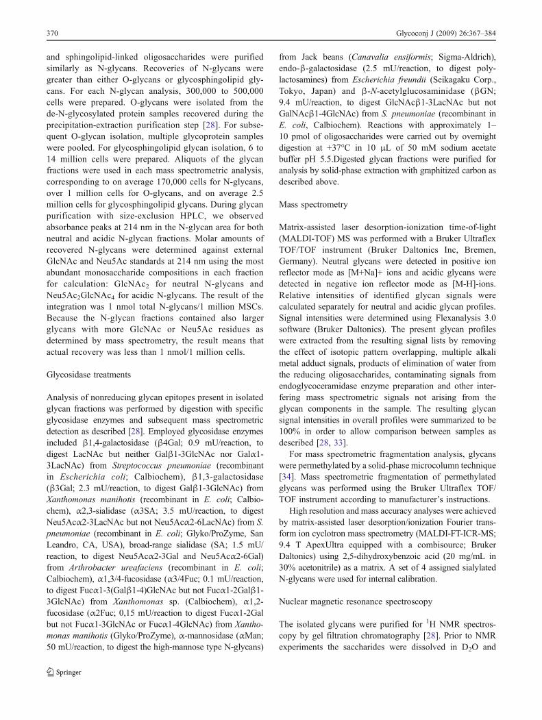
To study the overall glycome of MSCs, bone marrow-derived cells were either cultured in proliferative state ordifferentiated into osteogenic direction. N-glycans werereleased enzymatically from cell lysates by peptide: N-glycosidase F (PNGase F) digestion, O-glycans chemicallyby non-reductive β-elimination, and glycosphingolipidglycans enzymatically by endoglycoceramidase digestion.Mass spectrometric profiling of purified MSC N-glycans(Fig. 1a,b) showed 253 different signals, of which 50 majorneutral and 50 major acidic signals are listed in Table 1 aswell as visualized in Fig. 1c–f. Known polyhexosecontaminations [28], marked with asterisks, were discardedfrom the derived diagrams. The O-glycan profile of bonemarrow-derived MSCs revealed 42 different signals. Ofthese, 10 major acidic signals are listed in Table 1, and themost abundant neutral signal is visualized in Fig. 3. In thelipid glycan profile, 63 signals were observed and 15 majoracidic and 20 major neutral signals are listed in Table 1.
Using a mass matching approach, monosaccharidecompositions could be proposed for the different glycansignals (Table 1) and annotated in stem cell glycomeprofiles (Fig. 1e,f). The relative intensities for all thedetected glycan signals have been calculated. The relativeintensity of a single peak is expressed as percentages oftotal glycan profile, which has been given a value of 100%.We and others have documented that the percentagesdetermined in this way are useful for relative quantitativecomparison of glycan profile changes [28, 33]. The meanvalues of 5 different biological replicates are shown. Theglycan profiles were highly similar in the 5 different MSClines analyzed, as can be determined from the smalldeviations within the observed glycan profiles (Fig. 1c,d).The small profile differences between the 5 biologicalreplicates, also observed for the lineage-committed cells,indicates that MSCs have a very characteristic N-glycomeand that the glycome change during osteogenic differenti-ation occurs in a predetermined fashion.
N-glycans were grouped according to compositioncalculation into different glycan classes, e.g. mannose-type, complex-type and hybrid-type structures (Fig. 1e,f).Mannose-type N-glycans dominated in the neutral N-glycanprofile comprising 78% of MSC signals and 58% of osteo-blast signals. Complex-type N-glycans comprised 12% of
Glycoconj J (2009) 26:367–384 371
MSC signals and 25% of osteoblast signals, while only 9% ofMSC signals and 16% of osteoblast signals were determinedas hybrid-type. In the acidic N-glycan fraction, complex-typeN-glycans comprised the majority of the total glycan profile,
80% in MSCs and 86% in osteoblasts. Hybrid-type andmonoantennary N-glycans were less abundant. There werefew signals that could not be assigned to glycan structures bymass matching approach.
Fig 1 Mass spectrometric N-glycome profile of MSCs. Mass spectraof neutral (a) and acidic (b) N-glycans from bone marrow (BM)-derived MCSs are shown. Fifty major glycan peaks from five studiedMSC lines (black bars) as well as their osteoblast counterparts (greybars) are derived from mass spectrometric data (lines from a to c andb to d) and visualized according to their relative intensities (c neutralN-glycans; d acidic N-glycans). In panel e, neutral N-glycans and in
panel f acidic N-glycans are grouped according to their structuralclasses. Glycan species are annotated with proposed structures derivedby mass calculation. Blue square represents N-acetylglucosamine,green circle mannose, yellow circle galactose, red triangle fucose,pink diamond N-acetylneuraminic acid and P either sulfate orphosphate. Known polyhexose contaminations are marked withasterisks
372 Glycoconj J (2009) 26:367–384
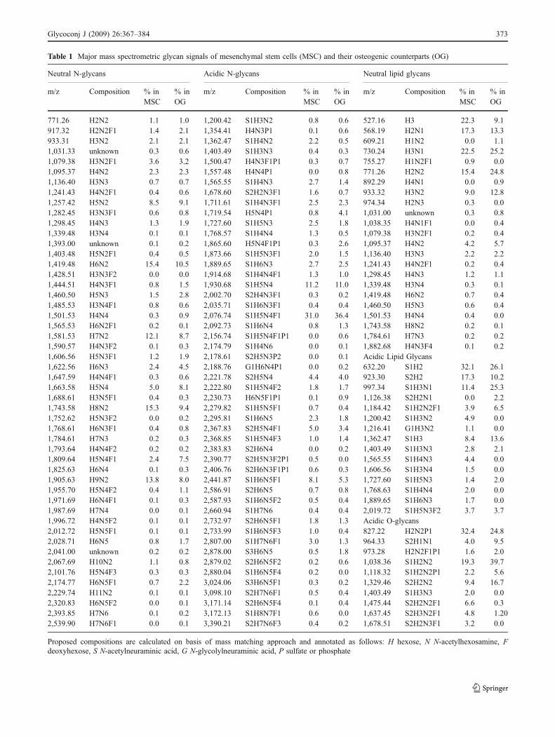
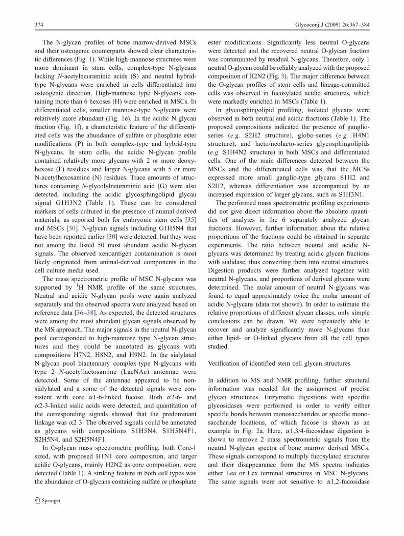
Table 1 Major mass spectrometric glycan signals of mesenchymal stem cells (MSC) and their osteogenic counterparts (OG)
Neutral N-glycans Acidic N-glycans Neutral lipid glycans
m/z Composition % inMSC
% inOG
m/z Composition % inMSC
% inOG
m/z Composition % inMSC
% inOG
771.26 H2N2 1.1 1.0 1,200.42 S1H3N2 0.8 0.6 527.16 H3 22.3 9.1917.32 H2N2F1 1.4 2.1 1,354.41 H4N3P1 0.1 0.6 568.19 H2N1 17.3 13.3933.31 H3N2 2.1 2.1 1,362.47 S1H4N2 2.2 0.5 609.21 H1N2 0.0 1.11,031.33 unknown 0.3 0.6 1,403.49 S1H3N3 0.4 0.3 730.24 H3N1 22.5 25.21,079.38 H3N2F1 3.6 3.2 1,500.47 H4N3F1P1 0.3 0.7 755.27 H1N2F1 0.9 0.01,095.37 H4N2 2.3 2.3 1,557.48 H4N4P1 0.0 0.8 771.26 H2N2 15.4 24.81,136.40 H3N3 0.7 0.7 1,565.55 S1H4N3 2.7 1.4 892.29 H4N1 0.0 0.91,241.43 H4N2F1 0.4 0.6 1,678.60 S2H2N3F1 1.6 0.7 933.32 H3N2 9.0 12.81,257.42 H5N2 8.5 9.1 1,711.61 S1H4N3F1 2.5 2.3 974.34 H2N3 0.3 0.01,282.45 H3N3F1 0.6 0.8 1,719.54 H5N4P1 0.8 4.1 1,031.00 unknown 0.3 0.81,298.45 H4N3 1.3 1.9 1,727.60 S1H5N3 2.5 1.8 1,038.35 H4N1F1 0.0 0.41,339.48 H3N4 0.1 0.1 1,768.57 S1H4N4 1.3 0.5 1,079.38 H3N2F1 0.2 0.41,393.00 unknown 0.1 0.2 1,865.60 H5N4F1P1 0.3 2.6 1,095.37 H4N2 4.2 5.71,403.48 H5N2F1 0.4 0.5 1,873.66 S1H5N3F1 2.0 1.5 1,136.40 H3N3 2.2 2.21,419.48 H6N2 15.4 10.5 1,889.65 S1H6N3 2.7 2.5 1,241.43 H4N2F1 0.2 0.41,428.51 H3N3F2 0.0 0.0 1,914.68 S1H4N4F1 1.3 1.0 1,298.45 H4N3 1.2 1.11,444.51 H4N3F1 0.8 1.5 1,930.68 S1H5N4 11.2 11.0 1,339.48 H3N4 0.3 0.11,460.50 H5N3 1.5 2.8 2,002.70 S2H4N3F1 0.3 0.2 1,419.48 H6N2 0.7 0.41,485.53 H3N4F1 0.8 0.6 2,035.71 S1H6N3F1 0.4 0.4 1,460.50 H5N3 0.6 0.41,501.53 H4N4 0.3 0.9 2,076.74 S1H5N4F1 31.0 36.4 1,501.53 H4N4 0.4 0.01,565.53 H6N2F1 0.2 0.1 2,092.73 S1H6N4 0.8 1.3 1,743.58 H8N2 0.2 0.11,581.53 H7N2 12.1 8.7 2,156.74 S1H5N4F1P1 0.0 0.6 1,784.61 H7N3 0.2 0.21,590.57 H4N3F2 0.1 0.3 2,174.79 S1H4N6 0.0 0.1 1,882.68 H4N3F4 0.1 0.21,606.56 H5N3F1 1.2 1.9 2,178.61 S2H5N3P2 0.0 0.1 Acidic Lipid Glycans1,622.56 H6N3 2.4 4.5 2,188.76 G1H6N4P1 0.0 0.2 632.20 S1H2 32.1 26.11,647.59 H4N4F1 0.3 0.6 2,221.78 S2H5N4 4.4 4.0 923.30 S2H2 17.3 10.21,663.58 H5N4 5.0 8.1 2,222.80 S1H5N4F2 1.8 1.7 997.34 S1H3N1 11.4 25.31,688.61 H3N5F1 0.4 0.3 2,230.73 H6N5F1P1 0.1 0.9 1,126.38 S2H2N1 0.0 2.21,743.58 H8N2 15.3 9.4 2,279.82 S1H5N5F1 0.7 0.4 1,184.42 S1H2N2F1 3.9 6.51,752.62 H5N3F2 0.0 0.2 2,295.81 S1H6N5 2.3 1.8 1,200.42 S1H3N2 4.9 0.01,768.61 H6N3F1 0.4 0.8 2,367.83 S2H5N4F1 5.0 3.4 1,216.41 G1H3N2 1.1 0.01,784.61 H7N3 0.2 0.3 2,368.85 S1H5N4F3 1.0 1.4 1,362.47 S1H3 8.4 13.61,793.64 H4N4F2 0.2 0.2 2,383.83 S2H6N4 0.0 0.2 1,403.49 S1H3N3 2.8 2.11,809.64 H5N4F1 2.4 7.5 2,390.77 S2H5N3F2P1 0.5 0.0 1,565.55 S1H4N3 4.4 0.01,825.63 H6N4 0.1 0.3 2,406.76 S2H6N3F1P1 0.6 0.3 1,606.56 S1H3N4 1.5 0.01,905.63 H9N2 13.8 8.0 2,441.87 S1H6N5F1 8.1 5.3 1,727.60 S1H5N3 1.4 2.01,955.70 H5N4F2 0.4 1.1 2,586.91 S2H6N5 0.7 0.8 1,768.63 S1H4N4 2.0 0.01,971.69 H6N4F1 0.1 0.3 2,587.93 S1H6N5F2 0.5 0.4 1,889.65 S1H6N3 1.7 0.01,987.69 H7N4 0.0 0.1 2,660.94 S1H7N6 0.4 0.4 2,019.72 S1H5N3F2 3.7 3.71,996.72 H4N5F2 0.1 0.1 2,732.97 S2H6N5F1 1.8 1.3 Acidic O-glycans2,012.72 H5N5F1 0.1 0.1 2,733.99 S1H6N5F3 1.0 0.4 827.22 H2N2P1 32.4 24.82,028.71 H6N5 0.8 1.7 2,807.00 S1H7N6F1 3.0 1.3 964.33 S2H1N1 4.0 9.52,041.00 unknown 0.2 0.2 2,878.00 S3H6N5 0.5 1.8 973.28 H2N2F1P1 1.6 2.02,067.69 H10N2 1.1 0.8 2,879.02 S2H6N5F2 0.2 0.6 1,038.36 S1H2N2 19.3 39.72,101.76 H5N4F3 0.3 0.3 2,880.04 S1H6N5F4 0.2 0.0 1,118.32 S1H2N2P1 2.2 5.62,174.77 H6N5F1 0.7 2.2 3,024.06 S3H6N5F1 0.3 0.2 1,329.46 S2H2N2 9.4 16.72,229.74 H11N2 0.1 0.1 3,098.10 S2H7N6F1 0.5 0.4 1,403.49 S1H3N3 2.0 0.02,320.83 H6N5F2 0.0 0.1 3,171.14 S2H6N5F4 0.1 0.4 1,475.44 S2H2N2F1 6.6 0.32,393.85 H7N6 0.1 0.2 3,172.13 S1H8N7F1 0.6 0.0 1,637.45 S2H3N2F1 4.8 1.202,539.90 H7N6F1 0.0 0.1 3,390.21 S2H7N6F3 0.4 0.2 1,678.51 S2H2N3F1 3.2 0.0
Proposed compositions are calculated on basis of mass matching approach and annotated as follows: H hexose, N N-acetylhexosamine, Fdeoxyhexose, S N-acetylneuraminic acid, G N-glycolylneuraminic acid, P sulfate or phosphate
Glycoconj J (2009) 26:367–384 373
The N-glycan profiles of bone marrow-derived MSCsand their osteogenic counterparts showed clear characteris-tic differences (Fig. 1). While high-mannose structures weremore dominant in stem cells, complex-type N-glycanslacking N-acetylneuraminic acids (S) and neutral hybrid-type N-glycans were enriched in cells differentiated intoosteogenic direction. High-mannose type N-glycans con-taining more than 6 hexoses (H) were enriched in MSCs. Indifferentiated cells, smaller mannose-type N-glycans wererelatively more abundant (Fig. 1e). In the acidic N-glycanfraction (Fig. 1f), a characteristic feature of the differenti-ated cells was the abundance of sulfate or phosphate estermodifications (P) in both complex-type and hybrid-typeN-glycans. In stem cells, the acidic N-glycan profilecontained relatively more glycans with 2 or more deoxy-hexose (F) residues and larger N-glycans with 5 or moreN-acetylhexosamine (N) residues. Trace amounts of struc-tures containing N-glycolylneuraminic acid (G) were alsodetected, including the acidic glycosphingolipid glycansignal G1H3N2 (Table 1). These can be consideredmarkers of cells cultured in the presence of animal-derivedmaterials, as reported both for embryonic stem cells [35]and MSCs [30]. N-glycan signals including G1H5N4 thathave been reported earlier [30] were detected, but they werenot among the listed 50 most abundant acidic N-glycansignals. The observed xenoantigen contamination is mostlikely originated from animal-derived components in thecell culture media used.
The mass spectrometric profile of MSC N-glycans wassupported by 1H NMR profile of the same structures.Neutral and acidic N-glycan pools were again analyzedseparately and the observed spectra were analyzed based onreference data [36–38]. As expected, the detected structureswere among the most abundant glycan signals observed bythe MS approach. The major signals in the neutral N-glycanpool corresponded to high-mannose type N-glycan struc-tures and they could be annotated as glycans withcompositions H7N2, H8N2, and H9N2. In the sialylatedN-glycan pool biantennary complex-type N-glycans withtype 2 N-acetyllactosamine (LacNAc) antennae weredetected. Some of the antennae appeared to be non-sialylated and a some of the detected signals were con-sistent with core α1-6-linked fucose. Both α2-6- andα2-3-linked sialic acids were detected, and quantitation ofthe corresponding signals showed that the predominantlinkage was α2-3. The observed signals could be annotatedas glycans with compositions S1H5N4, S1H5N4F1,S2H5N4, and S2H5N4F1.
In O-glycan mass spectrometric profiling, both Core-1sized, with proposed H1N1 core composition, and largeracidic O-glycans, mainly H2N2 as core composition, weredetected (Table 1). A striking feature in both cell types wasthe abundance of O-glycans containing sulfate or phosphate
ester modifications. Significantly less neutral O-glycanswere detected and the recovered neutral O-glycan fractionwas contaminated by residual N-glycans. Therefore, only 1neutral O-glycan could be reliably analyzed with the proposedcomposition of H2N2 (Fig. 3). The major difference betweenthe O-glycan profiles of stem cells and lineage-committedcells was observed in fucosylated acidic structures, whichwere markedly enriched in MSCs (Table 1).
In glycosphingolipid profiling, isolated glycans wereobserved in both neutral and acidic fractions (Table 1). Theproposed compositions indicated the presence of ganglio-series (e.g. S2H2 structure), globo-series (e.g. H4N1structure), and lacto/neolacto-series glycosphingolipids(e.g. S1H4N2 structure) in both MSCs and differentiatedcells. One of the main differences detected between theMSCs and the differentiated cells was that the MCSsexpressed more small ganglio-type glycans S1H2 andS2H2, whereas differentiation was accompanied by anincreased expression of larger glycans, such as S1H3N1.
The performed mass spectrometric profiling experimentsdid not give direct information about the absolute quanti-ties of analytes in the 6 separately analyzed glycanfractions. However, further information about the relativeproportions of the fractions could be obtained in separateexperiments. The ratio between neutral and acidic N-glycans was determined by treating acidic glycan fractionswith sialidase, thus converting them into neutral structures.Digestion products were further analyzed together withneutral N-glycans, and proportions of derived glycans weredetermined. The molar amount of neutral N-glycans wasfound to equal approximately twice the molar amount ofacidic N-glycans (data not shown). In order to estimate therelative proportions of different glycan classes, only simpleconclusions can be drawn. We were repeatedly able torecover and analyze significantly more N-glycans thaneither lipid- or O-linked glycans from all the cell typesstudied.
Verification of identified stem cell glycan structures
In addition to MS and NMR profiling, further structuralinformation was needed for the assignment of preciseglycan structures. Enzymatic digestions with specificglycosidases were performed in order to verify eitherspecific bonds between monosaccharides or specific mono-saccharide locations, of which fucose is shown as anexample in Fig. 2a. Here, α1,3/4-fucosidase digestion isshown to remove 2 mass spectrometric signals from theneutral N-glycan spectra of bone marrow derived MSCs.These signals correspond to multiply fucosylated structuresand their disappearance from the MS spectra indicateseither Lea or Lex terminal structures in MSC N-glycans.The same signals were not sensitive to α1,2-fucosidase
374 Glycoconj J (2009) 26:367–384

digestion (data not shown). The neutral N-glycan fractionwas further analyzed by specific galactosidase enzymes.Many diantennary-size N-glycans were digested by β1,4-galactosidase, whereas β1,3-galactosidase did not digestany of the glycans in question. Therefore, we suggest that itis more likely that the fucosylated antennae contain Lexthan Lea (data not shown).
Major findings with exoglycosidase digestions arepresented in Fig. 3 for the major glycan signals in each
separate glycan fraction. N-glycan signals proposed asmannose-type N-glycans were shown by α-mannosidasedigestion to have non-reducing terminal α-mannose resi-dues consistent with the initial assignment. Also theproposed hybrid-type signals were shown to be sensitiveto α-mannosidase treatment. All detected sialic acidresidues in O-glycans were α2-3-linked; whereas in N-glycans also α2-6-linkage was observed, as indicated bythe resistance of certain sialylated glycans to S. pneumoniae
Fig 2 Glycan structure verifica-tion by exoglycosidasedigestion, mass spectrometricfragmentation of permethylatedglycans and high resolution MS.α3/4-fucosidase digestion isshown (a) to remove annotatedglycan signals of MSCN-glycans. MS/MS fragmenta-tion is shown to verify a glyco-sphingolipid glycan structure(b). MALDI-FTICR MS analy-sis provides evidence for 5sulfated, but not phosphatedglycan species (c). Glycanannotations are as in Fig. 1
Glycoconj J (2009) 26:367–384 375
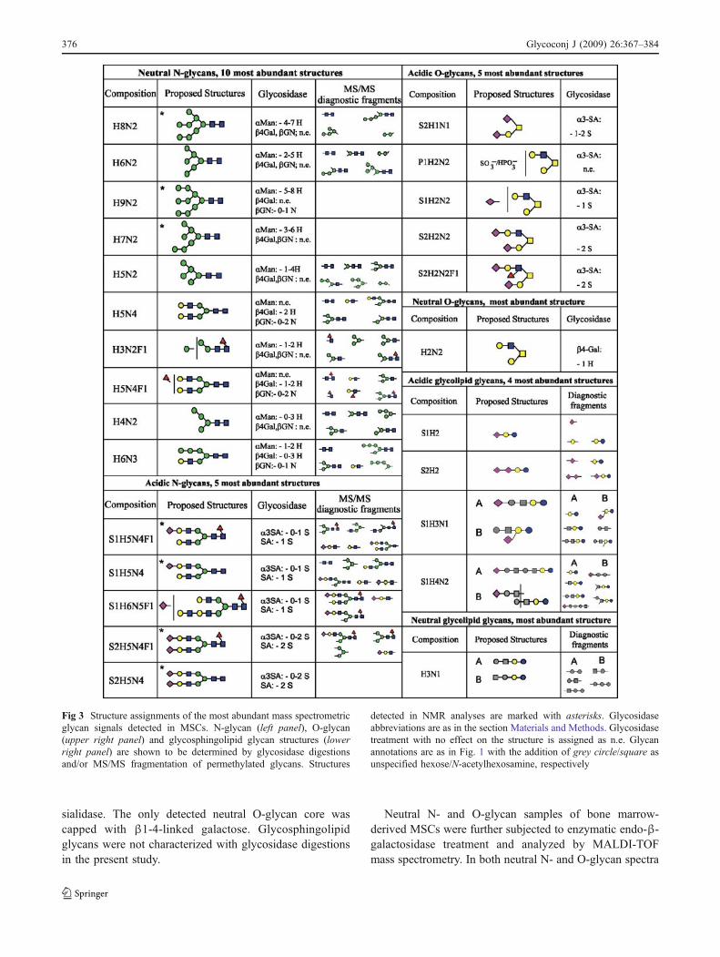
sialidase. The only detected neutral O-glycan core wascapped with β1-4-linked galactose. Glycosphingolipidglycans were not characterized with glycosidase digestionsin the present study.
Neutral N- and O-glycan samples of bone marrow-derived MSCs were further subjected to enzymatic endo-β-galactosidase treatment and analyzed by MALDI-TOFmass spectrometry. In both neutral N- and O-glycan spectra
Fig 3 Structure assignments of the most abundant mass spectrometricglycan signals detected in MSCs. N-glycan (left panel), O-glycan(upper right panel) and glycosphingolipid glycan structures (lowerright panel) are shown to be determined by glycosidase digestionsand/or MS/MS fragmentation of permethylated glycans. Structures
detected in NMR analyses are marked with asterisks. Glycosidaseabbreviations are as in the section Materials and Methods. Glycosidasetreatment with no effect on the structure is assigned as n.e. Glycanannotations are as in Fig. 1 with the addition of grey circle/square asunspecified hexose/N-acetylhexosamine, respectively
376 Glycoconj J (2009) 26:367–384
a signal at m/z 568 corresponding to H2N1 [M+Na]+ ionappeared after endo-β-galactosidase treatment (data notshown). This represents a characteristic cleavage product ofendo-β-galactosidase. Endo-β-galactosidase is specific forlinear poly-LacNAc substrates and the detected fragmentindicates a non-reducing terminal structure Gal-GlcNAcβ1-3Galβ1-4GlcNAc- (the released H2N1 fragment under-lined). The high relative intensity of the cleavage productindicates that it is derived from the breakdown of severalpoly-LacNAc containing glycans in both N- and O-glycans.
For further verification of glycan structures of bonemarrow-derived MSC glycan fractions were permethylatedfor MS/MS fragmentation analyses. The detected fragmentions indicative of specific glycan structures are shown inFig. 3. The fragmentation of a disialylated GD3-typeglycosphingolipid glycan is shown as an example inFig. 2b. Mass spectrometric fragmentation was shown tosupport the findings of glycome profiling and proposedcompositions based on mass matching (Fig. 3). Core fucosewas confirmed as the main fucosylation in N-glycans,although fucosylated antennae were also detected. Thefragmentation patterns of glycosphingolipid glycans wereconsistent with the common ganglioside structures GM3,GD3 (Fig. 2b) and GD1, globoside Gb4, and lacto/neolacto-series poly-LacNAc. All major glycosphingolipidclasses were therefore indicated to be expressed in MSCs(Fig. 3). MS/MS fragmentation patterns were also indica-tive of the presence of linear poly-LacNAc chains in MSCglycans. Candidate glycans carrying poly-LacNAc includethe acidic N-glycan S1H6N5F1 and the acidic glycosphingo-lipid glycan S1H4N2. The fragmentation pattern of the thirdmost abundant acidic N-glycan S1H6N5F1 was consistentwith a biantennary N-glycan structure in which one antennahad S1H1N1 composition (putative sialylated LacNAc)and the other H2N2 composition (putative linear poly-LacNAc). Especially the presence of a fragmentationproduct at m/z 2,141.7 (calc. m/z 2,142.1) corresponding toS1H4N3F1 [M+Na]+ ion supported the assignment. Thisfragment lacked the putative poly-LacNAc antenna andcontained a single free hydroxyl group indicating only one
branch point (Fig. 3). The detected fragments from theacidic glycosphingolipid glycan S1H4N2 indicated that themass spectrometric signal comprised at least 2 structures, ofwhich the structure A was consistent with a linear structurewith alternating hexose and N-acetylhexosamine residues(Fig. 3), corresponding to putative linear poly-LacNAc.Taken together, the data indicated presence of linear poly-LacNAc in N- and O-glycans, and possibly also inglycosphingolipids of bone marrow-derived MSCs.
Several glycan peaks with 80 Da mass addition wereobserved in acidic mass spectrometric glycome profiles ofMSCs and especially in the osteogenic cells derived fromthem (Table 1). To discriminate between putative sulfate orphosphate groups, the profiles were further analyzed usingMALDI-FTICR-MS in negative-ion mode (Fig. 2c). Here,the masses of sialylated N-glycans could be registered withan average mass deviation of 1.1 ppm. Five glycan specieswith potential sulfation or phosphorylation, which wereregistered in the same spectrum, were concluded to besulfated glycans with average mass deviation of 0.6 ppm.Phosphorylation could be ruled out on the basis of the largemass deviation (between 5.6 and 6.5 ppm) betweencalculated and registered masses (Fig. 2c inset). Unfortu-nately, acidic O-glycans could not be analyzed in this way,due to the limited amounts of O-glycans available foranalyses.
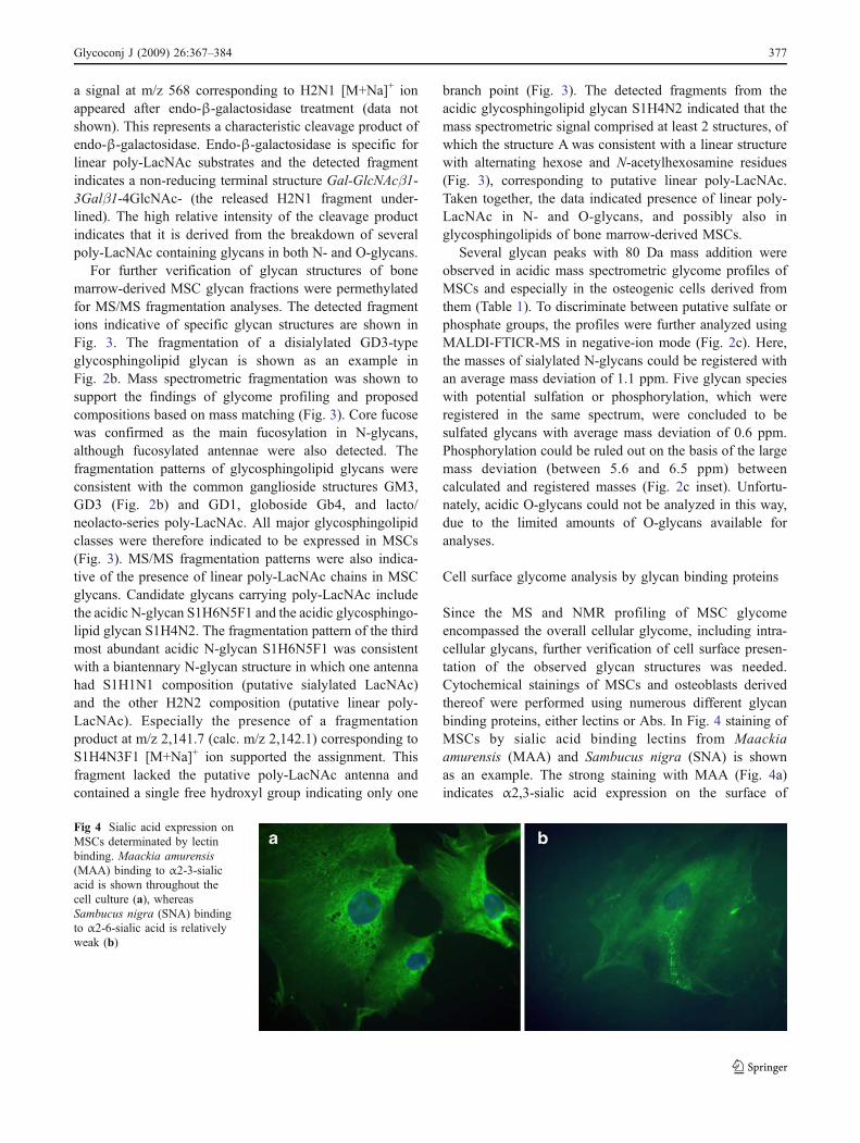
Cell surface glycome analysis by glycan binding proteins
Since the MS and NMR profiling of MSC glycomeencompassed the overall cellular glycome, including intra-cellular glycans, further verification of cell surface presen-tation of the observed glycan structures was needed.Cytochemical stainings of MSCs and osteoblasts derivedthereof were performed using numerous different glycanbinding proteins, either lectins or Abs. In Fig. 4 staining ofMSCs by sialic acid binding lectins from Maackiaamurensis (MAA) and Sambucus nigra (SNA) is shownas an example. The strong staining with MAA (Fig. 4a)indicates α2,3-sialic acid expression on the surface of
Fig 4 Sialic acid expression onMSCs determinated by lectinbinding. Maackia amurensis(MAA) binding to α2-3-sialicacid is shown throughout thecell culture (a), whereasSambucus nigra (SNA) bindingto α2-6-sialic acid is relativelyweak (b)
Glycoconj J (2009) 26:367–384 377
MSCs, in comparison with the relatively weak staining withSNA (Fig. 4b). Both NMR profiling and α2-3-linkagespecific sialidase digestion supported the finding that α2-3-linked sialic acids were abundant in MSCs (Fig. 3).
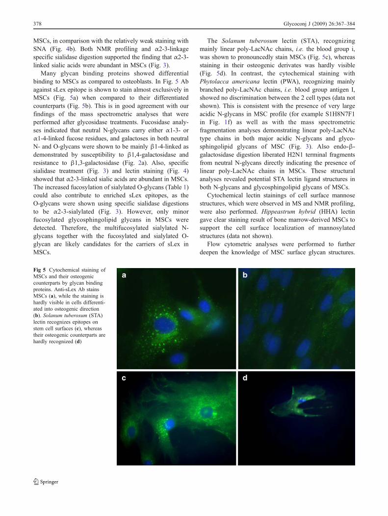
Many glycan binding proteins showed differentialbinding to MSCs as compared to osteoblasts. In Fig. 5 Abagainst sLex epitope is shown to stain almost exclusively inMSCs (Fig. 5a) when compared to their differentiatedcounterparts (Fig. 5b). This is in good agreement with ourfindings of the mass spectrometric analyses that wereperformed after glycosidase treatments. Fucosidase analy-ses indicated that neutral N-glycans carry either α1-3- orα1-4-linked fucose residues, and galactoses in both neutralN- and O-glycans were shown to be mainly β1-4-linked asdemonstrated by susceptibility to β1,4-galactosidase andresistance to β1,3-galactosidase (Fig. 2a). Also, specificsialidase treatment (Fig. 3) and lectin staining (Fig. 4)showed that α2-3-linked sialic acids are abundant in MSCs.The increased fucosylation of sialylated O-glycans (Table 1)could also contribute to enriched sLex epitopes, as theO-glycans were shown using specific sialidase digestionsto be α2-3-sialylated (Fig. 3). However, only minorfucosylated glycosphingolipid glycans in MSCs weredetected. Therefore, the multifucosylated sialylated N-glycans together with the fucosylated and sialylated O-glycan are likely candidates for the carriers of sLex inMSCs.
The Solanum tuberosum lectin (STA), recognizingmainly linear poly-LacNAc chains, i.e. the blood group i,was shown to pronouncedly stain MSCs (Fig. 5c), whereasstaining in their osteogenic derivates was hardly visible(Fig. 5d). In contrast, the cytochemical staining withPhytolacca americana lectin (PWA), recognizing mainlybranched poly-LacNAc chains, i.e. blood group antigen I,showed no discrimination between the 2 cell types (data notshown). This is consistent with the presence of very largeacidic N-glycans in MSC profile (for example S1H8N7F1in Fig. 1f) as well as with the mass spectrometricfragmentation analyses demonstrating linear poly-LacNActype chains in both major acidic N-glycans and glyco-sphingolipid glycans of MSC (Fig. 3). Also endo-β-galactosidase digestion liberated H2N1 terminal fragmentsfrom neutral N-glycans directly indicating the presence oflinear poly-LacNAc chains in MSCs. These structuralanalyses revealed potential STA lectin ligand structures inboth N-glycans and glycosphingolipid glycans of MSCs.
Cytochemical lectin stainings of cell surface mannosestructures, which were observed in MS and NMR profiling,were also performed. Hippeastrum hybrid (HHA) lectingave clear staining result of bone marrow-derived MSCs tosupport the cell surface localization of mannosylatedstructures (data not shown).
Flow cytometric analyses were performed to furtherdeepen the knowledge of MSC surface glycan structures.
Fig 5 Cytochemical staining ofMSCs and their osteogeniccounterparts by glycan bindingproteins. Anti-sLex Ab stainsMSCs (a), while the staining ishardly visible in cells differenti-ated into osteogenic direction(b). Solanum tuberosum (STA)lectin recognizes epitopes onstem cell surfaces (c), whereastheir osteogenic counterparts arehardly recognized (d)
378 Glycoconj J (2009) 26:367–384
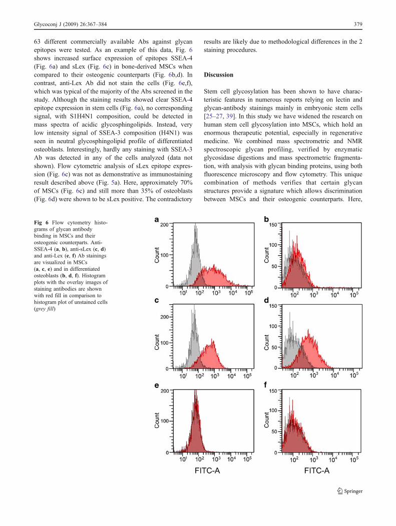
63 different commercially available Abs against glycanepitopes were tested. As an example of this data, Fig. 6shows increased surface expression of epitopes SSEA-4(Fig. 6a) and sLex (Fig. 6c) in bone-derived MSCs whencompared to their osteogenic counterparts (Fig. 6b,d). Incontrast, anti-Lex Ab did not stain the cells (Fig. 6e,f),which was typical of the majority of the Abs screened in thestudy. Although the staining results showed clear SSEA-4epitope expression in stem cells (Fig. 6a), no correspondingsignal, with S1H4N1 composition, could be detected inmass spectra of acidic glycosphingolipids. Instead, verylow intensity signal of SSEA-3 composition (H4N1) wasseen in neutral glycosphingolipid profile of differentiatedosteoblasts. Interestingly, hardly any staining with SSEA-3Ab was detected in any of the cells analyzed (data notshown). Flow cytometric analysis of sLex epitope expres-sion (Fig. 6c) was not as demonstrative as immunostainingresult described above (Fig. 5a). Here, approximately 70%of MSCs (Fig. 6c) and still more than 35% of osteoblasts(Fig. 6d) were shown to be sLex positive. The contradictory
results are likely due to methodological differences in the 2staining procedures.
Discussion
Stem cell glycosylation has been shown to have charac-teristic features in numerous reports relying on lectin andglycan-antibody stainings mainly in embryonic stem cells[25–27, 39]. In this study we have widened the research onhuman stem cell glycosylation into MSCs, which hold anenormous therapeutic potential, especially in regenerativemedicine. We combined mass spectrometric and NMRspectroscopic glycan profiling, verified by enzymaticglycosidase digestions and mass spectrometric fragmenta-tion, with analysis with glycan binding proteins, using bothfluorescence microscopy and flow cytometry. This uniquecombination of methods verifies that certain glycanstructures provide a signature which allows discriminationbetween MSCs and their osteogenic counterparts. Here,
Fig 6 Flow cytometry histo-grams of glycan antibodybinding in MSCs and theirosteogenic counterparts. Anti-SSEA-4 (a, b), anti-sLex (c, d)and anti-Lex (e, f) Ab stainingsare visualized in MSCs(a, c, e) and in differentiatedosteoblasts (b, d, f). Histogramplots with the overlay images ofstaining antibodies are shownwith red fill in comparison tohistogram plot of unstained cells(grey fill)
Glycoconj J (2009) 26:367–384 379
some of the glycan epitopes found to be characteristic ofMSCs and their differentiation are discussed.
Our data imply that a relatively high amount of highmannose glycans in the total glycan pool might be acommon feature of stem cell glycomes, including humanMSCs as shown in the present study, embryonic stem cells(unpublished data) and hematopoietic stem cells [28]. Itshould be realized that the total glycomes include alsointracellular glycans, and these typically contain more high-mannose glycans than e.g. individual secreted proteins orglycans from plasma membranes. The major ER and Golgitype glycans in the glycan profile might reflect themetabolic status of cells. However, in the case of culturedcells, such as the MSCs in the present study, cell cultureconditions may have a substantial effect on glycomes. Itwould be a major challenge to increase the sensitivity of theassay to enable the analysis of stem cells isolated directlyfrom tissues. So far this has been achieved only forhematopoietic stem cells, which were also shown to containrelatively high amounts of high-mannose glycans [28]. Theglycomes of the stem cells cultured under standardconditions are useful for the characterization of cells aimedfor therapy, even when the glycomes may not correspond tothe natural glycosylation of the cells.
We were able to observe increased expression of linearpoly-N-acetyllactosamine (poly-LacNAc) chains in MSCsas compared with osteoblasts by mass spectrometricfragmentation analysis, endo-β-galactosidase digestion,and Solanum tuberosum lectin (STA) staining. Poly-LacNAcs have been shown to undergo structural changesduring human development, wherein linear chains in fetalblood cells (i-antigen) are changed to branched chains (I-antigen) in the adult [40]. However, early embryonic cellsexpress large branched poly-LacNAc (embryoglycan),whereas the i-antigen appears in postimplantation embryos[21]. The SSEA-1 Lex structure, which is commonly usedas a marker for mouse embryonic stem cells, is expressedwithin embryoglycan, and even though human embryoniccells do not express SSEA-1, they contain embryoglycanconsisting of poly-LacNAc, that is considered to be anevolutionary conserved glycan backbone [21]. In general,poly-LacNAcs are thought to act as scaffolds presentingglycan epitopes to lectins in a multivalent fashion. Forexample, the galectins, a family of β-galactoside bindinglectins involved in a wide range of adhesion and signalingphenomena [41], have higher affinity for poly-LacNAcsthan for single LacNAc units [42–44]. Galectins arehypothesized to modulate their biological functions accord-ing to fine specificities for target glycans [44]. MSCsexpress galectins [45], and the immunomodulatory effectsof galectins have been shown to reduce GvHD in a mousemodel [46]. In addition, exposure to galectin-1 inducesskeletal muscle differentiation of human fetal MSCs [47].
The expression of SSEA-4, but not SSEA-3, on bonemarrow-derived MSCs was demonstrated in this study byantibody staining analyzed by flow cytometry. However,very low levels of either of the corresponding glycosphingo-lipid glycan signals were observed in mass spectrometricprofiles. The glycosphingolipid markers SSEA-3 and SSEA-4 are considered to be markers of human embryonic stemcells [27, 39, 48]. However, they do not seem to be essentialfor pluripotency [49]. Our results confirm the previouslyreported expression of SSEA-4 also on bone marrow MSCs[50–53]. Interestingly, one of the studies reported SSEA-4expression to markedly increase under serum free culturingconditions [51]. In contrast to the lack of SSEA-3expression reported here, evidence of SSEA-3 expressionin MSCs does exist [50, 52]. However, absence of bothSSEA-3 and SSEA-4 on adult bone marrow MSCs has alsobeen reported [54]. The contradictory results are likely dueto either cell line-specific expression of these epitopes ordifferences in culture conditions.
We observed the presence of multiple ganglioseriesglycosphingolipids on both MSCs and their differentiatedvariants. We detected changes in glycosphingolipid com-position during the differentiation of MSCs, such as atendency of smaller GM3/GD3 structures to change intolarger gangliosides, including GD1. Several ganglioseriesglycosphingolipids are expressed on human embryonicstem cells, and differentiation has been reported to inducea change in their ganglioside profile [39]. Especially,increased expression of disialylgangliosides, includingGD3 and GD2, after embryonic stem cell differentiation,has been reported [39]. GD2 has previously been reportedto be a potential marker of bone marrow-derived MSCs[55], but our results do not support this finding, since wedid not observe GD2 (composition S2H2N1) at all inMSCs, but rather in osteoblasts derived from them. Humanperiodontal bone marrow MSCs have been reported toexpress GM1 [56], which was found also in the presentstudy by MS/MS fragmentation, whereas after neuronaldifferentiation both GM1 and GT1b were detected [56]. Incontrast to the present study, no other gangliosides, likeGM3/GD3, could be detected [56]. The differences may becaused by methodological differences between the twostudies or different bone marrow sources used in them.
Sulfated carbohydrate epitopes, which we observed to bemarkedly enriched both in the N-glycan and O-glycanprofiles of osteogenically differentiated MSCs, are involvedin a variety of biological recognition events. Somegalectins, especially galectins 1–3, have been shown tobind 3′-sulfated LacNAcs better than their unsulfatedcounterparts [44]. Sulfation affects also the binding ofcertain siglecs, a family of sialic acid binding lectinsinvolved in the regulation of the immune system [57], totheir ligands. L-selectin ligand glycoproteins, which are
380 Glycoconj J (2009) 26:367–384

involved in leukocyte-endothelium interactions, are knownto carry sulfated sLex structures [58]. Keratan sulfateepitopes Tra-1–60 and Tra-1–81 have been reported asmarkers of embryonic stem cells [27, 39]. These sulfatedpoly-LacNAc structures (sulfated forms of the i-antigen) areclosely related to the structures observed enriched indifferentiated osteoblasts in the present study. The produc-tion of sulfated glycosaminoglycans, such as keratansulfate, is considered a hallmark of MSC differentiationcommitment into osteogenic lineage and it co-occurs withincreased expression of glycosaminoglycan biosyntheticenzymes [59]. It is also noteworthy that the bonesialoprotein, an osteoblast-associated marker, which isexpressed exclusively in bone and particularly during earlystages of bone formation [60], has been reported bymetabolic labeling to contain sulfated N- and O-glycans inrats [61]. However, sulfated glycans have not beenobserved in human bone sialoprotein [62].
In the present study, sialyl-Lewis x (sLex) is introducedas a novel characteristic glycan epitope expressed onhuman MSCs derived from bone marrow and to someextent also on osteogenically differentiated cells derivedfrom them. sLex is best known as a selectin ligand, and forits involvement in leukocyte homing as well as in cancermetastasis [63, 64]. In addition, sLex has been reported tomediate fucosylation-dependent homing of both hemato-poietic [65, 66] and mesenchymal [67] stem cells into bonemarrow. MSCs have also been shown to roll on endothelialcells in a P-selectin-dependent manner [68]. Interestingly, amajor selectin ligand PSGL-1 carries sLex on O-glycans[69], whereas selectin ligands carried on N-glycans havealso been reported [70], especially in hematopoietic andmesenchymal stem cells [67, 71]. In the present study weidentified both N- and O-glycans as potential carriers ofsLex epitope in MSCs.
We report here that α2-3-linked sialic acid, which is alsoterminal residue of the sLex epitope, is a typical structureon the surface of MSCs, Although these sialic acids arecommon terminal glycan structures, the ratio of α2-3- andα2-6-sialylation may affect intercellular interactions ordifferentiation of cells by modulating the binding of glycanreceptors, such as siglec 2 (CD22) [72, 73] or selectin–sLexadhesion. α2-3-sialylation has also been shown to regulateT-cell biology through inhibiting galectin-1 binding [74].Interestingly, terminal α2-3-sialic acid on a N-glycan hasbeen shown to inhibit binding of CD44 to hyaluronic acid[75]. As MSCs express α2-3-sialylated glycoform of CD44[67], α2-3-sialylation may regulate MSC adhesion to theextracellular matrix. We have previously shown α2-3-sialylation to be increased also on cord blood CD133+hematopoietic stem cells as compared to differentiatedCD133− cells [28]. In the present study, MSC O-glycanswere shown to contain sialic acid exclusively in α2-3
linkage, indicating differential regulation of sialylationbetween the different glycan classes.
In conclusion, we analyzed the glycome of bonemarrow-derived MSCs using MS and NMR profiling,further structure verification and a broad range of glycanbinding proteins. Comparison of the obtained data to theprofiles of osteogenically differentiated cells clearly indi-cated cell type specific variation in the glycosylationpatterns. Certain glycan structures seem to be indicative ofthe “stemness” nature of cells. The conservation of aspecific glycan structure between different stem cell typeswithin the same species, or conservation between differentspecies on a certain stem cell type, may be an indication ofan essential function for the glycan structure in multipotentcells [19]. At least to our knowledge, no glycosylationstudies on MSCs from different species have beenconducted, but we have demonstrated here similarities instem cell glycosylation between human MSCs and bothhuman embryonic stem cells [22–24] and hematopoieticstem cells [28]. It seems evident that glycan structures maybe”switched on” and “off” on cell surfaces several timesduring cellular differentiation in developmental processes,as implied by the transition between linear and branchedpoly-LacNAc structures [21]. Here, similar transition inmultiple glycan structures was demonstrated during MSCdifferentiation into osteogenic direction. The more precisebiological functions of these glycosylation characteristicsremain to be defined in the future.
Acknowledgments We would like to thank Teija Kupari and SirkkaMannelin for skillful technical assistance. We thank Dr. BogdanBogdanov for his assistance with the acquisition of the FTICR-MSdata. The present study was supported by the Finnish Funding Agencyfor Technology and Innovation (TEKES).
Open Access This article is distributed under the terms of theCreative Commons Attribution Noncommercial License, whichpermits any noncommercial use, distribution, and reproduction inany medium, provided the original author(s) and source are credited.
References
1. Moore, K.E., Mills, J.F., Thornton, M.M.: Alternative sources ofadult stem cells: a possible solution to the embryonic stem celldebate. Gend. Med. 3, 161–168 (2006). doi:10.1016/S1550-8579(06)80204-4
2. Pessina, A., Gribaldo, L.: The key role of adult stem cells:therapeutic perspectives. Curr. Med. Res. Opin. 22, 2287–2300(2006). doi:10.1185/030079906X148517
3. Porada, C.D., Zanjani, E.D., meida-Porad, G.: Adult mesenchy-mal stem cells: a pluripotent population with multiple applica-tions. Curr. Stem Cell Res. Ther. 1, 365–369 (2006)
4. Prockop, D.J.: Marrow stromal cells as stem cells for non-hematopoietic tissues. Science 276, 71–74 (1997). doi:10.1126/science.276.5309.71
Glycoconj J (2009) 26:367–384 381
5. Rochefort, G.Y., Delorme, B., Lopez, A., Herault, O., Bonnet, P.,Charbord, P., Eder, V., Domenech, J.: Multipotential mesen-chymal stem cells are mobilized into peripheral blood byhypoxia. Stem Cells 24, 2202–2208 (2006). doi:10.1634/stemcells.2006-0164
6. Bieback, K., Kern, S., Kluter, H., Eichler, H.: Critical parametersfor the isolation of mesenchymal stem cells from umbilical cordblood. Stem Cells 22, 625–634 (2004). doi:10.1634/stemcells.22-4-625
7. Erices, A., Conget, P., Minguell, J.J.: Mesenchymal progenitorcells in human umbilical cord blood. Br. J. Haematol. 109, 235–242 (2000). doi:10.1046/j.1365-2141.2000.01986.x
8. Katritsis, D.G., Sotiropoulou, P., Giazitzoglou, E., Karvouni, E.,Papamichail, M.: Electrophysiological effects of intracoronarytransplantation of autologous mesenchymal and endothelialprogenitor cells. Europace 9, 167–171 (2007). doi:10.1093/europace/eul184
9. Zaidi, N., Nixon, A.J.: Stem cell therapy in bone repair andregeneration. Ann. N YAcad.Sci. 1117, 62–72 (2007). doi:10.1196/annals.1402.074
10. Horwitz, E.M., Gordon, P.L., Koo, W.K., Marx, J.C., Neel, M.D.,McNall, R.Y., Muul, L., Hofmann, T.: Isolated allogeneic bonemarrow-derived mesenchymal cells engraft and stimulate growthin children with osteogenesis imperfecta: implications for celltherapy of bone. Proc. Natl. Acad. Sci. U S A 99, 8932–8937(2002). doi:10.1073/pnas.132252399
11. Le, B.K., Rasmusson, I., Sundberg, B., Gotherstrom, C., Hassan,M., Uzunel, M., Ringden, O.: Treatment of severe acute graft-versus-host disease with third party haploidentical mesenchymalstem cells. Lancet 363, 1439–1441 (2004). doi:10.1016/S0140-6736(04)16104-7
12. Moviglia, G.A., Fernandez, V.R., Brizuela, J.A., Saslavsky, J.,Vrsalovic, F., Varela, G., Bastos, F., Farina, P., Etchegaray, G.,Barbieri, M., Martinez, G., Picasso, F., Schmidt, Y., Brizuela, P.,Gaeta, C.A., Costanzo, H., Moviglia Brandolino, M.T., Merino,S., Pes, M.E., Veloso, M.J., Rugilo, C., Tamer, I., Shuster,G.S.: Combined protocol of cell therapy for chronic spinalcord injury. Report on the electrical and functional recovery oftwo patients. Cytotherapy 8, 202–209 (2006). doi:10.1080/14653240600736048
13. Varki, A.: Biological roles of oligosaccharides: all of the theories arecorrect. Glycobiology 3, 97–130 (1993). doi:10.1093/glycob/3.2.97
14. Haltiwanger, R.S., Lowe, J.B.: Role of glycosylation in develop-ment. Annu. Rev. Biochem. 73, 491–537 (2004). doi:10.1146/annurev.biochem.73.011303.074043
15. Ohtsubo, K., Marth, J.D.: Glycosylation in cellular mechanisms ofhealth and disease. Cell 126, 855–867 (2006). doi:10.1016/j.cell.2006.08.019
16. Yu, R.K., Yanagisawa, M.: Glycobiology of neural stem cells. CNSNeurol. Disord. Drug Targets 5, 415–423 (2006). doi:10.2174/187152706777950675
17. Marth, J.D.: Will the transgenic mouse serve as a Rosetta Stone toglycoconjugate function? Glycoconj. J. 11, 3–8 (1994). doi:10.1007/BF00732424
18. Lowe, J.B., Marth, J.D.: A genetic approach to mammalian glycanfunction. Annu. Rev. Biochem. 72, 643–691 (2003). doi:10.1146/annurev.biochem.72.121801.161809
19. Gagneux, P., Varki, A.: Evolutionary considerations in relatingoligosaccharide diversity to biological function. Glycobiology 9,747–755 (1999). doi:10.1093/glycob/9.8.747
20. Lanctot, P.M., Gage, F.H., Varki, A.P.: The glycans of stem cells.Curr. Opin. Chem. Biol. 11, 373–380 (2007). doi:10.1016/j.cbpa.2007.05.032
21. Muramatsu, T., Muramatsu, H.: Carbohydrate antigens expressedon stem cells and early embryonic cells. Glycoconj. J. 21, 41–45(2004). doi:10.1023/B:GLYC.0000043746.77504.28
22. Venable, A., Mitalipova, M., Lyons, I., Jones, K., Shin, S., Pierce,M., Stice, S.: Lectin binding profiles of SSEA-4 enriched,pluripotent human embryonic stem cell surfaces. BMC Dev. Biol.5, 15 (2005). doi:10.1186/1471-213X-5-15
23. Wearne, K.A., Winter, H.C., O’Shea, K., Goldstein, I.J.: Use oflectins for probing differentiated human embryonic stem cells forcarbohydrates. Glycobiology 16, 981–990 (2006). doi:10.1093/glycob/cwl019
24. Wearne, K.A., Winter, H.C., Goldstein, I.J.: Temporal changes inthe carbohydrates expressed on BG01 human embryonic stemcells during differentiation as embryoid bodies. Glycoconj. J. 25,121–136 (2008). doi:10.1007/s10719-007-9064-x
25. Badcock, G., Pigott, C., Goepel, J., Andrews, P.W.: The humanembryonal carcinoma marker antigen TRA-1–60 is a sialylatedkeratan sulfate proteoglycan. Cancer Res. 59, 4715–4719(1999)
26. Kannagi, R., Cochran, N.A., Ishigami, F., Hakomori, S., Andrews,P.W., Knowles, B.B., Solter, D.: Stage-specific embryonic anti-gens (SSEA-3 and -4) are epitopes of a unique globo-seriesganglioside isolated from human teratocarcinoma cells. EMBO J.2, 2355–2361 (1983)
27. Adewumi, O., Aflatoonian, B., hrlund-Richter, L., Amit, M.,Andrews, P.W., Beighton, G., Bello, P.A., Benvenisty, N., Berry,L.S., Bevan, S., Blum, B., Brooking, J., Chen, K.G., Choo, A.B.,Churchill, G.A., Corbel, M., Damjanov, I., Draper, J.S., Dvorak,P., Emanuelsson, K., Fleck, R.A., Ford, A., Gertow, K.,Gertsenstein, M., Gokhale, P.J., Hamilton, R.S., Hampl, A.,Healy, L.E., Hovatta, O., Hyllner, J., Imreh, M.P., Itskovitz-Eldor,J., Jackson, J., Johnson, J.L., Jones, M., Kee, K., King, B.L.,Knowles, B.B., Lako, M., Lebrin, F., Mallon, B.S., Manning, D.,Mayshar, Y., McKay, R.D., Michalska, A.E., Mikkola, M.,Mileikovsky, M., Minger, S.L., Moore, H.D., Mummery, C.L.,Nagy, A., Nakatsuji, N., O’Brien, C.M., Oh, S.K., Olsson, C.,Otonkoski, T., Park, K.Y., Passier, R., Patel, H., Patel, M.,Pedersen, R., Pera, M.F., Piekarczyk, M.S., Pera, R.A., Reubinoff,B.E., Robins, A.J., Rossant, J., Rugg-Gunn, P., Schulz, T.C.,Semb, H., Sherrer, E.S., Siemen, H., Stacey, G.N., Stojkovic, M.,Suemori, H., Szatkiewicz, J., Turetsky, T., Tuuri, T., van den,B.S., Vintersten, K., Vuoristo, S., Ward, D., Weaver, T.A., Young,L.A., Zhang, W.: Characterization of human embryonic stem celllines by the International Stem Cell Initiative. Nat. Biotechnol. 25,803–816 (2007). doi:10.1038/nbt1318
28. Hemmoranta, H., Satomaa, T., Blomqvist, M., Heiskanen, A.,Aitio, O., Saarinen, J., Natunen, J., Partanen, J., Laine, J.,Jaatinen, T.: N-glycan structures and associated gene expressionreflect the characteristic N-glycosylation pattern of humanhematopoietic stem and progenitor cells. Exp. Hematol. 35,1279–1292 (2007). doi:10.1016/j.exphem.2007.05.006
29. Leskelä, H.V., Risteli, J., Niskanen, S., Koivunen, J., Ivaska,K.K., Lehenkari, P.: Osteoblast recruitment from stem cells doesnot decrease by age at late adulthood. Biochem. Biophys. Res.Commun. 311, 1008–1013 (2003). doi:10.1016/j.bbrc.2003.10.095
30. Heiskanen, A., Satomaa, T., Tiitinen, S., Laitinen, A., Mannelin, S.,Impola, U., Mikkola, M., Olsson, C., Miller-Podraza, H., Blomqvist,M., Olonen, A., Salo, H., Lehenkari, P., Tuuri, T., Otonkoski, T.,Natunen, J., Saarinen, J., Laine, J.: N-glycolylneuraminic acidxenoantigen contamination of human embryonic and mesenchymalstem cells is substantially reversible. Stem Cells 25, 197–202 (2007).doi:10.1634/stemcells.2006-0444
31. Huang, Y., Mechref, Y., Novotny, M.V.: Microscale nonreductiverelease of O-linked glycans for subsequent analysis throughMALDI mass spectrometry and capillary electrophoresis. Anal.Chem. 73, 6063–6069 (2001). doi:10.1021/ac015534c
32. Miller-Podraza, H., Lanne, B., Angstrom, J., Teneberg, S., Milh,M.A., Jovall, P.A., Karlsson, H., Karlsson, K.A.: Novel bindingepitope for Helicobacter pylori found in neolacto carbohydrate
382 Glycoconj J (2009) 26:367–384
chains: structure and cross-binding properties. J. Biol. Chem. 280,19695–19703 (2005). doi:10.1074/jbc.M412688200
33. Aoki, K., Perlman, M., Lim, J.M., Cantu, R., Wells, L., Tiemeyer,M.: Dynamic developmental elaboration of N-linked glycancomplexity in the Drosophila melanogaster embryo. J. Biol.Chem. 282, 9127–9142 (2007). doi:10.1074/jbc.M606711200
34. Kang, P., Mechref, Y., Klouckova, I., Novotny, M.V.: Solid-phasepermethylation of glycans for mass spectrometric analysis. RapidCommun. Mass Spectrom. 19, 3421–3428 (2005). doi:10.1002/rcm.2210
35. Martin, M.J., Muotri, A., Gage, F., Varki, A.: Human embryonicstem cells express an immunogenic nonhuman sialic acid. Nat.Med. 11, 228–232 (2005). doi:10.1038/nm1181
36. Fu, D., Chen, L., O’Neill, R.A.: A detailed structural charac-terization of ribonuclease B oligosaccharides by 1H NMRspectroscopy and mass spectrometry. Carbohydr. Res. 261, 173–186 (1994). doi:10.1016/0008-6215(94)84015-6
37. Hard, K., Van, Z.G., Moonen, P., Kamerling, J.P., Vliegenthart,F.G.: The Asn-linked carbohydrate chains of human Tamm-Horsfall glycoprotein of one male. Novel sulfated and novel N-acetylgalactosamine-containing N-linked carbohydrate chains.Eur. J. Biochem. 209, 895–915 (1992). doi:10.1111/j.1432-1033.1992.tb17362.x
38. Helin, J., Maaheimo, H., Seppo, A., Keane, A., Renkonen, O.:Stepwise transfer of alpha-D-Galp-(1–>3)-beta-D-Galp-(1–>4)-beta-D-GlcpNAc sequences to 3-OH and 6-OH of distal galactoseresidues in bi-, tri-, and tetra-antennary asialo-glycans of N-linkedcomplex type. Carbohydr. Res. 266, 191–209 (1995). doi:10.1016/0008-6215(94)00272-H
39. Draper, J.S., Pigott, C., Thomson, J.A., Andrews, P.W.: Surfaceantigens of human embryonic stem cells: changes upon differen-tiation in culture. J. Anat. 200, 249–258 (2002). doi:10.1046/j.1469-7580.2002.00030.x
40. Fukuda, M., Fukuda, M.N., Hakomori, S.: Developmental changeand genetic defect in the carbohydrate structure of band 3glycoprotein of human erythrocyte membrane. J. Biol. Chem.254, 3700–3703 (1979)
41. Leffler, H., Carlsson, S., Hedlund, M., Qian, Y., Poirier, F.:Introduction to galectins. Glycoconj. J. 19, 433–440 (2004).doi:10.1023/B:GLYC.0000014072.34840.04
42. Leppänen, A., Stowell, S., Blixt, O., Cummings, R.D.: Dimericgalectin-1 binds with high affinity to alpha2,3-sialylated and non-sialylated terminal N-acetyllactosamine units on surface-boundextended glycans. J. Biol. Chem. 280, 5549–5562 (2005).doi:10.1074/jbc.M412019200
43. Stowell, S.R., as-Baruffi, M., Penttila, L., Renkonen, O., Nyame,A.K., Cummings, R.D.: Human galectin-1 recognition of poly-N-acetyllactosamine and chimeric polysaccharides. Glycobiology14, 157–167 (2004). doi:10.1093/glycob/cwh018
44. Stowell, S.R., Arthur, C.M., Mehta, P., Slanina, K.A., Blixt, O.,Leffler, H., Smith, D.F., Cummings, R.D.: Galectin-1, -2, and -3exhibit differential recognition of sialylated glycans and bloodgroup antigens. J. Biol. Chem. 283, 10109–10123 (2008). doi:10.1074/jbc.M709545200
45. Kadri, T., Lataillade, J.J., Doucet, C., Marie, A., Ernou, I., Bourin,P., Joubert-Caron, R., Caron, M., Lutomski, D.: Proteomic studyof Galectin-1 expression in human mesenchymal stem cells. StemCells Dev. 14, 204–212 (2005). doi:10.1089/scd.2005.14.204
46. Baum, L.G., Blackall, D.P., rias-Magallano, S., Nanigian, D., Uh,S.Y., Browne, J.M., Hoffmann, D., Emmanouilides, C.E., Territo,M.C., Baldwin, G.C.: Amelioration of graft versus host disease bygalectin-1. Clin. Immunol. 109, 295–307 (2003). doi:10.1016/j.clim.2003.08.003
47. Chan, J., O’Donoghue, K., Gavina, M., Torrente, Y., Kennea, N.,Mehmet, H., Stewart, H., Watt, D.J., Morgan, J.E., Fisk, N.M.:Galectin-1 induces skeletal muscle differentiation in human fetal
mesenchymal stem cells and increases muscle regeneration.Stem Cells 24, 1879–1891 (2006). doi:10.1634/stemcells.2005-0564
48. Thomson, J.A., Itskovitz-Eldor, J., Shapiro, S.S., Waknitz, M.A.,Swiergiel, J.J., Marshall, V.S., Jones, J.M.: Embryonic stem celllines derived from human blastocysts. Science 282, 1145–1147(1998). doi:10.1126/science.282.5391.1145
49. Brimble, S.N., Sherrer, E.S., Uhl, E.W., Wang, E., Kelly, S.,Merrill, A.H., Jr., Robins, A.J., Schulz, T.C.: The cell surfaceglycosphingolipids SSEA-3 and SSEA-4 are not essential forhuman ESC pluripotency. Stem Cells 25, 54–62 (2007).doi:10.1634/stemcells.2006-0232
50. Gang, E.J., Bosnakovski, D., Figueiredo, C.A., Visser, J.W.,Perlingeiro, R.C.: SSEA-4 identifies mesenchymal stem cells frombone marrow. Blood 109, 1743–1751 (2007). doi:10.1182/blood-2005-11-010504
51. Battula, V.L., Bareiss, P.M., Treml, S., Conrad, S., Albert, I., Hojak,S., Abele, H., Schewe, B., Just, L., Skutella, T., Buhring, H.J.:Human placenta and bone marrow derived MSC cultured in serum-free, b-FGF-containing medium express cell surface frizzled-9 andSSEA-4 and give rise to multilineage differentiation. Differentiation75, 279–291 (2007). doi:10.1111/j.1432-0436.2006.00139.x
52. Peiffer, I., Eid, P., Barbet, R., Li, M.L., Oostendorp, R.A.,Haydont, V., Monier, M.N., Milon, L., Fortunel, N., Charbord,P., Tovey, M., Hatzfeld, J., Hatzfeld, A.: A sub-population of highproliferative potential-quiescent human mesenchymal stem cells isunder the reversible control of interferon alpha/beta. Leukemia 21,714–724 (2007). doi:10.1038/sj.leu.2404589
53. Roubelakis, M.G., Pappa, K.I., Bitsika, V., Zagoura, D., Vlahou,A., Papadaki, H.A., Antsaklis, A., Anagnou, N.P.: Molecular andproteomic characterization of human mesenchymal stem cellsderived from amniotic fluid: comparison to bone marrowmesenchymal stem cells. Stem Cells Dev. 16, 931–952 (2007).doi:10.1089/scd.2007.0036
54. Guillot, P.V., Gotherstrom, C., Chan, J., Kurata, H., Fisk, N.M.:Human first-trimester fetal MSC express pluripotency markersand grow faster and have longer telomeres than adult MSC. StemCells 25, 646–654 (2007). doi:10.1634/stemcells.2006-0208
55. Martinez, C., Hofmann, T.J., Marino, R., Dominici, M., Horwitz,E.M.: Human bone marrow mesenchymal stromal cells expressthe neural ganglioside GD2: a novel surface marker for theidentification of MSCs. Blood 109, 4245–4248 (2007).doi:10.1182/blood-2006-08-039347
56. Kwak, D.H., Yu, K., Kim, S.M., Lee, D.H., Kim, S.M., Jung, J.U.,Seo, J.W., Kim, N., Lee, S., Jung, K.Y., You, H.K., Kim, H.A.,Choo, Y.K.: Dynamic changes of gangliosides expression duringthe differentiation of embryonic and mesenchymal stem cells intoneural cells. Exp. Mol. Med. 38, 668–676 (2006)
57. Varki, A., Angata, T.: Siglecs—the major subfamily of I-type lectins.Glycobiology 16, 1R–27R (2006). doi:10.1093/glycob/cwj008
58. Rosen, S.D.: Endothelial ligands for L-selectin: from lymphocyterecirculation to allograft rejection. Am. J. Pathol. 155, 1013–1020(1999)
59. Muller, B., Prante, C., Gastens, M., Kuhn, J., Kleesiek, K.,Gotting, C.: Increased levels of xylosyltransferase I correlate withthe mineralization of the extracellular matrix during osteogenicdifferentiation of mesenchymal stem cells. Matrix Biol. 27, 139–149 (2008). doi:10.1016/j.matbio.2007.09.005
60. Liu, F., Malaval, L., Aubin, J.E.: Global amplification polymerasechain reaction reveals novel transitional stages during osteopro-genitor differentiation. J. Cell Sci. 116, 1787–1796 (2003).doi:10.1242/jcs.00376
61. Midura, R.J., McQuillan, D.J., Benham, K.J., Fisher, L.W.,Hascall, V.C.: A rat osteogenic cell line (UMR 106–01)synthesizes a highly sulfated form of bone sialoprotein. J. Biol.Chem. 265, 5285–5291 (1990)
Glycoconj J (2009) 26:367–384 383
62. Zaia, J., Boynton, R., Heinegard, D., Barry, F.: Posttranslationalmodifications to human bone sialoprotein determined by massspectrometry. Biochemistry 40, 12983–12991 (2001). doi:10.1021/bi010887r
63. Ley, K.: The role of selectins in inflammation and disease. TrendsMol. Med. 9, 263–268 (2003). doi:10.1016/S1471-4914(03)00071-6
64. Magnani, J.L.: The discovery, biology, and drug development ofsialyl Lea and sialyl Lex. Arch. Biochem. Biophys. 426, 122–131(2004). doi:10.1016/j.abb.2004.04.008
65. Hidalgo, A., Frenette, P.S.: Enforced fucosylation of neonatalCD34+ cells generates selectin ligands that enhance the initialinteractions with microvessels but not homing to bone marrow.Blood 105, 567–575 (2005). doi:10.1182/blood-2004-03-1026
66. Xia, L.,McDaniel, J.M., Yago, T., Doeden, A.,McEver, R.P.: Surfacefucosylation of human cord blood cells augments binding to P-selectin and E-selectin and enhances engraftment in bone marrow.Blood 104, 3091–3096 (2004). doi:10.1182/blood-2004-02-0650
67. Sackstein, R., Merzaban, J.S., Cain, D.W., Dagia, N.M., Spencer, J.A., Lin, C.P., Wohlgemuth, R.: Ex vivo glycan engineering of CD44programs humanmultipotent mesenchymal stromal cell trafficking tobone. Nat. Med. 14, 181–187 (2008). doi:10.1038/nm1703
68. Ruster, B., Gottig, S., Ludwig, R.J., Bistrian, R., Muller, S.,Seifried, E., Gille, J., Henschler, R.: Mesenchymal stem cellsdisplay coordinated rolling and adhesion behavior on endothelialcells. Blood 108, 3938–3944 (2006). doi:10.1182/blood-2006-05-025098
69. Wilkins, P.P., McEver, R.P., Cummings, R.D.: Structures of the O-glycans on P-selectin glycoprotein ligand-1 fromHL-60 cells. J. Biol.Chem. 271, 18732–18742 (1996). doi:10.1074/jbc.271. 6.3255
70. Lenter, M., Levinovitz, A., Isenmann, S., Vestweber, D.: Mono-specific and common glycoprotein ligands for E- and P-selectinon myeloid cells. J. Cell Biol. 125, 471–481 (1994). doi:10.1083/jcb.125.2.471
71. Dimitroff, C.J., Lee, J.Y., Fuhlbrigge, R.C., Sackstein, R.: Adistinct glycoform of CD44 is an L-selectin ligand on humanhematopoietic cells. Proc. Natl. Acad. Sci. U S A 97, 13841–13846 (2000). doi:10.1073/pnas.250484797
72. Hennet, T., Chui, D., Paulson, J.C., Marth, J.D.: Immuneregulation by the ST6Gal sialyltransferase. Proc. Natl. Acad.Sci. USA 95, 4504–4509 (1998). doi:10.1073/pnas.95.8.4504
73. Takahata, M., Iwasaki, N., Nakagawa, H., Abe, Y., Watanabe, T.,Ito, M., Majima, T., Minami, A.: Sialylation of cell surfaceglycoconjugates is essential for osteoclastogenesis. Bone 41, 77–86 (2007). doi:10.1016/j.bone.2007.03.016
74. Priatel, J.J., Chui, D., Hiraoka, N., Simmons, C.J., Richardson, K.B., Page, D.M., Fukuda, M., Varki, N.M., Marth, J.D.: TheST3Gal-I sialyltransferase controls CD8+ T lymphocyte homeo-stasis by modulating O-glycan biosynthesis. Immunity 12, 273–283 (2000). doi:10.1016/S1074-7613(00)80180-6
75. Skelton, T.P., Zeng, C., Nocks, A., Stamenkovic, I.: Glycosylationprovides both stimulatory and inhibitory effects on cell surfaceand soluble CD44 binding to hyaluronan. J. Cell Biol. 140, 431–446 (1998). doi:10.1083/jcb.140.2.431
384 Glycoconj J (2009) 26:367–384