
Giovanni Di Salvo - echocardiography-course.com 3 G Di Salvo TEE... · • Intra-operative TEE is...
27
Giovanni Di Salvo MD,PhD,FESC Second University of Naples - Chair of Cardiology Monaldi Hospital
Transcript of Giovanni Di Salvo - echocardiography-course.com 3 G Di Salvo TEE... · • Intra-operative TEE is...

Giovanni Di SalvoMD,PhD,FESC
Second University of Naples - Chair of
Cardiology
Monaldi Hospital

The use of TEE and EpicardialcolorDoppler echo in the operating roomprovides information for assessing the outcome of heartsurgery immediatelyafter the surgicalprocedure.

Normal transducer sterilized by a cold gas technique
Non sterile transducer placed inside a steril plastic cover

Acquisition of data is made by the surgeon in the steril field.
A second person operates the instrument controls of the echo machine.

Major Advantages: its ability to provide multiple tomographic planes and its easy availability.
Limitations: poor visualization of the apex; interruption of the operation during the examinations; the pt can be studied only after thoracotomy.

• Intra-operative TEE is an important monitoring and diagnostic tool used during surgery for repair of CHD.
• In several studies TEE has been shown to provide additional intra-cardiac anatomic information.
• Its ability to be used intra-operatively before and after cardiac repair makes it a unique tool.
Garg R, Ann Card Anaesth 2009

• The first experience with pediatric intraoperative TEE was described by Cyran in 1989 in children 7.5 yr of age using an adult-sized probe.
• Before TEE was available for intra-operative use, significant residual abnormalities were frequently not detected.
• The result was often substantial post-operative morbidity and mortality and sometimes the need for re-operation.
Am J Cardiol 1989;63:594-8

Low risk: ASD, VSD, valve replacement, and extracardiac procedures.
Moderate risk: AVSD, combined ASD and VSD or combined VSD and pulmonary stenosis, valve reconstruction, subaortic stenosis resection.
High risk: Reoperation and neo-natal surgery, Fontan procedure, Fallot's tetralogy, Ebsteinanomaly.
Gallivan S, Eur J Cardiothor Surg 2001

Micro multiplane TEE (5
mm) with good resolution
enable us perform TEEs in
neonates ≈ 2.5kg.

A complete examination should be performed before surgical correction to confirm the diagnosis.
Baseline images should be stored in the echocardiography machine for comparison after surgical correction.

After surgical repair, TEE is done to see:
Residual shunts, Valve regurgitation, Gradients across ventricular inflows Gradients across ventricular outflows, Proper placement of the patch, Assessment of ventricular function.
Loading Conditions!!!

• Complications: 1.6%• Bleeding and arrhythmias
• No deaths or episodes of bacterial endocarditis attributable to this procedure have been reported in any pediatric series.
• Bacteremia: 7-17% of patients
Ann Card Anesth, 2009: 12-2: 173
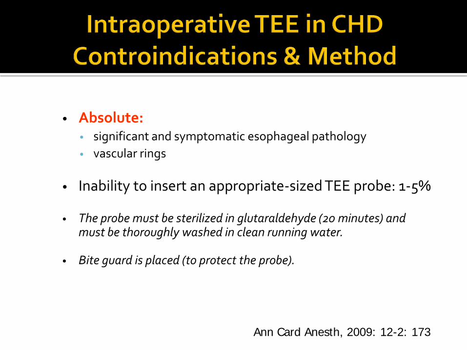
• Absolute:• significant and symptomatic esophageal pathology
• vascular rings
• Inability to insert an appropriate-sized TEE probe: 1-5%
• The probe must be sterilized in glutaraldehyde (20 minutes) and must be thoroughly washed in clean running water.
• Bite guard is placed (to protect the probe).
Ann Card Anesth, 2009: 12-2: 173

Tetralogy of Fallot
• Closure of the malalignment VSD
• Resection/patch repair of the infundibular obstruction.
• Residual VSD
• Assessment of residual RVOTO (desirable < 40 mmHg)
• Pulmonary regurgitation (not desirable!)
• Right ventricular size and contractility (related to hemodinamic conditions)
• Distal pulmonary arteries obstruction


PW Doppler sampling to estimate the gradients. (velocity <2-2.5 m/sec is not significant)Conduit regurgitation (relatively frequent)Morphology and movement of the conduit valve


Aortic Valvuloplasty

Intraoperative TEE:
Pratical Exemples
Ann Card Anesth, 2009: 12-2: 173

Ann Card Anesth, 2009: 12-2: 173

Atrial Repair of Transposition of the Great Arteries
• Performed if either the LV has regressed or there is a coronary anomaly making it unsuitable for ASO
• Pulmonary venous pathway (< 4-5 mmHg)• Sistemic venous pathway (phasic gradient,
< 6 mmHg)• Small baffle leaks (sist-to-pulm shunt,
desaturation)• Midbaffle obstruction (redundant or too
short)• LVOTO • Presence and severity of tricuspid
regurgitation.
post-operative Senning showing contrast injection in the SistemicVenous Atrium to check baffle leak

Intraoperative TEE:
D-TGA
Anatomic Correction of
Tratsposition of the Great
Arteries
•Neoaortic valvular insufficiency (degree)
•Supravalvular or branch pulmonary stenosis
•Assessment of coronary flow is
unsatisfactory
•Ventricular function

• Sub-aortic membrane• Circumferential membrane in the LVOT just before the aortic valve
causing a fixed sub-aortic obstruction. • Relation of this membrane to the aortic cusps • Aortic regurgitation.• Gradient across LVOT• Visualize the complete excision of the membrane
Ann Card Anesth, 2009: 12-2: 173

3D TEE is gaining popularity in assessing the intra-cardiac anatomy.
3D TEE has great impact in valve repairs in adult cardiac surgical patients.

• Intraoperative echo reduces the need for re-operation and then post-operative morbidity and mortality.
• Hemodynamic conditions influence the evaluation!!!

• Intra-operative TEE is a safe, reliable good diagnostictic modality, which provides a real-time assessment of surgical repair before removal of bypass cannulas and closure of the sternotomy.
• The introduction of a pediatric 3D TEE probe will help understand the complex intra-cardiac anatomy in a better way.

It is a team work
….Sometimes we are the eyes of our surgeons…
![Bi Rads Patologias 2 [Salvo Automaticamente]](https://static.fdocuments.us/doc/165x107/577c7a571a28abe05494cc50/bi-rads-patologias-2-salvo-automaticamente.jpg)


