Georgia Obstetrical and Gynecological Society TOOLKIT...
60
Optimizing Management of Obstetric Hemorrhage TOOLKIT Georgia Obstetrical and Gynecological Society Resources adapted from: The American Congress of Obstetricians and Gynecologists (ACOG) District II, Optimizing Protocols in Obstetrics California Maternal Quality Care Collaborative (CMQCC) Obstetric hemorrhage Toolkit Published March 2014
Transcript of Georgia Obstetrical and Gynecological Society TOOLKIT...

Optimizing Management of Obstetric Hemorrhage
TOOLKIT
Georgia Obstetrical and Gynecological Society
Resources adapted from: The American Congress of Obstetricians and Gynecologists (ACOG) District II, Optimizing Protocols in Obstetrics California Maternal Quality Care Collaborative (CMQCC) Obstetric hemorrhage Toolkit
Published March 2014


Optimizing Management of Obstetric Hemorrhage
Georgia Obstetrical and Gynecological Society 1
Dear Colleagues;
On behalf of the Georgia Obstetrical and Gynecological Society (GOGS), we are pleased to provide you with this toolkit on Optimizing Management of Postpartum Hemorrhage. Every day, 2 to 3 women die in the United States of pregnancy‐related complications, ranking the U.S. as number 46th in the world for maternal mortality.
Maternal mortality rates have steadily increased over the past decade (CDC, 2009). While some of the increase is due to improved data collection, these rates still have risen significantly. Georgia ranks among the highest in the United States with 35 maternal deaths per 100,000 live births in 2011, up from 20.5 from 2001 to 2006. Determined to reduce its maternal mortality rate, Dr. Brenda Fitzgerald, commissioner of the Georgia Department of Public Health (DPH) and an Obstetrician/Gynecologist, notes that many obstetric hemorrhage deaths are preventable. In response to rising maternal mortality, DPH, GOGS and a multidisciplinary team of experts from around the state have been diligently working on this very issue. In 2011, they formed the Georgia Maternal Mortality Review Committee (GA MMRC) to improve surveillance and understanding of pregnancy‐related deaths in Georgia.
In an effort to improve maternal mortality in this state, the Society continues to identify areas where improving policies, programs and services will impact the lives of all women and end preventable death and injury. Toward this goal, we are providing this postpartum hemorrhage (PPH) toolkit: Optimizing Management of Obstetric Hemorrhage to encourage all birthing hospitals in Georgia to review and implement the best practices and tools for managing obstetric hemorrhage. Enclosed in this toolkit you will find:
Core elements for the management of obstetric hemorrhage Recommendations to optimize the management of obstetric hemorrhage ACOG Practice Bulletin #76 – Postpartum Hemorrhage California Maternal Quality Care Collaborative (CMQCC) hemorrhage care checklist, flow chart,
table chart and training tools for the measurement of blood loss. Information on the importance of drills and sample PPH drill scenarios Additional information on postpartum hemorrhage
Providers are encouraged to review their hospital’s existing hemorrhage protocols and modify them if necessary to optimize the management of obstetrical hemorrhage. Standardization of health care processes and reduced variation in practice has been shown to improve outcomes and quality of care. Thank you for supporting this important initiative. If you have any questions regarding the enclosed materials, please contact Kaprice Welsh, Clinical Liaison for GOGS, 770‐904‐5288 or [email protected]. Sincerely,
Dr. Roland Matthews, MD President, Georgia OBGyn Society

Optimizing Management of Obstetric Hemorrhage
Georgia Obstetrical and Gynecological Society 2

Optimizing Management of Obstetric Hemorrhage
Georgia Obstetrical and Gynecological Society 3
Table of Contents
5 Improving Obstetric Hemorrhage in Georgia
6 Purpose of Toolkit
6 Resources for Optimizing Protocols in Obstetric Hemorrhage
6 Core Elements
7 Introduction to Obstetric Hemorrhage Management
7 California Maternal Quality Care Collaborative (CMQCC) Toolkit Overview
8 Recommendations to Optimize Management of Obstetrical Hemorrhage
10 Hemorrhage fact sheet
11 Issues with Hemorrhage Response in Obstetrics
13 ACOG Practice Bulletin No. 76, Postpartum Hemorrhage
23 Sample Hemorrhage Policy and Procedure
29 Obstetric Hemorrhage Care Guidelines Checklist 33 Obstetric Hemorrhage Care Summary Flow Chart 34 Obstetric Hemorrhage Care Summary Table Chart
35 Collaborative Guideline for Massive Transfusion
37 OB Hemorrhage: Carts, Kits, Trays
40 OB Hemorrhage Resources
42 Developing Training & Tools for Quantitative Measurement of Blood Loss
43 ACOG Patient Safety Checklist
45 Simulations & Drills 48 Importance of Drills 48 What you Need to Know for a Postpartum Hemorrhage Drill 49 Sample Postpartum Hemorrhage Drill 51 Sample Postpartum Hemorrhage Drill
55 Adopt a Systems Approach to Managing OB Hemorrhage
56 PPH Pearls
57 Reference Articles Relating to Obstetric Hemorrhage

Optimizing Management of Obstetric Hemorrhage
Georgia Obstetrical and Gynecological Society 4

Optimizing Management of Obstetric Hemorrhage
Georgia Obstetrical and Gynecological Society 5
Improving Obstetric Hemorrhage in Georgia Obstetric hemorrhage is the leading cause of maternal deaths in the United States with an estimate that 54-93 percent of these deaths are preventable. Unfortunately, our state’s maternal mortality ratio is 20.5 maternal deaths per 100,000 live births, which ranks 50th among all states in the U.S. The Georgia Obstetrical and Gynecological Society and the Department of Public Health would like to improve these statistics. To achieve this goal, we ask physicians and hospitals to use this toolkit to review and improve obstetric hemorrhage management in their own facilities. In addition, hospitals may apply to join a postpartum hemorrhage improvement initiative in which hospitals in Georgia, New Jersey and the District of Columbia have been invited to participate.
Georgia’s PPH Multi-Hospital Quality Improvement Initiative
The Georgia OBGyn Society is excited to lend its support to the Georgia Department of Public Health, Georgia Hospital Association, Association of Women’s Health, Obstetric and Neonatal Nurses (AWHONN), and Merck for Mothers, on a new Postpartum Hemorrhage Initiative to improve clinical practice and reduce errors related to postpartum hemorrhage. Georgia will be a part of this multi-state hospital quality improvement initiative to improve readiness, recognition, and response to postpartum hemorrhage.
Over the next three years, the collaborative will be working to improve outcomes for women and families in Georgia through participating birthing hospitals. We encourage your hospital to sign up to participate in the baseline survey about PPH practices at your institution. The survey can be accessed at www.pphproject.org. Hospitals that complete the baseline survey may apply to participate in a multi-hospital, multi-state learning collaborative. Hospitals selected to participate in the learning collaborative will work with local leaders and a group of national experts composed of nurses, physicians, and AWHONN staff to identify areas of improvement and work to change clinical practice at their facility.
Utilizing this Toolkit to Optimize Management of Obstetric Hemorrhage The Society has researched obstetric hemorrhage management resources from many excellent sources and included some of the best information in this toolkit. It is our hope that with this information and resources provided in this toolkit and with your leadership and encouragement, your hospital will review its hemorrhage protocols and implement the best practices for the women in your care.
If you have questions about the initiative, please contact: [email protected].

Optimizing Management of Obstetric Hemorrhage
Georgia Obstetrical and Gynecological Society 6
Purpose of Toolkit
This document reflects emerging clinical, scientific and patient safety advances as of the date issued and is subject to change. The information should not be construed as dictating an exclusive course of treatment or procedure to be followed. While the components of a particular protocol and/or checklist may be adapted to local resources, standardization of protocols and checklists within an institution is strongly encouraged.
Resources for Optimizing Protocols in Obstetric Hemorrhage
We have gathered materials from a number of sources in an effort to select the ideal requirements for a comprehensive approach to obstetrical hemorrhage. These included:
ACOG Practice Bulletin No. 76, Postpartum Hemorrhage
ACOG District II, Optimizing Protocols in Obstetrics, Management of Obstetrics Hemorrhage.
California Maternal Quality Care Collaborative, Obstetric Hemorrhage Care Guidelines and Compendium of Best Practices, CMQCC Obstetric Hemorrhage Toolkit.
Each hospital must take into account the resources available within its own institution and community to design a protocol that will assist them in the optimal management of obstetrical hemorrhage. Each institution is encouraged to review its existing policy and protocols, and modify them if necessary to provide safe patient care, or consider the creation of a policy that will optimize the management of obstetrical hemorrhage. Given the previous excellent work done in this area by the California Maternal Quality Care Collaborative (CMQCC), the American Congress of Obstetricians and Gynecologists (ACOG), and other organizations, we encourage individuals to utilize these extensive resources in the development of a hemorrhage protocol that will fit the needs of their individual institutions.
Core Elements
The following is a list of the components of any protocol that is created for the management of obstetrical hemorrhage. Hospitals should individualize their protocols based on an assessment of their own resources.
Definitions
Risk Factors/Etiology
Initial Interventions
Medical Treatment
Surgical Treatment
Defined Care Team and Escalation Role Clarity
Checklist Algorithm
Mass Transfusion Policy
Simulation Drills

Optimizing Management of Obstetric Hemorrhage
Georgia Obstetrical and Gynecological Society 7
Introduction to Obstetric Hemorrhage Management
Obstetric hemorrhage continues to cause maternal morbidity and mortality in Georgia and across the United States. Most of these cases occur in spite of women delivering in hospitals staffed by physicians, nurses and support personnel who are knowledgeable, highly motivated, and well trained. Often these cases occur in hospitals that have very well written obstetric hemorrhage protocols in place. Obstetric hemorrhage management is a time and team dependent performance that requires precise choreography. Having a “good protocol” that has never been practiced as a drill or dry run is similar to a football team that studies its plays but never works through the timing on the practice field, or a dance troupe that never rehearses before opening night.
We have evaluated and chosen resources that not only can be used to prevent serious harm associated with obstetric hemorrhage, but can be used to design very good hemorrhage protocols. However, to be effective, your protocol must have two things:
1. Each hemorrhage protocol must be designed and/or approved by the people who will execute it (and they must be given time and resources and permission needed to produce or thoroughly study the written protocol that will work specifically in the institution where it is designed).
2. Each hemorrhage protocol must be tested for feasibility within the institution and taught and rehearsed through dry runs or drills to improve its quality and the precision team work necessary to effectively manage obstetric hemorrhage.
California Maternal Quality Care Collaborative
The CMQCC toolkit provides excellent resources and can be viewed in complete form at https://www.cmqcc.org/ob_hemorrhage. The toolkit begins with a section on, “How To Use This Toolkit” (CMQMM pages 1‐2) followed by a compendium of evidence‐based, best practices related to obstetric hemorrhage. In this document we have included CMQCC Obstetric hemorrhage care guidelines which are presented in three forms starting with the most comprehensive “Checklist,” followed by the most streamlined version of the “Flowchart” and finally by a care summary “Table chart.” (See GOGS Toolkit pages 29–34, CMQCC pages 110–122.) The comprehensive “Checklist” delineates all topics the workgroup thought should be included in a protocol except for simulation/drills topic. A comprehensive document exists within the CMQCC tool kit related to obstetric hemorrhage drills and simulations. This document includes two detailed, ready to use scenarios which focus on both the technical management of obstetric hemorrhage, team function, communication, and role clarity. The document finishes with a Hospital Level Implementation Guide, which addresses practical planning for implementation of new evidence‐based protocols and guidelines for quality improvement (GOGS pages 45‐55, CMQCC pages 34‐47).

Optimizing Management of Obstetric Hemorrhage
Georgia Obstetrical and Gynecological Society 8
Recommendations to Optimize Management of Obstetric Hemorrhage
Here are PPH Toolkit examples that are either included in this GOGS toolkit or are part of the CMQCC Toolkit, including page numbers where they can be found:
Antepartum assessment is essential to identify women at risk for obstetrical hemorrhage.
Risk factor identification A prewritten order set for admission to L&D includes “risk scoring” for obstetric hemorrhage Definition checklist
Definitions & Early Recognition (CMQCC pages 3‐6) Incidence Risks & Diagnosis (CMQCC pages 22‐25)
Each institution should develop an effective written protocol for responding to maternal hemorrhage, including rapid emergency blood transfusion, which requires coordination among physicians, nurses, anesthesiologists and the blood bank.
Blood bank protocols should ensure that the institution has appropriate blood products for obstetric emergencies, and they should eliminate barriers to rapid blood access when needed. Sample Hemorrhage Policy (GOGS pages 23‐28, CMQCC pages 110‐115) Methods for developing training and tools for quantitative measurement of blood loss (GOGS page 42, CMQCC page 126)
Other suggestions include:
On initiation of the obstetric hemorrhage protocol, a complete set of prewritten orders should instantly be authorized and executed. The attending physician only will sign this order set after the emergency is completed.
Debriefings should occur after every drill and after every actual OB hemorrhage emergency. This allows for continuous quality improvement.
Flow charts, checklists, and other documentary materials needed for managing the OB hemorrhage emergency should be available to assist in the management. Toolkit examples
Surgical Treatment (CMQCC pages 72‐73) I. Literature Review (CMQCC pages 70‐71) II. Carts, Kits and Trays (GOGS pages 37‐41, CMQCC pages 26‐31)
Medical Treatment (CMQCC pages 74‐75) Checklist/Algorithms (GOGS pages 29‐34, CMQCC pages 86‐92)
Be vigilant regarding blood loss during pregnancy, labor, and delivery, and in the early postpartum period.
Nursing staff and physicians in the Labor, Delivery, Recovery and Postpartum areas must be trained in accurately assessing the degree of maternal hemorrhage.
When problems are identified, the nurse assigned must notify the physician immediately. See CMQCC toolkit for checklist example Toolkit examples
Definitions & Early Recognition (CMQCC pages 3‐6) Simulation & Drills (GOGS pages 45‐53, CMQCC pages 32‐47)

Optimizing Management of Obstetric Hemorrhage
Georgia Obstetrical and Gynecological Society 9
Use fluid resuscitation and transfusion based on the estimation of current blood loss and the expectation of continued bleeding, regardless of apparent maternal hemodynamic stability.
Accurately estimate blood loss Developing Training & Tools for Quantitative Measurement of Blood Loss (GOGS page 42, CMQCC page 126)
Toolkit examples Simulation & Drills (GOGS pages 45‐53, CMQCC pages 32‐47) Transfusion Policy (CMQCC pages 60‐69)
Work with hospital staff to conduct drills or simulation to ensure the most efficient management of obstetric hemorrhage.
Hospitals should run drills at different times of the day to ensure that appropriate hemorrhage team members are available at all times.
All members of the health care team should participate, including nurses, physicians and ancillary staff, as appropriate Simulation & Drills (GOGS pages 45‐53 , CMQCC pages 32‐47)
The maternal hemorrhage team should include, in addition to a team leader:
A surgeon with experience and expertise in controlling massive hemorrhage as well as operating room staff in case surgery is needed.
A critical care physician or anesthesiologist who is familiar with severe hemorrhage to help with assessment of organ perfusion and cardiovascular function.
A hematologist or clinical pathologist available on site to advise on appropriate blood products, and to coordinate and mobilize appropriate personnel to provide these products immediately.
Provide continuing medical education on hemorrhage for your entire medical team.
Ensure all hospital staff, including physicians, nurses, laboratory personnel and others are aware of the protocol related to dealing with maternal hemorrhage. Incorporate this protocol into your hospital’s mandatory annual educational programs and ensure all new staff is oriented to its content.
Findings from obstetrical quality improvement initiatives should be incorporated on an on‐going basis into improvements of the hemorrhage protocol.

Optimizing Management of Obstetric Hemorrhage
Georgia Obstetrical and Gynecological Society 10
Remains the major cause of obstetric morbidity and mortality
Hemorrhage >500ml (vaginal birth)= ~5-8%
Transfusion (vaginal birth)= ~0.5%
Transfusion (cesarean birth)= ~2%
Severe (massive) hemorrhage (>4units, >1500ml)= ~2/1,000 births
50-60% of severe morbidity in obstetrics
>60% of all postpartum maternal ICU admissions
The rate of severe hemorrhage is increasing, nearly doubling over the last decade
The greatest cause of maternal mortality by far, world-wide
: Amount of blood loss underestimated/ ignored until patient very unstable
: Expecting the bleeding “to stop soon”
: Hard to get the obstetrician back to the bedside for evaluation
: Repetitive use of the same procedure or medication (e.g. D&C, methergine) rather than
moving up the protocol (“scratched record”)
: Not using non-invasive procedures such as intrauterine balloons or B-Lynch sutures
Elliott Main, MD, Chair, California Pregnancy Associated Mortality Review Committee: personal communication (January 2009)

Optimizing Management of Obstetric Hemorrhage
Georgia Obstetrical and Gynecological Society 11
(from case review)
DENIAL, DELAY…
Poor quantification of blood loss
Lack of step-wise progression
Underutilization of non-pharmacologic approaches
Poor utilization of blood products:
“Too little, too late”
—Resuscitation v. Treatment
“Old wine in new bottles”—“Whole blood” v.
PRBCs
Step 1: Communication!


VOL. 108, NO. 4, OCTOBER 2006 OBSTETRICS & GYNECOLOGY 1039
ACOGPRACTICEBULLETIN
CLINICAL MANAGEMENT GUIDELINES FOROBSTETRICIAN–GYNECOLOGISTS
NUMBER 76, OCTOBER 2006
(Replaces Committee Opinion Number 266, January 2002)
This Practice Bulletin wasdeveloped by the ACOG Com-mittee on Practice Bulletins—Obstetrics with the assistanceof William N. P. Herbert, MD,and Carolyn M. Zelop, MD.The information is designed toaid practitioners in makingdecisions about appropriateobstetric and gynecologic care.These guidelines should not beconstrued as dictating an exclu-sive course of treatment or pro-cedure. Variations in practicemay be warranted based on theneeds of the individual patient,resources, and limitationsunique to the institution or typeof practice.
Postpartum HemorrhageSevere bleeding is the single most significant cause of maternal death world-wide. More than half of all maternal deaths occur within 24 hours of delivery,most commonly from excessive bleeding. It is estimated that, worldwide,140,000 women die of postpartum hemorrhage each year—one every 4 minutes(1). In addition to death, serious morbidity may follow postpartum hemorrhage.Sequelae include adult respiratory distress syndrome, coagulopathy, shock, lossof fertility, and pituitary necrosis (Sheehan syndrome).
Although many risk factors have been associated with postpartum hemor-rhage, it often occurs without warning. All obstetric units and practitionersmust have the facilities, personnel, and equipment in place to manage thisemergency properly. Clinical drills to enhance the management of maternalhemorrhage have been recommended by the Joint Commission on Accreditationof Healthcare Organizations (2). The purpose of this bulletin is to review theetiology, evaluation, and management of postpartum hemorrhage.
BackgroundThe physiologic changes over the course of pregnancy, including a plasma vol-ume increase of approximately 40% and a red cell mass increase of approxi-mately 25%, occur in anticipation of the blood loss that will occur at delivery(3). There is no single, satisfactory definition of postpartum hemorrhage. Anestimated blood loss in excess of 500 mL following a vaginal birth or a loss ofgreater than 1,000 mL following cesarean birth often has been used for thediagnosis, but the average volume of blood lost at delivery can approach theseamounts (4, 5). Estimates of blood loss at delivery are notoriously inaccurate,with significant underreporting being the rule. Limited instruction on estimat-ing blood loss has been shown to improve the accuracy of such estimates (6).Also, a decline in hematocrit levels of 10% has been used to define postpartumhemorrhage, but determinations of hemoglobin or hematocrit concentrationsmay not reflect the current hematologic status (7). Hypotension, dizziness, pal-
13
1040 ACOG Practice Bulletin Postpartum Hemorrhage OBSTETRICS & GYNECOLOGY
lor, and oliguria do not occur until blood loss is substan-tial—10% or more of total blood volume (8).
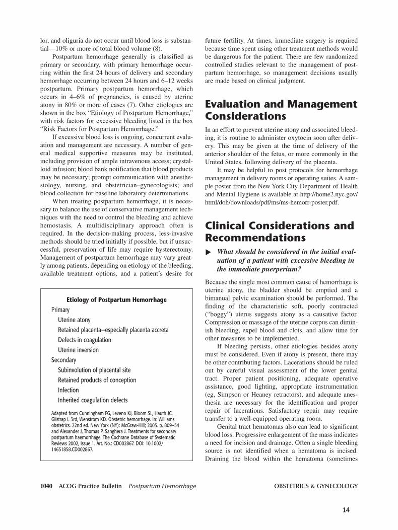
Postpartum hemorrhage generally is classified asprimary or secondary, with primary hemorrhage occur-ring within the first 24 hours of delivery and secondaryhemorrhage occurring between 24 hours and 6–12 weekspostpartum. Primary postpartum hemorrhage, whichoccurs in 4–6% of pregnancies, is caused by uterineatony in 80% or more of cases (7). Other etiologies areshown in the box “Etiology of Postpartum Hemorrhage,”with risk factors for excessive bleeding listed in the box“Risk Factors for Postpartum Hemorrhage.”
If excessive blood loss is ongoing, concurrent evalu-ation and management are necessary. A number of gen-eral medical supportive measures may be instituted,including provision of ample intravenous access; crystal-loid infusion; blood bank notification that blood productsmay be necessary; prompt communication with anesthe-siology, nursing, and obstetrician–gynecologists; andblood collection for baseline laboratory determinations.
When treating postpartum hemorrhage, it is neces-sary to balance the use of conservative management tech-niques with the need to control the bleeding and achievehemostasis. A multidisciplinary approach often isrequired. In the decision-making process, less-invasivemethods should be tried initially if possible, but if unsuc-cessful, preservation of life may require hysterectomy.Management of postpartum hemorrhage may vary great-ly among patients, depending on etiology of the bleeding,available treatment options, and a patient’s desire for
future fertility. At times, immediate surgery is requiredbecause time spent using other treatment methods wouldbe dangerous for the patient. There are few randomizedcontrolled studies relevant to the management of post-partum hemorrhage, so management decisions usuallyare made based on clinical judgment.
Evaluation and ManagementConsiderationsIn an effort to prevent uterine atony and associated bleed-ing, it is routine to administer oxytocin soon after deliv-ery. This may be given at the time of delivery of theanterior shoulder of the fetus, or more commonly in theUnited States, following delivery of the placenta.
It may be helpful to post protocols for hemorrhagemanagement in delivery rooms or operating suites. A sam-ple poster from the New York City Department of Healthand Mental Hygiene is available at http://home2.nyc.gov/html/doh/downloads/pdf/ms/ms-hemorr-poster.pdf.
Clinical Considerations andRecommendations
What should be considered in the initial eval-uation of a patient with excessive bleeding inthe immediate puerperium?
Because the single most common cause of hemorrhage isuterine atony, the bladder should be emptied and abimanual pelvic examination should be performed. Thefinding of the characteristic soft, poorly contracted(“boggy”) uterus suggests atony as a causative factor.Compression or massage of the uterine corpus can dimin-ish bleeding, expel blood and clots, and allow time forother measures to be implemented.
If bleeding persists, other etiologies besides atonymust be considered. Even if atony is present, there maybe other contributing factors. Lacerations should be ruledout by careful visual assessment of the lower genitaltract. Proper patient positioning, adequate operativeassistance, good lighting, appropriate instrumentation(eg, Simpson or Heaney retractors), and adequate anes-thesia are necessary for the identification and properrepair of lacerations. Satisfactory repair may requiretransfer to a well-equipped operating room.
Genital tract hematomas also can lead to significantblood loss. Progressive enlargement of the mass indicatesa need for incision and drainage. Often a single bleedingsource is not identified when a hematoma is incised.Draining the blood within the hematoma (sometimes
Etiology of Postpartum Hemorrhage
PrimaryUterine atonyRetained placenta—especially placenta accretaDefects in coagulationUterine inversion
SecondarySubinvolution of placental siteRetained products of conceptionInfection Inherited coagulation defects
Adapted from Cunningham FG, Leveno KJ, Bloom SL, Hauth JC,Gilstrap L 3rd, Wenstrom KD. Obstetric hemorrhage. In: Williamsobstetrics. 22nd ed. New York (NY): McGraw-Hill; 2005. p. 809–54and Alexander J, Thomas P, Sanghera J. Treatments for secondarypostpartum haemorrhage. The Cochrane Database of SystematicReviews 2002, Issue 1. Art. No.: CD002867. DOI: 10.1002/14651858.CD002867.
▲
14

VOL. 108, NO. 4, OCTOBER 2006 ACOG Practice Bulletin Postpartum Hemorrhage 1041
placing a drain in situ), suturing the incision, and ifappropriate, packing the vagina are measures usuallysuccessful in achieving hemostasis. Interventional radi-ology is another option for management of a hematoma.Genital tract hematomas may not be recognized untilhours after the delivery, and they sometimes occur in theabsence of vaginal or perineal lacerations. The mainsymptoms are pelvic or rectal pressure and pain.
The possibility that additional products of concep-tion remain within the uterine cavity should be consid-ered. Ultrasonography can help diagnose a retainedplacenta. Retained placental tissue is unlikely whenultrasonography reveals a normal endometrial stripe.Although ultrasonographic images of retained placentaltissue are inconsistent, detection of an echogenic mass inthe uterus is more conclusive. Ultrasound evaluation forretained tissue should be performed before uterineinstrumentation is undertaken (9). Spontaneous expul-sion of the placenta, apparent structural integrity oninspection, and the lack of a history of previous uterinesurgery (suggesting an increased risk of abnormal pla-centation) make a diagnosis of retained products of theplacenta less likely, but a curettage may identify a suc-centuriate lobe of the placenta or additional placental tis-sue. When a retained placenta is identified, a large, bluntinstrument, such as a banjo curette or ring forceps, guid-ed by ultrasonography, makes removal of the retainedtissue easier and reduces the risk of perforation.
Less commonly, postpartum hemorrhage may becaused by coagulopathy. Clotting abnormalities shouldbe suspected on the basis of patient or family history
Risk Factors for Postpartum Hemorrhage
Prolonged laborAugmented laborRapid laborHistory of postpartum hemorrhageEpisiotomy, especially mediolateralPreeclampsiaOverdistended uterus (macrosomia, twins, hydramnios)Operative deliveryAsian or Hispanic ethnicityChorioamnionitis
Data from Stones RW, Paterson CM, Saunders NJ. Risk factors formajor obstetric haemorrhage. Eur J Obstet Gynecol Reprod Biol1993;48:15–8 and Combs CA, Murphy EL, Laros RK. Factors associ-ated with hemorrhage in cesarean deliveries. Obstet Gynecol1991;77:77–82.
or clinical circumstances. Hemolysis, elevated liverenzymes, and low platelet count (HELLP) syndrome,abruptio placentae, prolonged intrauterine fetal demise,sepsis, and amniotic fluid embolism are associated withclotting abnormalities. Significant hemorrhage from anycause can lead to consumption of clotting factors.Observation of the clotting status of blood recently lostcan provide important information. When a coagulopa-thy is suspected, appropriate testing should be ordered,with blood products infused as indicated. In some situa-tions, the coagulopathy may be caused or perpetuated bythe hemorrhage. In such cases, simultaneous surgery andblood product replacement may be necessary.
Baseline studies should be ordered when excessiveblood loss is suspected and should be repeated periodi-cally as clinical circumstances warrant. Cliniciansshould remember that the results of some studies may bemisleading because equilibration may not have occurred.In addition, response to hemorrhage may be requiredbefore laboratory results are known. Baseline studiesinclude a complete blood count with platelets, a pro-thrombin time, an activated partial thromboplastin time,fibrinogen, and a type and cross order. The blood bankshould be notified that transfusion may be necessary.
The clot observation test provides a simple measureof fibrinogen (10). A volume of 5 mL of the patient’sblood is placed into a clean, red-topped tube andobserved frequently. Normally, blood will clot within8–10 minutes and will remain intact. If the fibrinogenconcentration is low, generally less than 150 mg/dL, theblood in the tube will not clot, or if it does, it will under-go partial or complete dissolution in 30–60 minutes.
What is the appropriate medical managementapproach for excessive postpartum bleeding?
Ongoing blood loss in the setting of decreased uterinetone requires the administration of additional uterotonicsas the first-line treatment for hemorrhage (Table 1).Some practitioners prefer direct injection of methyler-gonovine maleate and 15-methyl prostaglandin (PG) F2αinto the uterine corpus. Human recombinant factor VIIais a new treatment modality shown to be effective in controlling severe, life-threatening hemorrhage by actingon the extrinsic clotting pathway. Intravenous dosagesvary by case and generally range from 50 to 100 mcg/kgevery 2 hours until hemostasis is achieved. Cessation ofbleeding ranges from 10 minutes to 40 minutes afteradministration (11–14). Concern has been raised be-cause of apparent risk of subsequent thromboembolicevents following factor VIIa use (15). Compared withother agents, factor VIIa is extremely expensive.Additional clinical experience in all specialties will help
▲
15

1042 ACOG Practice Bulletin Postpartum Hemorrhage OBSTETRICS & GYNECOLOGY
available to control bleeding (Table 3). Hypogastricartery ligation is performed much less frequently than inyears past. Its purpose is to diminish the pulse pressure ofblood flowing to the uterus via the internal iliac(hypogastric) vessels. Practitioners are less familiar withthis technique, and the procedure has been found to beconsiderably less successful than previously thought(17). Bilateral uterine artery ligation (O’Leary sutures)accomplishes the same goal, and this procedure is quick-er and easier to perform (18, 19). To further diminishblood flow to the uterus, similar sutures can be placedacross the vessels within the uteroovarian ligaments.
The B-Lynch technique is a newer procedure forstopping excessive bleeding caused by uterine atony (20).The suture provides even pressure to compress the uterinecorpus and decrease bleeding. One study reported more
determine factor VIIa’s role in the treatment of patientswith postpartum hemorrhage.
When is packing or tamponade of the uterinecavity advisable?
When uterotonics fail to cause sustained uterine contrac-tions and satisfactory control of hemorrhage after vaginaldelivery, tamponade of the uterus can be effective indecreasing hemorrhage secondary to uterine atony (Table2). Such approaches can be particularly useful as a tem-porizing measure, but if a prompt response is not seen,preparations should be made for exploratory laparotomy.
Packing with gauze requires careful layering of thematerial back and forth from one cornu to the other usinga sponge stick, packing back and forth, and ending withextension of the gauze through the cervical os. The sameeffect often can be derived more easily using a Foleycatheter, Sengstaken-Blakemore tube, or, more recently,the SOS Bakri tamponade balloon (16), specifically tai-lored for tamponade within the uterine cavity in cases ofpostpartum hemorrhage secondary to uterine atony.
When are surgical techniques used to controluterine bleeding?
When uterotonic agents with or without tamponademeasures fail to control bleeding in a patient who hasgiven birth vaginally, exploratory laparotomy is indicat-ed. A midline vertical abdominal incision usually is pre-ferred to optimize exposure. Several techniques are
▲▲
Table 1. Medical Management of Postpartum Hemorrhage
Drug* Dose/Route Frequency Comment
Oxytocin (Pitocin) IV: 10–40 units in 1 liter Continuous Avoid undiluted rapid IV infusion,normal saline or lactated which causes hypotension.Ringer’s solution IM: 10 units
Methylergonovine IM: 0.2 mg Every 2–4 h Avoid if patient is hypertensive.(Methergine)
15-methyl PGF2α IM: 0.25 mg Every 15–90 min, Avoid in asthmatic patients;(Carboprost) 8 doses maximum relative contraindication if(Hemabate) hepatic, renal, and cardiac
disease. Diarrhea, fever, tachycardia can occur.
Dinoprostone Suppository: vaginal Every 2 h Avoid if patient is hypotensive.(Prostin E2) or rectal Fever is common. Stored frozen,
20 mg it must be thawed to room temperature.
Misoprostol 800–1,000 mcg rectally(Cytotec, PGE1)
Abbreviations: IV, intravenously; IM, intramuscularly; PG, prostaglandin.*All agents can cause nausea and vomiting.Modified from Dildy GA, Clark SL. Postpartum hemorrhage. Contemp Ob/Gyn 1993;38(8):21–9.
Table 2. Tamponade Techniques for Postpartum Hemorrhage
Technique Comment
Uterine tamponade
—Packing —4-inch gauze; can soak with 5,000 units of thrombin in 5 mL of sterile saline
—Foley catheter —Insert one or more bulbs; instill 60–80 mL of saline
—Sengstaken–Blakemore tube
—SOS Bakri tamponade balloon —Insert balloon; instill 300–500 mL of saline
16

VOL. 108, NO. 4, OCTOBER 2006 ACOG Practice Bulletin Postpartum Hemorrhage 1043
than 1,000 B-Lynch procedures with only seven failures(21). However, because the technique is new, many clini-cians have limited experience with this procedure (22).
Hemostatic multiple square suturing is another newsurgical technique for postpartum hemorrhage caused byuterine atony, placenta previa, or placenta accreta. Theprocedure eliminates space in the uterine cavity by sutur-ing both anterior and posterior uterine walls. One studyreported on this technique in 23 women after conserva-tive treatment failed. All patients were examined after 2months, and ultrasound findings confirmed normalendometrial linings and uterine cavities (23).
What are the clinical considerations for suspected placenta accreta?
Abnormal attachment of the placenta to the inner uterinewall (placenta accreta) can cause massive hemorrhage. Infact, accreta and uterine atony are the two most commonreasons for postpartum hysterectomy (24, 25). Risk factorsfor placenta accreta include placenta previa with or with-out previous uterine surgery, prior myomectomy, priorcesarean delivery, Asherman’s syndrome, submucousleiomyomata, and maternal age older than 35 years (26).
Prior cesarean delivery and the presence of placentaprevia in a current pregnancy are particularly important riskfactors for placenta accreta. In a multicenter study of morethan 30,000 patients who had cesarean delivery withoutlabor, the risk of placenta accreta was approximately 0.2%,0.3%, 0.6%, 2.1%, 2.3%, and 7.7% for women experienc-ing their first through sixth cesarean deliveries, respective-ly. In patients with placenta previa in the current pregnancy,the risk of accreta was 3%, 11%, 40%, 61%, and 67% forthose undergoing their first through their fifth or greatercesarean deliveries, respectively (27).
Women with placenta previa or placenta accretahave a higher incidence of postpartum hemorrhage andare more likely to undergo emergency hysterectomy
(28). In the multicenter study cited previously, hysterec-tomy was required in 0.7% for the first cesarean deliveryand increased with each cesarean delivery up to 9% forpatients with their sixth or greater cesarean delivery.
In the presence of previa or a history of cesareandelivery, the obstetric care provider must have a highclinical suspicion for placenta accreta and take appropri-ate precautions. Ultrasonography may be helpful inestablishing the diagnosis in the antepartum period.Color Doppler technology may be an additional adjunc-tive tool for suspected accreta (29). Despite advances inimaging techniques, no diagnostic technique affords theclinician complete assurance of the presence or absenceof placenta accreta.
If the diagnosis or a strong suspicion is formedbefore delivery, a number of measures should be taken:
• The patient should be counseled about the likelihoodof hysterectomy and blood transfusion.
• Blood products and clotting factors should be avail-able.
• Cell saver technology should be considered if avail-able.
• The appropriate location and timing for deliveryshould be considered to allow access to adequatesurgical personnel and equipment.
• A preoperative anesthesia assessment should beob-tained.
The extent (area, depth) of the abnormal attachmentwill determine the response—curettage, wedge resection,medical management, or hysterectomy. Uterine conserv-ing options may work in small focal accretas, but abdom-inal hysterectomy usually is the most definitive treatment.
Under what circumstances is arterialembolization indicated?
A patient with stable vital signs and persistent bleeding,especially if the rate of loss is not excessive, may be a can-didate for arterial embolization. Radiographic identifica-tion of bleeding vessels allows embolization with Gelfoam,coils, or glue. Balloon occlusion is also a technique used insuch circumstances. Embolization can be used for bleedingthat continues after hysterectomy or can be used as analternative to hysterectomy to preserve fertility.
When is blood transfusion recommended? Is there a role for autologous transfusions or directed donor programs?
Transfusion of blood products is necessary when theextent of blood loss is significant and ongoing, particu-larly if vital signs are unstable. Postpartum transfusion
▲
▲▲
Table 3. Surgical Management of Postpartum Hemorrhage
Technique Comment
Uterine curettage
Uterine artery ligation Bilateral; also can ligate uteroovarian vessels
B-Lynch suture
Hypogastric artery ligation Less successful than earlier thought; difficult technique; generally reserved for practitioners experienced in the procedure
Repair of rupture
Hysterectomy
17

1044 ACOG Practice Bulletin Postpartum Hemorrhage OBSTETRICS & GYNECOLOGY
rates vary between 0.4% and 1.6% (30). Clinical judg-ment is an important determinant, given that estimates ofblood loss often are inaccurate, determination of hemat-ocrit or hemoglobin concentrations may not accuratelyreflect the current hematologic status, and symptoms andsigns of hemorrhage may not occur until blood lossexceeds 15% (8). The purpose of transfusion of bloodproducts is to replace coagulation factors and red cells foroxygen-carrying capacity, not for volume replacement.To avoid dilutional coagulopathy, concurrent replace-ment with coagulation factors and platelets may be nec-essary. Table 4 lists blood components, indications fortransfusion, and hematologic effects.
Autologous transfusion (donation, storage, retrans-fusion) has been shown to be safe in pregnancy (31, 32).However, it requires anticipation of the need for transfu-sion, as well as a minimal hematocrit concentration oftenabove that of a pregnant woman. Autologous transfusiongenerally is reserved for situations with a high chance oftransfusion in a patient with rare antibodies, where thelikelihood of identifying compatible volunteer-providedblood is very low. Blood donated by directed donors hasnot been shown to be safer than blood from unknown,volunteer donors. Cell saver technology has been usedsuccessfully in patients undergoing cesarean delivery. Ina multicenter study of 139 patients using such devices, nountoward outcomes were noted when compared withcontrol patients (33).
What is the management approach for hemorrhage due to a ruptured uterus?
Rupture can occur at the site of a previous cesarean deliv-ery or other surgical procedure involving the uterine wallfrom intrauterine manipulation or trauma or from con-genital malformation (small uterine horn), or it can occurspontaneously. Abnormal labor, operative delivery, andplacenta accreta can lead to rupture. Surgical repair is
required, with the specific approach tailored to recon-struct the uterus, if possible. Care depends on the extentand site of rupture, the patient’s current clinical condi-tion, and her desire for future childbearing. Rupture of aprevious cesarean delivery scar often can be managed byrevision of the edges of the prior incision followed byprimary closure. In addition to the myometrial disrup-tion, consideration must be given to neighboring struc-tures, such as the broad ligament, parametrial vessels,ureters, and bladder. Regardless of the patient’s wishesfor the avoidance of hysterectomy, this procedure maybe necessary in a life-threatening situation.
What is the management approach for aninverted uterus?
Uterine inversion, in which the uterine corpus descendsto, and sometimes through, the uterine cervix, is associ-ated with marked hemorrhage. On bimanual examina-tion, the finding of a firm mass below or near the cervix,coupled with the absence of identification of the uterinecorpus on abdominal examination, suggests inversion. If the inversion occurs before placental separation,detachment or removal of the placenta should not beundertaken; this will lead to additional hemorrhage.Replacement of the uterine corpus involves placing thepalm of the hand against the fundus (now inverted andlowermost at or through the cervix), as if holding a ten-nis ball, with the fingertips exerting upward pressure circumferentially (34). To restore normal anatomy, re-laxation of the uterus may be necessary. Terbutaline,magnesium sulfate, halogenated general anesthetics, andnitroglycerin have been used for uterine relaxation.
Manual replacement with or without uterine relax-ants usually is successful. In the unusual circumstance inwhich it is not, laparotomy is required. Two procedureshave been reported to return the uterine corpus to theabdominal cavity. The Huntington procedure involves
▲
▲
Table 4. Blood Component Therapy
Product Volume (mL) Contents Effect (per unit)
Packed red cells 240 Red blood cells, Increase hematocrit 3 percentage white blood cells, plasma points, hemoglobin by 1 g/dL
Platelets 50 Platelets, red blood cells, Increase platelet count 5,000–white blood cells, plasma 10,000/mm3 per unit
Fresh frozen plasma 250 Fibrinogen, antithrombin III, Increase fibrinogen by 10 mg/dLfactors V and VIII
Cryoprecipitate 40 Fibrinogen, factors VIII and Increase fibrinogen by 10 mg/dLXIII, von Willebrand factor
Modified from Martin SR, Strong TH Jr. Transfusion of blood components and derivatives in the obstetric intensive carepatient. In: Foley MR, Strong TH Jr, Garite TJ, editors. Obstetric intensive care manual. 2nd ed. New York (NY): McGraw-Hill;2004. Produced with permission of The McGraw-Hill Companies.
18
progressive upward traction on the inverted corpus usingBabcock or Allis forceps (35). The Haultain procedureinvolves incising the cervical ring posteriorly, allowingfor digital repositioning of the inverted corpus, with sub-sequent repair of the incision (36).
What is the management approach for secondary postpartum hemorrhage?
Secondary hemorrhage occurs in approximately 1% ofpregnancies; often the specific etiology is unknown.Postpartum hemorrhage may be the first indication forvon Willebrand’s disease for many patients and shouldbe considered. The prevalence of von Willebrand’s dis-ease is reported to be 10–20% among adult women withmenorrhagia (37). Hence, testing for bleeding disordersshould be considered among pregnant patients with ahistory of menorrhagia because the risk of delayed orsecondary postpartum hemorrhage is high amongwomen with bleeding disorders (38, 39).
Uterine atony (perhaps secondary to retained prod-ucts of conception) with or without infection contrib-utes to secondary hemorrhage. The extent of bleeding usually is less than that seen with primary postpartumhemorrhage. Ultrasound evaluation can help identifyintrauterine tissue or subinvolution of the placental site.Treatment may include uterotonic agents, antibiotics,and curettage. Often the volume of tissue removed bycurettage is minimal, yet bleeding subsides promptly.Care must be taken in performing the procedure to avoidperforation of the uterus. Concurrent ultrasound assess-ment at the time of curettage can be helpful in prevent-ing this complication. Patients should be counseledabout the possibility of hysterectomy before initiatingany operative procedures.
What is the best approach to managingexcessive blood loss in the postpartum periodonce the patient’s condition is stable?
Regardless of the cause of postpartum hemorrhage, sub-sequent replacement of the red cell mass is important.Along with a prenatal vitamin and mineral capsule daily(which contains about 60 mg of elemental iron and 1 mg folate), two additional iron tablets (ferrous sulfate,300 mg, each yielding about 60 mg of elemental iron) willmaximize red cell production and restoration. Erythro-poietin can hasten red cell production in postpartum ane-mic patients to some extent, but it is not approved by theU.S. Food and Drug Administration for postoperative ane-mia, and it can be costly (40). Postpartum hemorrhage ina subsequent pregnancy occurs in approximately 10% ofpatients (8).
Summary ofRecommendations andConclusionsThe following recommendations and conclusionsare based primarily on consensus and expert opin-ion (Level C):
Uterotonic agents should be the first-line treatmentfor postpartum hemorrhage due to uterine atony.
Management may vary greatly among patients,depending on etiology and available treatmentoptions, and often a multidisciplinary approach isrequired.
When uterotonics fail following vaginal delivery,exploratory laparotomy is the next step.
In the presence of conditions known to be associat-ed with placenta accreta, the obstetric care providermust have a high clinical suspicion and take appro-priate precautions.
Proposed PerformanceMeasureIf hysterectomy is performed for uterine atony, thereshould be documentation of other therapy attempts.
References1. AbouZahr C. Global burden of maternal death and dis-
ability. Br Med Bull 2003;67:1–11. (Level III)
2. Preventing infant death and injury during delivery.Sentinel Event ALERT No. 30. Joint Commission onAccreditation of Healthcare Organizations. Available at:http://www.jointcommission.org/SentinelEvents/SentinelEventAlert/sea_30.htm. Retrieved June 12, 2006. (LevelIII)
3. Chesley LC. Plasma and red cell volumes during pregnan-cy. Am J Obstet Gynecol 1972;112:440–50. (Level III)
4. Pritchard JA, Baldwin RM, Dickey JC, Wiggins KM.Blood volume changes in pregnancy and the puerperium.Am J Obstet Gyencol 1962;84:1271–82. (Level III)
5. Clark SL, Yeh SY, Phelan JP, Bruce S, Paul RH.Emergency hysterectomy for obstetric hemorrhage.Obstet Gynecol 1984;64:376–80. (Level III)
6. Dildy GA 3, Paine AR, George NC, Velasco C. Estimatingblood loss: can teaching significantly improve visual esti-mation? Obstet Gynecol 2004;104:601–6. (Level III)
7. Combs CA, Murphy EL, Laros RK Jr. Factors associatedwith postpartum hemorrhage with vaginal birth. ObstetGynecol 1991;77:69–76. (Level II-2)
VOL. 108, NO. 4, OCTOBER 2006 ACOG Practice Bulletin Postpartum Hemorrhage 1045
▲
▲▲
▲▲
▲
19

1046 ACOG Practice Bulletin Postpartum Hemorrhage OBSTETRICS & GYNECOLOGY
25. Stanco LM, Schrimmer DB, Paul RH, Mishell DR Jr.Emergency peripartum hysterectomy and associated riskfactors. Am J Obstet Gynecol 1993;168:879–83. (LevelII-3)
26. Clark SL, Koonings PP, Phelan JP. Placenta previa/accreta and prior cesarean section. Obstet Gynecol1985;66:89–92. (Level III)
27. Silver RM, Landon MB, Rouse DT, Leveno KJ, Song CY,Thom EA, et al. Maternal morbidity associated with mul-tiple repeat cesarean delivery. Obstet Gynecol 2006;107:1226–32. (Level II-2)
28. Zaki ZM, Bahar AM, Ali ME, Albar HA, Gerais MA. Riskfactors and morbidity in patients with placenta previa ac-creta compared to placenta previa non-accreta. ActaObstet Gynecol Scand 1998;77:391–4. (Level II-3)
29. Kirkinen P, Helin-Martikainen HL, Vanninen R, PartanenK. Placenta accreta: imaging by gray-scale and contrast-enhanced color Doppler sonography and magnetic reso-nance imaging. J Clin Ultrasound 1998;26:90–4. (Level III)
30. Petersen LA, Lindner DS, Kleiber CM, Zimmerman MB,Hinton AT, Yankowitz J. Factors that predict low hemat-ocrit levels in the postpartum patient after vaginal deliv-ery. Am J Obstet Gynecol 2002;186:737–4. (Level II-2)
31. Kruskall MS, Leonard S, Klapholz H. Autologous blooddonation during pregnancy: analysis of safety and blooduse. Obstet Gynecol 1987;70:938–41. (Level III)
32. Herbert WN, Owen HG, Collins ML. Autologous bloodstorage in obstetrics. Obstet Gynecol 1988;72:166–70.(Level III)
33. Rebarber A, Lonser R, Jackson S, Copel JA, Sipes S. Thesafety of intraoperative autologous blood collection andautotransfusion during cesarean section. Am J ObstetGynecol 1998;179:715–20. (Level II-2)
34. Johnson AB. A new concept in the replacement of theinverted uterus and a report of nine cases. Am J ObstetGynecol 1949;57:557–62. (Level III)
35. Huntington JL, Irving FC, Kellogg FS. Abdominal reposi-tion in acute inversion of the puerperal uterus. Am JObstet Gynecol 1928;15:34–40. (Level III)
36. Haultain FW. The treatment of chronic uterine inversionby abdominal hysterotomy, with a successful case. BrMed J 1901;2:974–6. (Level III)
37. Demers C, Derzko C, David M, Douglas J. Gynaecolog-ical and obstetric management of women with inheritedbleeding disorders. Society of Obstetricians and Gyne-cologists of Canada. J Obstet Gynaecol Can 2005;27:707–32. (Level III)
38. Kadir RA, Aledort LM. Obstetrical and gynaecologicalbleeding: a common presenting symptom. Clin LabHaematol 2000 Oct;22 suppl 1:12–6; discussion 30–2.(Level III)
39. James AH. Von Willebrand disease. Obstet Gynecol Surv2006;61:136–45. (Level III)
40. Kotto-Kome AC, Calhoun DA, Montenegro R, Sosa R,Maldonado L, Christensen RD. Effect of administeringrecombinant erythropoietin to women with postpartumanemia: a meta-analysis. J Perinatol 2004;24:11–5.(Meta-analysis)
8. Bonnar J. Massive obstetric haemorrhage. Baillieres BestPract Res Clin Obstet Gynaecol 2000;14:1–18. (Level III)
9. Hertzberg BS, Bowie JD. Ultrasound of the postpartumuterus. Prediction of retained placental tissue. J Ultra-sound Med 1991;10:451–6. (Level III)
10. Poe MF. Clot observation test for clinical diagnosis of clot-ting defects. Anesthesiology 1959;20:825–9. (Level III)
11. Bouwmeester FW, Jonkhoff AR, Verheijen RH, van GeijnHP. Successful treatment of life-threatening postpartumhemorrhage with recombinant activated factor VII. ObstetGynecol 2003;101:1174–6. (Level III)
12. Tanchev S, Platikanov V, Karadimov D. Administration ofrecombinant factor VIIa for the management of massivebleeding due to uterine atonia in the post-placental period.Acta Obstet Gynecol Scand 2005;84:402–3. (Level III)
13. Boehlen F, Morales MA, Fontaana P, Ricou B, Irion O, deMoerloose P. Prolonged treatment of massive postpartumhaemorrhage with recombinant factor VIIa: case reportand review of the literature. BJOG 2004;111:284–7.(Level III)
14. Segal S, Shemesh IY, Blumental R, Yoffe B, Laufer N,Mankuta D, et al. The use of recombinant factor VIIa insevere postpartum hemorrhage. Acta Obstet GynecolScand 2004;83:771–2. (Level III)
15. O’Connell KA, Wood JJ, Wise RP, Lozier JN, Braun MM.Thromboembolic adverse events after use of recombinanthuman coagulation factor VIIa. JAMA 2006;295:293–8.(Level III)
16. Bakri YN, Amri A, Abdul Jabbar F. Tamponade-balloonfor obstetrical bleeding. Int J Gynaecol Obstet 2001;74:139–42. (Level III)
17. Clark AL, Phelan JP, Yeh SY, Bruce SR, Paul RH.Hypogastric artery ligation for obstetric hemorrhage.Obstet Gynecol 1985:66:353–6. (Level III)
18. O’Leary JL, O’Leary JA. Uterine artery ligation in thecontrol of intractable postpartum hemorrhage. Am JObstet Gynecol 1966;94:920–4. (Level III)
19. O’Leary JL, O’Leary JA. Uterine artery ligation for con-trol of postcesarean section hemorrhage. Obstet Gynecol1974;43:849–53. (Level III)
20. B-Lynch C, Coker A, Lawal AH, Abu J, Cowen MJ. TheB-Lynch surgical technique for the control of massivepostpartum haemorrhage: an alternative to hysterectomy?Five cases reported. Br J Obstet Gynaecol 1997;104:372–5. (Level III)
21. Allam MS, B-Lynch C. The B-Lynch and other uterinecompression suture techniques. Int J Gynaecol Obstet2005;89:236–41. (Level III)
22. Holtsema H, Nijland R, Huisman A, Dony J, van den BergPP. The B-Lynch technique for postpartum haemorrhage:an option for every gynaecologist. Eur J Obstet GynecolReprod Biol 2004;115:39–42. (Level III)
23. Cho JH, Jun HS, Lee CN. Hemostatic suturing techniquefor uterine bleeding during cesarean delivery. ObstetGynecol 2000;96:129–31. (Level III)
24. Zelop CM, Harlow BL, Frigoletto FD, Safon LE,Saltzman DH. Emergency peripartum hysterectomy. Am J Obstet Gynecol 1993;168:1443–8. (Level II-3)
20

VOL. 108, NO. 4, OCTOBER 2006 ACOG Practice Bulletin Postpartum Hemorrhage 1047
The MEDLINE database, the Cochrane Library, and theAmerican College of Obstetricians and Gynecologists’ owninternal resources and documents were used to conduct aliterature search to locate relevant articles published be-tween January 1901 and June 2006. The search was re-stricted to articles published in the English language.Priority was given to articles reporting results of originalresearch, although review articles and commentaries alsowere consulted. Abstracts of research presented at sympo-sia and scientific conferences were not considered adequatefor inclusion in this document. Guidelines published by or-ganizations or institutions such as the National Institutes ofHealth and ACOG were reviewed, and additional studieswere located by reviewing bibliographies of identified arti-cles. When reliable research was not available, expert opin-ions from obstetrician–gynecologists were used.
Studies were reviewed and evaluated for quality accordingto the method outlined by the U.S. Preventive Services TaskForce:
I Evidence obtained from at least one properly de-signed randomized controlled trial.
II-1 Evidence obtained from well-designed controlledtrials without randomization.
II-2 Evidence obtained from well-designed cohort orcase–control analytic studies, preferably from morethan one center or research group.
II-3 Evidence obtained from multiple time series with orwithout the intervention. Dramatic results in uncon-trolled experiments also could be regarded as thistype of evidence.
III Opinions of respected authorities, based on clinicalexperience, descriptive studies, or reports of expertcommittees.
Based on the highest level of evidence found in the data,recommendations are provided and graded according to thefollowing categories:
Level A—Recommendations are based on good and consis-tent scientific evidence.
Level B—Recommendations are based on limited or incon-sistent scientific evidence.
Level C—Recommendations are based primarily on con-sensus and expert opinion.
Copyright © October 2006 by the American College of Obstetriciansand Gynecologists. All rights reserved. No part of this publication maybe reproduced, stored in a retrieval system, posted on the Internet, ortransmitted, in any form or by any means, electronic, mechanical, pho-tocopying, recording, or otherwise, without prior written permissionfrom the publisher.
Requests for authorization to make photocopies should be directed toCopyright Clearance Center, 222 Rosewood Drive, Danvers, MA01923, (978) 750-8400.
The American College of Obstetricians and Gynecologists409 12th Street, SW, PO Box 96920, Washington, DC 20090-6920
12345/09876
Postpartum hemorrhage. ACOG Practice Bulletin No. 76. AmericanCollege of Obstetricians and Gynecologists. Obstet Gynecol 2006;108:1039–47.
21


OB Hemorrhage Toolkit
APPENDICES APPENDIX A. SAMPLE HEMORRHAGE POLICY AND PROCEDURE Obstetric Hemorrhage Care Guidelines: Sample Policy and Procedure
POLICY INDEX: O
Page 1 of X
POLICY TITLE: Obstetric Hemorrhage Care Guidelines DEPARTMENT AND USERS DISTRIBUTION: Maternal Child Health, Labor and Delivery, Emergency Department, Operating Room, Blood Bank, Intensive Care Unit, Post-Anesthesia Care Unit(s)
Original Date of Issue: ___________________________ Reviewed Date
Revised Date
PURPOSE The purpose of this protocol is to provide guidelines for the optimal response of the multidisciplinary team in the event of obstetric hemorrhage. This protocol will also aid in recognizing patients at risk for hemorrhage and identifying stages of hemorrhage and primary treatment goals. POLICY STATEMENTS Optimal response to obstetric hemorrhage requires the coordination of effort of team members from multiple disciplines and departments. • Obstetric unit, anesthesia department, blood bank, operating room, and other appropriate services
work together to identify necessary system supports and processes for mounting an efficient and coordinated response to obstetric hemorrhage.
• Obstetric physicians, obstetric RNs, certified nurse midwives, anesthesiologists, and other appropriately qualified clinicians are authorized to mobilize the team to respond to an obstetric hemorrhage.
• The OB hemorrhage critical pack/cart are always kept stocked, not expired, and available for an emergency in all areas of the hospital where women are treated for OB hemorrhage. Note: the assignments for stocking and checking the cart need to be clearly delineated by each hospital. For example: medications will be kept together in an emergency packet in the pharmacy cart on the unit; the emergency medication packet will be maintained by pharmacy; the adult resuscitation cart or a separate resuscitation cart will be designed with an OB hemorrhage supply component.
• The Obstetric (OB) Hemorrhage general and massive policies and procedures will be updated at least every three years.
CMQCC Obstetric Hemorrhage Tool-Kit, April 2009, www.cmqcc.org23

OB HEMORRHAGE TOOLKIT
111
DEFINITIONS General Hemorrhage: ≥500 ml blood loss for vaginal birth; ≥1000 ml blood loss for cesarean birth Massive Hemorrhage: ≥1500 ml blood loss for any birth MONITORING Perform annual assessment of readiness to respond to an obstetric hemorrhage. SUMMARY OF STAGES OF OBSTETRIC HEMORRHAGE AND PRIMARY TREATMENT GOALS Prenatal Screening and Treatment:
Risk assessment Aggressive treatment of anemia Risk appropriate blood work on admission
Stage 0: Prevention and Recognition of OB Hemorrhage in All Births Active Management of Third Stage Labor Ongoing Quantitative Evaluation of Blood Loss Ongoing Evaluation of Vital Signs
Stage 1: Cumulative Blood Loss >500 ml vaginal birth or >1000 ml cesarean birth –OR- Vital Signs>15% change or HR ≥110, BP ≤85/45, O2 sat <95% -OR- Increased bleeding during recovery or postpartum ACTIVATE HEMORRHAGE PROTOCOL, INITIATE PREPARATIONS, GIVE METHERGINE IM ONCE; IF NO RESPOSE, MOVE TO PROSTAGLANDINS (HEMABATE, CYTOTEC) (See Uterotonic Agent Information Table; Addendum A)
Stage 2: Continued Bleeding or Vital Sign instability and 1000-1500 ml cumulative blood loss SEQUENTIALLY ADVANCE THROUGH MEDICATIONS AND PROCEDURES; MOBILIZE HELP & BLOOD BANK SUPPORT; KEEP AHEAD WITH VOLUME AND BLOOD PRODUCTS
Stage 3: Cumulative Blood Loss >1500 ml, >2 units PRBCs given, Vital Signs unstable of suspicion for Disseminated Intravascular Coagulopathy ACTIVATE MASSIVE TRANSFUSION PROTOCOL AND INVASIVE SURGICAL APPROACHES TO CONTROL BLEEDING
CMQCC Obstetric Hemorrhage Tool-Kit, April 2009, www.cmqcc.org 24

OB HEMORRHAGE TOOLKIT
112
PROCEDURES Prenatal, Admission and Ongoing Risk Assessment • Identify and prepare for patients with special considerations: Placenta Preview/Accreta, Bleeding
Disorder, or those who decline blood products • Screen and aggressively treat severe anemia: if oral iron fails, initiate IV Iron Sucrose Protocol
(Best Practice: Iron Sucrose Protocol) to reach desired Hgb/Hct, especially for at-risk mothers
Admission Assessment & Planning Verify Type & Antibody Screen from prenatal record
If not available, Order Type & Screen (lab will notify if 2nd clot needed
for confirmation) If prenatal or current antibody screen positive (if not
low level anti-D from Rho-GAM), Type & Crossmatch 2 units PRBCs
All other patients, Send Clot to blood bank
Evaluate for Risk Factors (see below) If medium risk: Order Type & Screen Review Hemorrhage Protocol
If high risk: Order Type & Crossmatch 2 units PRBCs Review Hemorrhage Protocol Notify OB Anesthesia
Identify women who may decline transfusion Notify OB provider for plan of care Early consult with OB anesthesia Review Consent Form
*If admitted patients are started on magnesium sulfate they are at higher risk of postpartum hemorrhage.
Ongoing Risk Assessment Evaluate for development of additional risk factors in labor:
• Prolonged 2nd Stage labor • Prolonged oxytocin use • Active bleeding • Chorioamnionitis • Magnesium sulfate treatment
Increase Risk level (see below) and convert to Type & Screen or Type & Crossmatch
Treat multiple risk factors as High Risk
Admission Hemorrhage Risk Factor Evaluation Low (Clot only) Medium (Type and Screen) High (Type and Cross)
No previous uterine incision Prior cesarean birth(s) or uterine surgery Placenta previa, low lying placenta, Singleton pregnancy Multiple gestation Suspected placenta accreta or percreta ≤4 previous vaginal births >4 previous vaginal births Hematocrit <30 AND other risk factors No known bleeding disorder Chorioamnionitis Platelets <100,000 No history of PPH History of previous PPH Active bleeding (greater than show) on
admit Large uterine fibroids Known coagulopathy Estimated fetal weight greater than 4 kg Morbid obesity (BMI >35)
CMQCC Obstetric Hemorrhage Tool-Kit, April 2009, www.cmqcc.org 25

OB HEMORRHAGE TOOLKIT
113
PROCEDURES, CONTINUED STAGE 0 Prevention & Recognition of Hemorrhage during all births Active Management of Third Stage of Labor
1. Administer Oxytocin infusion: 10-20 Units/1000 ml solution for women with IV access. Note that the dosage and rates should be clearly specified by each hospital.
a. Titrate infusion rate to uterine tone. b. Use 10 units IM for women without IV access. c. Do not give oxytocin as IV push
2. Provide vigorous fundal massage for at least 15 seconds Ongoing Quantitative Measurement of Blood Loss at all Births
1. Assess blood loss at birth, prior to delivery of the placenta whenever possible. 2. Reassess cumulative blood loss after delivery of the placenta 3. Use formal methods to assess blood loss:
a. Use graduated under-buttock drapes b. Weigh blood soaked materials on gram scale (1 gm = 1ml)
i. Subtract known dry weight of materials ii. Use a hemorrhage report or Early Warning Chart (National Health Survey, NHS)
*NOTE: if a dry chux is used to protect scale from blood-soaked material, ZERO the scale after placing dry chux and prior to placing saturated item(s).
Ongoing Evaluation of Vital Signs and Clinical Triggers
STAGE 1 Cumulative Blood Loss >500 ml vaginal birth or >1000 ml C/S -OR- Vital Signs >15% change or HR ≥110, BP ≤85/45, O2 sat <95% -OR- Increased bleeding during recovery or postpartum Interventions: Follow Obstetric hemorrhage care guidelines checklist to mobilize response, act to mitigate bleeding, and move sequentially through treatment. Evaluate patient response to interventions:
1. If the patient is stable following Stage 1 interventions then perform increased postpartum surveillance.
CMQCC Obstetric Hemorrhage Tool-Kit, April 2009, www.cmqcc.org 26

OB HEMORRHAGE TOOLKIT
114
STAGE 2 Proceed to STAGE 2 for any of the following when cumulative blood loss is <1500 mL:
1. Continued bleeding 2. Continued vital sign instability
Evaluate patient response to interventions:
1. If stabilized during Stage 2 (<1500 ml cumulative blood loss) then perform increased postpartum surveillance
STAGE 3 Proceed to STAGE 3 if cumulative blood loss >1500 mL OR:
1. >2 units PRBCs administered 2. Unstable vital signs after stage 2 interventions 3. Suspicion of DIC
Evaluate patient response to interventions: 1. If stabilized during Stage 3 (cumulative blood loss >1500 ml) then perform increased
postpartum surveillance, consult with intensivist and/or transfer to ICU
Do not delay other interventions while waiting for response to medication(s). Do not wait for laboratory values to initiate transfusions:
1. Transfuse based on clinical signs and patient response. 2. Transfuse aggressively with a high ratio of Fresh Frozen Plasma to PRBCs for massive
hemorrhage (>1500 mL cumulative blood loss); key is high ratio of FFP to PRBC • Either 6:4:1 PRBCs:FFP:Platelets • Or 4:4:1 PRBCs:FFP:Platelets
COMMUNICATION and DOCUMENTATION
1. Verbally acknowledge actions you will take and orders received. 2. Provide ongoing updates about patient’s status with other departments. 3. Record intake and output records.
CMQCC Obstetric Hemorrhage Tool-Kit, April 2009, www.cmqcc.org 27

OB HEMORRHAGE TOOLKIT
115
.
1. CMQCC OB Hemorrhage Task Force: Care Guidelines and Compendium of Best Practices, OB Hemorrhage Care Guidelines Checklist: use the checklist to help think through possible etiologies and anticipate next steps and to identify Risk Factors: Prenatal, Admission and Ongoing Assessment
2. Lyndon, A., et al, Ongoing Quantitative Measurement of Blood Loss at all births 3. Casper, L., Lee, R., Carts, Kits and Trays 4. Gregory, K., et al, Definitions, Early Recognition, and Rapid Response Using Triggers
REFERENCES and RELATED DOCUMENTS: CMQCC Obstetric Hemorrhage Tool-Kit, April 2009, www.cmqcc.org
CMQCC Obstetric Hemorrhage Tool-Kit, April 2009, www.cmqcc.org 28
akowal
Typewritten Text
akowal
Typewritten Text
akowal
Typewritten Text
akowal
Typewritten Text
akowal
Typewritten Text

OB HEMORRHAGE TOOLKIT
APPENDIX B: CMQCC OB HEMORRHAGE CARE GUIDELINES CHECKLIST
29
CMQCC Obstetric Hemorrhage Tool-Kit, April 2009, www.cmqcc.orgCMQCC Obstetric Hemorrhage Tool-Kit, April 2009, www.cmqcc.orgCMQCC Obstetric Hemorrhage Tool-Kit, April 2009, www.cmqcc.org
CMQCC Obstetric Hemorrhage Tool-Kit, April 2009, www.cmqcc.org

OB HEMORRHAGE TOOLKIT
APPENDIX B: CHECKLIST, continued
30CMQCC Obstetric Hemorrhage Tool-Kit, April 2009, www.cmqcc.org

OB HEMORRHAGE TOOLKIT
APPENDIX B: CHECKLIST, continued
31CMQCC Obstetric Hemorrhage Tool-Kit, April 2009, www.cmqcc.org

OB HEMORRHAGE TOOLKIT
APPENDIX B: CHECKLIST, continued
32CMQCC Obstetric Hemorrhage Tool-Kit, April 2009, www.cmqcc.org

Blood Loss:
1000-1500 ml
Stage 2
Sequentially
Advance through
Medications &
Procedures
Pre-
Admission
Time of
admission
Identify patients with special consideration:
Placenta previa/accreta, Bleeding disorder, or
those who decline blood products
Follow appropriate workups, planning, preparing of
resources, counseling and notification
Screen All Admissions for hemorrhage risk:
Low Risk, Medium Risk and High Risk
Low Risk: Hold clot
Medium Risk: Type & Screen, Review Hemorrhage Protocol
High Risk: Type & Crossmatch 2 Units PRBCs; Review Hemorrhage
Protocol
All women receive active management of 3rd
stage
Oxytocin IV infusion or 10 Units IM
Vigorous fundal massage for 15 seconds minimum
Standard Postpartum
Management
Fundal Massage
Vaginal Birth:
Bimanual Fundal Massage
Retained POC: Dilation and Curettage
Lower segment/Implantation site/Atony: Intrauterine Balloon
Laceration/Hematoma: Packing, Repair as Required
Consider IR (if available & adequate experience)
Cesarean Birth:
Continued Atony: B-Lynch Suture/Intrauterine Balloon
Continued Hemorrhage: Uterine Artery Ligation
To OR (if not there);
Activate Massive Hemorrhage Protocol
Mobilize Massive Hemorrhage Team
TRANSFUSE AGGRESSIVELY
RBC:FFP:Plts à 6:4:1 or 4:4:1
Increased
Postpartum
Surveillance
Definitive Surgery
Hysterectomy
Conservative Surgery
B-Lynch Suture/Intrauterine Balloon
Uterine Artery Ligation
Hypogastric Ligation (experienced surgeon only)
Consider IR (if available & adequate experience)
Fertility Strongly Desired
Consider ICU
Care; Increased
Postpartum
Surveillance
Verify Type & Screen on prenatal
record;
if positive antibody screen on prenatal
or current labs (except low level anti-D
from Rhogam), Type & Crossmatch 2
Units PBRCs
CALL FOR EXTRA HELP
Give Meds: Hemabate 250 mcg IM -or-
Misoprostol 800-1000 mcg PR
Cumulative Blood Loss
>500 ml Vag; >1000 ml CS
>15% Vital Sign change -or-
HR ≥110, BP ≤85/45
O2 Sat <95%, Clinical Sx
Ongoing
Evaluation:
Quantification of
blood loss and
vital signs
Unresponsive Coagulopathy:
After 10 Units PBRCs and full
coagulation factor replacement,
may consider rFactor VIIa
HEMORRHAGE CONTINUES
Blood Loss:
>1500 ml
Stage 3
Activate
Massive
Hemorrhage
Protocol
Blood Loss:
>500 ml Vaginal
>1000 ml CS
Stage 1Activate
Hemorrhage
Protocol
NO
Stage 0
All Births
Transfuse 2 Units PRBCs per clinical
signs
Do not wait for lab values
Consider thawing 2 Units FFP
YES
YES NO
Ongoin
g C
um
ula
tive B
lood
Lo
ss E
va
luation
Cumulative Blood Loss
>1500 ml, 2 Units Given,
Vital Signs Unstable
YESIncrease IV rate (LR); Increase Oxytocin
Methergine 0.2 mg IM (if not hypertensive)
Continue Fundal massage; Empty Bladder; Keep Warm
Administer O2 to maintain Sat >95%
Rule out retained POC, laceration or hematoma
Order Type & Crossmatch 2 Units PRBCs if not already done
Activate Hemorrhage Protocol
CALL FOR EXTRA HELP
Continued heavy
bleeding
Increased
Postpartum
Surveillance
NO
NO
CONTROLLED
INCREASED BLEEDING
California Maternal Quality Care Collaborative (CMQCC), Hemorrhage Taskforce (2009) visit: www.CMQCC.org for details
This project was supported by Title V funds received from the State of California Department of Public Health, Center for Family Health; Maternal, Child and Adolescent Health Division
OBSTETRIC HEMORRHAGE CARE SUMMARY: FLOW CHART FORMAT
33

California Maternal Quality Care Collaborative (CMQCC): Hemorrhage Taskforce (2009) visit: www.CMQCC.org for details This Project was supported by Title V funds received from the State of California, Department of Public Health, Center for Family Health; Maternal, Child and Adolescent Health Division
Obstetric Hemorrhage Care Summary: Table Chart Format version 1.4
Assessments Meds/Procedures Blood Bank Stage 0 Every woman in labor/giving birth
Stage 0 focuses on risk assessment and active management of the third stage.
• Assess every woman for risk factors for hemorrhage
• Ongoing quantitative evaluation of blood loss on every birth
Active Management 3rd Stage:
• Oxytocin IV infusion or 10u IM
• Fundal Massage-vigorous, 15 seconds min.
• If Medium Risk:T&Scr • If High Risk: T&C 2 U • If Positive Antibody
Screen (prenatal or current, exclude low level anti-D from RhoGam):T&C 2 U
Stage 1 Blood loss: >500 ml vaginal or >1000 ml Cesarean, or VS changes (by >15% or HR ≥110, BP ≤85/45, O2 sat <95%)
Stage 1 is short: activate hemorrhage protocol, initiate preparations and give Methergine IM.
• Activate OB Hemorrhage Protocol and Checklist
• Notify Charge nurse, Anesthesia Provider
• VS, O2 Sat q5’ • Calculate cumulative
blood loss q5-15’ • Weigh bloody materials • Careful inspection with
good exposure of vaginal walls, cervix, uterine cavity, placenta
• IV Access: at least 18gauge • Increase IV fluid (LR) and
Oxytocin rate, and repeat fundal massage
• Methergine 0.2mg IM (if not hypertensive) May repeat if good response to first dose, BUT otherwise move on to 2nd level uterotonic drug (see below)
• Empty bladder: straight cath or place foley with urimeter
• T&C 2 Units PRBCs (if not already done)
Stage 2 Continued bleeding with total blood loss under 1500ml
Stage 2 is focused on sequentially advancing through medications and procedures, mobilizing help and Blood Bank support, and keeping ahead with volume and blood products.
OB back to bedside (if not already there) • Extra help: 2nd OB,
Rapid Response Team (per hospital), assign roles • VS & cumulative blood
loss q 5-10 min • Weigh bloody materials • Complete evaluation
of vaginal wall, cervix, placenta, uterine cavity • Send additional labs,
including DIC panel • If in Postpartum: Move
to L&D/OR • Evaluate for special
cases: -Uterine Inversion -Amn. Fluid Embolism
2nd Level Uterotonic Drugs: • Hemabate 250 mcg IM or • Misoprostol 800-1000 mcg
PR 2nd IV Access (at least 18gauge)
Bimanual massage Vaginal Birth: (typical order) • Move to OR • Repair any tears • D&C: r/o retained placenta • Place intrauterine balloon • Selective Embolization
(Interventional Radiology) Cesarean Birth: (still intra-op) (typical order) • Inspect broad lig, posterior
uterus and retained placenta
• B-Lynch Suture • Place intrauterine balloon
• Notify Blood Bank of OB Hemorrhage
• Bring 2 Units PRBCs to bedside, transfuse per clinical signs – do not wait for lab values • Use blood warmer for transfusion • Consider thawing 2 FFP (takes 35+min), use if transfusing >2u PRBCs • Determine availability of additional RBCs and other Coag products
Stage 3 Total blood loss over 1500ml, or >2 units PRBCs given or VS unstable or suspicion of DIC
Stage 3 is focused on the Massive Transfusion protocol and invasive surgical approaches for control of bleeding.
• Mobilize team -Advanced GYN surgeon -2nd Anesthesia Provider -OR staff -Adult Intensivist • Repeat labs including
coags and ABG’s • Central line • Social Worker/ family
support
• Activate Massive Hemorrhage Protocol • Laparotomy: -B-Lynch Suture -Uterine Artery Ligation -Hysterectomy • Patient support -Fluid warmer -Upper body warming device -Sequential compression stockings
Transfuse Aggressively Massive Hemorrhage Pack • Near 1:1 PRBC:FFP • 1 PLT pheresis pack per 6units PRBCs
Unresponsive Coagulopathy: After 10 units PRBCs and full coagulation factor replacement: may consider rFactor VIIa
34
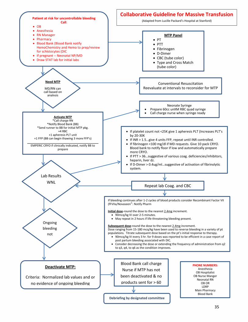
Patient at risk for uncontrollable bleeding Call:
OB Anesthesia RN Manager Pharmacy Blood Bank (Blood Bank notify
HemoChemistry and Hemo to prep/review for schistocytes (DIC
If pregnant -- Neonatal NP/MD Draw STAT lab for initial labs
MTP Panel PT PTT Fibrinogen D-Dimer CBC (tube color) Type and Cross Match
(tube color)
Need MTP
MD/RN can call based on
analysis
Conventional Resuscitation Reevaluate at intervals to reconsider for MTP
Activate MTP *Call charge RN
*Notify Blood Bank (BB) *Send runner to BB for initial MTP pkg.
+4 RBC +1 apheresis PLT unit
+1 FFP (BB can begin thawing 3 more FFP’s) ____________________________________________
EMPERIC CRYO if clinically indicated, notify BB to prepare
Lab Results
WNL
Ongoing
bleeding
not
anticipated
Deactivate MTP:
Criteria: Normalized lab values and or
no evidence of ongoing bleeding
Neonate Syringe Prepare 60cc unXM RBC quad syringe Call charge nurse when syringe ready
If platelet count not <25K give 1 apheresis PLT (Increases PLT’s by 20-30K
If INR > 1.5…give 4 units FFP, repeat until INR controlled. If fibrinogen <100 mg/dl if MD requests. Give 10 pack CRYO.
Blood bank to notify floor if low and automatically prepare more CRYO.
If PTT > 36…suggestive of various coag. deficiencies/inhibitors, heparin, liver dz.
If D-Dimer > 0.4ug/ml…suggestive of activation of fibrinolytic system.
Repeat lab Coag. and CBC
If bleeding continues after 1-2 cycles of blood products consider Recombinant Factor VII (fFVIIa/Novaseen: Notify Pharm Initial dose-round the dose to the nearest 2.4mg increment. 90mcq/kg IV over 2-5 minutes. May repeat in 2 hours if life threatening bleeding present.
Subsequent dose-round the dose to the nearest 2.4mg increment. Dose ranging from 15-180 mcq/kg have been used to reverse bleeding in a variety of pt populations. Titrate subsequent dose based on the pt’s initial response to therapy. 90mcq/kg IV every 3 hr. for 9 doses was reported to be efficient in a case report of
post partum bleeding associated with DIC. Consider decreasing the dose or extending the frequency of administration from q2
to q3, q4, to q6 as the condition improves.
Blood Bank call charge
Nurse if MTP has not
been deactivated & no
products sent for > 60
min.
Debriefing by designated committee
PHONE NUMBERS: Anesthesia
OB Hospitalist OB Nurse Manger
Neonatal RN OB OR LDRP
Main Pharmacy Blood Bank
Collaborative Guideline for Massive Transfusion (Adapted from Lucille Packard’s Hospital at Stanford)
35


Programs partially funded by grants from the California Department of Public Health, Center for Family Health, Maternal Child and Adolescent Health Division
OB HEMORRHAGE: CARTS, KITS, TRAYSLESLIE CASPER, MD, RICHARD LEE, MD San Diego Medical Center, Southern California Permanente Medical Group; Division of Maternal-Fetal Medicine, Women’s & Children’s Hospital, University of Southern California BACKGROUND Postpartum hemorrhage (PPH) is a commonly encountered emergency on labor and delivery units throughout California. Although medical management is often successful in treating PPH, the obstetrician may have to resort to surgical measures to treat it. In such settings, the care provider may find it practical to have rapid access to surgical instruments and tools designed to treat PPH. Equipment compiled on an obstetrical hemorrhage “cart” is designed to treat vaginal/cervical lacerations and provide the instruments for uterine tamponade or uterine/ovarian artery ligation. In short, the cart would have all the instruments necessary to treat PPH before hysterectomy is performed, if needed. The reader is referred to other guidelines that depict the use of these techniques. For more in-depth details about the hemorrhage “cart” the reader is referred to articles by T.F. Baskett (1, 2). RECOMMENDATION Labor and delivery units construct a sterile tray that provides rapid access to instruments used to surgically treat PPH. EDUCATIONAL TOOLS, SUPPORT DOCUMENTS OB Hemorrhage Cart: Recommended Instruments
• Set of vaginal retractors (long right angle); long weighted speculum • Sponge forceps (minimum: 2) • Sutures (for cervical laceration repair and B-Lynch [WEB LINK]) • Vaginal Packs • Uterine balloon [WEB-LINK] • Banjo curettes, several sizes • Long needle holder • Uterine forceps • Bright task light on wheels; behind ultrasound machine
Diagrams depicting various procedures (e.g. B-Lynch, uterine artery ligation, Balloon placement) OB Hemorrhage Medication Kit: Available in L&D and Postpartum Floor PYXIS/refrigerator
• Pitocin 20 units per liter NS 1 bag • Hemabate 250 mcg/ml 1 ampule • Cytotec 200mg tablets 5 tabs • Methergine 0.2 mg/ml 1 ampule
37

CARTS, KITS, TRAYS PAGE 2 OF 3 CASPER, L., LEE, R.
Programs partially funded by grants from the California Department of Public Health, Center for Family Health, Maternal Child and Adolescent Health Division
OB Hemorrhage Tray: Available on Postpartum Floor • IV start kit • 18 gauge angiocath • 1 liter bag lactated Ringers • IV tubing • Sterile Speculum • Urinary catheter kit with urimeter • Flash light • Lubricating Jelly • Assorted sizes sterile gloves
Labor and Delivery Emergency Hysterectomy Tray: Available in L&D OR Suite • 4 Towel Clips, Backhaus (perforating) 5 1/4" • 4 Mosquito,Curved, 5" • 2 Clamp,Mixter 9" • 2 Clamp, tonsil • 2 Clamp, Allis, Extra long 10" • 2 Clamp, Allis 6" • 2 Clamp, Babcock 8" • 2 Clamp, Babcock 6 1/4" • 2 Clamp, Lahey 6" • 2 Clamp, Heaney-Rezak, Straight, 8" • 8 Kelly, Curved 5 3/4" • 2 Kelly,Straight 5 3/4" • 8 Pean Curved, 6 1/4" • 2 Forceps, Debakey, 9 1/2" • 1 Forceps, Tissue with teeth 9 3/4" • 1 Forceps, Russian 8" • 1 Forceps, Smooth 8" • 1 Forceps, Fedrris Smith • 2 Forceps with Teeth, 6 " • 1 Forceps, Russian 6" • 2 Forceps, Adson with Teeth • 1 Forceps, Tissue, Smooth, 7"
• 2 Kocher, Straight, 8" • 6 Forceps, Heaney, Curved, 8 1/4" • NH, Mayo Hegar, 8" • 4 Sponge Stick, 9 1/2"
• 1 Scissor, Jorgensen, Curved, 9" • 1 Scissors, bandage 7" • 1 Scissors, curved dissecting, Metzenbaum • 1 Scissors, Mayo, curved • 1 Scissors, sharp/blunt, Straight, 5 1/2' • 1 Scissors, Curved Metzenbaum 12" • 1 Scissors, Mayo Straight 11" • 1 Scissors, Mayo Curved 11"
• 1 Knife Handle #3 • 1 Knife Handle #4 • 1 Knife Handle #3,Long
• 1 Retractor, Kelly, large • 1 Retractor, Deaver, Large, 3" x 12" • 1 Retractor, Deaver, Medium • 2 Retractor, Med/large Richardon • 1 Retractor, Balfour Blades • 2 Retractor,Goulet,7 1/2"
• 1 Suction, Yankauer Tip • 1 Suction, Pool Tip
38

CARTS, KITS, TRAYS PAGE 3 OF 3 CASPER, L., LEE, R.
Programs partially funded by grants from the California Department of Public Health, Center for Family Health, Maternal Child and Adolescent Health Division
EVIDENCE GRADING Level of Evidence: III. Opinions of respected authorities based on clinical experience, descriptive studies, or reports of expert committees. Level of Evidence: II-3. Evidence obtained from multiple time series with or without intervention. Dramatic results in uncontrolled experiments also could be regarded as this type of evidence. Strong quality improvement data such as statistical process control, or other well-designed analysis. REFERENCES 1. Baskett TF. Equipment tray for postpartum hemorrhage in A Textbook of PostPartum
Hemorrhage (ed C. B-Lynch et al.). Sapiens Publishing 2006 (III) 2. Baskett TF. Surgical management of severe obstetric hemorrhage: experience with an obstetric
hemorrhage equipment tray. J Obstet Gynaecol Can 2004; 26: 805-8. (II-3)
39

OB Hemorrhage Resources:
Fully stocked Obstetrical
Hemorrhage Cart
iStat Thromboelastogram
40
akowal
Typewritten Text

Optimizing Management of Obstetric Hemorrhage
Georgia Obstetrical and Gynecological Society
Thromboelastography (TEC) OVERVIEW
Thromboelastography measures the visco-elastic properties of whole blood as it clots
Shows the interaction of platelets with the coagulation cascade (aggregation, clot strengthening,
fibrin cross linking and fibrinolysis)
Assesses speed of clot formationƒ
Assesses potential for hypercoagulable state
Assesses accelerated clot breakdown/fibrinolysis
Assessment of clot strengthƒ
Partial assessment of components of clotting process (platelets and coagulation factors)
Lab Goals:
HCT >30
INR 1.4
Platelet count >50,000/ul
Fibrinogen > 100,000mg/dl
Base excess > -5 Normothermia 96.8 F or greater.
iStat System: Save Time! Point of Care Lab results
The i-STAT System™ rapid turnaround of test results could prove to be vital to decision making during PPH. The i-Stat System allows clinicians to access key test results on the spot, rather than wait for the 20-30 minutes it could take for sampling processing at the hospital lab.
Anesthesiologist are trained to use
Immediate lab results
Unit cost 12K (most ICU’s have them)
Cartridges are refrigerated: Cost ~$7
41

OB
HE
MO
RR
HA
GE
TO
OLK
IT
AP
PE
ND
IX E
.3. M
ET
HO
DS
FO
R D
EV
ELO
PIN
G T
RA
ININ
G A
ND
TO
OLS
FO
R Q
UA
NT
ITA
TIV
E M
EA
SU
RE
ME
NT
OF
BLO
OD
LO
SS
U
sed w
ith k
ind p
erm
issio
n o
f B
ev V
anderW
al, C
NS
Re
co
mm
en
de
d m
eth
od
s f
or
on
go
ing
qu
an
tita
tiv
e m
ea
su
rem
en
t o
f b
loo
d
los
s: 1.
Fo
rma
lly e
stim
ate
blo
od
lo
ss b
y r
eco
rdin
g p
erc
en
t (%
) sa
tura
tio
n o
f b
loo
d s
oa
ke
d ite
ms w
ith
th
e u
se
of
vis
ua
l cu
es s
uch
as p
ictu
res/p
oste
rs
to d
ete
rmin
e b
loo
d v
olu
me
eq
uiv
ale
nce
of
sa
tura
ted
/blo
od
so
ake
d
pa
ds,
ch
ux,
etc
. 2
. F
orm
ally
me
asu
re b
loo
d lo
ss b
y w
eig
hin
g b
loo
d s
oa
ke
d p
ad
s/c
hu
x
3.
Fo
rma
lly m
ea
su
re b
loo
d lo
ss b
y c
olle
ctin
g b
loo
d in
gra
du
ate
d
me
asu
rem
en
t co
nta
ine
rs
Qu
an
tify
ing
blo
od
lo
ss
by
we
igh
ing
(s
ee
im
ag
es
at
rig
ht
an
d b
elo
w)
•
Esta
blis
h d
ry w
eig
hts
of
co
mm
on
ite
ms
•
Sta
nd
ard
ize
use
of
pa
ds
•
Bu
ild w
eig
hin
g o
f p
ad
s in
to r
ou
tin
e p
ractice
•
De
ve
lop
wo
rksh
ee
t fo
r ca
lcu
latio
ns
Qu
an
tify
ing
blo
od
lo
ss
by
me
as
uri
ng
(s
ee
im
ag
e b
elo
w r
igh
t)
•
Use
gra
du
ate
d c
olle
ctio
n c
on
tain
ers
(C
/S a
nd
va
gin
al d
eliv
eri
es)
•
Acco
un
t fo
r o
the
r flu
ids (
am
nio
tic f
luid
, u
rin
e,
irri
ga
tio
n)
42

43

44

CMQCC OBSTETRIC HEMORRHAGE TOOLKIT OBSTETRIC HEMORRHAGE CARE GUIDELINES AND COMPENDIUM OF BEST PRACTICES REVIEWED BY CADPH-MCAH: 1/6/10
SIMULATIONS & DRILLS Leslie Casper, MD, San Diego Medical Center, Southern California Permanente Medical Group BACKGROUND AND LITERATURE REVIEW Medical simulation drills of obstetrical hemorrhage cases can assess system weaknesses and strengths, test policies and procedures for coping with hemorrhage and improve teamwork and communication skills of staff members. Drills that include all disciplines (obstetrics, anesthesia, pediatrics and nursing) can be especially effective in improving communication and coordination among team members. Drills are practice sessions of relatively uncommon but critical events, such as antenatal or postpartum hemorrhage and amniotic fluid embolism. Critical Event Training simulations for all physicians, midwives, anesthesiologists and nurses may improve neonatal outcomes (1). Implementing a rapid response team and addressing systemsʼ issues for management of obstetrical hemorrhage has been shown to decrease maternal mortality and improve outcomes (2). The Joint Commission recommends team training in their 2005 Executive Summary of Strategies to Improve the Medical Liability System and Prevent Patient Injury (3). Human factors training can improve communications and teamwork. Such training includes briefings, handoffs, time-outs and situational awareness for the team, which is a shared understanding of what is happening now and what happens next. Explicit communication skills to be taught include: addressing team members by name, making eye contact, repeating back orders and confirming that you are responding to an order, and not speaking to the room and assuming that you were heard. In addition, the concept of “Just Culture” or a similar environment should be implemented in all health care settings so that all team members feel respected and comfortable with asserting observations, suggestions and opinions. Improving team communications skills is one of the Joint Commission 2009 National Patient Safety Goals. Scenarios for simulation should be designed for the needs of the learners (nurses, physicians, residents, respiratory therapy, etc.) and tailored to available resources. Interdisciplinary training should include all disciplines involved in the care of obstetric patients. Simulation can be low tech—using live models—or high tech, using complex computerized simulators or a combination of both. The objective of simulation is to create situations that are as similar to “real life” as possible. Simulation in situ may improve ability to address systems issues and provides practice in oneʼs own hospital setting with familiar resources. Simulation in a computerized simulation center offers high technology in an environment similar to real life, but without the distractions of the hospital. The choice of high or low fidelity simulation is institution
45

CMQCC OBSTETRIC HEMORRHAGE TOOLKIT SIMULATIONS AND DRILLS REVIEWED BY CADPH-MCAH: 1/6/10 CASPER, L. PAGE 2 OF 17
dependent; both can work well for hemorrhage scenarios. For practicing complex events requiring a maternal cardiorespiratory arrest, high fidelity may be a better choice since chest compressions cannot be performed on a live model, for example. Some institutions use a combination of both types. Debriefing is appropriate both for simulation drills and for live events. Video taken during simulation serves as a realistic debriefing tool to explore what went well and what needs improvement after a scenario is performed. Evaluation tools such as checklists for expectations of each participant in their role and for team and individual performances can provide an objective approach to debriefing. Similarly, follow-up evaluation ensures that specific goals and objectives for each level of participant are met. The Ottawa Crisis Resource Management Global Rating Scale (4) and Mayo High Performance Teamwork Scale are examples (5). RECOMMENDATION All hospitals adopt regularly scheduled simulation drills for practicing response to obstetric hemorrhage. The choice of high or low fidelity drills is institution dependent; both can work well for hemorrhage scenarios. EDUCATIONAL TOOLS
1. Obstetric Hemorrhage Sample Scenario 1: Drill for Abruptio Placentae (attached) 2. Obstetric Hemorrhage Sample Scenario 2; Drill for Placenta Previa (attached) 3. Obstetric Hemorrhage Sample Scenario 3: Hemorrhage and Hypotension (attached) 4. Obstetric Hemorrhage Sample Scenario 4: Atonic Uterus (attached) 5. Kaiser Evaluation Form for Drills; Debriefing Tool: “Labor and Delivery/Family Centered
Care, Mock Obstetrical Hemorrhage, Roles and Responsibilities of Staff Skills Validation” (attached)
6. Dreyfus Model of Skill Acquisition (6) (attached) 7. Ottawa Crisis Resource Management Global Rating Scale (4) 8. Mayo High Performance Team Work Scale (5)
EVIDENCE GRADING Level of Evidence: B. Recommendations based on limited or inconsistent evidence. REFERENCES 1. Draycott,T, Simbanda,S, et al. Does training in obstetrical emergencies improve neonatal
outcome? BJOG 2006:113;177
46

CMQCC OBSTETRIC HEMORRHAGE TOOLKIT SIMULATIONS AND DRILLS REVIEWED BY CADPH-MCAH: 1/6/10 CASPER, L. PAGE 3 OF 17
2. Skupski,D, Lowenwirt,I, et al. Improving hospital systems for the care of women with major obstetric hemorrhage. Obstet Gynecol 2006: 107;977
3. Joint Commission on Accreditation of Healthcare Organizations: Healthcare at the Crossroads: Strategies For Improving The Medical Liability System and Preventing Patient Injury 2005. http://www.jointcommission.org/PublicPolicy/ (accessed February 2010)
4. Kim, J, et al. A Pilot study using high-fidelity simulation to formally evaluate performance in the resuscitation of critically ill patients: The University Ottawa Critical Care Medicine, High-Fidelity Simulation, and Crisis Resource Management I Study. Crit Care Med 2006; 34:2167-2174.
5. Malec, J, et al. The May High Performance Teamwork Scale: Reliability and Validity for Evaluating Key Crew Resource Management Skills. Simulation in Healthcare 2007; 2(1):4-10.
6. Dreyfus, S, Dreyfus, H, et al. A Five-Stage Model of the Mental Activities Involved in Directed Skill Acquisition. Storming Media. 1980.
www.stormingmedia.us/15/1554/A155480.html?searchTerms=dreyfus, (Accessed 4/2008).
47

Optimizing Management of Obstetric Hemorrhage
Georgia Obstetrical and Gynecological Society
Importance of Drills/Simulations Safety and QI Leader: Paul Preston, MD
“Medicine is the last high-risk industry that expects people to perform perfectly in complex, rare emergencies but does not support them with high-quality training and practice throughout their careers.”
“Certain individual and team skills require regular practice that
cannot ethically occur in routine care.”
What you need to know for a Postpartum Hemorrhage Drill
1. What are the risk factors for postpartum hemorrhage in the patient’s history?
2. Who is in charge?
3. Who should be called to assist with complications and how should they be called?
4. Where are the supplies (U/S machine, PRBC’s, platelets, cryoprecipitate, Hem abate)?
5. What needs to be documented?
6. Who will be documenting during the emergency?
7. What is communicated to the family?
8. How do you communicate with other staff members, i.e., RN’s, clerks, supervisors?
48

SIMULATIONS AND DRILLS CMQCC OBSTETRIC HEMORRHAGE TOOLKIT
CASPER, L. REVIEWED BY CADPH-MCAH: 1/6/10
SIMULATIONS AND DRILLS: EDUCATIONAL TOOL #2.
SAMPLE SCENARIO #2: PLACENTA PREVIA
Leslie Casper, MD
SCENARIO: a 21 year old gravida 2 para 1100 Caucasian woman presents at 37 weeks
estimated gestational age to Labor and Delivery in early labor with the onset of contractions
approximately one hour ago. She has had intermittent prenatal care starting at 12 weeks
estimated gestational age. Her records indicate she is carrying a singleton pregnancy in the vertex
presentation. Her past medical history is uncomplicated, she has no allergies, and she takes no
medications other than prenatal vitamins. She admits to smoking less than one-half pack per day
of cigarettes. Her prenatal labs are negative and her pregnancy has been uncomplicated except
for intermittent spotting in the last six weeks. An external fetal monitor is in place.
Physical examination reveals:
• Normal vital signs
• Uterus: longitudinal fetal lie, vertex presentation
• Cervix: dilatation 2 cm, effacement 10%, station -3, intact membranes
Fetal Monitor Output:
Fetal Heart Rate
• Baseline: 140 beats per minute
• Deviations from baseline: frequent accelerations throughout labor;
bradycardia and late decelerations occur late in labor, simultaneously with
frank hemorrhage
Fetal Heart Rate Variability
• Short-term: normal initially, demises as vaginal bleeding worsens
• Long-term: normal initially, demises as vaginal bleeding worsens
Maternal Uterine Activity
• Frequency: gradually increases to a rate of one contraction every two
minutes
• Duration: gradually increases to 60 seconds
• Intensity: gradually increasing to 100 mm Hg
CASE SUMMARY: Placenta previa is defined as a placenta that develops in the lower uterine
segment adjoining or covering the internal os. Three forms have been described. (8) In marginal
placenta previa the edge of the placenta is in contact with the margin of the cervical os but does
not cover it. Partial placenta previa incompletely covers the cervical os. Total placenta previa
completely covers the os. Varying degrees of bleeding occur during the third trimester as the lower
uterine segment matures in preparation for labor.
49

SIMULATIONS AND DRILLS CMQCC OBSTETRIC HEMORRHAGE TOOLKIT
CASPER, L. REVIEWED BY CADPH-MCAH: 1/6/10
The incidence of placenta previa is approximately 0.4%. Risk factors include previous cesarean
section and tobacco use. (9, 11) The management of a pregnancy complicated by placenta previa
is dependent on first recognition of the presence of the abnormally located placenta; this is usually
done by ultrasound (digital examination may inadvertently lead to severe hemorrhage). A history
of prior cesarean section or total placenta previa likely mandates operative delivery. In patients
with marginal or partial placenta previa, as in this case, vaginal delivery may be attempted,
provided an emergency cesarean section can be performed should uncontrollable hemorrhage
result. The descending fetal head often places pressure on the edge of the placenta and may act
to limit bleeding.
In this case fetal bradycardia and late decelerations ensue after frank hemorrhage develops late in
labor.
50

SIMULATIONS AND DRILLS CMQCC OBSTETRIC HEMORRHAGE TOOLKIT
CASPER, L. REVIEWED BY CADPH-MCAH: 1/6/10
SIMULATIONS AND DRILLS: EDUCATIONAL TOOL #4.
SAMPLE SCENARIO #4: ATONIC UTERUS AND POSTPARTUM HEMORRHAGE (12) Used with permission from Martin P. Eason, MD, JD
Participants: OB residents, FM residents.
Learning Objectives
At the end of the session the participants will be able to:
1) recognize the signs of uterine atony
2) appropriately treat uterine atony and hemorrhage
a. use correct medications
b. order appropriate labs
c. ensure resuscitation measures are instituted
d. make decision to treat surgically
Simulation Overview
This case involves the condition of uterine atony. It is the most common cause of significant
obstetric bleeding. It may be associated with placenta previa, placental abruption, retained
placenta, or occur alone. Factors associated with uterine atony include multiple gestation,
macrosomia, polyhydramnios, high parity, prolonged labor, excessive use of oxytocin, and
chorioamnionitis. An atonic uterus may contain up to 1L of blood. Although rarely life threatening,
uterine atony can cause severe postpartum hemorrhage and hypotension. The problem should be
recognized and treated quickly. Treatment should include administration of appropriate medication
to stimulate uterine contractions (oxytocin, Hemabate---prostglandin F2 alpha, ergonivine) and
management of hypovolemic shock. Postpartum hemorrhage should be treated as follows:
1. Initiate appropriate general resuscitation principals (ABC!s)
2. Ask for assistance
3. Place large bore intravenous lines
4. Order blood tests and blood products
5. Begin volume replacement with crystalloid and/or colloid solutions
6. Consider invasive monitoring
7. Treat bleeding disorders if present
8. Monitor urine output
9. Consider use of vasopressors
Patient History
Patient is a 32 year old G4P3 female with no prenatal care who presents in labor. She is crowning
and ready for delivery. She is a stat transfer from the ED.
51

SIMULATIONS AND DRILLS CMQCC OBSTETRIC HEMORRHAGE TOOLKIT
CASPER, L. REVIEWED BY CADPH-MCAH: 1/6/10
Patient History, continued
PMH: Asthma
PSH: None
Meds: albuterol 2 puffs BID prn
All: bee stings
SHx: married; husband is rushing from work. Smokes $ pack per day; no ETOH; she wants more
children
FHx: none
ROS: labor started about 2 hours ago. Water broke 45 minutes ago; clear
Physical examination (provided only if asked)
Cardiac: tachycardic otherwise normal
Lungs: clear bilaterally
Neck: supple
Labs:
None initially available
If ordered: CBC Hct 35% Platelets 235; WBC 8.2
Chem 7 WNL
PT/PTT 9.5/26 seconds
Simulation Parameters
Initial Parameters
BP: 135/78
Hr: 96
RR: 20
Sat: 98%
Heart Sounds: Normal
Lung sounds: clear
Scenario Run
The patient will present with the fetal head crowning; she will be wanted to push. The baby will be
delivered OA without complications. Immediately after the placenta is removed, blood will come
from the vaginal opening. If palpated the uterus will soft. It will remain so despite medications. The
blood pressure will drop from the initial readings over the next 10 minutes to 70/40. Oxytocics will
not cause uterine contraction. If Hemabate is given, the patient will complain of shortness of
breath and her sats will drop to 80%. If the lungs are auscultated, wheezing will be evident. Rise in
blood pressure will depend on replacement of volume. If large bore IVs are placed rapidly and put
on pressure bags, the pressure will initially improve to 80/45 but will slowly (over five minutes)
52

SIMULATIONS AND DRILLS CMQCC OBSTETRIC HEMORRHAGE TOOLKIT
CASPER, L. REVIEWED BY CADPH-MCAH: 1/6/10
decrease back to the 70!s. Decision to take patient to OR for operative intervention will end case.
If not, patient will expire.
Materials needed
Platform: Laerdal® or METI® monitor; Noelle birthing simulator modified with external tubing
attached to “blood” bag for bleeding
Diagnostic studies: CBC, coagulation studies
Props: L and D delivery drapes
Fetal monitor
IV equipment
Drugs
Personnel: one “L and D nurse”
Patient Voice
Expected actions by participants (checklist)
_____ Take appropriate history from patient
_____ Order IV access
_____ Order placement of fetal monitors
_____ Order placement of maternal monitor (BP, sat)
_____ Successfully deliver baby
_____ Recognize post-partum hemorrhage
_____ Assess uterine tone
_____ Order fundal massage
_____ Order labs to include CBC, type and screen, clotting studies
_____ Order placement of secondary IV access (large bore) with rapid fluid replacement
_____ Call for help
_____ Order vasopressors (correct drug in correct amount)
_____ Order blood to be administered
_____ Order oxytocin in appropriate dose
_____ Consider Hemabate® but hold its administration
_____ Order ergonovine in appropriate dose and route
_____ Makes decision to go to OR for surgical intervention
_____ Order surgical team to be called in
_____ Discusses the need for potential hysterectomy with patient and gets consent
53


Optimizing Management of Obstetric Hemorrhage
Georgia Obstetrical and Gynecological Society
Adopt a Systems Approach to Managing OB Hemorrhage
Department
OB Hemorrhage Protocol with stages
Hospital Massive Transfusion Protocol
Summary Flow algorithm
Graphic or tabular
Nursing checklist by stages
Documentation forms OB Hemorrhage Report
Worksheets to assist with assessment of blood
loss
Hemorrhage cart/kit
Instruction cards for new procedures in cart or operating room
Drills
55

Optimizing Management of Obstetric Hemorrhage
Georgia Obstetrical and Gynecological Society
PPH PEARLS
PREVENTION
Active management of the 3rd stage for all
Time Management: Manage the 3rd stage. Manage the Uterus
Do a formal assessment of blood loss
Identify vital sign triggers
Know your lab goals
“Move along” on uterotonic medications
DON’T DELAY
Activate the PPH Protocol
Bakri intrauterine balloon / B-Lynch suture
A new approach to the utilization of blood products. A role for rFactor
VIIa (Novoseven)
Avoid the Lethal Triad: Hypothermia, Acidosis and Coagulopathy
Realize the value of a formal protocol
56

Health Care Response to Obstetric Hemorrhage
Georgia Obstetrical and Gynecological Society
Reference Articles Relating to
Obstetric Hemorrhage
Please visit our website, www.georgiaobgyn.org, to access links to these resource articles and the online version of the GOGS Toolkit.
References used in the GOGS Tool Kit California Maternal Quality Care Collaborative Toolkit Lyndon A, Lagrew D, Shields L, Melsop K, Bingham D, & Main, E. (Eds.). (June 2010). Improving health
care response to obstetric hemorrhage. (California Maternal Quality Care Collaborative Toolkit to Transform Maternity Care) Developed under contract ##08-85012 with the California Department of Public Health; Maternal, Child and Adolescent Health Division; Published by the California Maternal Quality Care Collaborative. https://www.cmqcc.org/ob_hemorrhage. Accessed January 6, 2014.
OB Massive Transfusion Protocol Hiltner C. Blood Bank Shawnee Mission Medical Center. http://www.haabb.org/images/
Mass_Transfusion_Protocol-_Conna_Hiltner.pdf. Accessed December 18, 2013. Optimizing Protocols in Obstetrics: Management of Obstetric Hemorrhage
American Congress of Obstetricians and Gynecologists District II. Optimizing Protocols in Obstetrics: Series 2. October 2012. http://www.acog.org/About_ACOG/ACOG_Districts/District_II/ ~/media/Districts/District%20II/PDFs/Final_Hemorrhage_Web.pdf. Accessed January 7, 2014.
Patient Safety Checklist, Postpartum Hemorrhage From Vaginal Delivery
American College of Obstetricians and Gynecologists. Patient Safety Checklist Number 10. May 2013. Acog.org. Http://www.acog.org/~/media/Patient%20Safety%20Checklists/psc010.pdf?dmc= 1&ts=20140107T1252227469. Accessed January 6, 2014.
Postpartum Hemorrhage. ACOG Practice Bulletin No. 76
American College of Obstetrics and Gynecologists. Obstet Gynecol. 2006;108:1039-47. http://www.acog.org/Resources_And_Publications/Practice_Bulletins/Committee_on_Practice_Bulletins_--_Obstetrics/Postpartum_Hemorrhage. Accessed January 6, 2014.
Other References for Obstetric/Postpartum Hemorrhage Applying the Generic Errors Modeling System to Obstetric Hemorrhage Quality Improvement Efforts
Bingham D. The Association of Women’s Health, Obstetric, and Neonatal Nurses (AWHONN). JOGNN, 2012;00,1-11. DOI: 10.1111/j.1552-6909.2012.01373.x. http://jognn.awhonn.org. Accessed December 12, 2013.
57
akowal
Typewritten Text
akowal
Typewritten Text
akowal
Typewritten Text
akowal
Typewritten Text

Health Care Response to Obstetric Hemorrhage
Georgia Obstetrical and Gynecological Society
Maternal Death from Obstetric Hemorrhage.
Bingham D, Jones R. The Association of Women’s Health, Obstetric, and Neonatal Nurses (AWHONN). JOGNN. 2012;00,1-8. DOI: 10.1111/j.1552-6909.2012.01372.x. http://jognn.awhonn.org. Accessed December 12, 2013.
A State-Wide Obstetric Hemorrhage Quality Improvement Initiative
Bingham D, Lyndon A, Lagrew D, Mail EK, The American Journal of Maternal/Child Nursing. 2011;30(5),297-304. DOI:10.1097/NMC.0b013e318227c75f.
How we treat: Management of life-threatening primary postpartum hemorrhage with a standardized massive transfusion protocol
Burtelow M, Riley E, Druzin M, Fontaine M, Viele M, Goodnough LT. Transfusion. 2007 Sep;47(9):1564-72. PMID: 17725718
Hemabate (carboprost tromethamine) package insert. Pharmacia & Upjohn Co., Division of Pfizer Inc, NY, NY 10017. FDA rev date: march 2006 Rectally administered misoprostol for the treatment of postpartum hemorrhage unresponsive to
oxytocin and ergometrine: A descriptive study O'Brien P, et al. Obstet Gynecol. 1998 August;92(2):212-4. PMID: 9699753
58