Genome-wide association study identifies novel loci predisposing to
microorganisms
Article
Genome Insights into the Novel SpeciesMicrovirga brassicacearum, a Rapeseed Endophytewith Biotechnological Potential
Alejandro Jiménez-Gómez 1,2, Zaki Saati-Santamaría 1,2 , José M. Igual 3,4 , Raúl Rivas 1,2,3,Pedro F. Mateos 1,2,3 and Paula García-Fraile 1,2,3,*
1 Microbiology and Genetics Department, University of Salamanca, 37007 Salamanca, Spain;[email protected] (A.J.-G.); [email protected] (Z.S.-S.); [email protected] (R.R.); [email protected] (P.F.M.)
2 Spanish-Portuguese Institute for Agricultural Research (CIALE), Villamayor, 37185 Salamanca, Spain3 Associated R&D Unit, USAL-CSIC (IRNASA), 37007 Salamanca, Spain; [email protected] Institute of Natural Resourses and Agrobiology of Salamanca (IRNASA-CSIC), Salamanca 37008, Spain* Correspondence: [email protected]
Received: 30 June 2019; Accepted: 6 September 2019; Published: 14 September 2019�����������������
Abstract: Plants harbor a diversity of microorganisms constituting the plant microbiome. Manybioinoculants for agricultural crops have been isolated from plants. Nevertheless, plants are anunderexplored niche for the isolation of microorganisms with other biotechnological applications.As a part of a collection of canola endophytes, we isolated strain CDVBN77T. Its genome sequenceshows not only plant growth-promoting (PGP) mechanisms, but also genetic machinery to producesecondary metabolites, with potential applications in the pharmaceutical industry, and to synthesizehydrolytic enzymes, with potential applications in biomass degradation industries. Phylogeneticanalysis of the 16S rRNA gene of strain CDVBN77T shows that it belongs to the genus Microvirga,its closest related species being M. aerophila DSM 21344T (97.64% similarity) and M. flavescens c27j1T
(97.50% similarity). It contains ubiquinone 10 as the predominant quinone, C19:0 cycloω8c andsummed feature 8 as the major fatty acids, and phosphatidylcholine and phosphatidylethanolamine asthe most abundant polar lipids. Its genomic DNA G+C content is 62.3 (mol %). Based on phylogenetic,chemotaxonomic, and phenotypic analyses, we suggest the classification of strain CDVBN77T withina new species of the genus Microvirga and propose the name Microvirga brassicacearum sp. nov.(type strain CDVBN77T = CECT 9905T = LMG 31419T).
Keywords: plant–bacteria interactions; genome mining; PGPB; biofertilizer; CAZymes; secondarymetabolites; hydrolytic enzymes
1. Introduction
After millions of years of evolution, microorganisms have compiled a noteworthy genetic diversitywith biotechnological potential, which can be additionally enhanced by tools, such as genetic engineeringor heterologous gene expression. Thus, microbial isolates are essential for diverse biotechnology-basedindustries, such as pharmaceutical biotechnology (where they play important roles in the productionof antibiotics, antivirals, anticarcinogenic compounds, etc.), agricultural biotechnology (where theyare applied as biocontrol agents or probiotics for plant growth promotion), and environmentalbiotechnology (with variate applications such as lignocellulose breakdown or degradation of xenobioticcompounds). Moreover, according to recent estimations, potentially 99.999% of microbial taxa remainundiscovered [1], so there is a huge genetic potential yet undiscovered.
In the last couple of decades, the extensive use of the PCR and sequencing of the 16S rRNA genehas allowed a more accurate bacterial identification and served as the base for the discovery of new
Microorganisms 2019, 7, 354; doi:10.3390/microorganisms7090354 www.mdpi.com/journal/microorganisms
Microorganisms 2019, 7, 354 2 of 19
taxa, being the gold standard for prokaryote identification. According to Jongsik and collaborators [2],a bacterial strain with a similarity under 98.7% in the 16S rRNA gene sequence with the type strain ofits closest related species represents a novel species and, in case of a similarity over 98.7%, a valueunder 70% in total DNA–DNA hybridization (DDH) also indicates a novel species [3]. More recently,the quick development of DNA sequencing platforms that generate rapid and low-cost high-throughputsequencing data has spearheaded the widespread sequencing of bacterial genomes which, togetherwith the development of bioinformatics tools for genome sequence comparisons, facilitates the processof new species identification, by means of genome-to-genome sequence comparison [2].
Genome sequences also allow us to mine the genetic potential of a microorganism for differentpurposes, for example, plant growth promotion mechanisms [4] and the production of secondarymetabolites with applications in the pharmaceutical industry [5] or carbohydrate-active enzymes(CAZymes) with several biotechnologically based industry uses, such as biomass degradation, biodieselproduction, or bioremediation [6].
Plants are the ecological niche of a broad range of microorganisms which constitute theirmicrobiome [7]. Some of these microorganisms establish symbiotic associations with the plant,supplying their host with nutrients or improving its fitness by the production of phytohormones or theinduction of resistance to abiotic stresses [8]. These plant growth-promoting (PGP) microorganisms canbe applied as biofertilizers or plant probiotics to improve crop yields in an ecologically friendly mannerand thus, they are produced and formulated as bioinoculants in the industry. Nevertheless, plants arean underexplored niche as a source of microorganisms for other biotechnological applications differentfrom those of the agro-based industry.
The genus Microvirga is a cosmopolitan genus which as of now includes 17 validated speciesisolated from a wide diversity of niches, such as human stool [9], air [10], thermal water [11], natural,domestic, and contaminated soils [12–17], and even as legume nodule endophytes [18–20]. Somestudies report the genetic potential of some strains classified within the Microvirga genus for arsenicoxidation [15] and the production of pigments and amylolytic enzymes [21].
In this study, we propose the description of Microvirga brassicacearum sp. nov. isolated as arapeseed bacterial endophyte with the capability to promote plant growth. Moreover, we obtainedand mined the genome sequence of strain M. brassicacearum CDVBN77T, showing its genetic potentialnot only as a plant growth promoter but also as a producer of secondary metabolites with a potentialinterest in the pharmaceutical industry as well as of enzymatic machinery for biomass degradation.
2. Materials and Methods
2.1. Strain Isolation and Identification
Strain CDVBN77T was isolated as part of a collection of rapeseed bacterial root endophytesfrom the roots of rapeseed plants harvested in the locality of Castellanos de Villiquera (province ofSalamanca, Spain) [22]. Plants in the phenological state of rosette were collected and kept refrigerated tobe transported to the lab. Root endophytes were obtained after washing with sterile water. The plantswere extracted from the soils, kept refrigerated, and shipped to the laboratory. To isolate rapeseed rootbacterial endophytes, roots were washed in sterile Petri dishes containing sterile water (10 times) andthen sterilized by immersion in sodium hypochlorite (2%) for 2 min. Afterward, surfaced sterilizedroots were washed in sterile water (5 times) and dried without using sterile filter paper.
Surface sterilized roots were smashed with a pestle in a sterile mortar and the extract was seriallydiluted in sterile water. Then, 100 µL of the 10−2, 10−3, and 10−4 dilutions were plated onto Petri dishescontaining different media: YMA (Microkit®, Laboratorios Microkit, Madrid, Spain), Tryptic Soy Agar(TSA) (DifcoTM, Difco Laboratories, Le Pont de Claix, France), 869 medium (Tryptone (10 g/L), yeastextract (5 g/L), NaCl (5 g/L), D-glucose (1 g/L), CaCl2 (0.345 g/L), and agar (20 g/L)), and ten timesdiluted 869 medium (869(1/10) medium). Plates were incubated at 28 ◦C for 21 days. The emerging
Microorganisms 2019, 7, 354 3 of 19
bacterial colonies were regularly isolated to obtain pure cultures. Then, for long-term storage, isolatedstrains were stored in a sterile 20% glycerol solution at −80 ◦C.
2.2. DNA Extraction and 16S rRNA Amplification and Sequencing
CDVBN77T DNA for amplifying and sequencing 16S rRNA gen was extracted usingthe REDExtract-n-AmpTM Plant PCR Kit (Sigma-Aldrich, Darmstadt, Germany) following themanufacturer’s instructions. The amplification was performed using 2 µL of bacterial DNA (100 µg/mL)as the template, 6 µL of Milli-Q sterile water, 12.5 µL REDExtract Ready Mix, 2.5 µL 27F 1 µM(5′ GAGGGTGGCGGTTCT 3′), and 2,5 µL 1522R 1 µM (5′ AAGGAGGTGATCCANCCRCA 3′).The PCR conditions were the following: a first step of pre-heating at 95 ◦C for 9 min, 35 cycles ofdenaturing at 94 ◦C for 1 min; annealing at 56.5 ◦C for 1 min 30 s and extension 72 ◦C for 2 min, and afinal extension at 72 ◦C for 7 min.
PCR product was purified using DNA Cleanup Micro Kit (Thermo ScientificTM, Göteborg, Sweden).The sequence reaction was performed on an ABI PRISM sequencer (Applied BiosystemTM, Foster,CA, USA) using a Dye Terminator Cycle Sequencing Ready Reaction Kit (University of Salamanca,Salamanca, Spain). The isolate strain was identified using the following databases: NCBI’s BLASTnprogram [23] (https://blast.ncbi.nlm.nih.gov/Blast.cgi?PAGE_TYPE=BlastSearch) and EzTaxon tool [24].
2.3. Draft Genome Sequencing and Annotation
Bacterial DNA for genome sequencing was obtained from cells grown in YMA medium at 28 ◦Cfor five days. DNA extraction was performed with the Quick DNATM Fungal/Bacterial Miniprep Kit(Zymo Research®, Irvine, CA, USA) following the protocol provided by the manufacturer.
The draft genome sequence was obtained on an Illumina platform from the Genomics Serviceof the Institute of Functional Biology and Genomics (IBFG) (Salamanca, Spain). Sequences wereassembled using Velvet 1.12.10, [25]. Gene calling, annotation, and search of genes related to plantgrowth promotion capabilities were performed using RAST Pipeline [26]; the BlastKOALA tool wasalso used to perform KEGG Orthology (KO) assignment based on the KEGG database [27] in order tocomplete the mining of genes related to plant growth promotion; the dbCAN2 meta server [28] wasused to identify the coding sequences (CDSs) encoding carbohydrate-active enzymes (CAZymes) usingHMMER annotation; and the analysis of gene clusters related to secondary metabolites productionwas performed using antiSMASH 5.0 webserver [29]. This genome sequence was deposited at theDDBJ/EMBL/GenBank under the accession VCMV00000000. The version described in this paper isversion VCMV01000000.
2.4. Genotypic Analysis of Microvirga brassicacearum CDVBN77T
The GenBank accession number of the CDVBN77T 16S rRNA gene sequence is MK938302.Phylogenetic analysis of the CDVBN77T 16S rRNA gene sequence and the sequences of the type strainsof all the species included within the genus Microvirga was performed using MEGA7 software [30],based on the alignment of the sequences with Clustal_W [31]. The distances were calculated usingKimura’s two-parameter model [32], and the phylogenetic tree was inferred using neighbor-joining(NJ; [33]) analysis. The average nucleotide identity (ANI) and digital DNA–DNA hybridization (dDDH)values between the draft genome sequence of strain CDVBN77T and those of its closest related speciesM. aerophila c27j1T (GenBank/EMBL/DDBJ accession number NZ_QOIO00000000.1) and M. flavescensDSM 21344T (GenBank/EMBL/DDBJ accession number QMKH00000000) were calculated using the ANIcalculator (www.ezbiocloud.net/tools/ani) and the Genome-to-Genome Distance Calculator (GGDC 2.1)(http://ggdc.dsmz.de/distcalc2.php), respectively. The mol % G+C content of DNA was determinedfrom the draft genome sequence.
Microorganisms 2019, 7, 354 4 of 19
2.5. Chemotaxonomic Analysis of Microvirga brassicacearum CDVBN77T
For the analysis of fatty acid methyl esters (FAMEs), biomass was harvested as detailed byWeon et al. [8]. For the analyses of polar lipids and respiratory quinones, biomass was harvested aftercultivation of the bacterium for seven days in R2A broth medium (dextrose (0.5 g/L), dipotassiumphosphate (0.3 g/L), casamino acids (0.5 g/L), magnesium sulphate (0.05 g/L), proteose peptone (0.5 g/L),sodium pyruvate (0.3 g/L), starch (0.5 g/L), and yeast extract (0.5 g/L)) at 28 ◦C and 150 rpm shaking.Analysis of FAMEs was performed as previously described [34]. For the identification of polar lipidsand respiratory quinones, cells collected from the plates into sterile Falcon®tubes were freeze dried andsent to the Identification Service of the German Collection of Microorganisms and Cell Cultures DSMZ.
2.6. Microvirga brassicacearum CDVBN77T Bacterial Characterization
Catalase and oxidase tests [35] and Gram staining [36] were performed using previouslydescribed protocols.
Cell morphology and flagella were observed under a Zeiss EM209 transmission electron microscope,after growth in YMA medium at 28 ◦C for five days and cell treatment with 2% uranyl. To assay salttolerance, the bacterium was cultivated in YMB medium (mannitol (7.0 g/L), yeast extract (2 g/L),dipotassium phosphate (0.2g/L), and magnesium sulphate (0.2g/L)) supplemented with 0, 0.5, 1, 1.5,and 2% (w/v) NaCl. The same medium adjusted to a final pH range of 3–11 was used to examine thegrowth capability at different pHs; in both cases, flasks containing 50 mL of the different media wereinoculated with a 10 µl sterile plastic loop containing cells collected from a two-day plate culture ofthe bacterium. After the inoculation, flasks were cultivated in a shaker at 150 rpm for two days at28 ◦C. Plates containing YMA medium (YMB plus 2% agar) were incubated at 4, 12, 25 and 37 ◦C todetermine the temperature range for optimal growth. In all cases, the cultures were examined at 2, 5,10, and 15 days after inoculation.
Enzymatic assays were performed using the p-nitrophenyl (pNP) substrates listed in Table 1.Substrates were used at a concentration of 0.2% in 50 mM phosphate buffer (pH 7); however, thoserequiring pH 8.5 were prepared in Tris-HCl 0.2 M buffer and those requiring pH 5.0 were prepared inan acetate buffer (0.2 M). The enzymatic reactions were carried out in a multi-well plate mixing 50 µLof substrate prepared as specified above and 50 µL of a suspension of bacteria in sterile water with6 × 109 CFU/mL obtained from cells previously grown in YMA medium for 15 days at 28 ◦C. Plateswith the reaction mixture were incubated at 28 ◦C for 24 h and then revealed with 100 µL per well of a4% sodium carbonate solution. A positive reaction was evidenced by the appearance of a yellow colorwithin a few seconds.
To evaluate siderophore production, strain CDVBN77T was inoculated in M9-CAS-agar mediumplates [37] with the modifications suggested by Alexander and Zuberer [38] after 21 days of growth at28 ◦C. The addition of hexadecyltrimethylammonium bromide (HDTMA) as a cationic solvent wasused to stabilize the Fe-CAS complex, which provides the characteristic orange-colored halo aroundthe colonies when siderophores are produced.
The solubilization of non-soluble phosphates into soluble assimilable ions was analyzed inPikovskaya medium plates [39], which contain calcium phosphate (Ca2HPO4) as P source. The presenceof clear halos surrounding bacterial colonies are indicative of phosphate solubilization. StrainCDVBN77T was inoculated in Pikovskaya medium and phosphate solubilization was evaluated after21 days of growth at 28 ◦C.
Cellulose production was determined as described by Robledo et al. [40] in Nutrient Agar (NA)plates containing Congo Red colorant, which binds to cellulose polymers. Red-colored colonies areindicative of cellulose production. Strain CDVBN77T was inoculated in plates with Congo Red and thecolor of the colonies was evaluated after 21 days of growth at 28 ◦C.
Other phenotypic characters were tested by using the API 20NE and API ZYM (bioMerieux®)miniaturized galleries following the manufacturer’s instructions.
Microorganisms 2019, 7, 354 5 of 19
Table 1. Substrates for enzymatic enzymes.
Substrate Enzyme Assayed
PNP-α-L-arabinopyranoside α-L-arabinosidasePNP-β-L-arabinopyranoside β-L-arabinosidasePNP-β-D-cellobioside CellobiohydrolasePNP-phosphate (at pH 5.0) PhosphatasePNP-phosphate (at pH 7.0) PhosphatasePNP-phosphate (at pH 8.5) PhosphatasePNP-bis-phosphate (at pH 7) BisphosphatasePNP-bisphosphate (at pH 8.5) BisphosphatasePNP-α-L-fucopyranoside α-L-fucosidasePNP-β-D-fucopyranoside β-glucosidasePNP-α-D-galactopyranoside α-galactosidasePNP-β-D-galactopyranoside β-galactosidasePNP-β-D-galacturonide β-galacturonidasePNP-α-D-glucopyranoside α-glucosidasePNP-β-D-glucopyranoside β-glucosidasePNP-β-D-glucuronide β-glucuronidasePNP-β-D-lactopyranoside β-lactosidasePNP-α-D-mannopyranoside α-mannosidasePNP-β-D-mannopyranoside β-mannosidasePNP-α-L-rhamnopyranoside α-rhamnosidasePNP-N-thio-β-D-glucopyranoside β-glucosidasePNP-α-D-xylopyranoside α-xylosidasePNP-β-L-xylopyranoside β-xylosidase
3. Results
3.1. Bacterial Isolation and Identification
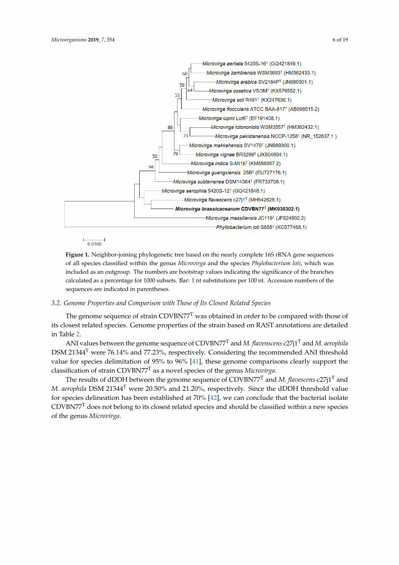
Strain CDVBN77T was isolated as part of a collection of rapeseed root endophytes from plantssampled at the municipality of Castellanos de Villiquera (province of Salamanca, Spain) [22].The comparison of the 16S rRNA gene sequence with those of type strains of described speciesavailable in GenBank and EzBiocloud databases showed that all strains but CDVBN77T showedsimilarities higher than 98% with their closest related type strains, whereas the similarity of strainCDVBN77T with M. aerophila DSM 21344T, its closest related type strain, was only 97.64%, whichaccording to the threshold value of 98.7% in the 16S rRNA gene sequence with the closest relatedspecies indicated by Jongsik and collaborators [2] indicates that the strain constitutes a new specieswithin the genus Microvirga.
Therefore, we performed a phylogenetic analysis of the 16S rRNA gene sequence of strainCDVBN77T and those of the type strains of all species included within the genus Microvirga.
Phylogenetic trees constructed with both Maximum Likelihood (ML) and Neighbor Joining(NJ) methods showed a similar topology (Figure 1) with strain CDVBN77T appearing in a separatedbranch with M. flavescens c27j1T and M. aerophila DSM 21344T as the closest related species. Thus,the phylogenetic analysis of the 16S rRNA gene supports the classification of the bacterium CDVBN77T
as a new species within the genus Microvirga.
Microorganisms 2019, 7, 354 6 of 19Microorganisms 2019, 7, x FOR PEER REVIEW 6 of 19
Microorganisms 2019, 7, x; doi: FOR PEER REVIEW www.mdpi.com/journal/microorganisms
Figure 1. Neighbor-joining phylogenetic tree based on the nearly complete 16S rRNA gene sequences of all species classified within the genus Microvirga and the species Phylobacterium loti, which was included as an outgroup. The numbers are bootstrap values indicating the significance of the branches calculated as a percentage for 1000 subsets. Bar: 1 nt substitutions per 100 nt. Accession numbers of the sequences are indicated in parentheses.
3.2. Genome Properties and Comparison with Those of Its Closest Related Species
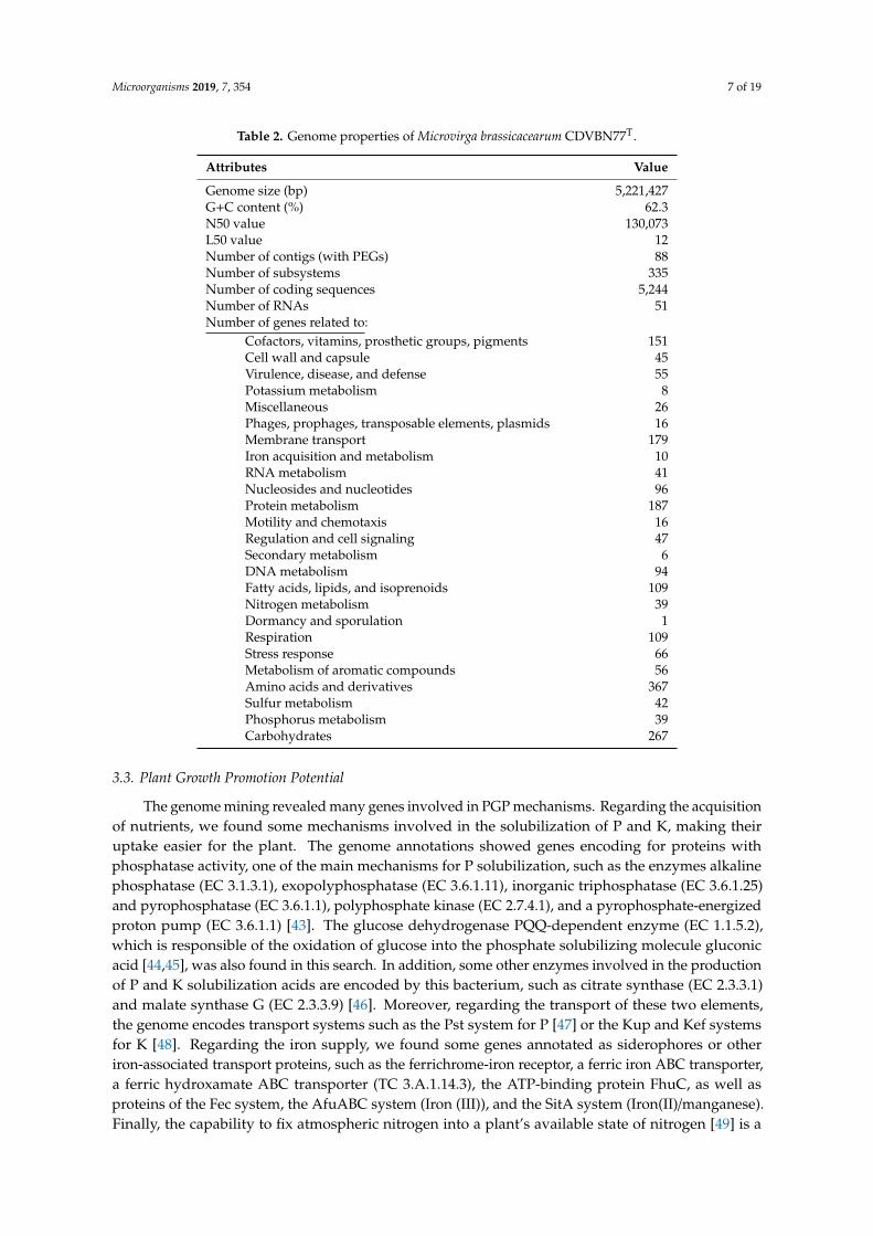
The genome sequence of strain CDVBN77T was obtained in order to be compared with those of its closest related species. Genome properties of the strain based on RAST annotations are detailed in Table 2.
Table 2. Genome properties of Microvirga brassicacearum CDVBN77T.
Attributes Value Genome size (bp) 5,221,427 G+C content (%) 62.3 N50 value 130,073 L50 value 12 Number of contigs (with PEGs) 88 Number of subsystems 335 Number of coding sequences 5,244 Number of RNAs 51 Number of genes related to:
Cofactors, vitamins, prosthetic groups, pigments 151 Cell wall and capsule 45 Virulence, disease, and defense 55 Potassium metabolism 8 Miscellaneous 26
Figure 1. Neighbor-joining phylogenetic tree based on the nearly complete 16S rRNA gene sequencesof all species classified within the genus Microvirga and the species Phylobacterium loti, which wasincluded as an outgroup. The numbers are bootstrap values indicating the significance of the branchescalculated as a percentage for 1000 subsets. Bar: 1 nt substitutions per 100 nt. Accession numbers of thesequences are indicated in parentheses.
3.2. Genome Properties and Comparison with Those of Its Closest Related Species
The genome sequence of strain CDVBN77T was obtained in order to be compared with those ofits closest related species. Genome properties of the strain based on RAST annotations are detailedin Table 2.
ANI values between the genome sequence of CDVBN77T and M. flavenscens c27j1T and M. aerophilaDSM 21344T were 76.14% and 77.23%, respectively. Considering the recommended ANI thresholdvalue for species delimitation of 95% to 96% [41], these genome comparisons clearly support theclassification of strain CDVBN77T as a novel species of the genus Microvirga.
The results of dDDH between the genome sequence of CDVBN77T and M. flavescens c27j1T andM. aerophila DSM 21344T were 20.50% and 21.20%, respectively. Since the dDDH threshold valuefor species delineation has been established at 70% [42], we can conclude that the bacterial isolateCDVBN77T does not belong to its closest related species and should be classified within a new speciesof the genus Microvirga.
Microorganisms 2019, 7, 354 7 of 19
Table 2. Genome properties of Microvirga brassicacearum CDVBN77T.
Attributes Value
Genome size (bp) 5,221,427G+C content (%) 62.3N50 value 130,073L50 value 12Number of contigs (with PEGs) 88Number of subsystems 335Number of coding sequences 5,244Number of RNAs 51Number of genes related to:
Cofactors, vitamins, prosthetic groups, pigments 151Cell wall and capsule 45Virulence, disease, and defense 55Potassium metabolism 8Miscellaneous 26Phages, prophages, transposable elements, plasmids 16Membrane transport 179Iron acquisition and metabolism 10RNA metabolism 41Nucleosides and nucleotides 96Protein metabolism 187Motility and chemotaxis 16Regulation and cell signaling 47Secondary metabolism 6DNA metabolism 94Fatty acids, lipids, and isoprenoids 109Nitrogen metabolism 39Dormancy and sporulation 1Respiration 109Stress response 66Metabolism of aromatic compounds 56Amino acids and derivatives 367Sulfur metabolism 42Phosphorus metabolism 39Carbohydrates 267
3.3. Plant Growth Promotion Potential
The genome mining revealed many genes involved in PGP mechanisms. Regarding the acquisitionof nutrients, we found some mechanisms involved in the solubilization of P and K, making theiruptake easier for the plant. The genome annotations showed genes encoding for proteins withphosphatase activity, one of the main mechanisms for P solubilization, such as the enzymes alkalinephosphatase (EC 3.1.3.1), exopolyphosphatase (EC 3.6.1.11), inorganic triphosphatase (EC 3.6.1.25)and pyrophosphatase (EC 3.6.1.1), polyphosphate kinase (EC 2.7.4.1), and a pyrophosphate-energizedproton pump (EC 3.6.1.1) [43]. The glucose dehydrogenase PQQ-dependent enzyme (EC 1.1.5.2),which is responsible of the oxidation of glucose into the phosphate solubilizing molecule gluconicacid [44,45], was also found in this search. In addition, some other enzymes involved in the productionof P and K solubilization acids are encoded by this bacterium, such as citrate synthase (EC 2.3.3.1)and malate synthase G (EC 2.3.3.9) [46]. Moreover, regarding the transport of these two elements,the genome encodes transport systems such as the Pst system for P [47] or the Kup and Kef systemsfor K [48]. Regarding the iron supply, we found some genes annotated as siderophores or otheriron-associated transport proteins, such as the ferrichrome-iron receptor, a ferric iron ABC transporter,a ferric hydroxamate ABC transporter (TC 3.A.1.14.3), the ATP-binding protein FhuC, as well asproteins of the Fec system, the AfuABC system (Iron (III)), and the SitA system (Iron(II)/manganese).Finally, the capability to fix atmospheric nitrogen into a plant’s available state of nitrogen [49] is a
Microorganisms 2019, 7, 354 8 of 19
common mechanism in rhizobial strains. In this case, we found the presence of genes implicated onFix and Nif systems.
A prior step to endophytic living is to attach and form biofilms over the root surface. Bacteriacommonly produce exopolysaccharides in order to colonize the root surface [50]. Genome annotationsrevealed a great number of genes related to the synthesis or transport of exopolysaccharides, for example,the exoD gene, which is associated with this process [51], lpx genes, responsible for the synthesis of thistype of molecules [52], or lpt [53] and Rf [54] transport systems. Once inside the plant, the bacteriummust evade the plant defense. The genome annotations revealed that the bacterium encodes genes forthe production of salicylate 1-hydroxylase (EC 1.14.13.1), an enzyme that degrades the salicylic acidinto catechol.
Moreover, strain CDVBN77T grew on chrome azurol S (CAS) indicator medium, where thecolonies were surrounded by a yellow-orange halo (2 mm radius around colonies), indicatingsiderophore production.
In addition, CDVBN77T solubilized phosphate on Pikovskaya’s agar, forming halos aroundits colonies (2 mm radius around colonies). Additionally, the results obtained using pNP-basedsubstrates showed the production of phosphatases (with activity at acid, alkaline, and neutral pH) andbisphosphatases (with activity at alkaline pH).
Furthermore, strain CDVBN77T colonies were red after seven days of incubation on plates containingCongo Red. The intensity of the color was comparable to that recorded for other strains, which indicatedthat CDVBN77T produces a moderate amount of a polysaccharide with 1,4-β-glycosidic bonds.
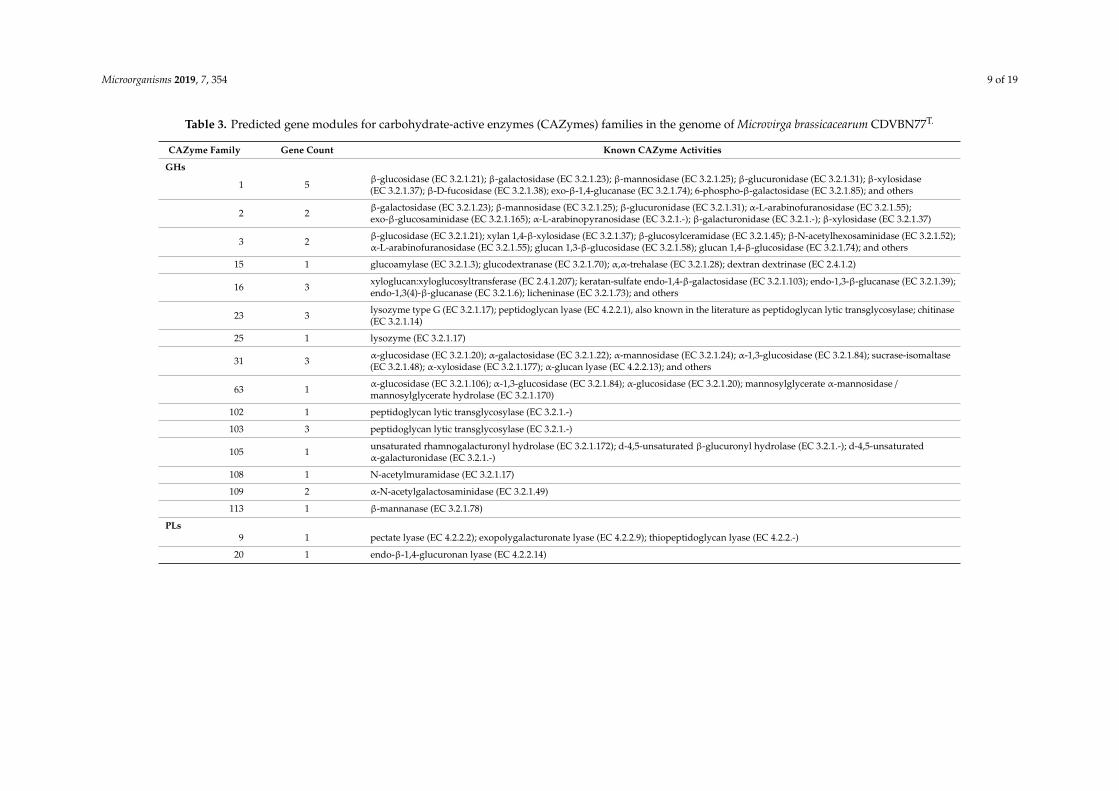
3.4. Potential for the Production of Enzymes with Biotechnological Potential
Analysis of the genome sequence of strain M. brassicacearum CDVBN77T with dbCAN2 showed115 genes encoding different CAZymes involved in degradation, modification, or creation ofglycosidic bonds.
In summary, we identified gene modules belonging to five different enzyme classes: (i) glycosidehydrolases (GHs), enzymes that catalyze the hydrolysis of glycosidic linkage of glucoside—30 genecounts in 15 different families; (ii) glycosyltransferases (GTs), involved in the formation of glycosidicbonds—46 gene counts in 11 different families; (iii) polysaccharide lyases (PLs), which performnon-hydrolytic cleavage of glycosidic bonds—2 gene counts in 2 different families; (iv) carbohydrateesterases (CEs), which hydrolyze carbohydrate esters—22 gene counts in 6 different families; and(v) auxiliary activities (AAs), redox enzymes that act in conjunction with CAZymes—15 gene counts in5 different families (Table 3).
Microorganisms 2019, 7, 354 9 of 19
Table 3. Predicted gene modules for carbohydrate-active enzymes (CAZymes) families in the genome of Microvirga brassicacearum CDVBN77T.
CAZyme Family Gene Count Known CAZyme Activities
GHs
1 5 β-glucosidase (EC 3.2.1.21); β-galactosidase (EC 3.2.1.23); β-mannosidase (EC 3.2.1.25); β-glucuronidase (EC 3.2.1.31); β-xylosidase(EC 3.2.1.37); β-D-fucosidase (EC 3.2.1.38); exo-β-1,4-glucanase (EC 3.2.1.74); 6-phospho-β-galactosidase (EC 3.2.1.85); and others
2 2 β-galactosidase (EC 3.2.1.23); β-mannosidase (EC 3.2.1.25); β-glucuronidase (EC 3.2.1.31); α-L-arabinofuranosidase (EC 3.2.1.55);exo-β-glucosaminidase (EC 3.2.1.165); α-L-arabinopyranosidase (EC 3.2.1.-); β-galacturonidase (EC 3.2.1.-); β-xylosidase (EC 3.2.1.37)
3 2 β-glucosidase (EC 3.2.1.21); xylan 1,4-β-xylosidase (EC 3.2.1.37); β-glucosylceramidase (EC 3.2.1.45); β-N-acetylhexosaminidase (EC 3.2.1.52);α-L-arabinofuranosidase (EC 3.2.1.55); glucan 1,3-β-glucosidase (EC 3.2.1.58); glucan 1,4-β-glucosidase (EC 3.2.1.74); and others
15 1 glucoamylase (EC 3.2.1.3); glucodextranase (EC 3.2.1.70); α,α-trehalase (EC 3.2.1.28); dextran dextrinase (EC 2.4.1.2)
16 3 xyloglucan:xyloglucosyltransferase (EC 2.4.1.207); keratan-sulfate endo-1,4-β-galactosidase (EC 3.2.1.103); endo-1,3-β-glucanase (EC 3.2.1.39);endo-1,3(4)-β-glucanase (EC 3.2.1.6); licheninase (EC 3.2.1.73); and others
23 3 lysozyme type G (EC 3.2.1.17); peptidoglycan lyase (EC 4.2.2.1), also known in the literature as peptidoglycan lytic transglycosylase; chitinase(EC 3.2.1.14)
25 1 lysozyme (EC 3.2.1.17)
31 3 α-glucosidase (EC 3.2.1.20); α-galactosidase (EC 3.2.1.22); α-mannosidase (EC 3.2.1.24); α-1,3-glucosidase (EC 3.2.1.84); sucrase-isomaltase(EC 3.2.1.48); α-xylosidase (EC 3.2.1.177); α-glucan lyase (EC 4.2.2.13); and others
63 1 α-glucosidase (EC 3.2.1.106); α-1,3-glucosidase (EC 3.2.1.84); α-glucosidase (EC 3.2.1.20); mannosylglycerate α-mannosidase /mannosylglycerate hydrolase (EC 3.2.1.170)
102 1 peptidoglycan lytic transglycosylase (EC 3.2.1.-)
103 3 peptidoglycan lytic transglycosylase (EC 3.2.1.-)
105 1 unsaturated rhamnogalacturonyl hydrolase (EC 3.2.1.172); d-4,5-unsaturated β-glucuronyl hydrolase (EC 3.2.1.-); d-4,5-unsaturatedα-galacturonidase (EC 3.2.1.-)
108 1 N-acetylmuramidase (EC 3.2.1.17)
109 2 α-N-acetylgalactosaminidase (EC 3.2.1.49)
113 1 β-mannanase (EC 3.2.1.78)
PLs9 1 pectate lyase (EC 4.2.2.2); exopolygalacturonate lyase (EC 4.2.2.9); thiopeptidoglycan lyase (EC 4.2.2.-)
20 1 endo-β-1,4-glucuronan lyase (EC 4.2.2.14)
Microorganisms 2019, 7, 354 10 of 19
Table 3. Cont.
CAZyme Family Gene Count Known CAZyme Activities
GTs
1 1 UDP-glucuronosyltransferase (EC 2.4.1.17); zeatin O-β-xylosyltransferase (EC 2.4.2.40); 2-hydroxyacylsphingosine 1-β-galactosyltransferase(EC 2.4.1.45); N-acylsphingosine galactosyltransferase (EC 2.4.1.47); and others
2 11 cellulose synthase (EC 2.4.1.12); chitin synthase (EC 2.4.1.16); dolichyl-phosphate β-D-mannosyltransferase (EC 2.4.1.83); dolichyl-phosphateβ-glucosyltransferase (EC 2.4.1.117); N-acetylglucosaminyltransferase (EC 2.4.1.-); and others
4 19 sucrose synthase (EC 2.4.1.13); sucrose-phosphate synthase (EC 2.4.1.14); α-glucosyltransferase (EC 2.4.1.52); lipopolysaccharideN-acetylglucosaminyltransferase (EC 2.4.1.56); phosphatidylinositol α-mannosyltransferase (EC 2.4.1.57); and others
19 1 lipid-A-disaccharide synthase (EC 2.4.1.182)
20 1 α,α-trehalose-phosphate synthase [UDP-forming] (EC 2.4.1.15); glucosylglycerol-phosphate synthase (EC 2.4.1.213); trehalose-6-Pphosphatase (EC 3.1.3.12); [retaining] GDP-valeniol: validamine 7-phosphate valeniolyltransferase (EC 2.-.-.-)
27 1 polypeptide α-N-acetylgalactosaminyltransferase (EC 2.4.1.41)
28 2 1,2-diacylglycerol 3-β-galactosyltransferase (EC 2.4.1.46); 1,2-diacylglycerol 3-β-glucosyltransferase (EC 2.4.1.157); UDP-GlcNAc:Und-PP-MurAc-pentapeptide β-N-acetylglucosaminyltransferase (EC 2.4.1.227); digalactosyldiacylglycerol synthase (EC 2.4.1.241)
30 1 CMP-β-KDO: α-3-deoxy-D-manno-octulosonic-acid (KDO) transferase (EC 2.4.99.-)
51 6 murein polymerase (EC 2.4.1.129)
83 1 undecaprenyl phosphate-α-L-Ara4N: 4-amino-4-deoxy-β-L-arabinosyltransferase (EC 2.4.2.43); dodecaprenyl phosphate-β-galacturonic acid:lipopolysaccharide core α-galacturonosyl transferase (EC 2.4.1.-)
94 2 GDP-Man: GlcA-β-1,2-Man-α-1,3-Glc-β-1,4-Glc-α-1-PP-undecaprenol β-1,4-mannosyltransferase (EC 2.4.1.251)
CEs
1 4 acetyl xylan esterase (EC 3.1.1.72); cinnamoyl esterase (EC 3.1.1.-); feruloyl esterase (EC 3.1.1.73); carboxylesterase (EC 3.1.1.1);S-formylglutathione hydrolase (EC 3.1.2.12); diacylglycerol O-acyltransferase (EC 2.3.1.20); trehalose 6-O-mycolyltransferase (EC 2.3.1.122)
4 8 acetyl xylan esterase (EC 3.1.1.72); chitin deacetylase (EC 3.5.1.41); chitooligosaccharide deacetylase (EC 3.5.1.-); peptidoglycan GlcNAcdeacetylase (EC 3.5.1.-); peptidoglycan N-acetylmuramic acid deacetylase (EC 3.5.1.-)
9 2 N-acetylglucosamine 6-phosphate deacetylase (EC 3.5.1.25); N-acetylglucosamine 6-phosphate deacetylase (EC 3.5.1.80)
10 6 arylesterase (EC 3.1.1.-); carboxyl esterase (EC 3.1.1.3); acetylcholinesterase (EC 3.1.1.7); cholinesterase (EC 3.1.1.8); sterol esterase (EC3.1.1.13); brefeldin A esterase (EC 3.1.1.-)
11 1 UDP-3-0-acyl N-acetylglucosamine deacetylase (EC 3.5.1.-)
14 1 N-acetyl-1-D-myo-inosityl-2-amino-2-deoxy-α-D-glucopyranoside deacetylase (EC 3.5.1.89); diacetylchitobiose deacetylase (EC 3.5.1.-);mycothiol S-conjugate amidase (EC 3.5.1.-)
AAs
3 5 cellobiose dehydrogenase (EC 1.1.99.18); glucose 1-oxidase (EC 1.1.3.4); aryl alcohol oxidase (EC 1.1.3.7); alcohol oxidase (EC 1.1.3.13);pyranose oxidase (EC 1.1.3.10)
4 6 vanillyl-alcohol oxidase (EC 1.1.3.38)
6 1 1,4-benzoquinone reductase (EC 1.6.5.6)
7 2 glucooligosaccharide oxidase (EC 1.1.3.-); chitooligosaccharide oxidase (EC 1.1.3.-)
12 1 pyrroloquinoline quinone-dependent oxidoreductase
Microorganisms 2019, 7, 354 11 of 19
The main enzymes acting in the lignocellulosic breakdown are GH. GHs in the genome sequenceof M. brassicacearum CDVBN77T belong to the families 1, 2, 3, 15, 16, 23, 25, 31, 63, 102, 103, 105,108, 109, and 113. Regarding enzymes related to biomass hydrolysis, we found five genes belongingGH family 1, which includes among other enzymes β-glucosidases (EC 3.2.1.21), exoglucanases(EC 3.2.1.74), and 1,4-β-xylosidases (EC 3.2.1.37), two genes belonging to GH family 2, which includesα-L-arabinofuranosidases (EC 3.2.1.55) and 1,4- β-xylosidases (EC 3.2.1.37), and two genes belonging toGH family 3, which includesβ-glucosidases (EC 3.2.1.21), exoglucanases (EC 3.2.1.74), 1,4-β-xylosidases(EC 3.2.1.37), and α-L-arabinofuranosidases (EC 3.2.1.55). The enzymes β-glucosidases (EC 3.2.1.21)and exoglucanases (EC 3.2.1.74) belong to the cellulase complex, and 1,4-β-xylosidases (EC 3.2.1.37)and α-L-arabinofuranosidases (EC 3.2.1.55) are implicated in hemicelluloses hydrolysis, celluloseand hemicelluloses being the most abundant plant polymers. β-glucosidases (EC 3.2.1.21) and1,4-β-xylosidases (EC 3.2.1.37) appear annotated in RAST and BlastKOALA annotations of theCDVBN77T genome sequence. The presence of genes encoding these enzymes suggests that thisbacterium could have biotechnological potential for the production of enzymes for lignocellulosicbiomass breakdown.
Moreover, enzymatic assays based on pNP-based substrates show the capability of the strainto synthesize β-galactosidase, β-glucosidase, and α-rhamnosidase. Nevertheless, in the conditionsassayed, we could not detect the production of xylosidases or arabinofuranosidases.
3.5. Genome Mining of Gene Clusters Associated to the Biosynthesis of Secondary Metabolites
AntiSMASH output revealed five biosynthetic gene clusters (BGCs) involved in the secondarymetabolism of the bacterium.
One of those clusters encodes an undescribed terpene BGC in which the core gene is a phytoenesynthase (EC 2.5.1.32), an enzyme involved in the first steps of the biosynthesis of carotenoids. Accordingto antiSMASH clusterblast, this cluster seems to be distributed among different Ochrobactrum andBrucella genomes, but it does not appear in the seven genomes of strains belonging to the Microvirgagenus included in the database at the moment of writing this work.
Another BGC found in Microvirga brassicacearum CDVBN77T is related to the synthesis of aN-acyl-homoserine lactone. This family of molecules is often involved in bacterial quorum sensing [55].In this case, the cluster is present in other Microvirga genomes, but it has never been tested for theproduction of any specific molecule.
Microvirga brassicacearum CDVBN77T also encodes two nonribosomal peptide synthetases (NRPSs)BGCs that are neither described for the production of an already known molecule. Moreover, none ofthem share similitude with any region of any other Microvirga strains’ genomes, although it appearsrelated to sequences of strains belonging to Pseudomonas and Chelatococcus genera (with 6%–20% genesshowing similarity).
In addition, a type III polyketide synthase (T3PKS) is present, a type of BGC involved in thesynthesis of a great diversity of molecules derived from the metabolism of fatty acids [56]. The regionencoding this BGC shares certain similitude with regions of some Pseudomonas genomes but not in thecore genes, and none of them has been described for the production of a known metabolite.
3.6. Colony and Cellular Morphology in Strain CDVBN77T
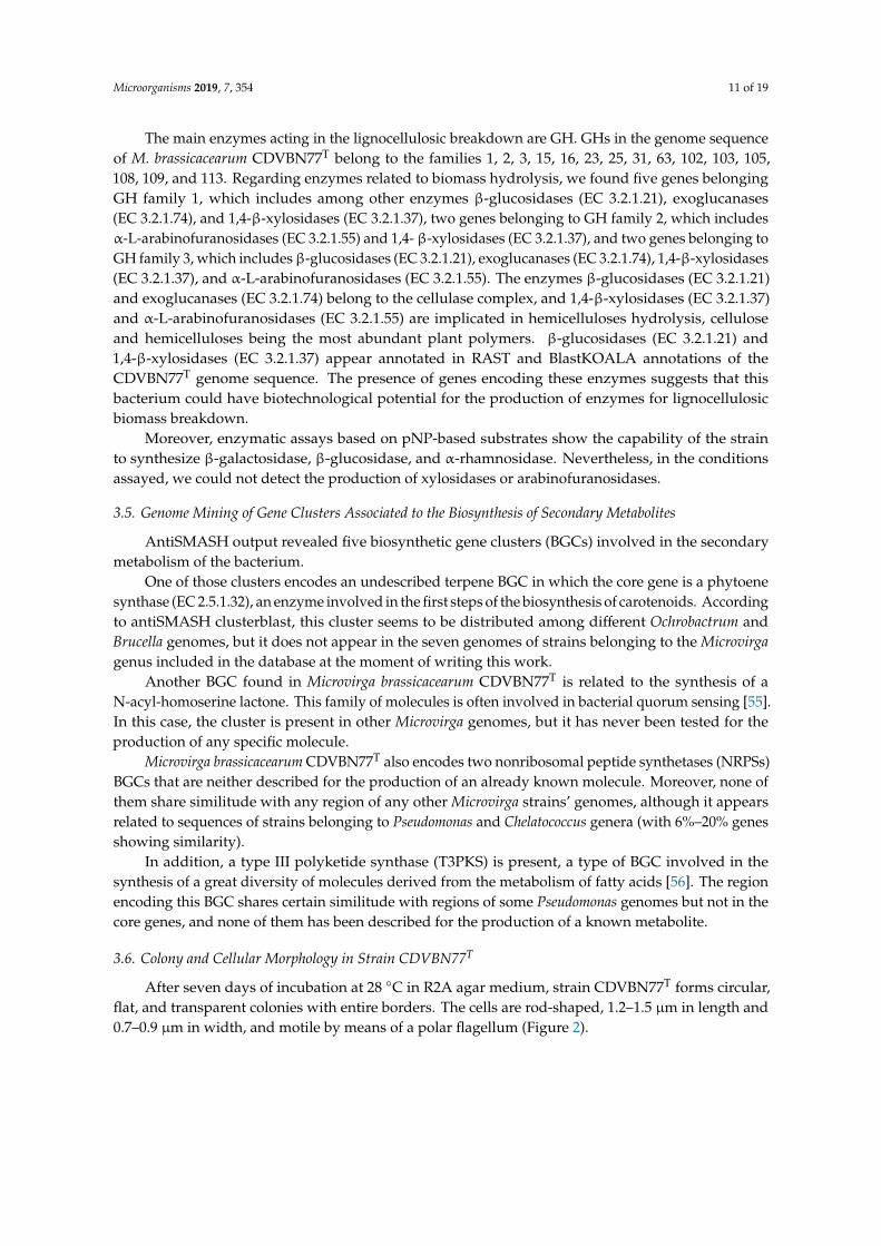
After seven days of incubation at 28 ◦C in R2A agar medium, strain CDVBN77T forms circular,flat, and transparent colonies with entire borders. The cells are rod-shaped, 1.2–1.5 µm in length and0.7–0.9 µm in width, and motile by means of a polar flagellum (Figure 2).
Microorganisms 2019, 7, 354 12 of 19
Microorganisms 2019, 7, x FOR PEER REVIEW 2 of 19
47 Figure 2. Electron TEM micrograph showing bacterial shape and size and the flagellum of Microvirga 48 brassicacearum CDVBN77T. 49
3.7. Phenotypic and Chemotaxonomic Characterization of Strain CDVBN77T 50 The analysis of respiratory quinones revealed that ubiquinone-10 (Q-10) is the major quinone in 51
CDVBN77T, and the main polar lipids of this strain are supporting the classification of the bacterium 52 within the genus Microvirga [11]. 53
Table 4. Cellular fatty acid composition (%) of Microvirga brassicacearum CDVBN77T (data from this 54 study) and its closest related species M. flavescens c27j1T (data from Zhang et al. [17]) and M. aerophila 55 DSM 21344T (data from Weon et al. [10]), as well as for the type species of the genus M. subterranea 56 DSM 14364T (data from Kanso and Patel [11]). Strains: 1, M. brassicacearum CDVBN77T; 2, M. flavescens 57 c27j1T; 3, M. aerophila DSM 21344T; 4, M. subterranea DSM 14364T. 58
Fatty Acid 1 2 3 4 C16:0 9.2 4.8 6.6 7.6 C17:0 tr 7.5 C18:0 6.1 2.0 3.1 5.5
C17:0 cyclo 3.8 3.5 2.1 tr 19:0 cyclo ω8c 24.3 57.7 11.8 27.9
C20:2 ω6,9c 1.1 tr 1.1 11-Methyl C18:1 ω7c 4.2 1.5 tr 1.5
C18:0 3-OH 1.7 2.1 1.1 1.7 Summed feature 2 4.5 5.0 1.4 3.2 Summed feature 3 4.5 1.1 5.2 1.1 Summed feature 8 39.3 18.5 64.8 38.5
The values are percentages of the total fatty acids. The values under 1% for all of the strains are not 59 included. tr: traces, values under 1%. Summed feature 2: C12:0 aldehyde and/or unknown 10.9525. 60 Summed feature 3: C16:1 ω7c and/or C16:1 ω6c. Summed feature 8: C18:1 ω7c and/or C18:1 ω6c. 61
Figure 2. Electron TEM micrograph showing bacterial shape and size and the flagellum of Microvirgabrassicacearum CDVBN77T.
3.7. Phenotypic and Chemotaxonomic Characterization of Strain CDVBN77T
The analysis of respiratory quinones revealed that ubiquinone-10 (Q-10) is the major quinone inCDVBN77T, and the main polar lipids of this strain are supporting the classification of the bacteriumwithin the genus Microvirga [11].
The major fatty acids in strain CDVBN77T are C19:0 cyclo ω8c (24.3%) and Summed Feature 8(39.3%) (Table 4), as it occurs for its closest related strains M. flavescens c27j1T [17] and M. aerophila DSM21344T [10], as well as for the type species of the genus M. subterranea DSM 14364T [11], supporting theclassification of the strain within the genus Microvirga. However, the fatty acid profile of this strainsignificantly differs from that of the closest related species (Table 4), supporting the classification ofCDVBN77T into a different species within the genus.
Table 4. Cellular fatty acid composition (%) of Microvirga brassicacearum CDVBN77T (data from thisstudy) and its closest related species M. flavescens c27j1T (data from Zhang et al. [17]) and M. aerophilaDSM 21344T (data from Weon et al. [10]), as well as for the type species of the genus M. subterraneaDSM 14364T (data from Kanso and Patel [11]). Strains: 1, M. brassicacearum CDVBN77T; 2, M. flavescensc27j1T; 3, M. aerophila DSM 21344T; 4, M. subterranea DSM 14364T.
Fatty Acid 1 2 3 4
C16:0 9.2 4.8 6.6 7.6C17:0 tr 7.5C18:0 6.1 2.0 3.1 5.5
C17:0 cyclo 3.8 3.5 2.1 tr19:0 cycloω8c 24.3 57.7 11.8 27.9
C20:2 ω6,9c 1.1 tr 1.111-Methyl C18:1 ω7c 4.2 1.5 tr 1.5
C18:0 3-OH 1.7 2.1 1.1 1.7Summed feature 2 4.5 5.0 1.4 3.2Summed feature 3 4.5 1.1 5.2 1.1Summed feature 8 39.3 18.5 64.8 38.5
The values are percentages of the total fatty acids. The values under 1% for all of the strains are not included. tr:traces, values under 1%. Summed feature 2: C12:0 aldehyde and/or unknown 10.9525. Summed feature 3: C16:1 ω7cand/or C16:1 ω6c. Summed feature 8: C18:1 ω7c and/or C18:1 ω6c.
Microorganisms 2019, 7, 354 13 of 19
Phenotypic features analyzed in strain CDVBN77T are detailed in the species protologue, and themain phenotypic differences with the closest related species and the type species of the genus aresummarized in Table 5.
Table 5. Phenotypic differences between Microvirga brassicacearum CDVBN77T (data from this study)and its closest related species M. flavescens c27j1T (data from Zhang et al. [17]) and M. aerophila DSM21344T (data from Weon et al. [10]), as well as for the type species of the genus M. subterranea DSM14364T (data from Kanso and Patel [11]) Strains: 1, M. brassicacearum CDVBN77T; 2, M. flavescens c27j1T;3, M. aerophila DSM 21344T; 4, M. subterranea DSM 14364T. +, positive; w, weakly positive; -, negative.
Characteristic 1 2 3 4
Isolation source Plant Soil Air Thermal aquiferColony color White Faint yellow Light pink Light pinkMotility + + − −
Oxidase + + + −
Nitrate reduction + + − +Hydrolysis of:
Gelatin + − − +Aesculin + − − −
Assimilation of:D-Glucose + + − −
L-Arabinose + + − +Production of:
β-glucoronidase − − + −
Esterase lipase (c8) + w + +Trypsin + − + w
pH range 6–10 6–10 7–10 6–9Salinity range (%) 0–1.5 0–1 0–2 0–1Temperature range (◦C) 12–37 15–37 10–35 25–45DNA G+C content (mol %) 62.3 62.2 62.1 65.1
4. Discussion
Many plant endophytic strains have been described as new taxa [57–60]. In this study, we isolatedstrain CDVBN77T, whose 16S rRNA gene sequence showed a relatively low similarity with its closestrelated species, M. aerophila DSM 21344T (97.64%) and M. flavescens c27j1T (97.50%), which suggestedthat it could belong to a new species within the genus Microvirga. Several Microvirga species, namely,M. lupini, M. lotononidis, M. zambiensis, M. ossetica, and M. vignae [18–20], were for the first time isolatedas legume endophytes. The phylogenetic analysis of this strain based on the 16S rRNA gene showedthat the bacterium appears in a separated branch within the genus Microvirga but clustered withits two closest related type strains M. flavescens c27j1T and M. aerophila DSM 21344T. The presenceof Q-10 as the major quinone and C19:0 cyclo ω8c and Summed Feature 8 as the main fatty acidssupports its classification within the genus Microvirga. However, this isolate differs from the closestspecies, M. aerophila and M. flavescens, in the capability to hydrolyze gelatin and aesculin, positivefor M. brassicacearum CDVBN77T and negative for the other two, and the color of the colonies inR2A agar medium, transparent in M. brassicacearum CDVBN77T and light yellow and pale pink inM. flavescens and M. aerophila, respectively. Unlike M. aerophila, it reduces nitrates to nitrites andassimilates D-glucose and L-arabinose, and it does not produce the enzyme β-glucoronidase; moreover,in contrast to M. flavescens, it produces trypsin. Thus, the results of the phylogenetic, chemotaxonomic,and phenotypic analysis clearly support the classification of strain CDVBN77T as a new species withinthe genus Microvirga, for which the name Microvirga brassicacearum sp. nov. is proposed.
The genus Microvirga belongs to Rhizobiales, a broadly studied order in the field of plant–bacteriainteractions because of its role as plant growth promoter. Five species of the genus Microvirga have beendescribed as plant endosymbionts capable of promoting plant growth, namely, M. lupini, M. lotononidis,M. zambiensis [18], M. vignae [20], and M. ossetica [19]. The genome mining of strain M. brassicacearum
Microorganisms 2019, 7, 354 14 of 19
CDVBN77T shows the presence of genes implicated in the nutrient provision to the plant, such asP and Fe, capabilities also proven in in vitro tests, as well as K and N. Moreover, both the analysisof the genome sequence as well as in vitro tests show the capability of this strain to synthesizeexopolysaccharides which may allow the attachment of the bacterial cells to the plant root surface inorder to establish an interaction with the host.
To live as plant endophytes, bacteria must have genetic machinery to use plant-synthesizedcompounds as a source of nutrients. Some of the enzymes implicated in plant compound degradationsare very interesting for industries based on biomass degradation, such as bioethanol production,pulp and paper industries, beverages industries, medicinal and analytical chemistry, fabricationof detergents, and textile de-sizing [6]. Thus, we explored the enzymatic machinery related tocarbohydrate degradation encoded by the M. brassicacearum CDVBN77T genome. As a result, weidentified genes related to the degradation and modification of biomass polymers, such as celluloseand hemicelluloses. β-glucosidase is the key enzyme in the cellulase system [34,61,62], since itconverts cellobiose to glucose, completing the final step during cellulose hydrolysis. In the genome ofM. brassicacearum CDVBN77T, we have found five genes belonging to family GH1 and three genesbelonging to family GH3; both enzyme families include enzymes, such as β-glucosidases (EC 3.2.1.21)or even exoglucanases (EC 3.2.1.74), enzymes which are also involved in the cellulose polymerdegradation [63].
The complete degradation of hemicelluloses requires the combined activity of xylanases,β-xylosidases, and several accessory enzymes such as α-arabinofuranosidases [64]. In the genome ofMicrovirga brassicacearum CDVBN77T, we have found gene counts in the GH1, GH2, and GH3 families,all of them including enzymes with xylan 1,4-β-xylosidase (EC 3.2.1.37) activity. Moreover, GH2 andG3 families include enzymes with α-arabinofuranosidase activity. However, we could not detectthe in vitro synthesis of xylosidases and arabinofuranosidases, either because the annotated genesbelonging to the GH1, GH2, and GH3 families have other different activities, or because they were notinduced under the assayed conditions.
On the other hand, we found how this bacterium is able to hydrolyze several substrates linked top-nitrophenol, which reveals that the bacterium synthesizesα-rhamnosidase, with potential applicationsin the clarification and debittering of citrus juices, the enhancement of wine aromas, or the synthesisof certain pharmaceutical compounds [65] and β-galactosidase, with potential application in foodprocessing industries for the hydrolysis of lactose in milk and milk by-products [66].
Regarding the secondary metabolism prospection of the bacterium, we found an interesting nichefor researching secondary metabolite BGCs that have not been very studied within the Microvirgagenus. AntiSMASH results showed that the CDVBN77T strain has the potential to encode at leastfive undescribed secondary metabolites; amongst those, a terpene, a PKS, and two NRPS havepotential applications in industry. NRPS are widely distributed among many taxa, and their products,nonribosomal peptides, are characterized because of their broad range of biological activities, such asbeing mainly antimicrobials, but also antitumoral compounds, antivirals, siderophores, etc. [67–70].The same fact happens with type III polyketides, in which we also found interesting activities [54,71,72].In addition, carotenoids (terpenes) are also used in the pharmaceutical and food industries because oftheir antioxidant activities or colorant properties [73,74].
Thus, in this study we found an interesting genetic potential in the genome of M. brassicacearumCDVBN77T to synthesize enzymes and secondary metabolites. Moreover, we detected in vitro theproduction of some of the predicted enzymes and metabolites. Nevertheless, the purification ofsuch gene products and the determination of their specific mechanisms of action in addition to theconditions of pH and temperature in which the molecules are active are required in order to determinethe possible industrial interest of those substances.
Microorganisms 2019, 7, 354 15 of 19
5. Description of Microvirga brassicacearum sp. nov.
Microvirga brassicacearum (bras.si.ca.ce.a.rum. M.L. fem. pl. gen. n. brassicacearum of the Brassicaceae,referring to its isolation from the rhizoplane of plants belonging to the genus Brassica). Cells are rod-shaped,1.2-1.5µm in length and 0.7-0.9µm in width, and motile by means of a polar flagellum. In 869(1/10) medium,colonies are transparent, circular, and flat, with an average size of 0.1–0.4 mm in diameter after sevendays of growth at 28 ◦C in R2A medium. The temperature growth ranges between 12 and 37 ◦C, withan optimum of 28 ◦C. The growth at different pH ranges occurs between 6 and 10 with an optimum of 7.It grows between 0 and 1.5% NaCl in 869(1/10) liquid medium. Catalase and oxidase positive. The mainpolar lipids are phosphatidylcholine and phosphatidylethanolamine. Ubiquinone-10 (Q-10) is themajor respiratory quinone and C19:0 cycloω8c and Summed Feature 8 are the main fatty acids. Resultsobtained in the API 20NE system indicate that the strain hydrolyzes aesculin, but it does not fermentD-glucose and does not hydrolyze arginine, urea, and gelatin. Reduction of nitrates to nitrites is positive.The assimilation of D-glucose, L-arabinose, D-mannitol, N-acetyl-glucosamine, D-maltose, potassiumgluconate, malic acid, and trisodium citrate is positive and that of D-mannose, capric acid, adipic acid,and phenylacetic acid is negative. Results obtained in the API ZYM system showed activity in theenzymes alkaline phosphatase, esterase (C 4), esterase lipase (C 8), leucine arylamidase, trypsin, acidphosphatase, and naphtol-AS-BI-phosphohydrolase, but activity was not shown in lipase (C14), valinearylamidase, cysteine arylamidase,α-chymotrypsin,α-galactosidase,β-galactosidase,β-glucuronidase,β-glucosidase, α-glucosidase, N-acetyl-β-glucosaminidase, α-mannosidase, and α-fucosidase.
The G+C base composition was 62.3 mol %. The type strain, CDVBN77T (= LMG 31419T= CECT9905T), was isolated as an endophyte from Brassica napus roots in Castellanos de Villiquera (Spain).
Author Contributions: Conceptualization, P.G.F.; methodology, P.G.-F., A.J.-G., Z.S.-S., J.M.I.; software, P.G.-F.,A.J.-G. and Z.S.-S.; validation, P.G.-F., A.J.-G. and Z.S.-S; formal analysis, P.G.-F., A.J.-G. and Z.S.-S.; investigation,P.G.-F., A.J.-G., Z.S.-S. and J.M.I.; resources, P.G.F., R.R. and P.F.M.; data curation, P.G.-F., A.J.-G. and Z.S.-S.;writing—original draft preparation, P.G.-F., A.J.-G. and Z.S.-S.; writing—review and editing, P.G.-F., A.J.-G.and Z.S.-S.; visualization, P.G.-F., A.J.-G. and Z.S.-S.; supervision, P.G.F.; project administration, P.G.F.; fundingacquisition, P.G.F.
Funding: This research was funded by the European Union’s Horizon 2020 research and innovation programmeunder the grant number 750795.
Acknowledgments: Authors thank the European Union’s Horizon 2020 research and innovation programme forthe research funding received by P.G.-F. A.J.-G. thanks the Central Spanish Government for an FPU predoctoralgrant and Z.S.-S. thanks the Junta de Castilla y Leon, Spanish Regional Government, for his predoctoral grant.
Conflicts of Interest: The authors declare that the research was conducted in the absence of any commercial orfinancial relationships that could be construed as a potential conflict of interest.
References
1. Locey, K.J.; Lennon, J.T. Scaling laws predict global microbial diversity. Proc. Natl. Acad. Sci. USA 2016, 113,5970–5975. [CrossRef] [PubMed]
2. Jongsik, C.; Oren, A.; Ventosa, A.; Christensen, H.; Ruiz, D.; Da Costa, M.; Rooney, A.P.; Yi, H.; Wei, X.;de Meyer, S.; et al. Proposed minimal standards for the use of genome data for the taxonomy of prokaryotes.Int. J. Syst. Evol. Microbiol. 2018, 68, 461–466.
3. Auch, A.F.; von Jan, M.; Klenk, H.P.; Göker, M. Digital DNA-DNA hybridization for microbial speciesdelineation by means of genome-to-genome sequence comparison. Stand. Genom. Sci. 2010, 2, 117–134.
4. López-Mondéjar, R.; Kostovcík, M.; Lladó, S.; Carro, L.; García-Fraile, P. Exploring the Plant MicrobiomeThrough Multi-omics Approaches. Probiotics Agroecosystem 2017, 233–268.
5. Saati-Santamaría, Z.; López-Mondéjar, R.; Jiménez-Gómez, A.; Díez-Méndez, A.; Vetrovský, T.; Igual, J.M.;Velázquez, E.; Kolarik, M.; Rivas, R.; García-Fraile, P. Discovery of phloeophagus beetles as a source ofPseudomonas strains that produce potentially new bioactive substances and description of Pseudomonasbohemica sp. nov. Front. Microbiol. 2018, 9. [CrossRef] [PubMed]
Microorganisms 2019, 7, 354 16 of 19
6. Fabryová, A.; Kostovcík, M.; Díez-Méndez, A.; Jiménez-Gómez, A.; Celador-Lera, L.; Saati-Santamaría, Z.;Sechovcová, H.; Menéndez, E.; Kolarik, M.; García-Fraile, P. On the bright side of a forest pest-the metabolicpotential of bark beetles’ bacterial associates. Sci. Total Environ. 2018, 619, 9–17. [CrossRef]
7. Gopal, M.; Gupta, A. Microbiome selection could spur next-generation plant breeding strategies.Front. Microbiol. 2016, 7, 1971. [CrossRef]
8. Menéndez, E.; Garcia-Fraile, P. Plant probiotic bacteria: Solutions to feed the world. Aims Microbiol. 2017, 3,502–524. [CrossRef]
9. Caputo, A.; Lagier, J.C.; Azza, S.; Robert, C.; Mouelhi, D.; Fournier, P.E.; Raoult, D. Microvirga massiliensis sp.nov., the human commensal with the largest genome. Microbiol. Open 2016, 5, 307–322. [CrossRef]
10. Weon, H.Y.; Kwon, S.W.; Son, J.A.; Jo, E.H.; Kim, S.J.; Kim, Y.S.; Kim, B.Y.; Ka, J.O. Description of Microvirgaaerophila sp. nov. and Microvirga aerilata sp. nov., isolated from air, reclassification of Balneimonas flocculansTakeda et al. 2004 as Microvirga flocculans comb. nov. and emended description of the genus Microvirga. Int. J.Syst. Evol. Microbiol. 2010, 60, 2596–2600. [CrossRef]
11. Kanso, S.; Patel, B.K. Microvirga subterranea gen. nov., sp. nov., a moderate thermophile from a deepsubsurface Australian thermal aquifer. Int. J. Syst. Evol. Microbiol. 2003, 53, 401–406. [CrossRef] [PubMed]
12. Zhang, J.; Song, F.; Xin, Y.H.; Zhang, J.; Fang, C. Microvirga guangxiensis sp. nov., a novel alphaproteobacteriumfrom soil, and emended description of the genus Microvirga. Int. J. Syst. Evol. Microbiol. 2009, 59, 1997–2001.[CrossRef] [PubMed]
13. Amin, A.; Ahmed, I.; Habib, N.; Abbas, S.; Hasan, F.; Xiao, M.; Hozzein, W.N.; Li, W.J. Microvirga pakistanensissp. nov., a novel bacterium isolated from desert soil of Cholistan, Pakistan. Arch. Microbiol. 2016, 198,933–939. [CrossRef] [PubMed]
14. Dahal, R.H.; Kim, J. Microvirga soli sp. nov., an alphaproteobacterium isolated from soil. Int. J. Syst. Evol.Microbiol. 2017, 67, 127–132. [PubMed]
15. Tapase, S.R.; Mawlankar, R.B.; Sundharam, S.S.; Krishnamurthi, S.; Dastager, S.G.; Kodam, K.M. Microvirgaindica sp. nov., an arsenite-oxidizing Alphaproteobacterium, isolated from metal industry waste soil. Int. J.Syst. Evol. Microbiol. 2017, 67, 3525–3531. [CrossRef] [PubMed]
16. Veyisoglu, A.; Tatar, D.; Saygin, H.; Inan, K.; Cetin, D.; Guven, K.; Tuncer, M.; Sahin, N. Microvirga makkahensissp. nov., and Microvirga arabica sp. nov., isolated from sandy arid soil. Antonie Van Leeuwenhoek 2016, 109,287–296. [CrossRef] [PubMed]
17. Zhang, X.J.; Zhang, J.; Yao, Q.; Zhu, H.H. Microvirga flavescens sp. nov., a novel bacterium isolated fromforest soil and emended description of the genus Microvirga. Int. J. Syst. Evol. Microbiol. 2019, 69, 667–671.[CrossRef]
18. Ardley, J.K.; Parker, M.A.; De Meyer, S.E.; Trengove, R.D.; O’Hara, G.W.; Reeve, W.G.; Yates, R.J.; Dilworth, M.J.;Willems, A.; Howieson, J.G. Microvirga lupini sp. nov., Microvirga lotononidis sp. nov., and Microvirga zambiensissp. nov. are Alphaproteobacterial root nodule bacteria that specifically nodulate and fix nitrogen withgeographically and taxonomically separate legume hosts. Int. J. Syst. Evol. Microbiol. 2012, 62, 2579–2588.[CrossRef]
19. Safronova, V.I.; Kuznetsova, I.G.; Sazanova, A.L.; Belimov, A.A.; Andronov, E.E.; Chirak, E.R.; Osledkin, Y.S.;Onishchuk, O.P.; Kurchak, O.N.; Shaposhnikov, A.I.; et al. Microvirga ossetica sp. nov., a species of rhizobiaisolated from root nodules of the legume species Vicia alpestris Steven. Int. J. Syst. Evol. Microbiol. 2017, 67,94–100.
20. Radl, V.; Simões-Araújo, J.L.; Leite, J.; Passos, S.R.; Martins, L.M.V.; Xavier, G.R.; Rumjanek, N.G.; Baldani, J.I.;Zilli, J.E. Microvirga vignae sp. nov., a root nodule symbiotic bacterium isolated from cowpea grown insemi-arid Brazil. Int. J. Syst. Evol. Microbiol. 2017, 64, 725–730. [CrossRef]
21. Samrot, A.V.; Rio, A.J.; Kumar, S.S.; Samanvitha, S.K. Bioprospecting studies of pigmenting Pseudomonasaeruginosa SU-1, Microvirga aerilata SU14 and Bacillus megaterium SU15 isolated from garden soil. Biocatal. Agric.Biotechnol. 2017, 11, 330–337. [CrossRef]
22. Jiménez-Gómez, A.; Saati-Santamaría, Z.; Menéndez, E.; Rivas, R.; Mateos, P.F.; Velázquez, E.; García-Fraile, P.Analysis of the biodiversity of Brassica napus bacterial endophytes and selection of potential efficientbiofertilizers for rapeseed crops. under review. Front. Microbiol.
23. Altschul, S.F.; Madden, T.L.; Schäffer, A.A.; Zhang, J.; Zhang, Z.; Miller, W.; Lipman, D.J. Gapped BLAST andPSI-BLAST: A new generation of protein database search programs. Nucleic Acids Res. 1997, 25, 3389–3402.[CrossRef] [PubMed]
Microorganisms 2019, 7, 354 17 of 19
24. Chun, J.; Lee, J.H.; Jung, Y.; Kim, M.; Kim, S.; Kim, B.K.; Lim, Y.W. EzTaxon: A web-based tool for theidentification of prokaryotes based on 16S ribosomal RNA gene sequences. Int. J. Syst. Evol. Microbiol. 2007,57, 2259–2261. [CrossRef] [PubMed]
25. Zerbino, D.R.; Birney, E. Velvet: Algorithms for de novo short read assembly using de Bruijn graphs.Genome Res. 2008, 18, 821–829. [CrossRef] [PubMed]
26. Aziz, R.K.; Bartels, D.; Best, A.A.; DeJongh, M.; Disz, T.; Edwards, R.A.; Meyer, F. The RAST Server: Rapidannotations using subsystems technology. BMC Genom. 2008, 9, 75. [CrossRef] [PubMed]
27. Kanehisa, M.; Sato, Y.; Morishima, K. BlastKOALA and GhostKOALA: KEGG tools for functionalcharacterization of genome and metagenome sequences. J. Mol. Biol. 2016, 428, 726–731. [CrossRef]
28. Zhang, H.; Yohe, T.; Huang, L.; Entwistle, S.; Wu, P.; Yang, Z.; Yin, Y. dbCAN2: a meta server for automatedcarbohydrate-active enzyme annotation. Nucleic Acids Res. 2018, 46, 95–101.
29. Blin, K.; Shaw, S.; Steinke, K.; Villebro, R.; Ziemert, N.; Lee, S.Y.; Weber, T. antiSMASH 5.0: Updates to thesecondary metabolite genome mining pipeline. Nucleic Acids Res. 2019, 47, 81–87. [CrossRef]
30. Kumar, S.; Stecher, G.; Tamura, K. MEGA7: Molecular evolutionary genetics analysis version 7.0 for biggerdatasets. Mol. Biol. Evol. 2016, 33, 1870–1874. [CrossRef]
31. Larkin, M.A.; Blackshields, G.; Brown, N.P.; Chenna, R.; McGettigan, P.A.; McWilliam, H.; Valentin, F.;Wallace, I.M.; Wilm, A.; Lopez, R.; et al. Clustal W and Clustal X version 2.0. Bioinformatics 2017, 23,2947–2948. [CrossRef] [PubMed]
32. Kimura, M. A simple method for estimating evolutionary rates of base substitutions through comparativestudies of nucleotide sequences. Mol. Biol. Evol. 1980, 16, 111–120. [CrossRef] [PubMed]
33. Saitou, N.; Nei, M. The neighbor-joining method: A new method for reconstructing phylogenetic trees.Mol. Biol. Evol. 1987, 4, 406–425. [PubMed]
34. Menéndez, E.; Ramírez-Bahena, M.H.; Fabryová, A.; Igual, J.M.; Benada, O.; Mateos, P.F.; Peix, A.; Kolarík, M.;García-Fraile, P. Pseudomonas coleopterorum sp. nov., a cellulase-producing bacterium isolated from the barkbeetle Hylesinus fraxini. Int. J. Syst. Evol. Microbiol. 2015, 65, 2852–2858.
35. Lladó, S.; Benada, O.; Cajthaml, T.; Baldrian, P.; García-Fraile, P. Silvibacterium bohemicum gen. nov. sp. nov.,an acidobacterium isolated from coniferous soil in the Bohemian Forest National Park. Syst. Appl. Microbiol.2016, 39, 14–19.
36. Doetsch, R.N. Determinative methods of light microscopy. Man. Meth. Gen. Bact. 1981, 21–33.37. Schwyn, B.; Neilands, J.B. Universal chemical assay for the detection and determination of siderophores.
Anal. Biochem. 1987, 160, 47–56. [CrossRef]38. Alexander, D.B.; Zuberer, D.A. Use of Chrome Azurol S reagents to evaluate siderophore production by
rhizosphere bacteria. Biol. Fertil. Soils 1991, 12, 39–45. [CrossRef]39. Pikovskaya, R.I. Mobilization of phosphorus in soil connection with the vital activity of some microbial
species. Microbiologiya 1948, 17, 362–370.40. Robledo, M.; Rivera, L.; Jiménez-Zurdo, J.I.; Rivas, R.; Dazzo, F.; Velázquez, E.; Martínez-Molina, E.;
Hirsch, A.; Mateos, P.F. Role of Rhizobium endoglucanase CelC2 in cellulose biosynthesis and biofilmformation on plant roots and abiotic surfaces. Microb. Cell Fact. 2012, 11, 125. [CrossRef]
41. Richter, M.; Rosselló-Móra, R. Shifting the genomic gold standard for the prokaryotic species definition.Proc. Natl. Acad. Sci. USA 2009, 106, 19126–19131. [CrossRef] [PubMed]
42. Meier-Kolthoff, J.P.; Auch, A.F.; Klenk, H.P.; Göker, M. Genome sequence-based species delimitation withconfidence intervals and improved distance functions. BMC Bioinform. 2013, 14, 60. [CrossRef] [PubMed]
43. Kwak, Y.; Jung, B.K.; Shin, J.H. Complete genome sequence of Pseudomonas rhizosphaerae IH5T (= DSM16299T), a phosphate-solubilizing rhizobacterium for bacterial biofertilizer. J. Biotechnol. 2015, 193, 137–138.[CrossRef] [PubMed]
44. de Werra, P.; Péchy-Tarr, M.; Keel, C.; Maurhofer, M. Role of gluconic acid production in the regulation ofbiocontrol traits of Pseudomonas fluorescens CHA0. Appl. Environ. Microbiol. 2009, 75, 41624174. [CrossRef][PubMed]
45. Goldstein, A.H. Involvement of the quinoprotein glucose dehydrogenase in the solubilization of exogenousphosphates by gram-negative bacteria. In Phosphate in microorganisms: Cellular and Molecular biology; ASMPress: Washington, DC, USA, 1994; pp. 197–203.
46. Etesami, H.; Emami, S.; Alikhani, H.A. Potassium solubilizing bacteria (KSB): Mechanisms, promotion ofplant growth, and future prospects A review. J. Soil Sci. Plant Nutr. 2017, 17, 897–911. [CrossRef]
Microorganisms 2019, 7, 354 18 of 19
47. Liu, W.; Wang, Q.; Hou, J.; Tu, C.; Luo, Y.; Christie, P. Whole genome analysis of halotolerant and alkalotolerantplant growth-promoting rhizobacterium Klebsiella sp. D5A. Sci. Rep. 2016, 6, 26710. [CrossRef]
48. Epstein, W. The roles and regulation of potassium in bacteria. Prog. Nucleic Acid Res. 2003, 75, 293–320.49. Velázquez, E.; García-Fraile, P.; Ramírez-Bahena, M.H.; Rivas, R.; Martínez-Molina, E. Bacteria involved in
nitrogen-fixing legume symbiosis: Current taxonomic perspective. Microbes Legume Improv. 2010, 1–25.50. Mateos, P.F.; Jimenez-Zurdo, J.I.; Chen, J.; Squartini, A.S.; Haack, S.K.; Martinez-Molina, E.; Hubbell, D.H.;
Dazzo, F.B. Cell-associated pectinolytic and cellulolytic enzymes in Rhizobium leguminosarum biovar trifolii.Appl. Environ. Microbiol. 1992, 58, 1816–1822.
51. Reed, J.W.; Walker, G.C. The exoD gene of Rhizobium meliloti encodes a novel function needed for alfalfanodule invasion. J. Bacteriol. 1991, 173, 664–677. [CrossRef]
52. Polissi, A.; Sperandeo, P. The lipopolysaccharide export pathway in Escherichia coli: Structure, organizationand regulated assembly of the Lpt machinery. Mar. Drugs 2014, 12, 1023–1042. [CrossRef]
53. Dong, H.; Zhang, Z.; Tang, X.; Paterson, N.G.; Dong, C. Structural and functional insights into thelipopolysaccharide ABC transporter LptB 2 FG. Nat. Commun. 2017, 8, 222. [CrossRef] [PubMed]
54. Pradel, E.; Parker, C.T.; Schnaitman, C.A. Structures of the rfaB, rfaI, rfaJ, and rfaS genes of Escherichia coliK-12 and their roles in assembly of the lipopolysaccharide core. J. Bacteriol. 1992, 174, 4736–4745. [CrossRef][PubMed]
55. Fuqua, C.; Greenberg, E.P. Signalling: Listening in on bacteria: Acyl-homoserine lactone signalling. Nat. Rev.Mol. Cell Biol. 2002, 3, 685. [CrossRef] [PubMed]
56. Yu, D.; Xu, F.; Zeng, J.; Zhan, J. Type III polyketide synthases in natural product biosynthesis. Iubmb Life2012, 64, 285–295. [CrossRef] [PubMed]
57. Rivas, R.; García-Fraile, P.; Mateos, P.F.; Martínez-Molina, E.; Velazquez, E. Paenibacillus cellulosilyticus sp.nov., a cellulolytic and xylanolytic bacterium isolated from the bract phyllosphere of Phoenix dactylifera. Int. J.Syst. Evol. Microbiol. 2006, 56, 2777–2781. [CrossRef] [PubMed]
58. Rivas, R.; Garcia-Fraile, P.; Zurdo-Pineiro, J.L.; Mateos, P.F.; Martinez-Molina, E.; Bedmar, E.J.; Sánchez-Raya, J.;Velazquez, E. Saccharibacillus sacchari gen. nov., sp. nov., isolated from sugar cane. Int. J. Syst. Evol. Microbiol.2008, 58, 1850–1854. [CrossRef]
59. García-Fraile, P.; Velázquez, E.; Mateos, P.F.; Martínez-Molina, E.; Rivas, R. Cohnella phaseoli sp. nov., isolatedfrom root nodules of Phaseolus coccineus in Spain, and emended description of the genus Cohnella. Int. J. Syst.Evol. Microbiol. 2008, 58, 1855–1859.
60. Flores-Félix, J.D.; Carro, L.; Velázquez, E.; Valverde, Á.; Cerda-Castillo, E.; García-Fraile, P.; Rivas, R.Phyllobacterium endophyticum sp. nov., isolated from nodules of Phaseolus vulgaris. Int. J. Syst. Evol. Microbiol.2013, 63, 821–826.
61. Mansfield, S.D.; Mooney, C.; Saddler, J.N. Substrate and enzyme characteristics that limit cellulose hydrolysis.Biotechnol. Prog. 1999, 15, 804–816. [CrossRef]
62. Sharma, B.; Sarkar, A.; Singh, P.; Singh, R.P. Agricultural utilization of biosolids: A review on potential effectson soil and plant grown. Waste Manag. 2017, 64, 117–132. [CrossRef]
63. Menéndez, E.; García-Fraile, P.; Rivas, R. Biotechnological applications of bacterial cellulases. AIMS Bioeng.2015, 2, 163–182. [CrossRef]
64. Collin, T.; Gerday, C.; Feller, G. Xylanase, xylanase families and extremophilic xylanase. FEMS Microbiol. Rev.2005, 29, 3–23. [CrossRef] [PubMed]
65. Yadav, V.; Yadav, P.K.; Yadav, S.; Yadav, K.D.S. α-L-Rhamnosidase: A review. Process Biochem. 2010, 45,1226–1235. [CrossRef]
66. Xavier, J.R.; Ramana, K.V.; Sharma, R.K. β-galactosidase: Biotechnological applications in food processing.J. Food Biochem. 2018, 42, e12564. [CrossRef]
67. Sieber, S.A.; Marahiel, M.A. Learning from nature’s drug factories: Nonribosomal synthesis of macrocyclicpeptides. J. Bacteriol. 2003, 185, 7036–7043. [CrossRef]
68. Finking, R.; Marahiel, M.A. Biosynthesis of nonribosomal peptides. Annu. Rev. Microbiol. 2004, 58, 453–488.[CrossRef]
69. Felnagle, E.A.; Jackson, E.E.; Chan, Y.A.; Podevels, A.M.; Berti, A.D.; McMahon, M.D.; Thomas, M.G.Nonribosomal peptide synthetases involved in the production of medically relevant natural products.Mol. Pharm. 2008, 5, 191–211. [CrossRef]
Microorganisms 2019, 7, 354 19 of 19
70. Agrawal, S.; Acharya, D.; Adholeya, A.; Barrow, C.J.; Deshmukh, S.K. Nonribosomal peptides from marinemicrobes and their antimicrobial and anticancer potential. Front. Pharm. 2017, 8, 828. [CrossRef]
71. Gross, F.; Luniak, N.; Perlova, O.; Gaitatzis, N.; Jenke-Kodama, H.; Gerth, K.; Müller, R. Bacterial type IIIpolyketide synthases: Phylogenetic analysis and potential for the production of novel secondary metabolitesby heterologous expression in pseudomonads. Arch. Microbiol. 2006, 185, 28–38. [CrossRef]
72. Katsuyama, Y.; Ohnishi, Y. Type III polyketide synthases in microorganisms. Methods Enzymol. 2012, 515,359–377.
73. Cardoso, L.A.; Karp, S.G.; Vendruscolo, F.; Kanno, K.Y.; Zoz, L.I.; Carvalho, J.C. Biotechnological productionof carotenoids and their applications in food and pharmaceutical products. Carotenoids 2017, 125.
74. Gateau, H.; Solymosi, K.; Marchand, J.; Schoefs, B. Carotenoids of microalgae used in food industry andmedicine. Mini Rev. Med. Chem. 2017, 17, 1140–1172. [CrossRef] [PubMed]
© 2019 by the authors. Licensee MDPI, Basel, Switzerland. This article is an open accessarticle distributed under the terms and conditions of the Creative Commons Attribution(CC BY) license (http://creativecommons.org/licenses/by/4.0/).