SymbiodiniumTranscriptomes: Genome Insights into the ...
14
Symbiodinium Transcriptomes: Genome Insights into the Dinoflagellate Symbionts of Reef-Building Corals Till Bayer 1. , Manuel Aranda 1. , Shinichi Sunagawa 2 , Lauren K. Yum 1 , Michael K. DeSalvo 3 , Erika Lindquist 4 , Mary Alice Coffroth 5 , Christian R. Voolstra 1 *, Mo ´ nica Medina 6 * 1 Red Sea Research Center, King Abdullah University of Science and Technology (KAUST), Thuwal, Saudi Arabia, 2 European Molecular Biology Laboratory, Heidelberg, Germany, 3 Department of Anesthesia, UCSF School of Medicine, University of California San Francisco, San Francisco, California, United States of America, 4 Department of Energy Joint Genome Institute, Walnut Creek, California, United States of America, 5 Graduate Program in Evolution, Ecology and Behavior, Department of Geology, State University of New York at Buffalo, Buffalo, New York, United States of America, 6 School of Natural Sciences, University of California Merced, Merced, California, United States of America Abstract Dinoflagellates are unicellular algae that are ubiquitously abundant in aquatic environments. Species of the genus Symbiodinium form symbiotic relationships with reef-building corals and other marine invertebrates. Despite their ecologic importance, little is known about the genetics of dinoflagellates in general and Symbiodinium in particular. Here, we used 454 sequencing to generate transcriptome data from two Symbiodinium species from different clades (clade A and clade B). With more than 56,000 assembled sequences per species, these data represent the largest transcriptomic resource for dinoflagellates to date. Our results corroborate previous observations that dinoflagellates possess the complete nucleosome machinery. We found a complete set of core histones as well as several H3 variants and H2A.Z in one species. Furthermore, transcriptome analysis points toward a low number of transcription factors in Symbiodinium spp. that also differ in the distribution of DNA-binding domains relative to other eukaryotes. In particular the cold shock domain was predominant among transcription factors. Additionally, we found a high number of antioxidative genes in comparison to non-symbiotic but evolutionary related organisms. These findings might be of relevance in the context of the role that Symbiodinium spp. play as coral symbionts. Our data represent the most comprehensive dinoflagellate EST data set to date. This study provides a comprehensive resource to further analyze the genetic makeup, metabolic capacities, and gene repertoire of Symbiodinium and dinoflagellates. Overall, our findings indicate that Symbiodinium possesses some unique characteristics, in particular the transcriptional regulation in Symbiodinium may differ from the currently known mechanisms of eukaryotic gene regulation. Citation: Bayer T, Aranda M, Sunagawa S, Yum LK, DeSalvo MK, et al. (2012) Symbiodinium Transcriptomes: Genome Insights into the Dinoflagellate Symbionts of Reef-Building Corals. PLoS ONE 7(4): e35269. doi:10.1371/journal.pone.0035269 Editor: Ahmed Moustafa, American University in Cairo, Egypt Received November 26, 2011; Accepted March 13, 2012; Published April 18, 2012 This is an open-access article, free of all copyright, and may be freely reproduced, distributed, transmitted, modified, built upon, or otherwise used by anyone for any lawful purpose. The work is made available under the Creative Commons CC0 public domain dedication. Funding: This study was supported through NSF (National Science Foundation) awards IOS 0644438 and IOS 0926906 (MM), OCE 0424994 (MAC), a KAUST AEA (King Abdullah University of Science and Technology) 3 Joint Collaborative Research award (CRV), and through a Collaborative Travel Fund to TB made by King Abdullah University of Science and Technology. The work conducted by the U.S. Department of Energy Joint Genome Institute is supported by the Office of Science of the U.S. Department of Energy under Contract No. DE-AC02-05CH11231. The funders had no role in study design, data collection and analysis, decision to publish, or preparation of the manuscript. Competing Interests: The authors have declared that no competing interests exist. * E-mail: [email protected] (CRV); [email protected] (MM) . These authors contributed equally to this work. Introduction Dinoflagellates are ubiquitous marine and freshwater unicellular eukaryotes. As photosynthetic plankton, they are responsible for much of the primary production of oceans, rivers, and lakes. As photosynthetic marine symbionts, they form mutualistic relation- ships with reef-building corals and other invertebrates [1]. Approximately half of the 4,000 known dinoflagellate species contain no plastids, and many species are mixotrophic [2]. Dinoflagellates belong to the Alveolata, a large eukaryotic clade that also comprises the ciliates, which are free-living, as well as the Apicomplexans, which all have parasitic lifestyles. In addition to their ecological diversification, dinoflagellates show some genetic traits that make them distinct from other eukaryotic lineages. In particular, dinoflagellates have extensively methylated nuclear DNA. About 12–70% of thymine bases are replaced by 5-hydroxymethyluracil, and varying levels of cytosine methylation have been observed [3,4]. Genome sizes are very large and remarkably variable within the group, with estimates ranging from 3–215 gigabases (Gb) in size [5,6]. The genomic DNA is present in up to several hundred chromosomes per species [7]. Dinoflagellate genomic DNA has been shown to occur in a crystal-like state [8], with chromosomes condensed throughout the cell cycle [9]. Some of these observations initially led authors to conclude that dinoflagellates lacked histones [9]. However, recent genome-enabled studies have confirmed the presence of histones H3 [10], H2A.X [11], and H4 [12] in members of this lineage. Dinoflagellate genomes may host some 40,000–90,000 genes, which might be partly due to high gene copy numbers [13]. Despite the high gene number, dinoflagellate genomes are assumed to consist mostly of non-coding DNA (98–99.9%) [13]. Another unique feature characteristic of the dinoflagellate PLoS ONE | www.plosone.org 1 April 2012 | Volume 7 | Issue 4 | e35269
Transcript of SymbiodiniumTranscriptomes: Genome Insights into the ...
Symbiodinium Transcriptomes: Genome Insights into theDinoflagellate Symbionts of Reef-Building CoralsTill Bayer1., Manuel Aranda1., Shinichi Sunagawa2, Lauren K. Yum1, Michael K. DeSalvo3,
Erika Lindquist4, Mary Alice Coffroth5, Christian R. Voolstra1*, Monica Medina6*
1 Red Sea Research Center, King Abdullah University of Science and Technology (KAUST), Thuwal, Saudi Arabia, 2 European Molecular Biology Laboratory, Heidelberg,
Germany, 3 Department of Anesthesia, UCSF School of Medicine, University of California San Francisco, San Francisco, California, United States of America, 4 Department
of Energy Joint Genome Institute, Walnut Creek, California, United States of America, 5 Graduate Program in Evolution, Ecology and Behavior, Department of Geology,
State University of New York at Buffalo, Buffalo, New York, United States of America, 6 School of Natural Sciences, University of California Merced, Merced, California,
United States of America
Abstract
Dinoflagellates are unicellular algae that are ubiquitously abundant in aquatic environments. Species of the genusSymbiodinium form symbiotic relationships with reef-building corals and other marine invertebrates. Despite their ecologicimportance, little is known about the genetics of dinoflagellates in general and Symbiodinium in particular. Here, we used454 sequencing to generate transcriptome data from two Symbiodinium species from different clades (clade A and clade B).With more than 56,000 assembled sequences per species, these data represent the largest transcriptomic resource fordinoflagellates to date. Our results corroborate previous observations that dinoflagellates possess the completenucleosome machinery. We found a complete set of core histones as well as several H3 variants and H2A.Z in onespecies. Furthermore, transcriptome analysis points toward a low number of transcription factors in Symbiodinium spp. thatalso differ in the distribution of DNA-binding domains relative to other eukaryotes. In particular the cold shock domain waspredominant among transcription factors. Additionally, we found a high number of antioxidative genes in comparison tonon-symbiotic but evolutionary related organisms. These findings might be of relevance in the context of the role thatSymbiodinium spp. play as coral symbionts. Our data represent the most comprehensive dinoflagellate EST data set todate. This study provides a comprehensive resource to further analyze the genetic makeup, metabolic capacities, and generepertoire of Symbiodinium and dinoflagellates. Overall, our findings indicate that Symbiodinium possesses some uniquecharacteristics, in particular the transcriptional regulation in Symbiodinium may differ from the currently known mechanismsof eukaryotic gene regulation.
Citation: Bayer T, Aranda M, Sunagawa S, Yum LK, DeSalvo MK, et al. (2012) Symbiodinium Transcriptomes: Genome Insights into the Dinoflagellate Symbionts ofReef-Building Corals. PLoS ONE 7(4): e35269. doi:10.1371/journal.pone.0035269
Editor: Ahmed Moustafa, American University in Cairo, Egypt
Received November 26, 2011; Accepted March 13, 2012; Published April 18, 2012
This is an open-access article, free of all copyright, and may be freely reproduced, distributed, transmitted, modified, built upon, or otherwise used by anyone forany lawful purpose. The work is made available under the Creative Commons CC0 public domain dedication.
Funding: This study was supported through NSF (National Science Foundation) awards IOS 0644438 and IOS 0926906 (MM), OCE 0424994 (MAC), a KAUST AEA(King Abdullah University of Science and Technology) 3 Joint Collaborative Research award (CRV), and through a Collaborative Travel Fund to TB made by KingAbdullah University of Science and Technology. The work conducted by the U.S. Department of Energy Joint Genome Institute is supported by the Office ofScience of the U.S. Department of Energy under Contract No. DE-AC02-05CH11231. The funders had no role in study design, data collection and analysis, decisionto publish, or preparation of the manuscript.
Competing Interests: The authors have declared that no competing interests exist.
* E-mail: [email protected] (CRV); [email protected] (MM)
. These authors contributed equally to this work.
Introduction
Dinoflagellates are ubiquitous marine and freshwater unicellular
eukaryotes. As photosynthetic plankton, they are responsible for
much of the primary production of oceans, rivers, and lakes. As
photosynthetic marine symbionts, they form mutualistic relation-
ships with reef-building corals and other invertebrates [1].
Approximately half of the 4,000 known dinoflagellate species
contain no plastids, and many species are mixotrophic [2].
Dinoflagellates belong to the Alveolata, a large eukaryotic clade
that also comprises the ciliates, which are free-living, as well as the
Apicomplexans, which all have parasitic lifestyles.
In addition to their ecological diversification, dinoflagellates
show some genetic traits that make them distinct from other
eukaryotic lineages. In particular, dinoflagellates have extensively
methylated nuclear DNA. About 12–70% of thymine bases are
replaced by 5-hydroxymethyluracil, and varying levels of cytosine
methylation have been observed [3,4]. Genome sizes are very
large and remarkably variable within the group, with estimates
ranging from 3–215 gigabases (Gb) in size [5,6]. The genomic
DNA is present in up to several hundred chromosomes per species
[7]. Dinoflagellate genomic DNA has been shown to occur in a
crystal-like state [8], with chromosomes condensed throughout the
cell cycle [9]. Some of these observations initially led authors to
conclude that dinoflagellates lacked histones [9]. However, recent
genome-enabled studies have confirmed the presence of histones
H3 [10], H2A.X [11], and H4 [12] in members of this lineage.
Dinoflagellate genomes may host some 40,000–90,000 genes,
which might be partly due to high gene copy numbers [13].
Despite the high gene number, dinoflagellate genomes are
assumed to consist mostly of non-coding DNA (98–99.9%) [13].
Another unique feature characteristic of the dinoflagellate
PLoS ONE | www.plosone.org 1 April 2012 | Volume 7 | Issue 4 | e35269
molecular machinery is the trans-splicing of spliced leader
sequences [12,14]. In this process, a highly conserved spliced
leader (SL) is transplanted to the 59 end of mRNAs. SL trans-
splicing acts to convert polycistronic mRNAs to monocistronic
mRNAs and has also been suggested to regulate gene expression
[15].
Symbiodinium spp. (Alveolata: Dinophycea) – often referred to as
zooxanthellae – are a specific group of dinoflagellates that are
intracellular symbionts of many marine invertebrates including
scleractinian corals. Although initially considered a single
symbiotic species, molecular phylogenetics has uncovered major
Symbiodinium clades [16] that are separated from each other by tens
of millions of years [17–19]. Through photosynthesis, Symbiodinium
algae supply much of their hosts’ dietary needs and in return
receive shelter, a light-rich environment, and inorganic nutrients
[20]. In most cases this symbiotic relationship is reestablished
during each host generation [21]. Recent transcriptome-wide
efforts have been mainly devoted towards the understanding of the
molecular and cellular processes involved in the onset of symbiosis
from the host perspective [22–25]. From the symbiont’s
perspective, a relatively small number of ESTs has been analyzed
by Leggat et al. [26] and Voolstra et al. [27]. Voolstra et al. [27]
compared orthologous cDNA sequences from cultured and
symbiotic species (i.e. Symbiodinium CassKB8 and Symbiodinium
C3, respectively), providing some preliminary insight into the
genes that might be involved in Symbiodinium symbiosis. In a similar
fashion, studies have focused on the biochemical and transcrip-
tomic responses to the breakdown of symbiosis (i.e. coral
bleaching) in adult corals [23,27–30]. However, thus far there
are no comprehensive Symbiodinium genome-enabled investigations
that can shed light onto the complement of genes associated with
susceptibility to bleaching.
In this study, we sequenced EST libraries from two Symbiodinium
species that are known to establish stable symbioses with coral
hosts (clade A: Symbiodinium sp. CassKB8 and clade B: Symbiodinium
sp. Mf1.05b). These data represent the largest dinoflagellate EST
data set available to date with more than 56,000 assembled
transcripts per species. Annotation of these transcripts yielded new
insights into the complex gene repertoire of dinoflagellates, and
the mechanisms of nuclear organization of DNA and transcrip-
tional regulation among others.
Materials and Methods
Cultures, RNA isolation and sequencingTwo different species of Symbiodinium spp., CassKB8 (clade A)
and Mf1.05b (clade B), were exposed to a range of different
conditions (heat, cold, light, and dark) for 3–6 days to induce
expression of a maximum number of genes. Cultures were grown
in f/2 medium [31], for Mf1.05b cultures antibiotics were added
to the medium to combat bacterial contamination [32]. ‘Hot’ and
‘cold’ cultures were grown at 30–31uC and 19uC, respectively, all
other cultures at 27uC. All treatments were subject to a diurnal
light cycle (14:10 hrs) of approximately 50 mmol photons/m2/s,
except the ‘dark’ treatment, for which cultures were grown in
darkness for 6 days. For the ‘light’ treatment the light intensity was
increased to approximately 120 mmol photons/m2/s. Treated
Symbiodinium were harvested during the exponential growth phase
(approx. 106 cells/mL), pelleted and then snap frozen in liquid
nitrogen. Symbiodinium sp. CassKB8 was originally isolated from
Cassiopeia sp. in Kaneohe Bay, Hawaii by Robert Kenzie (personal
communication). Symbiodinium sp. Mf1.05b was isolated from
Montastraea faveolata, Florida Keys by M.A. Coffroth. Frozen pellets
were ground into a fine powder using a pre-chilled mortar and
pestle, and powder was added directly to Qiazol lysis reagent
(Qiagen, Hilden, Germany). Total RNAs were precipitated with
isopropanol, and RNA pellets were washed with 80% ethanol and
redissolved in water. Total RNAs were cleaned with RNeasy Mini
columns (Qiagen) and pooled in equal amounts for each species.
Library preparation for sequencing was carried out differently for
both strains. For CassKB8 the RNA was used to construct cDNA
libraries using the cDNA Rapid Library Preparation Method as
outlined in the Roche kit (Roche 454 Life Sciences, Branford,
USA), followed by normalization using the protocol provided for
the Evrogen Normalization kit (Evrogen, Moscow, Russia). The
normalized dscDNA was then used to construct 454 libraries using
the 454 library construction protocol provided in the 454 FLX
Titanium Roche kit (Roche, Branford, USA) and then sequenced
using the 454 GS-FLX platform. For Mf1.05b cDNA was
generated using an oligo-dT primer followed by template
switching (Clontech, Mountain View, USA) and subsequently
normalized using the same kit as above and sequenced as detailed
for CassKB8.
Data and AssemblyThe reads were assembled using version 3.2.1 of MIRA [33]
with settings appropriate for transcriptome assembly (–job = de-
novo,est,normal,454 COMMON_SETTINGS -GE:not = 8
454_SETTINGS -CL:qc = no:cpat = yes:msvs = yes -AS:mrpc = 1
-OUT:sssip = yes:stsip = yes). Adaptors were searched and marked
with SSAHA2 [34], and the locations included in the MIRA input
files to enable clipping. As MIRA assembles transcripts (not genes),
size sorted contigs and singlets were clustered using the UCLUST
algorithm as implemented in USEARCH 4.2.66 [35] in both
directions with an identity cutoff of 90% in order to estimate the
number of genes (Suppl. Table S1). The cutoff was empirically
chosen as a conservative estimate to account for sequencing errors
and mRNA editing. In the following, clustered contigs and singlets
are referred to as genes. To test the effect of clustering on gene
families, all contigs belonging to the actin gene family were
determined by searching a full length actin sequence from
Symbiodinium (accession no. AB231899, [36]) against all CassKB8
contigs, and comparing to the clustering of these contigs (Suppl.
Table S2). All raw reads are available in the NCBI Short Read
Archive (SRA) under the accession numbers SRX076710,
SRX076709, and SRX076696. The assembled and annotated
sequences are available for download at http://medinalab.org/
zoox. In most of the cases, we were not able to identify a SL
sequence in our dataset. However, PCRs with a SL and gene
specific primer for three genes (actin, Glyceraldehyde 3-phosphate
dehydrogenase and b-tubulin) showed that the SL sequence is
present in all three genes in CassKB8 (data not shown). Absence of
the SL from the transcriptome sequences may be a library
preparation or sequencing artifact.
AnnotationAssembled transcriptome data were annotated as follows: 1) by
BLASTX homology search against protein databases, 2) by
mapping to pathways using the KEGG annotation service KAAS
[37], and 3) by searching for protein domains with InterProScan
[38]. The BLASTX homology search was conducted against the
Swissprot, TrEMBL [39] and NCBI nr non-redundant protein
databases (all as of May 2011) in that order, and the first hit with
an e-value below 1025 was retained for annotation. For KAAS
pathway annotation and analysis, we used the single-directional
best hit (SBH) method to query the set of organisms representative
for ‘genes’ as suggested on the KAAS website, with the default
bitscore threshold of 60. Determination of completeness of the
Transcriptome Analysis of Two Symbiodinium Species
PLoS ONE | www.plosone.org 2 April 2012 | Volume 7 | Issue 4 | e35269

transcriptome data was also based on the KEGG annotation and
manual analysis of the pathways and complexes identified. Protein
domains were annotated using the InterProScan software in
version 4.6 with all possible applications and in all reading frames
[38]. The ‘sig’ and ‘SignalPHMM’ databases were excluded from
the InterProScan results, as they do not represent functional
protein domains.
Codon usageWe searched all contigs and singlets against the NCBI nr
database using BLASTX to ensure that only codons in the proper
reading frame were used to calculate codon usage statistics. For all
calculations we extracted and used only the nucleotide sequences
corresponding to the best HSP in hits with an e-value of equal or
less than 10210. This procedure yielded a total of 4,224,266 and
2,525,073 codons for CassKB8 and Mf1.05b, respectively.
Transcriptome data were analyzed for codon usage and the
effective number of codons (Nc) [40] with the programs cusp and
chips from the EMBOSS package [41]. The maximum number
for Nc is 61, which indicates uniform codon usage whereas lower
values signify codon bias. We analyzed Nc in relation to the GC
content of the third codon position (GC3) through an Nc plot (i.e.
a plot of Nc versus GC3s for all genes) to determine whether
codon usage heterogeneity exists among different genes in our
transcriptome data. In order to look at major differences between
genes in relation to codon usage, we performed Correspondence
Analysis – a multivariate statistic that displays the greatest variance
in codon usage in a two dimensional plot. Correspondence
analysis of codon usage was calculated with the software CodonW
[42]. One group of transcripts formed a distinct cloud of points in
this analysis. In order to analyze this group in more detail, we
chose a visual cutoff to separate the member transcripts. We
summarized the putative functions for these transcripts by
clustering at 90% similarity (as described earlier) and by
subsequently counting genes with the same annotation. To ensure
accurate results, we counted only transcripts with more than 100
analyzed codons.
Histones and Nucleosome-Associated ProteinsHistone and histone-associated genes were identified based on
gene annotation. Genes were annotated according to the best
annotation hit in the corresponding transcript cluster (Suppl.
Table S3). Putative histone transcripts with less than 30 amino
acids length were excluded from further analysis. Only full-length
amino acid sequences of histones (Suppl. Table S3) were
considered for phylogenetic analysis. Histone sequences for
different H2A, H3 and H3.3 variants were downloaded from
the NCBI databases. We preferentially selected sequences from
closer and further related species for which more than one histone
variant was present. Sequences were aligned using the MUSCLE
[43] implementation in Mega5 v.5.05 with standard settings [44].
Phylogenetic trees were reconstructed using maximum likelihood
(ML) and Bayesian analysis. ML analysis was performed using the
PhyLM v3.0 software [45] available at the ‘‘ATGC South of
France bioinformatics platform’’ (http://www.atgc-montpellier.fr).
Analyses were performed using the WAG substitution model (as
determined by Mr. Bayes mixed model). Tree improvement was
assessed using both, Subtree Pruning and Regrafting topological
moves (SPR) and simultaneous Nearest Neighbor Interchanges
(NNI) algorithms, branch support was assessed via nonparametric
bootstrapping using 1,000 replicates. Bayesian analysis was
performed using MrBayes v3.1.2. [46] using the following settings:
nchains = 4, one cold and three heated chains, with the exception
of codon models were two chains were used; the number of
steps = generations was set to 1,000,000 with sampfreq = 100 and
burnin = 2,500. Convergence was assessed using Tracer v.1.5 [47]
and by examining the PSRF values and standard deviation of split
frequencies. The best substitution model was assessed using mixed
model as recommended by MrBayes and the WAG model was
used for subsequent analysis based on the highest posterior
probability.
Transcription factorsWe used the comprehensive set of annotated, sequence-specific
DNA/RNA binding domains described in [48] to search for
transcription factors in our transcriptome data. We included the
AP2 domain, which is common in plants, but has recently also
been found in apicomplexans [49]. Our analysis was based on
Pfam domains with an e-value cutoff of 1026 as provided by
HMMER [50] following the approach of Ryu et al. [51]. All
contigs and singlets were translated in all reading frames to obtain
all possible peptide sequences using transeq from the EMBOSS
package [41]. To estimate transcription factor numbers at the gene
level, any domain was counted only once 1) per transcript cluster,
and 2) per transcript if the transcript contained multiple domains
of the same type. In addition, all dinoflagellate ESTs from the
NCBI Genbank dbEST database (as of June 2011) were
downloaded and analyzed as described above (total number of
sequences: 165,532). Finally, all protein sequences from selected
outgroup taxa were included in the analysis: Plasmodium falciparum
and P. vivax from PlasmoDB [52], Paramecium tetraurelia from
ParameciumDB [53], and Thalassiosira pseudonana, Arabidopsis
thaliana, Drosophila melanogaster and human from BioMart [54].
Outgroup protein sequences were analyzed with HMMER as
described above.
Antioxidative responsePutative antioxidant genes were identified in a similar manner
as the transcription factors. Briefly, we screened our data set for
antioxidant-associated genes using a list of pertinent Pfam domains
[55] as compiled by Reitzel et al. [56]. We additionally included
Pfam motifs for Peroxiredoxin (PF10417), Glutaredoxin2_C
(PF04399), Alkylhydroperoxide reductase (PF00578), and ex-
changed the listed An_peroxidase (PF03098) for peroxidase
(PF00141). For outgroup comparisons, we included all protein
sequences from Arabidopsis thaliana, Physcomitrella patens, Thalassiosira
pseudonana and Phaeodactylum tricornutum available through the
BioMart database [54]. To estimate numbers at the gene level,
domains were counted as previously described in the transcription
factor analysis.
Results
Transcriptome Data SetWe obtained approximately one million reads of around 400 nt
in length from each of the Symbiodinium CassKB8 (clade A) and
Mf1.05b (clade B) transcriptomes (Table 1). Assembly of the reads
yielded 72,152 and 76,284 contigs and singlets for CassKB8 and
Mf1.05b, respectively. We clustered all contigs and singlets at 90%
identity in order to estimate the true gene number rather than the
number of transcripts. This clustering resulted in 57,676 and
56,198 potential genomically encoded genes. The clustering
yielded a conservative gene number estimate, as closely related
genes from gene families were clustered in one group. For
instance, for the actin gene family cluster, 36 contigs clustered into
14 groups with as many as 7 contigs in one group (Suppl. Table
S2).
Transcriptome Analysis of Two Symbiodinium Species
PLoS ONE | www.plosone.org 3 April 2012 | Volume 7 | Issue 4 | e35269

Using BLAST against three protein databases we could only
annotate 41% and 31% of all contigs and singlets for CassKB8
and Mf1.05b, respectively. Using KAAS these values were even
lower with 15% and 11%. Protein domains could be identified
with InterProScan in 34% and 25% of all contigs and singlets
(Table 1).
When examining the distribution of hits to the KEGG database
in the highest category of the KEGG Brite hierarchy for pathways
[57], both transcriptomes showed a similar distribution of genes
among categories (Suppl. Table S4). For instance, the highest
number of genes had a function in ‘Metabolism’, followed in
second place by the ‘Organismal System’ category, and thirdly by
the group of genes that are relevant to human diseases. The
distribution of genes among these categories and their subcatego-
ries is similar to that seen in P. falciparum, P. tetraurelia and A. thaliana
(data not shown).
In order to estimate the completeness of our sequenced
transcriptomes, we searched the KEGG annotation for compo-
nents of essential metabolic pathways and protein complexes
(Table 2). In addition, we searched for gene families that exist
universally in single copy across the tree of life (Suppl. Table S5,
[58]). We found the majority of genes for the pathways and
complexes analyzed as well as the majority of single copy genes,
although the Mf1.05b transcriptome displayed lower gene
numbers for the Pentosephosphate pathway, TCA cycle, and the
proteasome and spliceosome complexes (Table 2).
Codon UsageGC content values showed a marked difference between both
species. The coding GC content in CassKB8 was about 6% higher
than in Mf1.05b (Table 3). In particular, values were much lower
than previously reported (,78%) for the third codon position in
the dinoflagellate Alexandrium tamarense [11,59], but closer to those
reported for the dinoflagellate Karenia brevis (53.5%) [60].
The analyzed Symbiodinium species show some codon bias with
Nc values of 51.36 for CassKB8 and 55.56 for Mf1.05b,
respectively. In comparison, codon bias is higher in A. tamarense
with 43.64 [59]. In the Nc plots (Figure 1 A, B), the absence of
codon usage bias as a null hypothesis (NcH0) is displayed as a solid
curve [40], and genes which lie below this line have a stronger
codon bias than expected based purely on their GC3. In both
species most genes have an Nc value lower than NcH0, indicating
codon bias and that codon usage is not determined by GC content
alone (GC3) (Figure 1 A, B).
The distribution of genes on the two axes on the correspon-
dence plots (Figure 1 C, D) showed one cluster of genes around
zero on both axes, and a secondary cluster of genes offset on axis 1.
To separate these ‘outlier’ genes, we visually chose a cutoff of
, = 20.75 for CassKB8 and , = 20.5 for Mf1.05b, which
yielded 270 and 431 genes, respectively. The genes in these
separated clusters have much less GC in the third codon base than
the majority of genes in both species. Most of these contigs and
singlets represented genes encoded by the chloroplast genome,
which has been shown to exist in the form of short circular DNA
molecules, termed minicircles, in peridinin-containing dinoflagel-
lates such as Symbiodinium [61,62] (Table 4). In addition to
chloroplast genes, the list includes cytochrome oxidase subunit 1, a
mitochondrial gene, and single copies of a diverse group of other
genes that did not seem to be related to each other in function.
Histones and Nucleosome-Associated ProteinsThe absence of histones was in the past perceived as one of the
peculiarities of dinoflagellate genetics. Recent analyses of diverse
dinoflagellate ESTs, however, revealed nucleosome components,
Table 1. Overview of the sequencing data, assembly,clustering, and annotation statistics.
CassKB8 Mf10.5b
Raw read data
No. of useable reads 1,103,642 940,418
Average read length 401 365
Total no. of bases 443,465,967 343,473,807
Assembly
No. of contigs 53,374 48,942
No. of singlets 18,778 27,342
Total bases 61,920,532 45,335,163
Average contig length 1,029 769
Clustering (90% identity)
Clusters (no. contigs and singlets) 8,483 (22,959) 11,407 (31,493)
Unclustered contigs and singlets 49,193 44,791
Total genes estimate 57,676 56,198
Annotation (percent genes with hits)
BLASTX (swissprot, trembl, nr) 41.38% 31.17%
KEGG/KAAS 15.51% 11.10%
InterProScan 34.18% 25.19%
doi:10.1371/journal.pone.0035269.t001
Table 2. Annotation of pathways and complexes in thetranscriptome data (values are numbers of genes, i.e. contigsand singlets clustered at 90% similarity).
Pathway/complex Known genes Identified genes
CassKB8 Mf1.05b
Glycolysis 10 10 10
Pentosephosphate pathway 7 7 6
TCA cycle 11 10 9
Calvin Cycle 11 11 11
Proteasome 33 31 25
Spliceosome 72 66 63
Universal single copy genes 40 38* 38*
*COG0096 and COG0552 were not identified.doi:10.1371/journal.pone.0035269.t002
Table 3. GC content in predicted coding regions of geneswith BLASTX e-values,10210.
CassKB8 Mf1.05b
coding %GC 56.41 50.57
3rd position %GC 68.90 54.96
Nc* 51.36 55.56
No of codons 4,224,266 2,525,073
*Nc = number of effectively used codons.doi:10.1371/journal.pone.0035269.t003
Transcriptome Analysis of Two Symbiodinium Species
PLoS ONE | www.plosone.org 4 April 2012 | Volume 7 | Issue 4 | e35269

including representatives of the four nucleosome core histones
[14]. We have found a total of 20 histone-encoding genes, 53
histone-modifying enzymes as well as several nucleosome- and
chromatin-remodeling associated genes in both Symbiodinium
transcriptomes (Table 5). Symbiodinium sp. CassKB8 contains
several copies of each of the four core histones H2A, H2B, H3,
and H4. Histones H2A, H2B, and H3 include members of more
than one subfamily, such as orthologs of the minor histone variants
H2A.Z as well as putative H3.3 and H3.4 orthologs (Table 5). In
Mf1.05b, we found three H2A.Z-like transcripts but no H2A.X
ortholog. H3 was represented by two genes similar to the H3.3-like
minor histone and a H3-like centromeric protein CSE4. Only one
copy of histone H4 and none for H2B were detected in Mf1.05b
(Table 5).
Phylogenetic analysis of H2A-like full-length sequences grouped
with strong support one of the identified CassKB8 genes with the
previously identified dinoflagellate H2A.X sequences from Alexan-
drium tamarense [11] and Crypthecodinium cohnii [63] (Figure 2A). The
classification of this genes as of dinoflagellate origin was further
confirmed by the presence of the H2A.X signature motif ‘SEQY‘
in the full-length sequence encoded in the contig kb8_rep_c81. In
general H2A.X sequences did not cluster by variant. This was
expected since H2A.X genes are known to have arisen multiple
times during evolution of the H2A gene family [64,65]. In contrast
to that, the Symbiodinium H2A.Z-like sequences were clearly
separated from the H2A.X sequences and formed a group with
the H2A.Z sequences from other species, thus reflecting the single
evolutionary origin of the H2A.Z protein [64]. The histone H3
family is a diverse histone family [66]. In line with that, we found
the highest number of gene copies for H3-like histones.
Phylogenetic analysis of the putative Symbiodinium H3 genes places
them within well-supported dinoflagellate H3 histone clades
(Figure 2B) [64]. However, the putative H3 genes identified here
cannot be clearly classified into subfamilies based on phylogenetic
grouping since the different variants do not resolve into distinctive
groups as is the case for H2A.Z (Figure 2B). This is expected as the
different H3 variants evolved multiple times independently in
Figure 1. Nc and correspondence analysis of codon usage plots. (A, B) Plots of the effective number of codons (Nc) plotted versus thirdcodon position GC content (GC3) in CassKB8 and Mf1.5b respectively. The red points are the same genes as in C and D, respectively. The yellow linerepresents the neutral expectation for Nc. (C, D) Correspondence analysis of codon usage. The genes separated from the main cloud are marked red.doi:10.1371/journal.pone.0035269.g001
Transcriptome Analysis of Two Symbiodinium Species
PLoS ONE | www.plosone.org 5 April 2012 | Volume 7 | Issue 4 | e35269
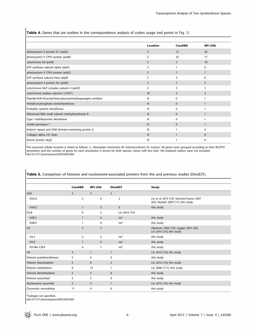
Table 4. Genes that are outliers in the correspondence analysis of codon usage (red points in Fig. 1).
Location CassKB8 Mf1.05b
photosystem II protein D1 (psbA) C 12 20
photosystem II CP47 protein (psbB) C 25 17
cytochrome b6 (petB) C 3 10
ATP synthase subunit alpha (atpA) C 1 9
photosystem II CP43 protein (psbC) C 7 7
ATP synthase subunit beta (atpB) C 5 6
photosystem II protein D2 (psbD) C 1 5
cytochrome b6/f complex subunit 4 (petD) C 5 3
cytochrome oxidase subunit I (COX1) M 3 2
Peptide-N(4)-(N-acetyl-beta-glucosaminyl)asparagine amidase N 0 1
Histidinol-phosphate aminotransferase N 0 1
Probable cysteine desulfurase N 0 1
Ribosomal RNA small subunit methyltransferase B N 0 1
Type I iodothyronine deiodinase N 0 1
Ureide permease 1 N 0 1
Ankyrin repeat and SAM domain-containing protein 6 N 1 0
Collagen alpha-1(I) chain N 1 0
Sensor protein degS N 1 0
The assumed cellular location is noted as follows: C, chloroplast minicircles; M, mitochrondrium; N, nucleus. All genes were grouped according to their BLASTXannotation and the number of genes for each annotation is shown for both species. Genes with less than 100 analyzed codons were not included.doi:10.1371/journal.pone.0035269.t004
Table 5. Comparison of histones and nucleosome-associated proteins from this and previous studies (DinoEST).
CassKB8 Mf1.05b DinoEST Study
H2A 3 3 2
H2A.X 2 0 2 Lin et al 2010 [14]; Sanchez-Puerta 2007[63]; Hackett 2005 [11]; this study
H2A.Z 1 3 0 this study
H2.B 0 2 Lin 2010 [14]
H2B.2 1 0 na* this study
H2B.4 1 0 na* this study
H3 3 3 Okamoto 2003 [10]; Leggat 2007 [26];Lin 2010 [14]; this study
H3.3 2 2 na* this study
H3.4 1 0 na* this study
H3-like CSE4 0 1 na* this study
H4 3 1 1 Lin 2010 [14]; this study
Histone acetyltransferases 2 4 0 this study
Histone deacetylation 5 8 2 Lin 2010 [14]; this study
Histone methylation 9 15 1 Lin 2008 [117]; this study
Histone demethylation 5 5 0 this study
Histone associated 3 2 0 this study
Nucleosome assembly 2 3 1 Lin 2010 [14]; this study
Chromatin remodeling 11 9 0 this study
*Subtype not specified.doi:10.1371/journal.pone.0035269.t005
Transcriptome Analysis of Two Symbiodinium Species
PLoS ONE | www.plosone.org 6 April 2012 | Volume 7 | Issue 4 | e35269

Transcriptome Analysis of Two Symbiodinium Species
PLoS ONE | www.plosone.org 7 April 2012 | Volume 7 | Issue 4 | e35269

different lineages, including plants, animals, ciliates and apicom-
plexans [64].
Apart from the nucleosome core histones, we identified a variety
of histone-modifying proteins including histone acetyltransferases,
deacetylases, methylases, and demethylases as well as several
nucleosome assembly and histone binding proteins in both species
(Table 5). Furthermore, we found the histone-associated chaper-
one ASF1 in CassKB8 and the Chromatin assembly factor 1
(CAF1) in Mf1.05, which have important roles in chromatin
transactions [67,68]. We found more histone-modifying genes in
Mf1.05b than in CassKB8, 32 and 21 genes, respectively. Histone
methylases appear to be the most common type of histone-
modifying proteins in both species, followed by deacetylases and
demethylases (Table 5).
Transcription factors in SymbiodiniumWhile histones take part in gene regulation at the genome level,
the most important proteins that influence transcription of
individual genes are transcription factors (TFs). We found a low
number of such domains in Symbiodinium. In the whole dataset,
only 156 and 87 genes contained at least one known protein
domain for sequence-specific DNA-binding activity in CassKB8
and Mf1.05b, respectively. These numbers correspond to only
0.27% and 0.15% of all genes (as determined from clusters at the
90% similarity level) (Table 6). A similar result was obtained when
the same analysis was conducted on the collection of all
dinoflagellate sequences available in the Genbank EST database
dbEST, with a percentage of 0.29% of all clustered EST sequences
containing at least one transcription factor domain.
Not only is the overall number of TF domains low, but the
distribution of domains was also different than in other organisms.
For instance, Zinc finger C2H2 domain TFs, which make up the
largest fraction of TFs in many eukaryotes such as human and
Drosophila, were completely absent from the dinoflagellate
sequences analyzed here (Suppl. Figure S1). The distribution of
the most common TF domains is distinct from the apicomplexans
P. falciparum and P. vivax, the ciliate P. tetraurelia, the heterokont
diatom T. pseudonana, the plant A. thaliana, and the insect D.
melanogaster as well as from human (Figure 3). The most common
domain in Symbiodinium was the ‘cold shock factor’ DNA-binding
domain, making up more than 60% of the transcription factor
domains of CassKB8 and Mf1.05b. This domain is a b-barrel
domain present in most organisms from all three domains of life.
This type of transcription factor also appeared to be among the
most common in all dinoflagellates as assessed from dbEST
(Figure 3). This domain also occurs in all non-dinoflagellate species
studied, though only a few genes contained it (Suppl. Table S6).
Antioxidative responseGiven the importance of Reactive Oxygen Species (ROS) in the
bleaching-associated breakdown of the symbiotic relationship
between Symbiodinium and their coral host, we screened our data
for genes associated with the antioxidative response. We used a
Pfam protein domain-based approach to assess the antioxidant
gene repertoire in both Symbiodinium species as well as in four
photosynthetic outgroup taxa for which whole genome data were
available, namely the land plant A. thaliana, the bryophyte P. patens,
and the diatoms T. pseudonana and P. tricornutum. We chose plant
species for the comparative analysis because land plants are known
to possess an efficient antioxidant enzymatic machinery, which
allows them to deal with extreme climates and stresses [69,70]. T.
pseudonana and P. tricornutum, in turn, represented outgroup species
more closely related to dinoflagellates that share a similar marine
lifestyle with dinoflagellates.
The Symbiodinium transcriptomes encoded higher numbers of
some proteins involved in the antioxidative response when
compared to plants and diatoms, specifically, those containing
the Nickel-containing SODs (Sod_Ni), Thioredoxin (Trx), and
glutaredoxin 2 (Grx2) domains (Table 7). Interestingly, in contrast
to plants, CassKB8 and Mf1.05b possess Sod_Ni, which are
common in prokaryotes. Four of the Sod_Ni encoding genes in
CassKB8 (kb8_rep_c6308, kb8_rep_c17584, kb8_rep_c1869 and
kb8_rep_c6458) and five in Mf1.05b (mf105_rep_c13460,
mf105_rep_c40857, mf105_rep_c42288, mf105_rep_c543 and
mf105_s69277) were annotated as being Ubiquitin orthologs
based on BLASTX. A protein domain analysis confirmed that
these genes encoded both a conserved Sod_Ni and an ubiquitin
domain. Due to this unexpected result, we searched our sequences
against the NCBI nr and dbEST using BLAST to analyze whether
this domain composition was restricted to a certain set of species.
We found genes encoding both domains in eukaryotic lineages
such as the stramenopiles Phaeodactylum tricornutum
Table 6. Number of transcription factor domains found in Symbiodinium genes (based on 90% similarity clustering of contigs andsinglets) and of all dinoflagellate ESTs available in Genbank dbEST.
CassKB8 Mf1.05b All dino ESTs from dbEST
No. of genes with transcription factor domain 156 87 272
Total no. of genes with Pfam annotation 18,564 13,495 24,098
% contigs with transcription factor domains of all Pfam annotated 0.84 0.64 1.13
Total no. of clusters 57,676 56,198 92,308
% contigs with transcription factor domains of all clusters/genes 0.27 0.15 0.29
doi:10.1371/journal.pone.0035269.t006
Figure 2. Phylogenetic analysis of histone sequences. H2A- and H3-like sequences from Symbiodinium sp. CassKB8, Mf1.05b, and otherorganisms were used to calculate phylogenetic trees. The trees were inferred using contigs and singlets with full-length amino acid sequences of (A)H2A and (B) H3-like genes using Maximum-Likelihood and Bayesian analysis. Bootstrap values and posterior probabilities are provided as ML/MB fornodes with support above 50% or 0.5. The singlet and contig names are provided for Symbiodinium sp. CassKB8 and Mf1.05b sequences (in bold),other taxa are shown as species name followed by GenBank accession number. The H2 tree was rooted for H2A.Bbd sequences whereas the H3 treewas rooted for Homo sapiens H3.doi:10.1371/journal.pone.0035269.g002
Transcriptome Analysis of Two Symbiodinium Species
PLoS ONE | www.plosone.org 8 April 2012 | Volume 7 | Issue 4 | e35269

(XP_002183736), Chaetoceros neogracile (EL622395) and Aureococcus
anophagefferens (EGB03009), as well as in different dinoflagellate
species including Karlodinium brevis (EX871806), Karlodinium micrum
(EC161447), Karlodinium veneficum (GH269044), and Heterocapsa
triquetra (EU153190) where these genes appear to be common. The
only eukaryotic genes outside the chromalveolates displaying this
domain signature were found in Micromonas sp. (XM_002506486,
XM_003063226), and none were found in prokaryotes.
The thioredoxin (Trx) superfamily comprises different groups of
proteins that share a common structural motif. These include
thioredoxins (Trx) and protein disulfide isomerases (PDI) as well as
glutathione peroxidases (GSHPx) and glutaredoxins, the last two
of which are represented separately in this study and are therefore
not addressed as Trx here. Comparison of genes encoding putative
Trx domains across the six species analyzed here revealed an
unexpected high number of genes in both Symbiodinium species
(Table 7). In CassKB8 we identified a total of 106 genes encoding
a Trx domain, which is substantially higher than what is found in
the plants Arabidopsis thaliana and Physcomitrella patens (79 and 70),
while 73 putative Trx genes were identified in Mf1.05b. This result
is in stark contrast to the comparably low number in the diatoms
Thalassiosira pseudonana and Phaeodactylum tricornutum, where only 55
and 41 Trx domain encoding genes appear to be present in the
genomes (Table 7).
Table 7. Comparison of the antioxidant gene repertoire between Arabidopsis thaliana, Phycomitrella patens, Symbiodinium sp.CassKB8, Symbiodinium sp. Mf1.05b, Thalassosira pseudonana, and Phaeodactylum tricornutum based on Pfam domains associatedwith antioxidant function.
Function Type PFAM A. thaliana P. patens CassKB8 Mf1.05b T. pseudonana P. tricornutum
Sod_Cu Superoxide dismutase PF00080.14 4 7 3 0 0 1
Sob_Fe_N Superoxide dismutase PF00081.16 5 4 4 2 4 2
Sod_Fe_C Superoxide dismutase PF02777.12 5 3 5 6 3 2
Sod_Ni Superoxide dismutase PF09055.5 0 0 5 10 0 1
Catalase Catalase PF00199.13 3 7 0 0 0 1
Peroxidase Peroxidase PF00141.17 82 65 27 24 16 10
GSHPx Glutathione peroxidase PF00255.13 9 4 5 1 2 3
Thioredoxin Thioredoxin PF00085.14 79 70 106 73 55 41
Glutaredoxin Glutathione reductase PF00462.18 52 28 29 17 13 11
Ferritin Ferritin PF00210.18 6 4 2 0 0 1
1-cysPrx_C peroxiredoxin PF10417.3 3 4 4 2 1 2
Glutaredoxin2_C Glutaredoxin2_C PF04399.7 0 0 2 5 1 1
AhpC-TSA Alkylhydroperoxide reductase PF00578.15 45 43 28 18 19 22
doi:10.1371/journal.pone.0035269.t007
Figure 3. Transcription factor domain composition. The relative fraction of the most abundant transcription factor domains in theSymbiodinium transcriptomes, all dinoflagellate ESTs from the NCBI dbEST database, and other eukaryotes. Searches were performed by usingHMMER to search domain models for DNA binding domains, with an e-value cutoff of , = 1e26. Domains which make up less than 5% were groupedin the ‘others’ category.doi:10.1371/journal.pone.0035269.g003
Transcriptome Analysis of Two Symbiodinium Species
PLoS ONE | www.plosone.org 9 April 2012 | Volume 7 | Issue 4 | e35269

Discussion
Assembly and completenessThe sequence data reported in this study comprises the largest
transcriptome of dinoflagellates to date, and surpasses the available
number of dinoflagellate sequences currently in public databases.
The number of genes that can be estimated from the data is
around 56,000, more than twice the number of genes predicted for
the human genome. However, this number is not too far from the
40,000 estimated genes in Symbiodinium genomes, based on genome
size and its correlation with gene number [13]. One caveat with
shotgun sequenced transcriptome data is the possibility of
assembling fragmented transcripts. These would artificially
increase the gene count, a general problem that is hard to
evaluate in a transcriptome where most genes do not have
orthologs in fully sequenced genomes. However, our gene number
estimate is similar to an estimate based on Illumina sequencing
data with much higher coverage (unpublished data), which yields
more than 43,000 genes of 500 bp and larger. Furthermore, it is
known that dinoflagellates have large gene families with some very
closely related members [71,72]. Such closely related genes may
be grouped together in the clustering process performed for this
study. In the example tested here, the actin gene family in
CassKB8 has 36 contigs and singlets as members, which were
grouped into only 14 clusters. Thus, this method makes our gene
number estimate more conservative.
As found in other dinoflagellate sequencing data sets, the
majority of transcripts do not have similarity to sequences in
GenBank or KEGG [11,14,27,60]. This novelty could be expected
of an organism which is evolutionary distant from most model
organisms. Most of the KEGG-based annotations (10–15% of all
genes) fall into the metabolic pathways category. The complete-
ness analysis showed that the majority of the standard ‘‘house-
keeping’’ genes in the pathways and complexes are present. The
difference between the two species, namely the lower coverage in
Mf1.05b, probably arose due to the differences in the sequencing
read length, which influences the assembly process and may reflect
the different sequencing library generation protocols. In addition,
all but two of the genes that belong to universal single-copy gene
families were found. Ribosomal protein S8 (COG0096) and signal
recognition particle GTPase (COG0552) were not identified in
either of the two species. Although this absence is not conclusive
without the availability of completely sequenced genomes, it will
be interesting to see whether and when a lineage-specific loss of
these gene families has occurred.
The genus Symbiodinium is comprised of a large number of
species encompassed in nine major lineages (clades A–I) [17,73].
Symbiodinium spp. are crucial components of coral reef ecosystems
as endosymbionts of corals and other marine invertebrates.
However, few analyses exist which identify genes that might play
a role in physiological differences, e.g. susceptibility to bleaching of
the different symbiont species or clades [26,27]. Using two
Symbiodinium species, it becomes possible to conduct an evolution-
ary screen for candidate adaptive genes involved in symbiosis as
has been recently conducted for two coral species – i.e. Acropora
millepora and A. palmata [74,75]. Identifying adaptively evolving
genes – via the ratio of the relative rates of synonymous and
nonsynonymous substitutions (dN/dS) of ortholog genes, [76,77]
can be a powerful strategy to narrow gene lists to a few candidates.
However, the synonymous nucleotide changes per synonymous
site (dS) we calculated for the two species analyzed here far
exceeded 1 (avg dS 26.3 as estimated by PAML, a value that gets
increasingly inaccurate if dS.1), indicating that multiple substitu-
tions may have occurred at a single site. For this reason, we did not
conduct an evolutionary analysis. Nonetheless, having the benefit
of two transcriptome data sets that were sequenced at similar
depths, we were able to independently confirm all of our findings
in both Symbiodinium species. In this regard, our estimate of gene
numbers, the paucity of transcription factors as estimated from
DNA binding domains, the presence of full sets of histones, etc.
seem to be general hallmarks of Symbiodinium biology rather than
clade- or species-specific adaptations. Many of these questions will
be answered more definitively in the near future as several whole
genome Symbiodinium sequencing projects are currently underway.
Codon usageWe estimated the GC content of the third codon position from a
large number of codons in our dataset. GC3 has been found to be
a good predictor of the overall genomic GC content [78]. EST
data for dinoflagellates in the literature already suggest that GC
content in the third codon position is highly variable, from 50% to
77% [11,59,60]. It is surprising, however, that we find a large
difference of about 14% in GC3 also in two species within the
genus Symbiodinium. It is usually assumed that differences in GC
content between species stem from genome wide mutational bias
[79]. If this is true, and if GC richness is homogeneous across the
genome, then it stands to reason that there are indeed different
mutational mechanisms at work across even closely related
dinoflagellate species.
GC content also influences codon usage bias, in addition to a
range of other factors such as mutational bias, selective pressures
depending on expression strength, abundance of tRNA genes, and
environmental factors, and can also be a means of transcriptional
control (see [80] for review). Both Symbiodinium species have a more
relaxed codon bias than A. tamarense. Codon usage bias is often
coupled with growth rate, as not all tRNAs are present in the cell
in equal amounts [81]. Accordingly, the variable codon bias
detected between Symbiodinium sp. CassKB8 and Mf1.05b may
reflect physiological differences (e.g. growth rate, thermal
tolerance).
The effective number of used codons is mostly dependent on the
overall GC value in the coding sequences. It is shifted towards
lower values in Mf1.05b, as this strain has a lower overall GC
content. Overall the picture looks similar to the one found
previously for A. tamarense [59], where the majority of genes are
below the Nc expected for neutral evolution of codon usage. This
suggests that these genes may underlie selective processes that
favor certain codons, as is known from highly expressed genes.
The correspondence analysis of codon usage (Figure 1 C, D) shows
a group of genes that is separated from the main cloud, almost all
of these are encoded in the chloroplast and mitochondrial
genomes. The chloroplast genome in dinoflagellates deviates from
the organization that is found in most plants and algae; many
genes seem to have transferred to the nucleus, and the genes that
are still present in the chloroplast are encoded on several small
DNA molecules, termed minicircles, that contain only one or a few
genes (see Lila Koumandou et al. [82] for review). Codon usage in
these genes has been found to be different than in nuclear genes,
and some contain unusual start codons [62]. Most of the genes that
are known to reside on minicircles are grouped together with those
genes whose codon usage deviates from the norm. However, none
of the chloroplast genes that have been found to be transferred to
the nucleus in dinoflagellates are in that group. This supports the
data presented in the literature so far, and makes it unlikely that
more genes remain encoded in the chloroplast minicircles than
currently known for peridinin-containing dinoflagellates, including
the Symbiodinium species analyzed here.
Transcriptome Analysis of Two Symbiodinium Species
PLoS ONE | www.plosone.org 10 April 2012 | Volume 7 | Issue 4 | e35269
Histones and Nucleosome-Associated ProteinsUntil now, no complete set of the nucleosome core histones has
been identified in a single dinoflagellate species. Here, we report
the complete set of nucleosome core histones as well as the minor
histones H2A.Z and several H3 variants in a single dinoflagellate
species (Symbiodinium sp. CassKB8). Our results indicate that
dinoflagellates possess not only a basic nucleosome machinery, but
also specialized histones that are known to be involved in
transcriptional and epigenetic regulation, e.g. H2A.Z [83–85]
and H3.3 [64,86,87].
In contrast to the histones H2B and H4, the histones H2A and
H3 have highly conserved ubiquitously expressed variants with
specialized functions. H2A.Z is associated with the promoter
region of actively transcribed genes linked to transcriptional
competence [83,84], and is also involved in epigenetic regulation
[85]. Studies in A. thaliana have shown that DNA methylation and
H2A.Z incorporation are mutually exclusive. DNA methylation
plays a pivotal role in establishing and maintaining an inactive
state of a gene, suggesting that the transcriptional activity
promoted by H2A.Z might be conferred through the inhibition
of DNA methylation [85]. This is in agreement with studies on
Amphidinium carterae and Symbiodinium microadriaticum, whose ge-
nomes appear to be hypermethylated [88]. Furthermore, it has
been shown that the methylation status of important photosyn-
thesis genes is correlated with their transcriptional activity [89].
Hence, dinoflagellates might be able to use the nucleosome
machinery for transcriptional regulation through the regulation of
the methylation status of specific loci. The variation we observed
in Symbiodinium H3-like proteins is also suggestive of the role of the
nucleosome machinery in transcriptional regulation, with possible
subfunctionalization of the multiple variants. Finally, we found
that Symbiodinium species contain various genes for the modification
of histones including methylation and acetylation as well as
orthologs of the histone-specific chaperones ASF1 and CAF1.
ASF1 is involved in the modulation of local chromatin structure
during gene-selective silencing in Drosophila [67], whereas CAF1 is
mainly associated with processes involving DNA, such as DNA
replication and DNA repair [68].
Overall, our results provide corroborating evidence for the
presence of a functional nucleosome machinery in dinoflagellates.
We found specialized histones and histone-associated proteins that
are known to be involved in transcriptional and epigenetic
regulation. Given that histones appear to be rare in the
dinoflagellates nucleus [14,90], one might speculate that dinofla-
gellates may employ the nucleosome machinery for transcriptional
regulation rather than chromatin packaging. In that case, one
would expect that the transcriptional regulation of genes is mainly
regulated at the level of accessibility thereby generically switching
transcription on or off. Consequently, activated loci would likely
display very similar transcription levels. Previous studies have
shown that transcriptional levels across many genes indeed appear
to be similar [14,91,92].
Transcription factors in SymbiodiniumThe unusual chromatin structure, low concentration of proteins
in the nucleus, and the very large genomes of dinoflagellates raise
the question whether gene regulation is realized with the same
mechanisms as in other eukaryotes. Transcriptional regulation
might play a minor role in dinoflagellates as opposed to other
mechanisms of regulation. Here, we analyzed the number and
composition of transcription factors in Symbiodinium to get a better
understanding of gene regulation. We identified only a small
number of proteins with sequence specific nucleic acid binding
domains (i.e. putative transcription factors). Transcription factors
have been shown to scale with genome size [93], and make up 6–
9% of all genes of higher eukaryotic transcriptomes (Suppl. Table
S7). The percentages found here for dinoflagellates are much
lower than those for other protists such as Plasmodium, even though
Plasmodium has a reduced genome due to its parasitic lifestyle.
The assemblage of transcription factors in Symbiodinium seems to
be completely different from other eukaryotes; common domains
such as zinc fingers, helix loop helix, AP2, or homeobox domains
are rare or absent. This is also true for the other dinoflagellates
represented in the set of ESTs analyzed here, as the set of
transcription factor domains and their abundances are quite
similar to those found in Symbiodinium. This low abundance of
transcription factors appears to be a genomic signature of the
dinoflagellate clade.
A similar conclusion about an apparent low number of
transcription factors in P. falciparum and apicomplexans in general
[94,95] was challenged through the discovery of the ApiAP2
family of transcription factors in this group [49]. The 27 members
of the ApiAP2 family in P. falciparum fill the gap of the apparent
lack of transcription factors, and many have now been investigated
in detail and shown to be conserved the Apicomplexa [96]. It
seems plausible that dinoflagellates may also contain yet
undescribed transcription factor families that would represent
part of the ‘‘missing’’ transcriptional regulatory machinery. Thus,
it is interesting that more than 60% of the putative transcription
factors identified carry a ‘cold shock’ domain (CSD). This domain
is not very common in other eukaryotes, suggesting a lineage-
specific expansion in dinoflagellates. Such lineage-specific expan-
sions of different transcription factor domains have been found in
multiple taxa throughout the tree of life [97,98]. Originally
identified as a reaction to cold shock in E. coli, proteins with CSD
domains have now been associated with a wide range of functions.
They can act as transcription factors by binding DNA (i.e. Y-box
factors), but many interact with RNA rather than DNA. They are
involved in regulation of transcription, splicing, and translation,
and influence mRNA stability as RNA chaperones (see [99] for
review). This observation fits with the notions that 1) regulation in
dinoflagellates may take place after transcription, and 2) that RNA
editing is widespread [100]. Thus, proteins with cold shock
domains may be responsible for much of the transcriptional
regulation in dinoflagellates. Considering that Symbiodinium under-
goes a dramatic change in its environment and lifestyle upon
entering invertebrate hosts, a need for efficient regulation of a
large number of genes might be advantageous. However, as all
data gathered here are based on Symbiodinium grown in cultures, it
is possible that more or different types of transcription factors are
expressed in the symbiotic state. As Symbiodinium genomes are
currently being sequenced, this question can be conclusively
answered in the near future as the genome sequence becomes
available.
Antioxidative responseThe impact of ROS on the symbiosis of Symbiodinium and its
marine invertebrate hosts is likely to affect mechanisms to cope
with photosynthesis-generated ROS in order to prevent the
breakdown of the symbiotic relationship. Our analysis shows that
Symbiodinium also possesses a rich antioxidant gene repertoire, but
surprisingly appears to lack or transcribe below detection limit the
enzyme catalase (note that catalase activity has been shown by
Merle et al. [101]), one of the central enzymes in eukaryotic
cellular redox-chemistry. In contrast, catalase was among the most
abundant transcripts in Aiptaisa pallida, an anemone host for
Symbiodinium [102]. However, since the transcriptome sequences
analyzed here were derived from cultured zooxanthellae it cannot
Transcriptome Analysis of Two Symbiodinium Species
PLoS ONE | www.plosone.org 11 April 2012 | Volume 7 | Issue 4 | e35269
be excluded that the catalase gene is only expressed in hospite. One
of the main differences between Symbiodinium and diatoms or plants
is the presence of several prokaryotic Ni-type SODs in both
Symbiodinium species, which were not present in the plant species
and were only represented by a single gene in the diatom T.
pseudonana. The presence of bacterial proteins is not surprising
given that lateral gene transfer between prokaryotes and
eukaryotes, especially protists, is common [103,104]. Furthermore,
several genes of bacterial origin have already been identified in
Symbiodinium [26]. To our surprise, we found that some of the Ni-
type SOD genes identified here also encode an additional
ubiquitin domain. The ubiquitin domain is a 76 amino acid
domain found in eukaryotes, whereas the SOD_Ni domain is
supposedly of prokaryotic origin [105], suggesting that these
transcripts might represent fusions of prokaryotic and eukaryotic
genes.
In contrast to heterotrophic organisms, plants contain a large
family of Trx and Trx-like proteins, although the reasons for their
abundance remain unclear [106,107]. We found that Symbiodinium
species appear to possess an unexpectedly high number of Trx
domain encoding genes comparable to plants, and in stark contrast
to the substantially smaller number found in diatoms. The Trx
superfamily proteins fulfill diverse cellular functions. These include
the maintenance of cell homeostasis and the regulation of the
redox state of the cell [108,109]. They play key roles in the
oxidative stress response [110,111] and have been shown to be
differentially expressed in response to high temperature, salinity,
and ultraviolet radiation in corals [28,112,113]. Members of these
groups are also involved in photosystem repair in response to ROS
in plants and algae [114,115]. Although the function of the large
number of Trx containing genes can currently not be assessed, this
high abundance might be indicative of a complex redox regulatory
system in Symbiodinium as in plants [116]. The general similarity of
the antioxidant gene repertoire of Symbiodinium and plants suggests
that Symbiodinium might well be adapted to high oxidative stress.
The comparably low amount of antioxidant genes found in both
diatom species, which supposedly share more similarities with
dinoflagellates than plants, raises the question whether Symbiodi-
nium might have evolved a richer repertoire of antioxidant genes as
an adaptation to its symbiotic lifestyle. More comprehensive
transcriptome studies in other dinoflagellates are necessary to
determine whether the features revealed in this study are specific
to Symbiodinium or common among dinoflagellates.
This study greatly enhances the available sequence data for
dinoflagellates in general, and Symbiodinium in particular. Our data
highlight interesting aspects of the genetics of Symbiodinium, and
provides the basis for deeper insights into dinoflagellate biology.
Supporting Information
Figure S1 Relative fraction of all transcription factor domains in
the Symbiodinium transcriptomes, all Dinoflagellate ESTs from the
NCBI database, and other eukaryotes. Values shown were arcsine
transformed. Searches were performed by using HMMER to
query the Pfam models for DNA binding domains with an e-value
cutoff of 1e26.
(PDF)
Table S1 Cluster numbers and contigs and singlets contained in
the clusters.
(XLS)
Table S2 Contigs and singlets of the actin gene family and their
grouping into contigs (90% similarity).
(XLS)
Table S3 Histone genes and sequences.
(XLS)
Table S4 Distribution of contigs and singlets among KEGG
categories and subcategories.
(XLS)
Table S5 Universal single-copy clusters of orthologous groups
(COGs) that were used for transcriptome completeness analysis in
this study. COGs not identified in Symbiodinium transcriptomes are
boldfaced.
(XLS)
Table S6 Number of transcription factor domains found in
Symbiodinium and other eukaryotes.
(XLS)
Table S7 Transcription factor domain counts in Symbiodinium
CassKB8 and Mf1.05b, all dinoflagellate ESTs, Plasmodium
falciparum and P. vivax, Paramecium tetraurelia, Thalassiosira pseudonana,
Arabidopsis thaliana, Drosophila melanogaster and human.
(XLS)
Acknowledgments
We would like to thank Robert A. Kinzie III for initial donation of the
Symbiodinium sp. CassKB8 culture, the Aquarium of Niagara for seawater
and three anonymous reviewers for valuable suggestions.
Author Contributions
Conceived and designed the experiments: TB MA SS CRV MM.
Performed the experiments: TB MA CRV MM MAC LKY EL. Analyzed
the data: TB MA SS CRV. Contributed reagents/materials/analysis tools:
MKD MAC. Wrote the paper: TB MA CRV.
References
1. Muscatine L, McCloskey LR, Marian RE (1981) Estimating the daily
contribution of carbon from zooxanthellae to coral animal respiration.
Limnology and Oceanography 26: 601–611.
2. Graham LE, Wilcox L (2000) Algae (vol 36, pg 788, 2000). Journal of
Phycology 36: 975–975.
3. Blank RJ, Huss VAR, Kersten W (1988) Base composition of DNA from
symbiotic dinoflagellates: a tool for phylogenetic classification. Archives of
Microbiology 149: 515–520.
4. Rae PMM (1976) Hydroxymethyluracil in eukaryote DNA: A natural feature
of the pyrrophyta (Dinoflagellates). Science 194: 1062–1064.
5. Veldhuis MJW, Cucci TL, Sieracki ME (1997) Cellular DNA content of
marine phytoplankton using two new fluorochromes: taxonomic and ecological
implications. Journal of Phycology 33: 527–541.
6. LaJeunesse TC, Lambert G, Andersen RA, Coffroth MA, Galbraith DW
(2005) Symbiodinium (pyrrhophyta) genome sizes (DNA content) are smallest
among dinoflagellates. Journal of Phycology 41: 880–886.
7. Dodge J (1966) The Dinophyceae. In: Godward MBE, ed. The chromosomes
of the algae. New York,: St. Martin’s Press. ix p212 p.
8. Bouligand Y, Norris V (2001) Chromosome separation and segregation indinoflagellates and bacteria may depend on liquid crystalline states. Biochimie
83: 187–192.
9. Rizzo P (1987) Biochemistry of the dinoflagellate nucleus. In: Taylor F, ed. TheBiology of dinoflagellates. Oxford: Blackwell Scientific Publications.
10. Okamoto OK, Hastings JW (2003) Novel dinoflagellate clock-related genes
identified through microarray analysis. Journal of Phycology 39: 519–526.
11. Hackett J, Scheetz T, Yoon H, Soares M, Bonaldo M, et al. (2005) Insights intoa dinoflagellate genome through expressed sequence tag analysis. BMC
Genomics 6: 80.
12. Lin S (2011) Genomic understanding of dinoflagellates. Research in
Microbiology 162: 551–569.
13. Hou Y, Lin S (2009) Distinct gene number-genome size relationships foreukaryotes and non-eukaryotes: gene content estimation for dinoflagellate
genomes. PLoS ONE 4: e6978–e6978.
14. Lin S, Zhang H, Zhuang Y, Tran B, Gill J (2010) Spliced leader–basedmetatranscriptomic analyses lead to recognition of hidden genomic features in
dinoflagellates. Proceedings of the National Academy of Sciences 107:
20033–20038.
Transcriptome Analysis of Two Symbiodinium Species
PLoS ONE | www.plosone.org 12 April 2012 | Volume 7 | Issue 4 | e35269

15. Zhang H, Hou Y, Miranda L, Campbell DA, Sturm NR, et al. (2007) Splicedleader RNA trans-splicing in dinoflagellates. Proceedings of the National
Academy of Sciences of the United States of America 104: 4618–4623.
16. Rowan R, Powers DA (1991) A molecular genetic classification of
zooxanthellae and the evolution of animal-algal symbioses. Science 251:
1348–1351.
17. Pochon X, Gates RD (2010) A new Symbiodinium clade (Dinophyceae) from
soritid foraminifera in Hawai’i. Molecular Phylogenetics and Evolution 56:492–497.
18. Pochon X, LaJeunesse TC, Pawlowski J (2004) Biogeographic partitioning andhost specialization among foraminiferan dinoflagellate symbionts (Symbiodi-
nium; Dinophyta). Marine Biology 146: 17–27.
19. Lajeunesse TC (2005) ‘‘Species’’ radiations of symbiotic dinoflagellates in theAtlantic and Indo-Pacific since the Miocene-Pliocene transition. Mol Biol Evol
22: 570–581.
20. Muscatine L, Falkowski PG, Porter JW, Dubinsky Z (1984) Fate of
photosynthetic fixed carbon in light- and shade-adapted colonies of the
symbiotic coral Stylophora pistillata. Proceedings of the Royal Society ofLondon Series B Biological Sciences 222: 181–202.
21. Harrison P, Wallace C (1990) Reproduction, dispersal and recruitment ofscleractinian corals. In: Dubinsky Z, ed. Ecosystems of the world: coral reefs.
Amsterdam; New York: Elsevier.
22. Schnitzler CE, Weis VM (2010) Coral larvae exhibit few measurable
transcriptional changes during the onset of coral-dinoflagellate endosymbiosis.
Marine Genomics 3: 107–116.
23. Weis VM (2008) Cellular mechanisms of Cnidarian bleaching: stress causes the
collapse of symbiosis. Journal of Experimental Biology 211: 3059.
24. Voolstra CR, Schwarz JA, Schnetzer J, Sunagawa S, Desalvo MK, et al. (2009)
The host transcriptome remains unaltered during the establishment of coral-algal symbioses. Molecular Ecology 18: 1823–1833.
25. Yuyama I, Watanabe T, Takei Y (2011) Profiling differential gene expression
of symbiotic and aposymbiotic corals using a high coverage gene expressionprofiling (HiCEP) analysis. Marine Biotechnology 13: 32–40.
26. Leggat W, Hoegh-Guldberg O, Dove S, Yellowlees D (2007) Analysis of anEST library from the dinoflagellate (Symbiodinium sp.) symbiont of reef-
building corals. Journal of Phycology 43: 1010–1021.
27. Voolstra CR, Sunagawa S, Schwarz JA, Coffroth MA, Yellowlees D, et al.
(2009) Evolutionary analysis of orthologous cDNA sequences from cultured
and symbiotic dinoflagellate symbionts of reef-building corals (Dinophyceae:Symbiodinium). Comparative Biochemistry and Physiology Part D: Genomics
and Proteomics 4: 67–74.
28. Desalvo MK, Voolstra CR, Sunagawa S, Schwarz JA, Stillman JH, et al.
(2008) Differential gene expression during thermal stress and bleaching in theCaribbean coral Montastraea faveolata. Molecular Ecology 17: 3952–3971.
29. Portune KJ, Voolstra CR, Medina M, Szmant AM (2010) Development and
heat stress-induced transcriptomic changes during embryogenesis of thescleractinian coral Acropora palmata. Marine Genomics 3: 51–62.
30. DeSalvo MK, Sunagawa S, Fisher PL, Voolstra CR, Iglesias-Prieto R, et al.(2010) Coral host transcriptomic states are correlated with Symbiodinium
genotypes. Molecular Ecology 19: 1174–1186.
31. Guillard RR, Ryther JH (1962) Studies of marine planktonic diatoms. I.Cyclotella nana Hustedt, and Detonula confervacea (cleve) Gran.
Can J Microbiol 8: 229–239.
32. Polne-Fuller M (1991) A novel technique for preparation of axenic cultures of
Symbiodinium (Pyrrophyta) through selective digestion by amoebae. Journal ofPhycology 27: 552–554.
33. Chevreux B, Pfisterer T, Drescher B, Driesel AJ, Muller WE, et al. (2004)
Using the miraEST assembler for reliable and automated mRNA transcriptassembly and SNP detection in sequenced ESTs. Genome Res 14: 1147–1159.
34. Ning Z, Cox AJ, Mullikin JC (2001) SSAHA: a fast search method for largeDNA databases. Genome Res 11: 1725–1729.
35. Edgar RC (2010) Search and clustering orders of magnitude faster thanBLAST. Bioinformatics 26: 2460–2461.
36. Watanabe T, Kii S-i, Tanaka J, Takishita K, Maruyama T (2006) cDNA
cloning and phylogenetic and expression analyses of actin in symbioticdinoflagellates (Symbiodinium spp.). Journal of Applied Phycology 18:
219–225.
37. Moriya Y, Itoh M, Okuda S, Yoshizawa AC, Kanehisa M (2007) KAAS: an
automatic genome annotation and pathway reconstruction server. NucleicAcids Research 35: 182–185.
38. Zdobnov EM, Apweiler R (2001) InterProScan – an integration platform for
the signature-recognition methods in InterPro. Bioinformatics 17: 847–848.
39. Boeckmann B, Bairoch A, Apweiler R, Blatter M-C, Estreicher A, et al. (2003)
The SWISS-PROT protein knowledgebase and its supplement TrEMBL in2003. Nucleic Acids Research 31: 365–370.
40. Wright F (1990) The ‘effective number of codons’ used in a gene. Gene 87:23–29.
41. Rice P, Longden I, Bleasby A (2000) EMBOSS: the European Molecular
Biology Open Software Suite. Trends in Genetics 16: 276–277.
42. Peden JF (1999) Analysis of Codon Usage: University of Nottingham.
43. Edgar RC (2004) MUSCLE: multiple sequence alignment with high accuracyand high throughput. Nucleic Acids Research 32: 1792–1797.
44. Tamura K, Peterson D, Peterson N, Stecher G, Nei M, et al. (2011) MEGA5:molecular evolutionary genetics analysis using maximum likelihood, evolu-
tionary distance, and maximum parsimony methods. Molecular Biology andEvolution.
45. Guindon S, Dufayard J-F, Lefort V, Anisimova M, Hordijk W, et al. (2010)New algorithms and methods to estimate maximum-likelihood phylogenies:
assessing the performance of PhyML 3.0. Systematic Biology 59: 307–321.
46. Ronquist F, Huelsenbeck JP (2003) MrBayes 3: bayesian phylogeneticinference under mixed models. Bioinformatics 19: 1572–1574.
47. Drummond A, Rambaut A (2007) BEAST: Bayesian evolutionary analysis bysampling trees. BMC Evolutionary Biology 7: 214.
48. Wilson D, Charoensawan V, Kummerfeld SK, Teichmann SA (2007) DBD–taxonomically broad transcription factor predictions: new content and
functionality. Nucleic Acids Research 36: D88-D92–D88-D92.
49. Balaji S, Babu MM, Iyer LM, Aravind L (2005) Discovery of the principalspecific transcription factors of Apicomplexa and their implication for the
evolution of the AP2-integrase DNA binding domains. Nucleic Acids Research33: 3994–4006.
50. Finn RD, Clements J, Eddy SR (2011) HMMER web server: interactivesequence similarity searching. Nucleic Acids Research 39: W29–W37.
51. Ryu T, Mavromatis C, Bayer T, Voolstra C, Ravasi T (2011) Unexpected
complexity of the reef-building coral Acropora millepora transcription factornetwork. BMC Systems Biology 5: 58–58.
52. Aurrecoechea C, Brestelli J, Brunk BP, Dommer J, Fischer S, et al. (2009)PlasmoDB: a functional genomic database for malaria parasites. Nucleic Acids
Research 37: D539-543-D539-543.
53. Arnaiz O, Cain S, Cohen J, Sperling L (2007) ParameciumDB: a communityresource that integrates the Paramecium tetraurelia genome sequence with
genetic data. Nucleic Acids Research 35: D439-D444-D439-D444.
54. Haider S, Ballester B, Smedley D, Zhang J, Rice P, et al. (2009) BioMart
Central Portal–unified access to biological data. Nucleic Acids Research 37:W23-27-W23-27.
55. Finn RD, Tate J, Mistry J, Coggill PC, Sammut SJ, et al. (2008) The Pfam
protein families database. Nucleic Acids Res 36: D281–288.
56. Reitzel AM, Sullivan JC, Traylor-Knowles N, Finnerty JR (2008) Genomic
survey of candidate stress-response genes in the estuarine anemoneNematostella vectensis. The Biological Bulletin 214: 233–233.
57. Kanehisa M, Goto S (2000) KEGG: Kyoto Encyclopedia of Genes andGenomes. Nucleic Acids Research 28: 27–30.
58. Ciccarelli FD, Doerks T, von Mering C, Creevey CJ, Snel B, et al. (2006)
Toward automatic reconstruction of a highly resolved tree of Life. Science 311:1283–1287.
59. Hsiao Y-Y, Lin C-H, Liu J-K, Wong T-Y, Kuo J (2010) Analysis of codonusage patterns in toxic dinoflagellate Alexandrium tamarense through
expressed sequence tag data. Comparative and Functional Genomics 2010:138538.
60. Lidie KB, Ryan JC, Barbier M, Van Dolah FM (2005) Gene expression in
Florida red tide dinoflagellate Karenia brevis: analysis of an expressedsequence tag library and development of DNA microarray. Marine
Biotechnology 7: 481–493.
61. Moore RB, Ferguson KM, Loh WKW, Hoegh-Guldberg O, Carter DA (2003)
Highly organized structure in the non-coding region of the psbA minicirclefrom clade C Symbiodinium. International Journal of Systematic and
Evolutionary Microbiology 53: 1725–1734.
62. Zhang Z, Green BR, Cavalier-Smith T (1999) Single gene circles indinoflagellate chloroplast genomes. Nature 400: 155–159.
63. Sanchez-Puerta MV, Lippmeier JC, Apt KE, Delwiche CF (2007) Plastid genesin a non-photosynthetic dinoflagellate. Protist 158: 105–117.
64. Malik HS, Henikoff S (2003) Phylogenomics of the nucleosome. Nat StructMol Biol 10: 882–891.
65. Gonzalez-Romero R, Ausio J, Mendez J, Eirin-Lopez JM (2008) Early
evolution of histone genes: prevalence of an ‘orphon’ H1 lineage inprotostomes and birth-and-death process in the H2A family. Journal of
molecular evolution 66: 505–518.
66. Pusarla R-H, Bhargava P (2005) Histones in functional diversification. FEBS
Journal 272: 5149–5168.
67. Moshkin YM, Kan TW, Goodfellow H, Bezstarosti K, Maeda RK, et al. (2009)
Histone chaperones ASF1 and NAP1 differentially modulate removal of active
histone marks by LID-RPD3 complexes during NOTCH silencing. MolecularCell 35: 782–793.
68. Ramirez-Parra E, Gutierrez C (2007) The many faces of chromatin assemblyfactor 1. Trends in Plant Science 12: 570–576.
69. Gupta SD (2010) Reactive oxygen species and antioxidants in higher plants.
Boca Raton, FL: Science Publishers; Distributed by CRC Press.
70. Gill SS, Tuteja N (2010) Reactive oxygen species and antioxidant machinery in
abiotic stress tolerance in crop plants. Plant Physiology and Biochemistry 48:909–930.
71. Kim S, Bachvaroff TR, Handy SM, Delwiche CF (2011) Dynamics of actinevolution in dinoflagellates. Mol Biol Evol 28: 1469–1480.
72. Moustafa A, Evans AN, Kulis DM, Hackett JD, Erdner DL, et al. (2010)
Transcriptome profiling of a toxic dinoflagellate reveals a gene-rich protist anda potential impact on gene expression due to bacterial presence. PLoS ONE 5:
e9688.
73. LaJeunesse TC (2001) Investigating the biodiversity, ecology, and phylogeny of
endosymbiotic dinoflagellates in the genus Symbiodinium using the ITS region:in search of a ‘‘species’’ level marker. Journal of Phycology 37: 866–880.
Transcriptome Analysis of Two Symbiodinium Species
PLoS ONE | www.plosone.org 13 April 2012 | Volume 7 | Issue 4 | e35269

74. Voolstra CR, Sunagawa S, Matz MV, Bayer T, Aranda M, et al. (2011) Rapid
evolution of coral proteins responsible for interaction with the environment.
PLoS ONE 6: e20392.
75. Iguchi A, Shinzato C, Foret S, Miller DJ (2011) Identification of fast-evolving
genes in the scleractinian coral Acropora using comparative EST analysis.
PLoS ONE 6: e20140.
76. Goldman N, Yang Z (1994) A codon-based model of nucleotide substitution for
protein-coding DNA sequences. Molecular Biology and Evolution 11:
725–736.
77. Nielsen R, Yang Z (1998) Likelihood models for detecting positively selected
amino acid sites and applications to the HIV-1 envelope gene. Genetics 148:
929–936.
78. Eyre-Walker A, Hurst LD (2001) The evolution of isochores. Nat Rev Genet 2:
549–555.
79. Hershberg R, Petrov DA (2008) Selection on Codon Bias. Annual Review of
Genetics 42: 287–299.
80. Plotkin JB, Kudla G (2011) Synonymous but not the same: the causes and
consequences of codon bias. Nat Rev Genet 12: 32–42.
81. Dong H, Nilsson L, Kurland CG (1996) Co-variation of tRNA abundance and
codon usage in Escherichia coli at different growth rates. Journal of Molecular
Biology 260: 649–663.
82. Lila Koumandou V, Nisbet RER, Barbrook AC, Howe CJ (2004)
Dinoflagellate chloroplasts - where have all the genes gone? Trends in
Genetics 20: 261–267.
83. Mavrich TN, Ioshikhes IP, Venters BJ, Jiang C, Tomsho LP, et al. (2008) A
barrier nucleosome model for statistical positioning of nucleosomes throughout
the yeast genome. Genome Res 18: 1073–1083.
84. Mavrich TN, Jiang C, Ioshikhes IP, Li X, Venters BJ, et al. (2008) Nucleosome
organization in the Drosophila genome. Nature 453: 358–362.
85. Zilberman D, Coleman-Derr D, Ballinger T, Henikoff S (2008) Histone H2A.Z
and DNA methylation are mutually antagonistic chromatin marks. Nature 456:
125–129.
86. Ahmad K, Henikoff S (2002) Histone H3 variants specify modes of chromatin
assembly. Proc Natl Acad Sci U S A 99 Suppl 4: 16477–16484.
87. Hake SB, Allis CD (2006) Histone H3 variants and their potential role in
indexing mammalian genomes: The ‘‘H3 barcode hypothesis’’. Proceedings of
the National Academy of Sciences 103: 6428–6435.
88. Lohuis MR, Miller DJ (1998) Hypermethylation at CpG-motifs in the
dinoflagellates Amphidinium carterae (Dinophyceae) and Symbiodinium
microadriaticum (Dinophyceae): evidence from restriction analyses, 5-azacy-
tidine and ethionine treatment. Journal of Phycology 34: 152–159.
89. Lohuis MR, Miller DJ (1998) Light-Regulated Transcription of Genes
Encoding Peridinin Chlorophyll a Proteins and the Major Intrinsic Light-
Harvesting Complex Proteins in the DinoflagellateAmphidinium carterae
Hulburt (Dinophycae). Plant Physiology 117: 189–196.
90. Kellenberger E, Arnold-Schulz-Gahmen B (1992) Chromatins of low-protein
content: special features of their compaction and condensation. FEMS
Microbiology Letters 79: 361–370.
91. Moustafa A, Evans AN, Kulis DM, Hackett JD, Erdner DL, et al. (2010)
Transcriptome profiling of a toxic dinoflagellate reveals a gene-rich protist and
a potential impact on gene expression due to bacterial presence. PLoS ONE 5:
9688.
92. Erdner D, Anderson D (2006) Global transcriptional profiling of the toxic
dinoflagellate Alexandrium fundyense using Massively Parallel Signature
Sequencing. BMC Genomics 7: 88.
93. van Nimwegen E (2003) Scaling laws in the functional content of genomes.
Trends in Genetics 19: 479–484.
94. Aravind L, Iyer LM, Wellems TE, Miller LH (2003) Plasmodium biology:
genomic gleanings. Cell 115: 771–785.
95. Iyer LM, Anantharaman V, Wolf MY, Aravind L (2008) Comparative
genomics of transcription factors and chromatin proteins in parasitic protists
and other eukaryotes. International Journal for Parasitology 38: 1–31.
96. Painter HJ, Campbell TL, Llinas M (2011) The Apicomplexan AP2 family:
integral factors regulating Plasmodium development. Molecular and Biochem-ical Parasitology 176: 1–7.
97. Aravind L, Watanabe H, Lipman DJ, Koonin EV (2000) Lineage-specific loss
and divergence of functionally linked genes in eukaryotes. Proceedings of theNational Academy of Sciences of the United States of America 97:
11319–11324.98. Lespinet O, Wolf YI, Koonin EV, Aravind L (2002) The Role of lineage-
specific gene family expansion in the evolution of eukaryotes. Genome
Research 12: 1048–1059.99. Mihailovich M, Militti C, Gabaldon T, Gebauer F (2010) Eukaryotic cold
shock domain proteins: highly versatile regulators of gene expression. BioEssays32: 109–118.
100. Lin S, Zhang H, Gray MW (2007) RNA editing in dinoflagellates and itsimplications for the evolutionary history of the editing machinery. Smith H, ed.
RNA and DNA Editing: Molecular Mechanisms and Their Integration into
Biological Systems: JohnWiley & Sons, Inc. pp 280–309.101. Merle PL, Sabourault C, Richier S, Allemand D, Furla P (2007) Catalase
characterization and implication in bleaching of a symbiotic sea anemone. FreeRadic Biol Med 42: 236–246.
102. Sunagawa S, Wilson EC, Thaler M, Smith ML, Caruso C, et al. (2009)
Generation and analysis of transcriptomic resources for a model system on therise: the sea anemone Aiptasia pallida and its dinoflagellate endosymbiont.
BMC Genomics 10: 258.103. Keeling PJ, Palmer JD (2008) Horizontal gene transfer in eukaryotic evolution.
Nat Rev Genet 9: 605–618.104. Andersson JO (2005) Lateral gene transfer in eukaryotes. Cellular and
Molecular Life Sciences 62: 1182–1197.
105. Schmidt A, Gube M, Schmidt A, Kothe E (2009) In silico analysis of nickelcontaining superoxide dismutase evolution and regulation. Journal of Basic
Microbiology 49: 109–118.106. Hisabori T, Motohashi K, Hosoya-Matsuda N, Ueoka-Nakanishi H,
Romano PGN (2007) Towards a functional dissection of thioredoxin networks
in plant cells. Photochemistry and Photobiology 83: 145–151.107. Chibani K, Wingsle G, Jacquot J-P, Gelhaye E, Rouhier N (2009)
Comparative Genomic Study of the Thioredoxin Family in PhotosyntheticOrganisms with Emphasis on Populus trichocarpa. Molecular Plant 2:
308–322.108. Eckardt NA (2007) Oxidation Pathways and Plant Development: Crosstalk
between Thioredoxin and Glutaredoxin Pathways. The Plant Cell Online 19:
1719–1721.109. Papp E, Nardai G, Soti C, Csermely P (2003) Molecular chaperones, stress
proteins and redox homeostasis. BioFactors 17: 249–257.110. Vieira Dos Santos C, Rey P (2006) Plant thioredoxins are key actors in the
oxidative stress response. Trends in Plant Science 11: 329–334.
111. Niwa T (2007) Protein glutathionylation and oxidative stress. Journal ofChromatography B 855: 59–65.
112. Aranda M, Banaszak AT, Bayer T, Luyten JR, Medina M, et al. (2011)Differential sensitivity of coral larvae to natural levels of ultraviolet radiation
during the onset of larval competence. Molecular Ecology 20: 2955–2972.113. Edge SE, Morgan MB, Gleason DF, Snell TW (2005) Development of a coral
cDNA array to examine gene expression profiles in Montastraea faveolata
exposed to environmental stress. Mar Pollut Bull 51: 507–523.114. Nishiyama Y, Allakhverdiev SI, Murata N (2011) Protein synthesis is the
primary target of reactive oxygen species in the photoinhibition of photosystemII. Physiologia Plantarum 142: 35–46.
115. Kim J, Mayfield SP (1997) Protein Disulfide Isomerase as a Regulator of
Chloroplast Translational Activation. Science 278: 1954–1957.116. Dietz K-J, Pfannschmidt T (2011) Novel regulators in photosynthetic redox
control of plant metabolism and gene expression. Plant Physiology 155:1477–1485.
117. Lin S, Zhang H (2008) Dinoflagellate meta-transcriptomics enabled by spliced
leader. In: Ho K, Zhou M, Qi Y, eds. Hong Kong: Environmental PublicationHouse Hong Kong. pp 166–170.
Transcriptome Analysis of Two Symbiodinium Species
PLoS ONE | www.plosone.org 14 April 2012 | Volume 7 | Issue 4 | e35269