Fundamental Clinical Brain MR Imaging Applications and ...mri/CE/slides/NeuroCETalkMRI.pdf ·...
21
9/8/2009 1 Continuing Education Seminar for Radiologic Technologists Fundamental Clinical Brain MR Imaging – Applications and Protocols Darren P. O’Neill, MD Indiana University Neuroradiology Objectives • Review fundamental clinical cases that illustrate the wide array and utility of available brain MR imaging techniques / applications • Gain insight into the rationale behind MR imaging protocols • Develop a greater understanding of potential points of patient care impact Outline • Review fundamental clinical cases that illustrate the wide array and utility of available brain MR imaging techniques: • Gradient echo • Diffusion • FLAIR • IR-SPGR / MP-RAGE • MRA / MRV • Spectroscopy • Perfusion • Review potential points of patient care impact: – Additional patient history? – Are the referring physician and/or neuroradiologist aware of the findings? – Would an additional study(s) or different protocol be better? – Would intravenous contrast be helpful? Brain – Screen • Indications – Screen, Altered mental status, Dementia, Psychiatric disorder, Headaches • Sequences – 3 PL LOC – Sag T1 SE – Sag T2 FLAIR – Ax T2 FLAIR – Ax T1 SE – Ax T2 TSE FS – Ax DWI EPI – Cor T2 TSE • Comments – Axial scans should be parallel to the AC-PC line 25 year-old pregnant female with mental status change Potential Points of Impact • Patient history? – Do we know more than “mental status change”? – Onset of symptoms? – Hypertension? Pregnancy? Steroids? • Sequences to consider anticipating? – MRI brain without contrast => evaluate for acute ischemia – Avoid contrast with pregnancy!
Transcript of Fundamental Clinical Brain MR Imaging Applications and ...mri/CE/slides/NeuroCETalkMRI.pdf ·...

9/8/2009
1
Continuing Education Seminar for Radiologic Technologists
Fundamental Clinical Brain MR Imaging – Applications and
Protocols
Darren P. O’Neill, MD
Indiana University Neuroradiology
Objectives
• Review fundamental clinical cases that illustrate the wide array and utility of available brain MR imaging techniques / applications
• Gain insight into the rationale behind MR imaging protocols
• Develop a greater understanding of potential points of patient care impact
Outline• Review fundamental clinical cases that illustrate the wide array and
utility of available brain MR imaging techniques:• Gradient echo• Diffusion• FLAIR• IR-SPGR / MP-RAGE• MRA / MRV• Spectroscopy• Perfusion
• Review potential points of patient care impact:– Additional patient history?– Are the referring physician and/or neuroradiologist aware of the
findings?– Would an additional study(s) or different protocol be better?– Would intravenous contrast be helpful?
Brain – Screen
• Indications– Screen, Altered mental status, Dementia, Psychiatric disorder,
Headaches
• Sequences– 3 PL LOC– Sag T1 SE– Sag T2 FLAIR– Ax T2 FLAIR– Ax T1 SE– Ax T2 TSE FS– Ax DWI EPI– Cor T2 TSE
• Comments– Axial scans should be parallel to the AC-PC line
25 year-old pregnant female with mental status change
Potential Points of Impact
• Patient history?
– Do we know more than “mental status change”?
– Onset of symptoms?
– Hypertension? Pregnancy? Steroids?
• Sequences to consider anticipating?
– MRI brain without contrast => evaluate for acute ischemia
– Avoid contrast with pregnancy!

9/8/2009
2
Findings
• CT Head:– Posterior > anterior cerebral hemispheric white
matter hypodensity– Bilateral cerebellar hypodensity
• MRI Brain:– Abnormal T2 / FLAIR hyperintense signal within
bilateral cerebral hemispheric white mater (parieto-occipital > frontal) and bilateral cerebellar hemispheres
– Small amount of diffusion restriction within right cerebellar hemisphere
Posterior Reversible Encephalopathy Syndrome (PRES)
• Associated with a multitude of diverse clinical entities:– acute glomerulonephritis, preeclampsia / eclampsia, SLE,
thrombotic thrombocytopenic purpura, and hemolytic-uremic syndrome, as well as drug toxicity (e.g. cyclosporine, tacrolimus, cisplatin, and erythropoietin)
• Most cases manifest with acute to subacute hypertension, and seizures are also frequent
• Two pathophysiologic mechanisms:– Cerebral vasospasm and resulting ischemia within the involved
territories– Breakdown in cerebrovascular autoregulation with ensuing
interstitial extravasation of fluid
• Diffusion MR imaging - used to discriminate – Cytotoxic edema of cerebral ischemia demonstrates decreased
water mobility– Vasogenic edema due to cerebrovascular autoregulatory
dysfunction results in increased water mobility
Middle-aged female with new onset parasthesias

9/8/2009
3
Potential Points of Impact
• Patient history?
– Do we know more than “parasthesias”?
– Onset of symptoms? Prior history?
– Neurologic deficits?
• Sequences to consider anticipating?
– Sagittal FLAIR imaging (eg. multiple sclerosis)
– Post-contrast images
Axial T1
Axial T2 Axial FLAIR
Sagittal T1 Sagittal FLAIR

9/8/2009
4
Findings
• MRI Brain:
– Hyperintense FLAIR signal scattered
throughout the frontal-parietal white matter
• Involves corpus callosum, immediate pericallosal white matter, and callosal-septal junction on the sagittal FLAIR sequence
Multiple Sclerosis
• Inflammatory demyelinating condition of the central nervous system (CNS) that is generally considered to be autoimmune in nature
• White matter tracts are affected, including those of the cerebral hemispheres, infratentorium, and spinal cord
• Clinical diagnosis supported by radiologic findings
3 month-old male with obtundation
Potential Points of Impact
• Patient history?– Do we know more than “obtundation”?
– Onset of symptoms?
– History of cardiopulmonary arrest?
• Are the referring physician and/or neuroradiologist aware?
• Other studies to consider anticipating?– MRI => confirm suspected acute ischemia

9/8/2009
5
Findings
• Diffuse reversal of gray/white matter densities
– Decreased density of cerebral cortical gray matter
– Relatively increased density of thalami, brainstem, and cerebellum
• Relatively decompressed ventricles, with diffuse loss of sulcation and effaced suprasellar cistern
– Indicative of diffuse cerebral edema and early transtentorial herniation
Diffuse hypoxic-ischemic cerebral injury• Major cause of morbidity in children
– Clinical discrepancies should raise possibility of nonaccidental trauma
• Several possible reasons for anoxic injury: – Anoxic anoxia - not enough oxygen – uncommon– Anemic anoxia - not enough blood or hemoglobin
• acute hemorrhage • chronic anemia • carbon monoxide poisoning
– Stagnant (ischemic) anoxia (hypoxic-ischemic injury) -not enough blood flow – most common• localized (such as ischemic strokes) • generalized (circulatory collapse / arrhythmias /
cardiac arrest) • CT reversal sign (reversal of gray/white matter densities)
Headache
Potential Points of Impact
• Patient history?
– Do we know more than “headache”?
– Onset of symptoms? Recent trauma?
– Neurologic deficits?
• Are the referring physician and/or neuroradiologist aware?

9/8/2009
6
Findings
• MRI Brain:
– Hyperintense FLAIR signal within scattered
cerebral sulci
– Unremarkable on other sequences
– Patent basilar cisterns
Subarachnoid hemorrhage
• Differential diagnosis of hyperintense FLAIR signal within subarachnoid space:– Subarachnoid hemorrhage
– Meningitis / pus
– Carcinomatosis
– Supplemental oxygen• Caution: Most sedated / general anesthesia
patients will have this finding– be careful not to overcall in this setting!
Brain – Tumor or Infection• Indications
– Tumor, Infection, Meningitis
• Sequences– 3PL LOC– SAG T1 SE– SAG T2 FLAIR– AX T2 FLAIR– AX T1 SE
– _Inject_– AX T2 TSE FS– AX DW EPI– SAG T1 IRSPGR 3D +C– AX T1 IRSPGR 3D +C MPR– COR T1 SE FS +C– SAG T1 SE +C OPT– AX T1 SE +C OPT
• Optional– SPECT – Single Voxel– SPECT – Multi Voxel
Mental status change
Potential Points of Impact
• Patient history?
– Do we know more than “mental status change”?
– Onset of symptoms?
– Neurologic deficits? Fever? Elevated WBC?
• Other studies to consider anticipating?
– MRI brain => include post-contrast images
– Include ADC map to assess diffusion restriction

9/8/2009
7
Findings
• MRI Brain:
– Left frontal lobe intra-axial lesion with fluid-
like signal characteristics
• Bright T2, low FLAIR, low T1
• Partial low T2 rim, peripheral enhancement
• Large area of central restricted diffusion
– Marked surrounding vasogenic edema
Cerebral abscess
• Key characteristics: central diffusion restriction; rim enhancement; vasogenic edema
• Differential diagnosis:– Non-Hodgkin’s lymphoma
• Can have rim enhancement and restricted diffusion, but usually iso-to low T2 signal
– GBM• Usually more heterogeneous, without large fluid center or diffusion
restriction
– Tumefactive MS– Metastasis– Resolving hematoma– Infarct
Mental status change
Potential Points of Impact
• Patient history?
– Do we know more than “mental status
change”?
– Onset of symptoms?
– Neurologic deficits? Fever? Elevated WBC?
• Other studies to consider anticipating?
– MRI brain => include post-contrast images, particularly if infectious process is suspected

9/8/2009
8
Findings
• CT Head:– Layering debris within dilated ventricles
– Gross pus on ventriculostomy
• MR Brain:– Extensive leptomeningeal enhancement (including brainstem,
cord) and ependymal enhancement
– Parenchymal enhancement along lenticulostriate distribution
– Scattered small infarcts
– Diffusion restriction within layering intraventricular pus
– Hydrocephalus

9/8/2009
9
Meningitis / Ventriculitis
• Debris in ventricles with ependymal enhancement ventriculitis
• Can have ependymal enhancement with NHL, spread of GBM, etc. but usually more nodular/focal and w/o debris
• Often associated with abscess rupture or indwelling shunt catheter
• 40-80% mortality but often indolent
45 year-old woman who presented with severe headache; recent lumbar puncture
performed for suspected meningitis
Potential Points of Impact
• Patient history?
– Do we know more than “severe headache”?
– Any neurologic symptoms?
– Recent surgery?
• Other studies to consider anticipating?
– MRI brain WITH contrast – concern for meningitis

9/8/2009
10
Findings
• MRI Brain without and with contrast:
– Diffuse thin pachymeningeal / dural
enhancement
– Depressed appearance of brain and brainstem
– Small bilateral extra-axial fluid collections which are T1 isointense and T2 hyperintense (3 mm maximum diameter)
Intracranial Hypotension• Result of low CSF volume caused by:
– Head trauma– Tear in spinal nerve root sheath, perineural cyst, or spinal
arachnoid diverticulum– Iatrogenic causes
• Lumbar puncture• Overdraining ventricular or spinal shunts
– Spontaneous• Results from rupture of spinal arachnoid membrane, which
allows CSF passage into subdural or epidural space• MR imaging:
– Diffuse, smooth dural / pachymeningeal enhancement• Contrast material accumulates in engorged dural veins and in
interstitium of dura – Subdural fluid collections
• Mostly bilateral and without significant mass effect– Brain descent
• Inferior displacement relative to incisural line
Brain – Trauma / Hemorrhage
• Indications– Trauma, Hemorrhage
• Sequences– 3 PL LOC– Sag T1 SE– Sag T2 FLAIR– Ax T2 FLAIR– Ax T1 SE– Ax T2 TSE FS– Ax DWI EPI– Cor T2 TSE– Ax GRE
• Comments– Axial GRE should have TE>25
7-month old male with scalp swelling
9/8/2009
11
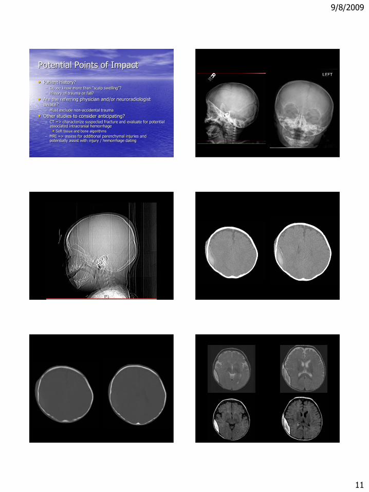
Potential Points of Impact
• Patient history?– Do we know more than “scalp swelling”?
– History of trauma or fall?
• Are the referring physician and/or neuroradiologist aware?– Must exclude non-accidental trauma
• Other studies to consider anticipating?– CT => characterize suspected fracture and evaluate for potential
associated intracranial hemorrhage
• Soft tissue and bone algorithms
– MRI => assess for additional parenchymal injuries and potentially assist with injury / hemorrhage dating

9/8/2009
12
Findings• Skull radiographs:
– Suspected oblique right parietal skull fracture
• CT Head without contrast:– Asymmetric mild right frontoparietal scalp soft tissue swelling,
with suspected nondisplaced oblique right parietal skull fracture – Heterogeneously hyperdense lenticular right parietal extra-axial
fluid collection (2 cm maximum thickness), with relative effacement of adjacent sulci and right lateral ventricle
• MRI Brain without contrast:– Heterogeneously lenticular right parietal extra-axial fluid
collection (T1 hyperintense; T2/FLAIR mildly hyperintense), with relative effacement of adjacent sulci and right lateral ventricle
Non-accidental trauma
• CT - recommended in initial evaluation of non-accidental trauma– High sensitivity in detecting acute intracranial bleed, fractures, cerebral
edema and hypoxic-ischemic injury – Attenuation of subdural / epidural hematoma varies by chronicity:
• Acute - hyperdense • Subacute – isodense• Chronic – hypodense
– Various factors such as low hematocrit and active hemorrhage may affect overall density
• MRI - essential second investigation– Best performed 5-10 days after insult– Can reliably differentiate between acute and chronic subdural hematoma – Most sensitive modality for detecting early ischemic changes – Clearly delineates anatomical locations that are difficult to image with CT
• posterior fossa, anterior part of middle cranial fossa, close to inner table of skull
80 year-old male with dementia that has progressed over the past 4 years
Potential Points of Impact
• Patient history?
– Do we know more than “dementia”?
– Previous CVA symptoms? Risk factors?
– Neurologic deficits?
• Other studies to consider anticipating?
– GRE imaging – evaluate for previous hemorrhage associated with infarcts
Figure 3a. Sensitivity of GRE imaging for hemosiderin in an 80-year-old man with dementia that has progressed over the past 4 years
Chao C P et al. Radiographics 2006;26:1517-1531
©2006 by Radiological Society of North America

9/8/2009
13
Figure 3b. Sensitivity of GRE imaging for hemosiderin in an 80-year-old man with dementia that has progressed over the past 4 years
Chao C P et al. Radiographics 2006;26:1517-1531
©2006 by Radiological Society of North America
Figure 4a. Recurrent CAA-related ICH in a 65-year-old woman with progressive aphasia, right visual field deficits, and headache
Chao C P et al. Radiographics 2006;26:1517-1531
©2006 by Radiological Society of North America
Figure 4b. Recurrent CAA-related ICH in a 65-year-old woman with progressive aphasia, right visual field deficits, and headache
Chao C P et al. Radiographics 2006;26:1517-1531
©2006 by Radiological Society of North America
Findings
• MRI Brain:
– Multiple foci of signal loss / “blooming” in
cortical-subcortical locations
– Consistent with chronic microhemorrhages
Cerebral Amyloid Angiopathy• Cerebrovascular disorder characterized by
deposition of β-amyloid protein in the media and adventitia of small and medium-sized vessels
• Both sporadic and hereditary forms may occur
• Manifests radiologically as part or all of a constellation of findings including acute or chronic ICHs in a distinctive cortical-subcortical distribution, leukoencephalopathy, and atrophy
Brain – Seizure / Dysplasia
• Indications– Seizure, Dysplasia, Mesial temporal sclerosis
• Sequences– 3 PL LOC– Sag T1 SE– Ax T2 FLAIR– Ax T1 SE– Ax T2 TSE FS– Ax DWI EPI– Cor T2 TSE (angled perpendicular to temporal lobes)– Cor FLAIR (angled perpendicular to temporal lobes)– Ax IR-SPGR / MP-RAGE or Cor IR-SPGR / MP-RAGE
• Comments– Coronal sequences should be thin section perpendicular to the
long axis of the hippocampus

9/8/2009
14
2 year-old female with seizures
Potential Points of Impact
• Patient history?
– Do we know more than “seizures”?
– Onset of symptoms?
– Prior studies / previous surgery / trauma?
• Pulse sequences to consider anticipating?
– Axial IR-SPGR / MP-RAGE
– Coronal T2 and FLAIR through hippocampi
Findings
• MRI Brain:– In a normal brain, white matter is in the interior, and
gray matter is mostly on the surface
– In patients with periventricular nodular heterotopia, clumps of gray matter, called nodules, appear deep within the brain, instead of on the surface
– Image courtesy of Bernard Chang, MD, Beth Israel Deaconess Medical Center (via Internet for teaching)
Periventricular Nodular Heterotopia
• In a normal brain, much of the gray matter (consisting mostly of nerve cells) appears on the brain's surface, while white matter (consisting mostly of nerve fibers, or "wiring" interconnecting areas of gray matter) runs deeper in the brain
• In PNH, a migrational abnormality occurs during development - portions of gray matter sit deep in the brain's core, in the white matter, having failed to migrate out to the surface– May serve as elliptogenic foci

9/8/2009
15
Brain – Advanced Protocols
• Dural venous sinus thrombosis – Cor 2D TOF SPGR– Sag 2D TOF SPGR (slight oblique angle)
• Stroke, TIA, Vertebrobasilar infarct, Aneurysm– Ax 3D TOF SPGR– Ax Perfusion
• Tumor, Metabolic abnormality– Single voxel spectroscopy (short and long echo; eg.
TE 35 and 144) on all new mass lesions– Multi voxel spectroscopy - suspected gliomas– Perfusion
• Gd – 20 ml @ 3-5 ml/s
30 year-old female with mental status changes
Potential Points of Impact
• Patient history?– Do we know more than “mental status changes”?
– Any neurologic symptoms?
– Recent surgery? Dehydration?
• Are the referring physician and/or neuroradiologist aware?
• Other studies to consider anticipating?– CTV head + contrast; reconstructions / MIP images
– MRI brain – evaluation for ischemia / hemorrhage

9/8/2009
16
Findings
• CT Head without contrast:– Tubular hyperdensity within superior sagittal sinus, torcula, and
left frontal cortical veins
• CT Venography with contrast:– Extensive irregular filling defects within superior sagittal sinus
extending to torcula, left transverse sinus, and left sigmoid sinus
– Irregular filling defects within left frontal cortical veins
• MRI Brain without contrast:– Tubular T1 hyperintense signal and loss of normal flow void
within superior sagittal sinus, torcula, left transverse sinus, and left frontal cortical veins

9/8/2009
17
Dural Venous Thrombosis
• “empty delta sign”– With CT venography, the thrombus does not enhance
but dura enhances – Triangular defect can be demonstrated within
superior sagittal and transverse sinuses– Seen in 25–75% of cases– Because of volume averaging, may fail to
demonstrate a hyperattenuating sinus or an empty delta sign in horizontal segment of superior sagittal sinus or transverse sinus
– Can disappear in chronic stages with enhancement of organized clot
Dural Venous Thrombosis• "dense triangle sign”
– Hyperdense triangular shaped structure along posterior aspect of superior sagittal sinus
• “cord sign” – Hyperdense tubular superficial cortical vein or
dural venous sinus
• Represent intravascular acute blood clots– Takes approximately 1–2 weeks to disappear– Caution
• Similarly increased attenuation of cerebral venous sinuses may also represent polycythemia
• Nonmyelinated brain in neonates makes sinuses appear hyperattenuating
Dural Venous Thrombosis
• MRI– Main sign is lack of expected signal flow void
on standard T1- and T2-weighted images
– Challenging diagnosis in acute stage• Hypointense signal of acute thrombus mimics
normal flow void on T2-weighted images
– Absence of flow void on T1-weighted images must be carefully sought because thrombus may be isointense / mildly hyperintense to brain tissue
69 year-old female with chronic headaches
Potential Points of Impact
• Patient history?
– Do we know more than “headaches”?
– Any neurologic symptoms?
• Concern for cerebral aneurysm?
• Other studies to consider anticipating?
– CTA head - reconstructions / MIP images
– Cerebral angiogram – if warranted

9/8/2009
18

9/8/2009
19
Findings
• CT head angiography:– Large aneurysm projects superiorly from basilar tip
• 20 mm CC, 13 mm transverse, and 23 mm AP
– Origins of both posterior cerebral arteries and superior cerebellar arteries appear incorporated into aneurysm neck
– Aneurysm projects superiorly into third ventricle and compresses foramen of Monroe
• Cerebral angiography:– Large, lobulated aneurysm at basilar artery tip
– Left P1 segment arises from base of aneurysm
– Right P1 segment appears clear
Basilar Tip Aneurysm
• Occurs at distal bifurcation of basilar artery, between origin of two posterior cerebral arteries
• Intracranial aneurysm distribution: – 30-35% => anterior communicating artery – 30-35% => posterior communicating artery origin – 20% => middle cerebral artery bifurcation – 5% => basilar artery bifurcation or tip – 1-5% => other posterior fossa vessels
• Conventional angiography – remains gold standard for detection and characterization of cerebral aneurysms
• CTA can detect more than 95% of aneurysms identified on conventional angiography
50 year-old male with brain tumor found on an outside hospital MRI study
Potential Points of Impact
• Patient history?
– Do we know more than “tumor”?
– Previous surgery?
– Neurologic deficits?
– Outside images available for radiologist review?
• Other studies to consider anticipating?
– MR spectroscopy
– MR perfusion
– Post-contrast IR-SPGR (for radiation therapy)
9/8/2009
20
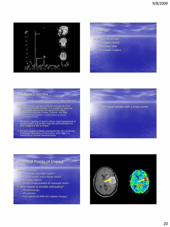
Findings
• MR Spectroscopy:
– Increased choline
– Decreased NAA
– Decreased creatine
MR Spectroscopy
• Useful in tumor evaluation and surgical / biopsy planning
• Although water and fat contribute virtually all of the signal in proton MR imaging, it is possible to suppress these signals and assess the signal from other metabolites including choline, creatine, and NAA – Altered in concentration in various disease processes,
particularly tumors
• Metabolic mapping of spectra allows rapid assessment of spectral peaks and choline map also demonstrates the most malignant site to biopsy
• Elevated choline probably represents the cell membrane breakdown secondary to the tumor, while NAA is a metabolite of normal neuronal tissue
Middle-aged female with a brain tumor
Potential Points of Impact
• Patient history?
– Do we know more than “tumor”?
– Previous surgery and/or biopsy results?
– Neurologic deficits?
– Outside images available for radiologist review?
• Other studies to consider anticipating?
– MR spectroscopy
– MR perfusion
– Post-contrast IR-SPGR (for radiation therapy)

9/8/2009
21
Findings
• MR Brain:
– Large avidly enhancing mass centered within
the left insula
• MR Perfusion:
– Significant patchy hyperperfusion – increased CBF compared with surrounding cortex /
juxtacortical white matter
MR Perfusion
• Useful in brain tumor evaluation and surgical / biopsy planning
• Uses contrast which has slightly different magnetic characteristics from blood - causes a disturbance in the localized magnetic field
• Signals are analyzed mathematically and expressed as an image (e.g. CBF, CBV, MTT maps). By offsetting the changes in shape in the flow of the contrast bolus against time, it is possible to calculate how much blood is reaching the area of concern within the brain.
THANK YOU!
HAVE A GREAT DAY!