Extracts of Adhatoda vasica Nees. and vasicine inhibits...
13
IOSR Journal of Pharmacy and Biological Sciences (IOSR-JPBS) e-ISSN:2278-3008, p-ISSN:2319-7676. Volume 13, Issue 2 Ver. I (Mar. – Apr. 2018), PP 37-49 www.iosrjournals.org DOI: 10.9790/3008-1302013749 www.iosrjournals.org 37 | Page Extracts of Adhatoda vasica Nees. and vasicine inhibits biofilm formation of Klebsiella pneumoniae Lovely Rahaman 1 , Sushanta Ghosh 1 , Nandita Nath 1 , Aparna Sen 2 , Kiran Sharma 3 and Bipin Kumar Sharma 1* 1 (Microbial Ecology Laboratory, Department of Microbiology, Tripura University, Tripura, India) 2 (Department of Microbiology, Lady Brabourne College, Kolkata, West Bengal, India) 3 (Department of Anaesthesia, Kalpana Chawla Govt. Medical College, Karnal, Haryana, India) *Corresponding author: Dr. Bipin Kumar Sharma Abstract: Adhatoda vasica Nees. are used traditionally in Tripura to cure pneumonia, cough and cold. Pneumonia is mainly caused by bacteria like Klebsiella pneumoniae. Moreover, many of these pathogenic strains are showing acute resistance for many commercially available antibiotics due to its biofilm formation ability. The present study evaluates the antimicrobial activity of Adhatoda vasica Nees. extracts against the biofilms of Klebsiella pneumoniae and their free-living forms. After extraction and phytochemical screening of Adhatoda vasica Nees, the presence of vasicine in extracts was ascertained by thin layer chromatography. The antimicrobial activity for all the extracts and commercially available vasicine against planktonic forms of bacteria were determined using the agar well diffusion method with tetracycline as control. The minimum inhibitory concentration (MIC) and the minimum bactericidal concentration (MBC) values were evaluated by a macrobroth dilution technique. The anti-biofilm effects were assessed by microtitre plate method. The cell viability assay was assessed by EtBr/AO staining. Results shows that the A. vasica Nees. leaf extract have remarkable zone of inhibition for Klebsiella pneumoniae as compared to standard vasicine and tetracycline. Similarly, the MIC values confirmed its outstanding ability for inhibition of planktonic as well as biofilm form of the bacteria, and same is also been visualized and established further by cell viability studies and SEM analysis for the biofilm of Klebsiella pneumoniae . Key words: A. vasica Nees, Vasicine, Biofilm, Klebsiella pneumoniae --------------------------------------------------------------------------------------------------------------------------------------- Date of Submission: 28-02-2018 Date of acceptance: 15-03-2018 --------------------------------------------------------------------------------------------------------------------------------------- I. Introduction Tripura is a small hilly state of North-east India with a rich source of flora and fauna. The tropical climate of Tripura supports the growth of many medicinal plants and forest resources starting from the hills to the plains. Even today, most of the tribal population extract their livelihood from forests and depend on traditional herbal treatment practices. Many medicinal plants are used by the local practitioners to combat infectious disease like pneumonia. Herbal medicine is based on the premise that plants contain natural bioactive substances that can promote health and alleviate illness. Adhatoda vasica Nees. (Family- Acanthaceae) is a perennial, evergreen shrub found widely throughout the tropical regions of Southeast Asia. The name J. adhatoda (L.) Nees and Adhatoda zeylanica Medic are used synonymously. It is commonly known as Vasaka in Tripura. The plant is highly branched shrub (1.0 m to 2.5 mm height) with unpleasant smell and bitter taste. It has opposite ascending branches with white, pink or purple flowers (Dhankar et al., 2011). It is used in Ayurvedic medicinal plant to treat cold, cough, asthma and tuberculosis (Sharma et al., 1992). The main action is expectorant and antispasmodic (bronchodilator) (Karthikeyan et al., 2009). Moreover, the importance of Vasaka plant in the treatment of respiratory disorders can be understood from the ancient Indian saying, “No man suffering from phthisis need despair as long as the Vasaka plant exists” ( Dymock et al., 1893). Thus the frequent use of Adhatoda vasica Nees. has resulted in its inclusion in the WHO manual “The Use of Traditional Medicine in Primary Health Care” which is intended for health workers in South-east Asia to keep them informed of the restorative utility of their surrounding flora (WHO fact sheets., 1990). The major alkaloids of the plant found to be biologically active are vasicine and vasicinone (Dhankar et al., 2011).
Transcript of Extracts of Adhatoda vasica Nees. and vasicine inhibits...
IOSR Journal of Pharmacy and Biological Sciences (IOSR-JPBS)
e-ISSN:2278-3008, p-ISSN:2319-7676. Volume 13, Issue 2 Ver. I (Mar. – Apr. 2018), PP 37-49
www.iosrjournals.org
DOI: 10.9790/3008-1302013749 www.iosrjournals.org 37 | Page
Extracts of Adhatoda vasica Nees. and vasicine inhibits biofilm
formation of Klebsiella pneumoniae
Lovely Rahaman1, Sushanta Ghosh
1, Nandita Nath
1, Aparna Sen
2, Kiran
Sharma3 and Bipin Kumar Sharma
1*
1(Microbial Ecology Laboratory, Department of Microbiology, Tripura University, Tripura, India)
2(Department of Microbiology, Lady Brabourne College, Kolkata, West Bengal, India)
3(Department of Anaesthesia, Kalpana Chawla Govt. Medical College, Karnal, Haryana, India)
*Corresponding author: Dr. Bipin Kumar Sharma
Abstract: Adhatoda vasica Nees. are used traditionally in Tripura to cure pneumonia, cough and cold.
Pneumonia is mainly caused by bacteria like Klebsiella pneumoniae. Moreover, many of these pathogenic
strains are showing acute resistance for many commercially available antibiotics due to its biofilm formation
ability. The present study evaluates the antimicrobial activity of Adhatoda vasica Nees. extracts against the
biofilms of Klebsiella pneumoniae and their free-living forms. After extraction and phytochemical screening of
Adhatoda vasica Nees, the presence of vasicine in extracts was ascertained by thin layer chromatography. The
antimicrobial activity for all the extracts and commercially available vasicine against planktonic forms of
bacteria were determined using the agar well diffusion method with tetracycline as control. The minimum
inhibitory concentration (MIC) and the minimum bactericidal concentration (MBC) values were evaluated by a
macrobroth dilution technique. The anti-biofilm effects were assessed by microtitre plate method. The cell
viability assay was assessed by EtBr/AO staining. Results shows that the A. vasica Nees. leaf extract have
remarkable zone of inhibition for Klebsiella pneumoniae as compared to standard vasicine and tetracycline.
Similarly, the MIC values confirmed its outstanding ability for inhibition of planktonic as well as biofilm form
of the bacteria, and same is also been visualized and established further by cell viability studies and SEM
analysis for the biofilm of Klebsiella pneumoniae .
Key words: A. vasica Nees, Vasicine, Biofilm, Klebsiella pneumoniae
----------------------------------------------------------------------------------------------------------------------------- ----------
Date of Submission: 28-02-2018 Date of acceptance: 15-03-2018
----------------------------------------------------------------------------------------------------------------------------- ----------
I. Introduction
Tripura is a small hilly state of North-east India with a rich source of flora and fauna. The tropical
climate of Tripura supports the growth of many medicinal plants and forest resources starting from the hills to
the plains. Even today, most of the tribal population extract their livelihood from forests and depend on
traditional herbal treatment practices. Many medicinal plants are used by the local practitioners to combat
infectious disease like pneumonia. Herbal medicine is based on the premise that plants contain natural bioactive
substances that can promote health and alleviate illness. Adhatoda vasica Nees. (Family- Acanthaceae) is a
perennial, evergreen shrub found widely throughout the tropical regions of Southeast Asia. The name J.
adhatoda (L.) Nees and Adhatoda zeylanica Medic are used synonymously. It is commonly known as Vasaka in
Tripura. The plant is highly branched shrub (1.0 m to 2.5 mm height) with unpleasant smell and bitter taste. It
has opposite ascending branches with white, pink or purple flowers (Dhankar et al., 2011). It is used in
Ayurvedic medicinal plant to treat cold, cough, asthma and tuberculosis (Sharma et al., 1992). The main action
is expectorant and antispasmodic (bronchodilator) (Karthikeyan et al., 2009). Moreover, the importance of
Vasaka plant in the treatment of respiratory disorders can be understood from the ancient Indian saying, “No
man suffering from phthisis need despair as long as the Vasaka plant exists” (Dymock et al., 1893). Thus the
frequent use of Adhatoda vasica Nees. has resulted in its inclusion in the WHO manual “The Use of Traditional
Medicine in Primary Health Care” which is intended for health workers in South-east Asia to keep them
informed of the restorative utility of their surrounding flora (WHO fact sheets., 1990). The major alkaloids of
the plant found to be biologically active are vasicine and vasicinone (Dhankar et al., 2011).

Extracts of Adhatoda vasica Nees. and vasicine inhibits biofilm formation of Klebsiella pneumoniae
DOI: 10.9790/3008-1302013749 www.iosrjournals.org 38 | Page
Figure: 1. A. Adhatoda vasica Nees. plant. B. Vasicine (Sigma)
The anti-inflammatory and antimicrobial properties of pyrroloquinazoline alkaloids from Adhatoda
vasica Nees. have been reported by Singh, 2013 and Sheeba, 2012 (Singh et al., 2015; Sheeba et al., 2012). The
antimicrobial property of leaf extracts of J. adhatoda (L.) in comparison with vasicine was shown by Rashmi Pa
in 2012 (Rashmi et al., 2012). The leaves, flowers and roots of this plant used in herbal drugs against tubercular
activities (Barry et al., 1955), cancer (Pandey., 2002) and possessed to have anti-helminthic properties (Ayyanar
et al., 2008). In many cases, pneumonia is treated at home. It often clears up in 2 to 3 weeks. But older adults,
babies, and immune compromised patients may develop serious complications. It can be a community-
associated pneumonia or healthcare-associated pneumonia. Pneumonia has bacterial, viral, fungal, and other
primary causes. Amongst the pathogenic bacteria, Klebsiella pneumoniae is the causative agents of bacterial
pneumonia and the leading cause of infant mortality globally (WHO fact sheets., 2013) due to acute resistant to
many commercially available antibiotics and the formation of stringent biofilms. A number of intervention
strategies are under-trial against infection associated pneumonic bacterial biofilms, most of the strategies are
disadvantageous in terms of their impact on host systems. Application of phytochemicals independently or in
combination with existing therapeutics might offer an important implication in this regard.
However, it will be interesting to evaluate the antibacterial/ antibiofilm properties of Adhatoda vasica Nees. to
validate scientifically its traditional usage against pneumonia.
II. Materials And Methods 2.1 Preparation of standard vasicine
Vasicine (10 mg) was procured from Sigma Aldrich. A solution of vasicine (5mg/ml) was prepared using
methanol as solvent.
2.2 Plant sample collection and identification
The fresh leaves of Adhatoda vasica Nees. were collected from Sepahijala Botanical Garden (23°39’52’’N &
91°18’42’’E). The plants are identified with the help of Department of Botany, Tripura University, India.
2.3 Culture and maintenance of test microorganisms for antimicrobial studies
Strain of Klebsiella pneumoniae MTCC 3384 was bought from IMTECH, Chandigarh. The strain was revived
in Luria Bertanni broth (Hi- media) and stored as 50 % glycerol stock at -80ºC refrigerator.
2.4 Processing of plant materials and extraction
Fresh leaves of plant Adhatoda vasica Nees. were collected and washed thoroughly 2-3 times with running
water and once with sterile distilled water. Then the leaves were shade dried and placed in hot air oven at a
temperature of 50ºC for 4 - 5 days till the weight became constant. Plant materials were regularly examined to
check any fungal growth or rotting. The dried leaves were powdered to obtain a very fine particle size using
mechanical mixer grinder.
The dried powder of leaves (125g) was soaked successively in petroleum ether, ethanol, methanol and water in
the sample: solvent ratio of 1: 10 according to their polarity index. The mixture was subjected to intermittent
shaking for 7 days. The extracts were filtered through Whatman filter paper no.1 and concentrated at 50º C
using rotary evaporator (Ika, Japan). The concentrated extracts were stored in air tight container at 4º C
refrigerator. For further experiments the extracts were dissolved in dimethyl sulphoxide (DMSO) to a
concentration of 1mg/ml (Malik et al., 2013).
A B

Extracts of Adhatoda vasica Nees. and vasicine inhibits biofilm formation of Klebsiella pneumoniae
DOI: 10.9790/3008-1302013749 www.iosrjournals.org 39 | Page
2.5 Determination of percentage yield The yield percentage of each crude extract was determined by percentage of weight of the dissolved extract after
evaporation divided by the initial weight of the extract before addition of solvent.
Weight of the dissolved extract after evaporation
Yield percentage = × 100
Initial weight of the extract before addition of solvent
2.6 Biochemical characterisation of crude extracts
2.6.1 Detection of alkaloids The presence of alkaloids in the extract was determined by Mayer’s test, Dragendroff’s test and Wagner’s test
(Joseph et al., 2013).
2.6.2 Detection of carbohydrates The presence of carbohydrates was determined by Molisch’s test, Benedict’s test and Fehling’s test (Joseph et
al., 2013).
2.6.3 Detection of glycosides The presence of cardiac glycosides was determined by Legal’s test (Joseph et al., 2013).
2.6.4 Detection of saponins The presence of saponins was determined by froth test and foam test (Joseph et al., 2013).
2.6.5 Detection of phytosterols The presence of phytosterols was determined by Salkowski’s test (Joseph et al., 2013).
2.6.6 Detection of phenols The presence of phenols was determined by Ferric chloride test (Joseph et al., 2013).
2.6.7 Detection of tannins The presence of tannins was determined by Lead acetate test and Gelatin tests (Joseph et al., 2013).
2.6.8 Detection of flavonoids The presence of flavonoids was determined by alkaline reagent test and Lead acetate tests (Joseph et al., 2013).
2.6.9 Detection of proteins and amino acids The presence of protein and amino acids was determined by Xanthoproteic test and Ninhydrin test. (Joseph et
al., 2013)
2.7 Thin layer chromatography with commercial vasicine:
Thin layer chromatography has been done with the extracts and commercially available vasicine (Sigma-
Aldrich) as standard to compare and see the presence of vasicine in the extracts. The petroleum ether extract,
ethanolic extract, methanolic extract, water extract of leaves of Adhatoda vasica Nees. and standard vasicine
were dissolved in chloroform and the mobile phase contained chloroform, methanol and ethyl acetate in the
ratio 6:2:1 (Srivastava et al., 2001; Soni et al., 2008). The extracts were chromatographed on silica coated TLC
plates (Sigma- Aldrich).
2.8 Biological evaluation:
2.8.1 Determination of antimicrobial activity
Agar well-diffusion method was done to determine the antimicrobial activity of the different extracts of A.
vasica Nees leaves and commercially available vasicine (Sigma) using tetracycline as control (Sen et al., 2012).
Luria Bertanni (LB) agar were swabbed (sterile cotton swabs) with 24 hour old - broth culture of Klebsiella
pneumoniae .Wells were made in each of these plates using sterile cork borer. Stock solution of the plant extract
was prepared at a concentration of 20 mg/ml viz. Petroleum Ether, Ethanol, Methanol, and Water. Vasicine
(1mg/ml) and tetracycline (20mg/ml) were also prepared. About 100 µl of the plant extracts, vasicine and
tetracycline were added into the wells and allowed to diffuse at room temperature for 2hrs. The plates were
incubated at 37°C for 24 hours.
2.8.2 Determination of Minimum Inhibitory Concentration (MIC) and Minimum Bactericidal
Concentration (MBC)
Minimum Inhibitory Concentration (MIC) and Minimum Bactericidal Concentration (MBC) assay was
performed. The test strain Klebsiella pneumoniae MTCC 3384 were grown for overnight on Luria Bertanni
Broth (Hi-media) respectively. 10 μl of the seed culture was inoculated into 5 ml of fresh respective broths in
test tubes and incubated with shaking for 12 h. These broth cultures were diluted 1:500 in fresh broths. 20 μl of
the diluted culture containing 1.2 × 10⁵ to 2 × 10
5 CFU of each test strain was inoculated into 2 ml of broth
containing the vasicine (Sigma Aldrich) and the extracts and incubated with shaking for 24 h. The concentration
of the extracts for Klebsiella pneumoniae MTCC 3384 the extracts were taken in concentration of 0, 40,
80,120,160, 200 µg/ml. 100 μl of the each culture was then inoculated onto solid medium and incubated for 36
Extracts of Adhatoda vasica Nees. and vasicine inhibits biofilm formation of Klebsiella pneumoniae
DOI: 10.9790/3008-1302013749 www.iosrjournals.org 40 | Page
h. The incubation temperature will be 37°C, throughout the experiment. The MBC will be defined as that
concentration at which no viable bacteria will be recovered (Provenzano et al., 2000). For each phytochemical-
treated system Dimethyl sulphoxide (DMSO) was used as control.
2.8.3 Biofilm formation assay
The bacterial strains Klebsiella pneumoniae MTCC 3384 was grown over-night on Luria Bertanni agar plates at
37ºC supplemented with 1 % dextrose. Colonies of each strain were resuspended in Luria Bertanni Broth (Hi-
media) at OD600 of 0.6. A 1:100 dilution of these suspensions were inoculated in fresh broth containing 1 %
dextrose in 96-well polystyrene cell culture plates (Tarsons) for 3 hrs and then after 3 hrs extracts and vasicine
(Sigma Aldrich) were added in the concentration of 0xMIC, 0.25xMIC, 0.50xMIC, 0.75xMIC and 1xMIC
µg/ml and incubated for 24 h at 37°C. Subsequently, the respective media were removed carefully from the
wells and the wells were rinsed twice with sterile distilled water. Then sterile distilled water was removed
carefully from the wells and dried for 15 mins. Then the wells were filled with 1 % crystal violet stain ((Hi-
media). After 5 min, the stain was removed from the wells and subsequently rinsed twice with sterile distilled
water. The biofilm-associated crystal violet was resuspended with 99 % ethanol and the OD620 of the resulting
suspension was measured (Christensen et al., 1985) using Synergy H1 Hybrid reader.
2.8.4 Microscopic study of biofilm inhibition by the crude extract and compound
The bacterial strain Klebsiella pneumoniae MTCC 3384 was grown over-night on Luria Bertanni Broth (Hi-
media) supplemented with 1 % dextrose respectively at 37°C. A 1:100 dilution of these suspensions were
inoculated in fresh broth containing 1 % dextrose in test tubes (Borosil) of capacity 50 ml supplemented with
glass coverslips (Borosil) for 3 hrs and then after 3 hrs extracts and vasicine (Sigma Aldrich) were added and
incubated for 24 h at 37°C respectively. Subsequently, the respective media were removed carefully from the
tubes and the tubes were rinsed twice with sterile distilled water. Then sterile distilled water was removed
carefully from the tubes and dried for 15 mins. Then the tubes were filled with 5 ml of 1 % crystal violet stain
((Hi-media). Then the cover slips with biofilm associated crystal violet were taken out and examined under the
compound microscope (Magnus) and photographed with digital camera (Allegruci et al., 2006).
2.8.5 Bacterial viability study of biofilm inhibition using EtBr/AO staining method
The bacterial strain Klebsiella pneumoniae MTCC 3384 was grown over-night on Luria Bertanni Broth (Hi-
media) at 37°C. A 1:100 dilution of these suspensions were inoculated in fresh broth containing 1 % dextrose in
glass petridishes (Borosil) supplemented with glass coverslips (Borosil) in presence and absence of the extracts
and vasicine (Sigma Aldrich) and incubated for 24 h at 37°C respectively. Subsequently, the respective media
were removed carefully from the plates and the plates were rinsed twice with sterile distilled water. Then sterile
distilled water was removed carefully from the plates and dried for 15 mins. Then the cover slips with biofilm
were taken out and 100 µl of Ethidium bromide/ Acridine orange (1:1) solution was added to the coverslips and
observed under fluorescent microscope (Shin et al., 2015).
2.8.6 SEM analysis
To evaluate the action of the compounds on the integrity of biofilm of the bacteria K. pneumoniae MTCC 3384,
the morphological analysis of the bacteria was carried out with the antimicrobial compounds with treatment.
The bacterial strain Klebsiella pneumoniae MTCC 3384 was grown over-night on Luria Bertanni Broth (Hi-
media) at 37°C. A 1:100 dilution of these suspensions were inoculated in fresh broth containing 1 % dextrose in
glass petridishes (Borosil) supplemented with glass coverslips (Borosil) in presence and absence of the extracts
and vasicine (Sigma Aldrich) and incubated for 24 h at 37°C respectively. Subsequently, the respective media
were removed carefully from the plates and the plates were rinsed twice with sterile distilled water. Then sterile
distilled water was removed carefully from the plates and dried for 15 mins. The biofilms were fixed with 2.5%
gluteraldehyde solution. After fixation for a minimum of 1 hr, samples are washed twice. The biofilms were
then fixed by immersion in 1% osmium tetroxide in PBS for 30 min then, washed with the same buffer and
dehydrated in a graded alcohol series of 50%, 70%, 95% and 100% ethanol consecutively for 5 min each. Then
the cover slips were mounted on aluminum stubs by using double-sided carbon tape and coated with gold for
scanning electron microscope (JEOL JSM –6390LV) in 15 kV accelerating voltage according to the method
described by Kalchayanand, Dunne, and Ray (Kalchayanand et al., 2004).
III. Results
In this investigation, we were mainly focussing on the effect of different extracts of fresh leaves of
Adhatoda vasica Nees. on biofilm formation of Klebsiella pneumoniae MTCC 3384 in comparison to the
commercially available vasicine (Sigma- Aldrich). So the fresh leaves were collected, identified and washed
properly with tap water and then sterile distilled water. And then it was subjected to extraction in petroleum

Extracts of Adhatoda vasica Nees. and vasicine inhibits biofilm formation of Klebsiella pneumoniae
DOI: 10.9790/3008-1302013749 www.iosrjournals.org 41 | Page
ether, ethanol, methanol and water. After extraction, the percentage yield of the extracts was calculated and is
shown in Table 1.
Table 1: Showing yield percentage of the different extracts of Adhatoda vasica Nees.
Figure 2: Showing yield percentage of the different extracts of Adhatoda vasica Nees.
The different biochemical tests of all the extracts of leaf of A. vasica Nees. were performed using the
standard protocol to analyse the presence of phytochemicals in the different extracts. The results suggested that
most of the phytochemicals were present in methanolic extract of the leaf of the plant. Total carbohydrates were
found to be present in all the extracts but reducing sugar was present in petroleum ether and ethanolic extracts as
compared to methanolic and water extracts. In case of alkaloids, it was seen that alkaloids were absent in only
pet ether extract as compared to others. Cardiac glycosides were found to be present in only methanolic extract,
saponins were absent in only ethanolic extract, phytosterols were present in all the extracts except water extract,
tannins were present in all the extracts except ethanolic extract, whereas flavonoids were found to be present in
all the extracts and proteins and amino acids were found to be present in only petroleum ether extracts. The
results were shown in Table 2.
Phytochemicals Tests Adhatoda vasica Nees.
Petroleum ether Ethanol Methanol Water
Carbohydrates
Molisch’s + + + +
Benedict’s + + - -
Fehling’s + + - -
Alkaloids
Mayer’s - + + +
Dragendroff’s - + + +
Wagner’s - - + +
Glycosides Legal’s - - + -
Saponins Froth + - + +
Foam + - + +
Phytosterols Salkowski’s + + + -
Phenols Ferric chloride - + + +
Tannins Lead acetate + - + +
Gelatin + - + +
Flavonoids Alkaline reagent + + + +
Lead acetate + + + +
Proteins and aminoacids Xanthoproteic + - - -
Ninhydrin + - - -
Table 2: Phytochemical screening of the different extracts of Adhatoda vasica Nees.
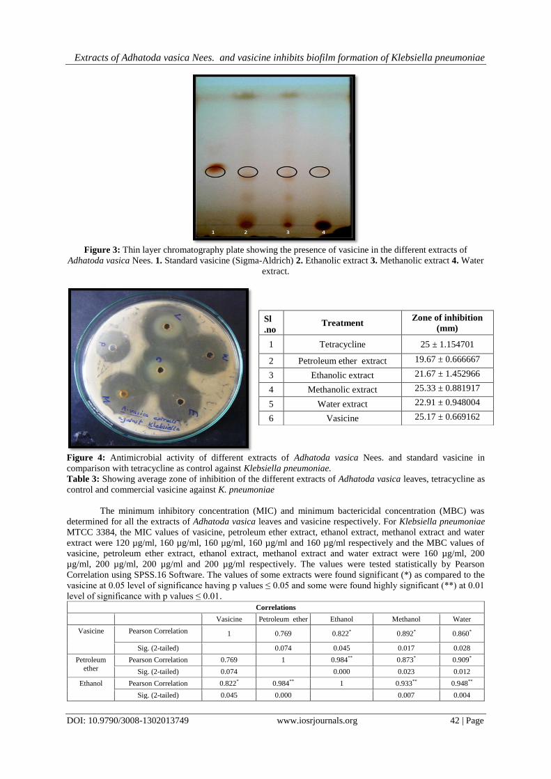
Thin layer chromatography was carried out to verify the presence of vasicine in the extracts. The result
showed that the Rf value of the spot obtained in the case of ethanolic, methanolic and water extracts were same
as the spot obtained in standard vasicine (Rf ~ 0.70). The image of the TLC plate was shown in figure no.2
Solvent used A. vasica Nees. (%)
Petroleum ether 2.12
Ethanol 7.136
Methanol 9.096
Water 8.608
Extracts of Adhatoda vasica Nees. and vasicine inhibits biofilm formation of Klebsiella pneumoniae
DOI: 10.9790/3008-1302013749 www.iosrjournals.org 42 | Page
Figure 3: Thin layer chromatography plate showing the presence of vasicine in the different extracts of
Adhatoda vasica Nees. 1. Standard vasicine (Sigma-Aldrich) 2. Ethanolic extract 3. Methanolic extract 4. Water
extract.
Figure 4: Antimicrobial activity of different extracts of Adhatoda vasica Nees. and standard vasicine in
comparison with tetracycline as control against Klebsiella pneumoniae.
Table 3: Showing average zone of inhibition of the different extracts of Adhatoda vasica leaves, tetracycline as
control and commercial vasicine against K. pneumoniae
The minimum inhibitory concentration (MIC) and minimum bactericidal concentration (MBC) was
determined for all the extracts of Adhatoda vasica leaves and vasicine respectively. For Klebsiella pneumoniae
MTCC 3384, the MIC values of vasicine, petroleum ether extract, ethanol extract, methanol extract and water
extract were 120 µg/ml, 160 µg/ml, 160 µg/ml, 160 µg/ml and 160 µg/ml respectively and the MBC values of
vasicine, petroleum ether extract, ethanol extract, methanol extract and water extract were 160 µg/ml, 200
µg/ml, 200 µg/ml, 200 µg/ml and 200 µg/ml respectively. The values were tested statistically by Pearson
Correlation using SPSS.16 Software. The values of some extracts were found significant (*) as compared to the
vasicine at 0.05 level of significance having p values ≤ 0.05 and some were found highly significant (**) at 0.01
level of significance with p values ≤ 0.01.
Correlations
Vasicine Petroleum ether Ethanol Methanol Water
Vasicine Pearson Correlation 1 0.769 0.822* 0.892* 0.860*
Sig. (2-tailed) 0.074 0.045 0.017 0.028
Petroleum
ether
Pearson Correlation 0.769 1 0.984** 0.873* 0.909*
Sig. (2-tailed) 0.074 0.000 0.023 0.012
Ethanol Pearson Correlation 0.822* 0.984** 1 0.933** 0.948**
Sig. (2-tailed) 0.045 0.000 0.007 0.004
Sl
.no Treatment
Zone of inhibition
(mm)
1 Tetracycline 25 ± 1.154701
2 Petroleum ether extract 19.67 ± 0.666667
3 Ethanolic extract 21.67 ± 1.452966
4 Methanolic extract 25.33 ± 0.881917
5 Water extract 22.91 ± 0.948004
6 Vasicine 25.17 ± 0.669162

Extracts of Adhatoda vasica Nees. and vasicine inhibits biofilm formation of Klebsiella pneumoniae
DOI: 10.9790/3008-1302013749 www.iosrjournals.org 43 | Page
Methanol Pearson Correlation 0.892* 0.873* 0.933** 1 0.989**
Sig. (2-tailed) 0.017 0.023 0.007 0.000
Water Pearson Correlation 0.860* 0.909* 0.948** 0.989** 1
Sig. (2-tailed) 0.028 0.012 0.004 0.000
*. Correlation is significant at the 0.05 level (2-tailed).
**. Correlation is significant at the 0.01 level (2-tailed).
N= 6
Table 5: Showing Pearson correlation between the different extracts of Adhatoda vasica Nees and standard
vasicine against Klebsiella pneumoniae using SPSS 16.
Graph 1: Showing MIC values of different extracts of leaf of Adhatoda vasica Nees and standard vasicine on
planktonic K. pneumoniae
The biofilm formation assay with crystal violet stain was studied for all the extracts of Adhatoda vasica
Nees. leaves and vasicine (Figure 5). It was found that all the extracts and vasicine have a significant effect on
the biofilm formation of the organism. The biofilm inhibitory studies have been conducted for commercial
vasicine and different extracts (petroleum ether, ethanol, methanol, water) of A. vasica Nees. The commercial
vasicine was taken as a control and the study was conducted for different concentration of 0, 30 60, 90 and 120
µg/ml and the proportional decline of values are 0, 2.89, 23.14, 33.88 and 46.28 % respectively. The different
concentrations of petroleum ether extract (0, 40, 80, 120 and 160 µg/ml) shows remarkable percentage biofilm
inhibition and the values are 0, 4.56, 25.36, 27.35 and 31.62 % respectively. Similar studies were carried out
with ethanol, methanol and water extract for the same concentration. The values for ethanol extract are 0, 29.87,
40.27, 50 and 53.36 %, methanol extract are 0, 27.21, 39.8, 43.2 and 47.28 % and for water extract are 0, 35.26,
46.8, 55.13 and 57.37 % respectively which shows notable biofilm intervention of the organism (Graph 3)
The microscopic examination of the biofilm adherence was done with organisms adhered on sterile
glass coverslips incubated in presence and absence of the treatment with commercial vasicine and different A.
vasica extracts (petroleum ether, ethanol, methanol and water). Then the coverslips were recovered and stained
with 1 % crystal violet stain which shows considerable inhibition of streptococcal biofilm adherence by the
different extracts of Adhatoda vasica Nees and commercial vasicine (Figure 6).Similar biofilm intervention was
seen in case of cell viability study with EtBr/ AO staining. The green cells take up acridine orange as they are
live cells but in case of dead cells ethidium bromide can enter into the cell and stain it red. So live cells and dead
cells can be differentiated easily (Figure 7). Similarly, biofilm inhibition was seen in case of SEM analysis of
the biofilm in presence of extracts of Adhatoda vasica Nees, vasicine and control antibiotic tetracycline (Figure
8)

Extracts of Adhatoda vasica Nees. and vasicine inhibits biofilm formation of Klebsiella pneumoniae
DOI: 10.9790/3008-1302013749 www.iosrjournals.org 44 | Page

Extracts of Adhatoda vasica Nees. and vasicine inhibits biofilm formation of Klebsiella pneumoniae
DOI: 10.9790/3008-1302013749 www.iosrjournals.org 45 | Page

Extracts of Adhatoda vasica Nees. and vasicine inhibits biofilm formation of Klebsiella pneumoniae
DOI: 10.9790/3008-1302013749 www.iosrjournals.org 46 | Page

Extracts of Adhatoda vasica Nees. and vasicine inhibits biofilm formation of Klebsiella pneumoniae
DOI: 10.9790/3008-1302013749 www.iosrjournals.org 47 | Page
IV. Discussion Pneumonia causing and biofilm forming bacterial agents like Klebsiella pneumoniae develops
characteristics that offer tough antibiotic resistance due to EPS formation. The traditional use of Adhatoda
vasica Nees. is very common in Tripura for herbal healing of pneumonia. It has been reported that the active
major alkaloid extracted from Adhatoda vasica Nees. are vasicine and vasicinone. Along with this other
phytocompounds are adhatodine, vasicolinone, vasicoline, anisotine, isoniazid, ethambutol, pyrazinamide etc
(Jha et al., 2012).

Extracts of Adhatoda vasica Nees. and vasicine inhibits biofilm formation of Klebsiella pneumoniae
DOI: 10.9790/3008-1302013749 www.iosrjournals.org 48 | Page
The petroleum ether, ethanol, methanol, water extracts of Adhatoda vasica Nees and its
phytochemical screening for carbohydrates, alkaloids, glycosides, saponins, phytosterols, phenols tannins,
flavonoids, protein and amino acids indicates that the phytocompounds extracted in petroleum ether is non
alkaloid, non glycosidic and non phenolic compound. The compound extracted in ethanol is non glycosidic, non
saponin, non protein and amino acid. Whereas the compound in methanolic and water extract is a non protein
and amino acid compound (Table 1). As there is no alkaloid compound in petroleum ether extract, it infers that
the compound extracted is not a vasicine. To check the presence of vasicine in ethanol, methanol, and water thin
layer chromatography is performed. The similarity in Rf value indicated the presence of vasicine in all of them
(Figure 3).
The antimicrobial activity of the extracts of Adhatoda vasica Nees are being compared in three
replicates taking tetracycline as control and vasicine as standard. The result depicted higher activity in
Klebsiella pneumoniae for the methanolic extract. However the activity against Klebsiella pneumoniae is
recorded to be higher than the standard vasicine and control tetracycline (Figure 4). This might be due to the
presence of other alkaloids along with vasicine in the methanolic extract showing combine effect. Next to
methanol the commercial vasicine is showing good effects followed by water, ethanolic and petroleum ether
extract of Adhatoda vasica Nees.
The MIC values of vasicine, petroleum ether extract, ethanol extract, methanol extract and water
extract were 120 μg/ml, 160 μg/ml, 160 μg/ml, 160 μg/ml and 160 μg/ml respectively against Klebsiella
pneumoniae MTCC 3384 (Graph-1). The MIC determined for the extract shows similar concentration for
petroleum ether, methanol, ethanol and water extract whereas the concentration for vasicine (commercial) was
lower. The MBC values also follow the same trend. However, the MBC values for pure commercial vasicine is
160μg/ml which is less than petroleum ether, methanol, ethanol and water extract. It might be attributed to the
purity and efficacy of vasicine in pure form towards microorganisms. The correlation analysis among the
different extracts and commercial vasicine were found highly significant (Table-2). This implies the similar
trend of effect for all extracts and the commercial vasicine statistically. Same patterns of results are also being
seen for the antimicrobial activity by agar well diffusion method.
Further the compounds are being checked for its ability of Klebsiella pneumoniae biofilm
intervention. The percentage inhibition in biofilm formation at different concentration for all the extracts and
commercial vasicine is depicted in graph 2. The percentage reduction in biofilm formation was observed to be
uniformly increasing as per the concentration in commercial vasicine and petroleum ether, ethanol, methanol
and water extract. So, it implies that, the trend of increase in rate of inhibition is directly proportional to the
increase in concentration of the extracts. Same trend is also been observed in microscopical studies using
Crystal violet staining (Figure-6) and EtBr/ AO staining (Figure-7) However, the maximum inhibition in biofilm
formation was observed in water extract of Adhatoda vasica followed by ethanol and methanol. The percentage
inhibition of Klebsiella pneumoniae biofilm with water extract 11% higher than the commercially available
vasicine (positive control) which can be attributed to some additional active fraction of compound along with
vasicine showing synergistic effect. However, the petroleum ether showed minimum inhibition. Similarly it was
confirmed by SEM analysis (Figure -8) that water extract of Adhatoda vasica Nees has the maximum effect. It
infers that unknown active fraction of the compound is commendably water-soluble and has remarkable effect
on gram negative Klebsiella pneumoniae. Further fractional analysis of water extract and detail studies of the
active faction in the compound may lead to some promising antibiofilm drugs for gram negative pneumonia
causing Klebsiella pneumoniae.
V. Conclusion The study concluded that the water extracts of A. vasica is more active than commercially available
vasicine towards gram negative Klebsiella pneumoniae. The MIC and MBC values of water and ethanol extracts
are found promising against Klebsiella pneumoniae in its planktonic form as well as in the biofilm forms.
However, it indicates that, there is an unidentified active compound in water and ethanol extract of A. vasica
showing more efficiency towards gram negative Klebsiella pneumoniae biofilm intervention than vasicine even
at below MIC concentration, and it needs further studies to know the compound in detail.
Acknowledgement We would like to thank the Department of Science and Technology for funding the project under
Women scientist -A programme. We would also like to thank to the Department of Microbiology and State
Biotech Hub, Tripura University for giving us lab facilities. We would like to thank Department of
Microbiology, Lady Brabourne College, Kolkata for their lab facilities.
Extracts of Adhatoda vasica Nees. and vasicine inhibits biofilm formation of Klebsiella pneumoniae
DOI: 10.9790/3008-1302013749 www.iosrjournals.org 49 | Page
References [1]. S. Dhankhar, R. Kaur, S. Ruhil, M. Balhara, S. Dhankhar, A.K. Chhillar, A review on Justicia adhatoda: A potential source of
natural medicine, African Journal of Plant Science, 5(11), 2011, 620-627.
[2]. M.P. Sharma, J. Ahmad, A. Hussain, S. Khan , Folklore medicinal plants of Mewat (Gurgaon district), Haryana, Indi, International
Journal of Pharmacognosy, 30, 1992, 129-134. [3]. A. Karthikeyan, V. Shanthi, A. Nagasathaya, Preliminary phytochemical and antibacterial screening of crude extract of the leaf of
Adhatoda vasica. L, International Journal of Green Pharmacy, 3, 2009, 78-80.
[4]. W. Dymock, J.H. Warden and D. Hooper, Pharmacographia indica: A History of the Principal Drugs of the Vegetable Origin. Kegsm Paul, Trench, Trubner & Co. Ltd, London 1893, (1893) pp. 261– 265.
[5]. WHO (1990) The use of traditional medicine in primary health care. A manual for health workers in South-East Asia, SEARO
Regional Health Papers, No. 19. New Delhi, pp. 1-2. [6]. B. Singh and R.A. Sharma, Anti-inflammatory and antimicrobial properties of pyrroloquinazoline alkaloids from Adhatoda vasica
Nees, Phytomedicine, 20 (5), 2015, 441-5.
[7]. J.B. Sheeba and S.T. Mohan, Antimicrobial activity of Adhatoda vasica against clinical pathogens. Asian Journal of Plant Science Research, 2(2), 2012, 83-88.
[8]. Pa Rashmi and L. Mathew, Antimicrobial activity of leaf extracts of Justicia adhatoda L. incomparison with vasicine, Asian Pacific
Journal of Tropical Biomedicine, 2012, 1556-1560. [9]. V.C. Barry, M.L. Conalty, H.J.R. Rylance, Antitubercular effects of an extract of Adhatoda vasica, Nature, 1955, 176: 119-120.
[10]. G. Pandey, Anticancer herbal drugs of India with special reference to Ayurveda. Sri Satguru Publications; Delhi, 2002, pp. 18-121.
[11]. M. Ayyanar, S. Ignacimuthu, Medicinal uses and pharmacological Actions of five commonly used Indian Medicinal plants: A mini-review. Iranian Journal of Pharmacology and Therapeutics, 2008, 7, 107-114.
[12]. Global action plan for pneumonia & diarrhoea (GAPPD), WHO and UNICEF (2013)
www.who.int/maternal_child_adolescent/documents /global_ action _ plan_pneumonia_diarrhoea. [13]. J.K. Malik, A. Sharma, S. Singh and S. Jain, Nanosuspension of vasicine from Adhatoda vasica: Isolation and characterization,
Drug Invention Today, 2013, 5, 32-38. [14]. B.S. Joseph, P.H. Kumbhare and M.C. Kale, Preliminary phytochemical screening of selected Medicinal Plants. International
Research Journal of Science & Engineering, 2013, 1, 2, 55-62.
[15]. S. Srivastava, K.V. Ram, M.G. Madan, C.S. Subhash, S. Kumar, HPLC determination of vasicine and vasicinone in Adhatoda vasica with photo diode array detection. Journal of Liquid Chromatography and Related Technologies, 2001, 24, 153–9.
[16]. S. Soni, S. Anandjiwala, G. Patel, and M. Rajani, Validation of Different Methods of Preparation of Adhatoda vasica Leaf Juice by
Quantification of Total Alkaloids and Vasicine. Indian Journal of Pharmaceutical Sciences , 2008, 70, 1, 36–42. [17]. A. Sen and A. Batra, Evaluation of antimicrobial activity of different solvent extracts of medicinal plant: Melia azedarach L,
International Journal of Current Pharmaceutical Research, 2012, 4, 2, 67-73.
[18]. D. Provenzano and K.E. Klose, Altered expression of ToxR regulated porins, OmpU and OmpT diminishes V. cholerae bile resistance, virulence factor expression and intestinal colonization. Proceedings of the National Academy of Science, 2000,
97, 10220–10224.
[19]. G.D. Christensen, W.A. Simpson, J.J. Younger, L.M. Baddour, F.F. Barrett, D.M. Melton, Adherence of coagulase-negative staphylococci to plastic tissue culture plates: a quantitative model for the adherence of staphylococci to medical devices. Journal of
Clinical Microbiology, 1985, 22, 996–1006.
[20]. M. Allegrucci, F. Z. Hu, K. Shen, J. Hayes, G. D. Ehrlich, J. C. Post, K. Sauer, Phenotypic characterization of Streptococcus pneumoniae biofilm development. Journal of Bacteriology, 2006, 188, 2325–2335.
[21]. J.M. Shin, I. Ateia, J. R. Paulus, H. Liu, J.C. Fenno, A.H. Rickard and Y.L. Kapila, Antimicrobial nisin acts against saliva derived
multi-species biofilms without cytotoxicity to human oral cells. Frontiers in Microbiology, 2015, 6, 617. [22]. D.K. Jha, L. Panda, P. Lavanya, S. Ramaiah, A. Anbarasu, Detection and Confirmation of Alkaloids in Leaves of Justicia
adhatoda and Bioinformatics Approach to Elicit Its Anti-tuberculosis Activity. Appled Biochemistry and Biotechnology, 2012,
168, 5, 980–990. [23]. N. Kalchayanand , P. Dunne, & B. Ray . Viability loss and morphology change of foodborne pathogens following exposure to
hydrostatic pressures in the presence and absence of bacteriocins. International Journal of Food Microbiology,2004, 91, 991-998.
Dr. Bipin Kumar Sharma "Extracts of Adhatoda vasica Nees. and vasicine inhibits biofilm
formation of Klebsiella pneumoniae." IOSR Journal of Pharmacy and Biological Sciences
(IOSR-JPBS) 13.2 (2018): 37-49.


![Research Journal of Pharmaceutical, Biological and ...3)/[19].pdf · Research Journal of Pharmaceutical, Biological and Chemical ... Adhatoda Vasica leaves have been used extensively](https://static.fdocuments.us/doc/165x107/5a770d817f8b9a0d558da31c/research-journal-of-pharmaceutical-biological-and-319pdf-research.jpg)