RESEARCH Open Access Indirect shoot organogenesis from ... · PDF fileRESEARCH Open Access...
8
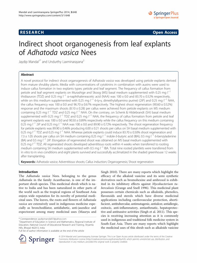
RESEARCH Open Access Indirect shoot organogenesis from leaf explants of Adhatoda vasica Nees Jaydip Mandal 1* and Undurthy Laxminarayana 2 Abstract A novel protocol for indirect shoot organogenesis of Adhatoda vasica was developed using petiole explants derived from mature shrubby plants. Media with concentrations of cytokinins in combination with auxins were used to induce callus formation in two explants types: petiole and leaf segment. The frequency of callus formation from petiole and leaf segment explants on Murashige and Skoog (MS) basal medium supplemented with 0.25 mg l -1 thidiazuron (TDZ) and 0.25 mg l -1 α-naphthaleneacetic acid (NAA) was 100 ± 0.0 and 83.70 ± 0.52% respectively, while on this medium supplemented with 0.25 mg l -1 6-(γ-γ, dimethylallyamino purine) (2iP) and 0.25 mg l -1 NAA, the callus frequency was 100 ± 0.0 and 96.70 ± 0.67% respectively. The highest shoot regeneration (90.60 ± 0.52%) response and the maximum shoots (8.10 ± 0.28) per callus were achieved from petiole explants on MS medium containing 0.25 mg l -1 TDZ and 0.25 mg l -1 NAA. On the contrary, on Schenk & Hildebrandt (SH) basal medium supplemented with 0.25 mg l -1 TDZ and 0.25 mg l -1 NAA, the frequency of callus formation from petiole and leaf segment explants was 100 ± 0.0 and 90.50 ± 0.89% respectively while the callus frequency on this medium containing 0.25 mg l -1 2iP and 0.25 mg l -1 NAA was 100 ± 0.0 and 89.90 ± 0.72% respectively. The shoot regeneration frequency for petiole explants was 89.90 ± 0.46% producing 6.00 ± 0.21 shoots per callus on SH basal medium supplemented with 0.25 mg l -1 TDZ and 0.25 mg l -1 NAA. Whereas petiole explants could induce 83.70 ± 0.50% shoot regeneration and 7.3 ± 1.05 shoots per callus on SH medium containing 0.25 mg l -1 indole-3-butyric acid (IBA), 0.5 mg l -1 6-benzyladenine (BA) and 0.5 mg l -1 2iP. Elongation of regenerated shoot was obtained on MS basal medium supplemented with 0.25 mg l -1 TDZ. All regenerated shoots developed adventitious roots within 4 weeks when transferred to rooting medium containing SH medium supplemented with 0.5 mg l -1 IBA. Total nine rooted plantlets were transferred from in vitro to in vivo conditions and eight plants survived and successfully acclimatized in the shaded greenhouse 12 weeks after transplanting. Keywords: Adhatoda vasica; Adventitious shoots; Callus induction; Organogenesis; Shoot regeneration Introduction The Adhatoda vasica Nees. belonging to the genus Adhatoda in the family Acanthaceae, is one of the im- portant shrub species. This medicinal shrub which is na- tive to India and has been naturalized in other parts of the world such as the tropical regions of Southeast Asia enjoys wide reputation for its novelty of potential medi- cinal uses. The leaves, the roots and flowers of Adhatoda vasica are extensively used in indigenous medicine espe- cially as bronchodilator, antidiabetic, anti-jaundice and expectorant among many medicinal uses (Maurya and Singh 2010). There are many reports which highlight the efficacy of the alkaloid vasicine and its semi synthetic derivatives such as bromohexine and ambroxol is exhib- ited in its inhibitory effects against Mycobacterium tu- berculosis (Grange and Snell 1996). This medicinal plant possesses certain chemicals such as alkaloids, phenolics, flavonoids and sterols which have diverse medicinal applications including cardiovascular protection, aborti- facient, antitubercular, antimutagenic, antiulcer, antiallergic, oxitocic, anti-inflammatory, antiasthmatic, hepatoprotec- tive and antitussive activities (Singh et al. 2011). This spe- cies is receiving increasing attention as it is commonly used in indigenous and traditional folk medicine system in South-East Asia. There are many reports which highlight the medicinal uses of this shrub such as alkaloids vasicine * Correspondence: [email protected] 1 Department of Education in Science and Mathematics, Regional Institute of Education, National Council of Educational Research and Training, Shyamla Hills, Bhopal 462013, India Full list of author information is available at the end of the article a SpringerOpen Journal © 2014 Mandal and Laxminarayana; licensee Springer. This is an Open Access article distributed under the terms of the Creative Commons Attribution License (http://creativecommons.org/licenses/by/4.0), which permits unrestricted use, distribution, and reproduction in any medium, provided the original work is properly credited. Mandal and Laxminarayana SpringerPlus 2014, 3:648 http://www.springerplus.com/content/3/1/648
Transcript of RESEARCH Open Access Indirect shoot organogenesis from ... · PDF fileRESEARCH Open Access...
a SpringerOpen Journal
Mandal and Laxminarayana SpringerPlus 2014, 3:648http://www.springerplus.com/content/3/1/648
RESEARCH Open Access
Indirect shoot organogenesis from leaf explantsof Adhatoda vasica NeesJaydip Mandal1* and Undurthy Laxminarayana2
Abstract
A novel protocol for indirect shoot organogenesis of Adhatoda vasica was developed using petiole explants derivedfrom mature shrubby plants. Media with concentrations of cytokinins in combination with auxins were used toinduce callus formation in two explants types: petiole and leaf segment. The frequency of callus formation frompetiole and leaf segment explants on Murashige and Skoog (MS) basal medium supplemented with 0.25 mg l−1
thidiazuron (TDZ) and 0.25 mg l−1 α-naphthaleneacetic acid (NAA) was 100 ± 0.0 and 83.70 ± 0.52% respectively,while on this medium supplemented with 0.25 mg l−1 6-(γ-γ, dimethylallyamino purine) (2iP) and 0.25 mg l−1 NAA,the callus frequency was 100 ± 0.0 and 96.70 ± 0.67% respectively. The highest shoot regeneration (90.60 ± 0.52%)response and the maximum shoots (8.10 ± 0.28) per callus were achieved from petiole explants on MS mediumcontaining 0.25 mg l−1 TDZ and 0.25 mg l−1 NAA. On the contrary, on Schenk & Hildebrandt (SH) basal mediumsupplemented with 0.25 mg l−1 TDZ and 0.25 mg l−1 NAA, the frequency of callus formation from petiole and leafsegment explants was 100 ± 0.0 and 90.50 ± 0.89% respectively while the callus frequency on this medium containing0.25 mg l−1 2iP and 0.25 mg l−1 NAA was 100 ± 0.0 and 89.90 ± 0.72% respectively. The shoot regeneration frequencyfor petiole explants was 89.90 ± 0.46% producing 6.00 ± 0.21 shoots per callus on SH basal medium supplemented with0.25 mg l−1 TDZ and 0.25 mg l−1 NAA. Whereas petiole explants could induce 83.70 ± 0.50% shoot regeneration and7.3 ± 1.05 shoots per callus on SH medium containing 0.25 mg l−1 indole-3-butyric acid (IBA), 0.5 mg l−1 6-benzyladenine(BA) and 0.5 mg l−1 2iP. Elongation of regenerated shoot was obtained on MS basal medium supplemented with0.25 mg l−1 TDZ. All regenerated shoots developed adventitious roots within 4 weeks when transferred to rootingmedium containing SH medium supplemented with 0.5 mg l−1 IBA. Total nine rooted plantlets were transferred fromin vitro to in vivo conditions and eight plants survived and successfully acclimatized in the shaded greenhouse 12 weeksafter transplanting.
Keywords: Adhatoda vasica; Adventitious shoots; Callus induction; Organogenesis; Shoot regeneration
IntroductionThe Adhatoda vasica Nees. belonging to the genusAdhatoda in the family Acanthaceae, is one of the im-portant shrub species. This medicinal shrub which is na-tive to India and has been naturalized in other parts ofthe world such as the tropical regions of Southeast Asiaenjoys wide reputation for its novelty of potential medi-cinal uses. The leaves, the roots and flowers of Adhatodavasica are extensively used in indigenous medicine espe-cially as bronchodilator, antidiabetic, anti-jaundice andexpectorant among many medicinal uses (Maurya and
* Correspondence: [email protected] of Education in Science and Mathematics, Regional Institute ofEducation, National Council of Educational Research and Training, ShyamlaHills, Bhopal 462013, IndiaFull list of author information is available at the end of the article
© 2014 Mandal and Laxminarayana; licensee SpCommons Attribution License (http://creativecoreproduction in any medium, provided the orig
Singh 2010). There are many reports which highlight theefficacy of the alkaloid vasicine and its semi syntheticderivatives such as bromohexine and ambroxol is exhib-ited in its inhibitory effects against Mycobacterium tu-berculosis (Grange and Snell 1996). This medicinal plantpossesses certain chemicals such as alkaloids, phenolics,flavonoids and sterols which have diverse medicinalapplications including cardiovascular protection, aborti-facient, antitubercular, antimutagenic, antiulcer, antiallergic,oxitocic, anti-inflammatory, antiasthmatic, hepatoprotec-tive and antitussive activities (Singh et al. 2011). This spe-cies is receiving increasing attention as it is commonlyused in indigenous and traditional folk medicine system inSouth-East Asia. There are many reports which highlightthe medicinal uses of this shrub such as alkaloids vasicine
ringer. This is an Open Access article distributed under the terms of the Creativemmons.org/licenses/by/4.0), which permits unrestricted use, distribution, andinal work is properly credited.
Mandal and Laxminarayana SpringerPlus 2014, 3:648 Page 2 of 8http://www.springerplus.com/content/3/1/648
and vasicinone from leaves and roots as antidiabetic, bron-chodilator, expectorant, antispasmodic and oxitocic ac-tivity (Dinesh and Parameswaran 2009) besides havingpotential to inhibit human immunodeficiency virus (HIV)(Nath and Buragohain 2005). The oil from leaves, flowersand roots of Adhatoda vasica plant exhibits high activityagainst tubercle bacilli (Mycobacterium tuberculosis)(Grange and Snell 1996). The leaf extract shows chemo-preventive efficacy especially stimulative for liver, lung andforestomach (Singh et al. 2000). Moreover, plant-derivedmedicines such as reserpine (antihypertensive), digoxin(cardiotonic), vinblastine and paclitaxel (antineoplasic),morphine (analgesic), codeine (antitussive), artimisinin(antimalarial) etc. constitute potential health care sys-tems (Gomez-Galera et al. 2007). Besides, biochemicalconstituents of extracts from different parts of medicinalplants are subject to alteration and vary in concentrationdepending on the physicochemical properties of speciesthat directly or indirectly in communion with environ-mental factors (Murch et al. 2000). The advent of PlantBiotechnology has ushered in the production and com-mercialization of bioengineered (modified) plants withthe inbuilt capacity to expressing novel genes conferringbeneficial effects on food security, human health care,the environment and conservation of biodiversity (Vasil2008).The propagation of Adhatoda vasica is restricted due
to poor seed sets, low potential for seed germination andthrough shoot cuttings which solely rely on season formultiplication (Anand and Bansal 2002; Maurya andSingh 2010). In vitro propagation through axillary shootproliferation is a useful technique for producing clonalplantlets while shoot regeneration via adventitious budinduction is a promising tool in order to explore vari-ability, to bring forth new characteristics of agronomicvalue and to develop new varieties through genetictransformation (Corredoira et al. 2008). In vitro studiesfor shoot regeneration of Adhatoda vasica using nodalsegment and shoot tip explants (Abhyankar and Reddy2007; Nath and Buragohain 2005; Khalekuzzaman et al.2008) have been reported. In other species of the familyAcanthaceae, successful multiple shoot initiation andshoot regeneration were achieved from split nodal halveof Adhatoda beddomei (Sudha and Seeni 1994) and ma-ture explants of Beloperone plumbaginifolia (Shameeret al. 2009). Successful adventitious bud regeneration inAdhatoda vasica was limited to reports of shoot forma-tion from callus through internode segments of matureplants (Azad and Amin 1998) and young leaves andcotyledon explants (Amin et al. 1997; Azad et al. 1999).Therefore, in present study an efficient regeneration
system is devised for a rapid propagation in Adhatodavasica through callus mediated induction of adventitiousbuds and shoots in leaf explants derived from mature
plant. Consequently, it will meet the pharmaceuticalrequirements besides strengthening solidarity to the in-creasing concern to conserve biodiversity through pres-ervation of germplasm of this medicinal shrub. Inaddition, this protocol of shoot bud regeneration systemof Adhatoda vasica could provide the target organs foruse in gene transformation experiments for further im-provement of proven value genotypes.
Materials and methodsPlant material and ex vitro derived leavesMature plants of A.vasica growing on the experimentalgarden of Regional Institute of Education campus wereused as explants sources. The first formed 7–10 day oldleaves of 9.0 cm × 2.0 cm length from a single genotypewere collected on November 1st 2010 and surface disin-fested by immersing in 0.1% HgCl2 (w/v) with constantstirring for 5 min and then rinsing five times, each of2 min with sterile double distilled water under asepticcondition. Explants were prepared by transverse cut witha sterile surgical blade to petiole explant of 2.0 cm ×2.0 cm and leaf segment of 2.0 cm × 1.0 cm.
Callus induction and maintenanceTwo different basal media were used to induce callusformation: MS (Murashige and Skoog 1962) and SH(Schenk and Hildebrandt 1972). Each excised explantwas incubated into 100 ml conical flasks (Borosil, India)containing 20 ml medium supplemented with variouscombinations of auxins and cytokinins. The mediumwas supplemented with: 0.25 or 1.0 mg l−1 indole-3-butyric acid (IBA), α-naphthaleneacetic acid (NAA) or2,4-dichlorophenoxyacetic acid (2,4-D) in combinationwith: 0.25 or 1.0 mg l−1 6-benzyladenine (BA), thidia-zuron (TDZ) or 6-(γ-γ, dimethylallyamino purine)(2iP).The pH of the medium was adjusted to pH 5.8 with 1 NNaOH and 0.1 N HCl prior to the addition of 7.5 g l−1
agar, the gelling agent and autoclaved at 121°C and 103.5kPa for 20 min. Each experiment consisted of 5 conicalflasks per treatment and 4 explants of each type: LSWPand LS per flask. All the explants were cultured on themedia in a growth chamber at 25°C under 16 h light/8 hdark photoperiod provided by white fluorescent tubes at35 μmol m−2 s−1. Leaf cultures were maintained on thetreatment medium for 4 weeks in a growth room at thesame culture conditions. Callus induced at the petioleexplants and around the cut surface of leaf segmentexplants was excised (2–3 mm diameter pieces) withscalpel after 4 weeks of inoculation and transferred totreatments for induction of organogenic callus. Effect ofvarious auxin/cytokinin combinations on callus induc-tion was evaluated based on the appearance and consist-ence of callus formation in each treatment.
Mandal and Laxminarayana SpringerPlus 2014, 3:648 Page 3 of 8http://www.springerplus.com/content/3/1/648
Shoot differentiation and proliferationAfter 4 weeks of callus induction, only well developedregenerating calli (2–3 mm diameter pieces) were trans-ferred to two basal media: Murashige and Skoog (MS)medium and Schenk & Hildebrandt (SH) medium andsupplemented with 0.25 mg l−1 NAA or IBA in combin-ation with 0.25 mg l−1 BA, 2iP or TDZ. The regeneratingcallus cultures were maintained in growth chambers onthe same medium with controlled conditions (25°C and16 h light and 8 h dark photoperiod) for 8 weeks forshoot induction. There were four replicate culture flaskswith 5 explants (callus pieces of 1.0-2.5 g fwt) each pertreatment and the experiment was repeated twice. Thenumber of well developed shoots regenerated per calluswas counted after 4 weeks of incubation. Regeneratedshoots were placed onto MS medium supplemented with0.25 mg l−1 TDZ and incubated for 4 weeks to initiateshoot elongation and proliferation of multiple shoots.The adventitious shoots were subcultured on the sameculture medium every 3 weeks.
In vitro rooting and acclimatizationOne to two cm long shoots were isolated from shootclusters of elongation medium and transferred individu-ally to SH medium supplemented with IBA or NAA (0.5or 1.0 mg l−1) and incubated for 4 weeks for root devel-opment. After 4 weeks of incubation in light the rootedshoots were carefully removed from culture vessels andwashed in running tap water to free agar. Healthy rootedshoots were transplanted to garden soil in plastic pots
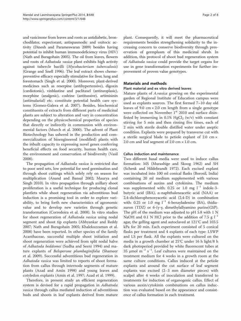
Figure 1 Mean % of callus induction of leaf segment and petiole expregulators after 4 weeks. Different letter(s) indicate a significant differenc
and kept jacketed with polyethylene bags in growthroom under the same culture conditions as for shoot dif-ferentiation. The plantlets were maintained under highhumidity for 8 weeks by spraying with liquid ½ strengthMS in every alternate day and slowly weaned to lowerhumidity by making small holes around the polyethylenebags of potted plantlets for a period of further 12 weeks.All the surviving plants (90%) were transferred to greenhouse with removal of polyethylene bag before exposureto the experimental field under natural photoperiod of9–11 h and a temperature of 20-29°C.
Statistical analysisAll the experiments described above were conducted ina completely randomized design and repeated twice.Means and standard error of means for all dependentvariables such as callus induction, shoot regeneration,shoot number and shoot length under different plantgrowth regulator concentrations were computed and de-termined the significant differences between means usingTukey test.
ResultsIncubation of petiole and leaf segment explants of Adha-toda vasica either on MS or SH medium supplementedwith different concentrations of NAA, IBA or 2,4-D (0.25or 1.0 mg l−1) in combination with TDZ, BA or 2iP (0.25or 1.0 mg l−1) produced callus within 2 weeks (Figures 1and 2). Callus primarily originated in petiole explantswhereas callus development was observed on cut off
lants of Adhatoda vasica on MS medium containing plant growthe between treatments at P ≤0.05 according to Tukey test.
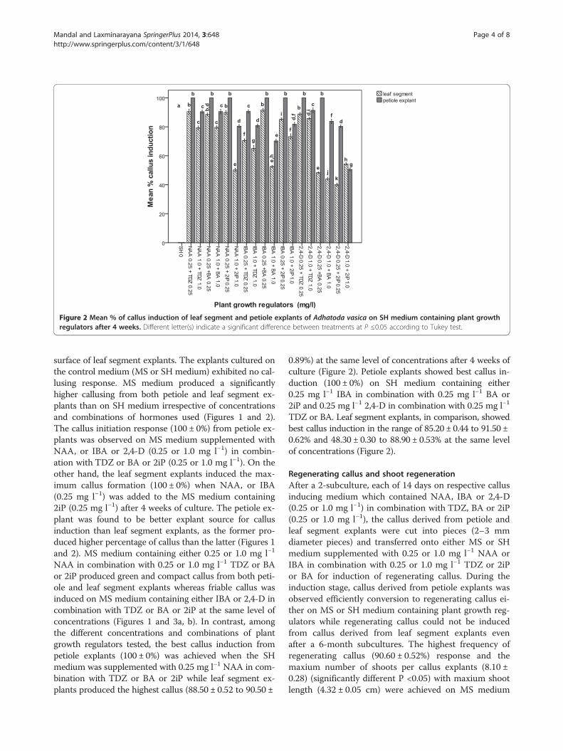
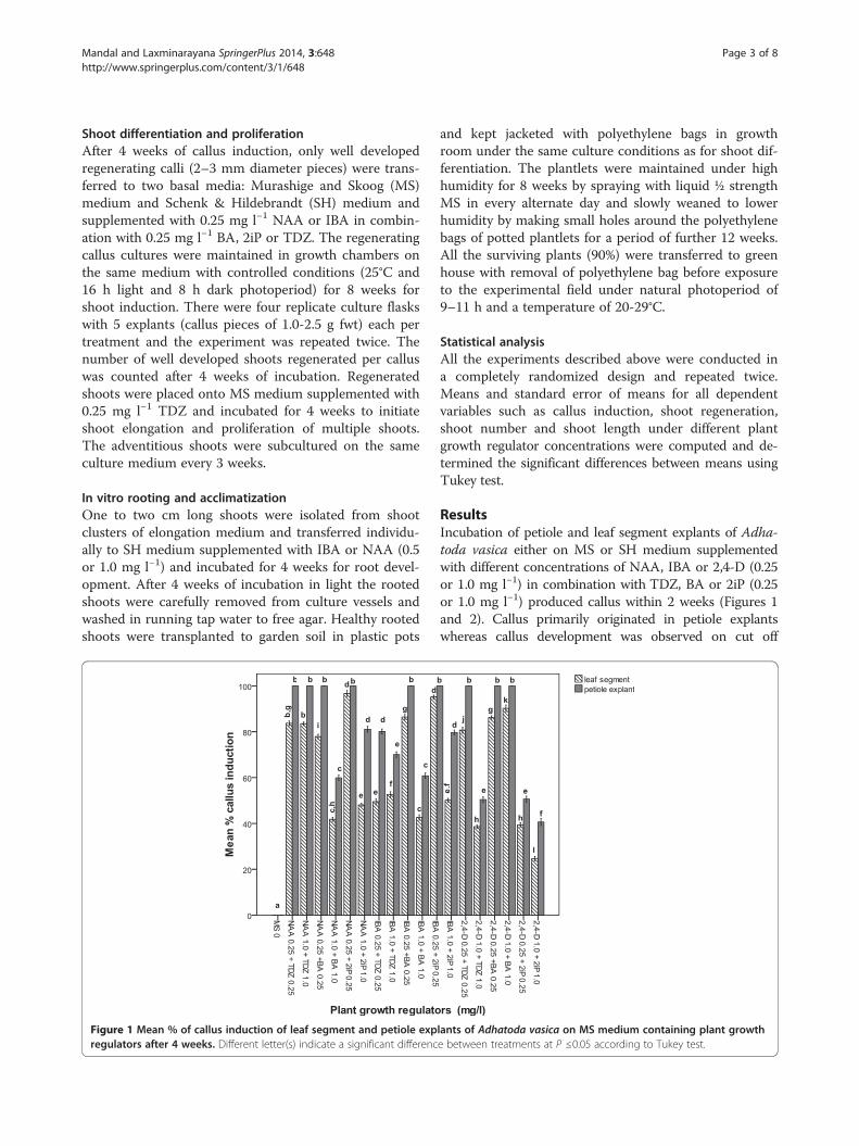
Figure 2 Mean % of callus induction of leaf segment and petiole explants of Adhatoda vasica on SH medium containing plant growthregulators after 4 weeks. Different letter(s) indicate a significant difference between treatments at P ≤0.05 according to Tukey test.
Mandal and Laxminarayana SpringerPlus 2014, 3:648 Page 4 of 8http://www.springerplus.com/content/3/1/648
surface of leaf segment explants. The explants cultured onthe control medium (MS or SH medium) exhibited no cal-lusing response. MS medium produced a significantlyhigher callusing from both petiole and leaf segment ex-plants than on SH medium irrespective of concentrationsand combinations of hormones used (Figures 1 and 2).The callus initiation response (100 ± 0%) from petiole ex-plants was observed on MS medium supplemented withNAA, or IBA or 2,4-D (0.25 or 1.0 mg l−1) in combin-ation with TDZ or BA or 2iP (0.25 or 1.0 mg l−1). On theother hand, the leaf segment explants induced the max-imum callus formation (100 ± 0%) when NAA, or IBA(0.25 mg l−1) was added to the MS medium containing2iP (0.25 mg l−1) after 4 weeks of culture. The petiole ex-plant was found to be better explant source for callusinduction than leaf segment explants, as the former pro-duced higher percentage of callus than the latter (Figures 1and 2). MS medium containing either 0.25 or 1.0 mg l−1
NAA in combination with 0.25 or 1.0 mg l−1 TDZ or BAor 2iP produced green and compact callus from both peti-ole and leaf segment explants whereas friable callus wasinduced on MS medium containing either IBA or 2,4-D incombination with TDZ or BA or 2iP at the same level ofconcentrations (Figures 1 and 3a, b). In contrast, amongthe different concentrations and combinations of plantgrowth regulators tested, the best callus induction frompetiole explants (100 ± 0%) was achieved when the SHmedium was supplemented with 0.25 mg l−1 NAA in com-bination with TDZ or BA or 2iP while leaf segment ex-plants produced the highest callus (88.50 ± 0.52 to 90.50 ±
0.89%) at the same level of concentrations after 4 weeks ofculture (Figure 2). Petiole explants showed best callus in-duction (100 ± 0%) on SH medium containing either0.25 mg l−1 IBA in combination with 0.25 mg l−1 BA or2iP and 0.25 mg l−1 2,4-D in combination with 0.25 mg l−1
TDZ or BA. Leaf segment explants, in comparison, showedbest callus induction in the range of 85.20 ± 0.44 to 91.50 ±0.62% and 48.30 ± 0.30 to 88.90 ± 0.53% at the same levelof concentrations (Figure 2).
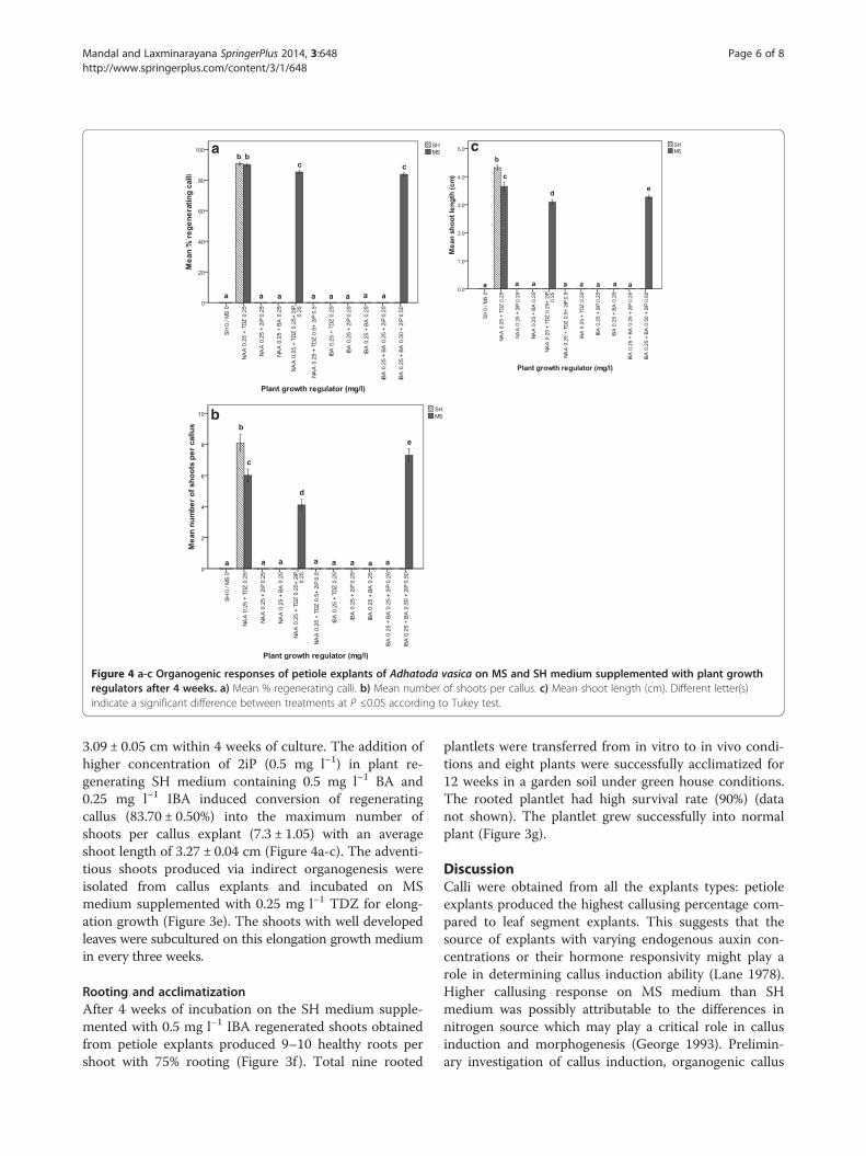
Regenerating callus and shoot regenerationAfter a 2-subculture, each of 14 days on respective callusinducing medium which contained NAA, IBA or 2,4-D(0.25 or 1.0 mg l−1) in combination with TDZ, BA or 2iP(0.25 or 1.0 mg l−1), the callus derived from petiole andleaf segment explants were cut into pieces (2–3 mmdiameter pieces) and transferred onto either MS or SHmedium supplemented with 0.25 or 1.0 mg l−1 NAA orIBA in combination with 0.25 or 1.0 mg l−1 TDZ or 2iPor BA for induction of regenerating callus. During theinduction stage, callus derived from petiole explants wasobserved efficiently conversion to regenerating callus ei-ther on MS or SH medium containing plant growth reg-ulators while regenerating callus could not be inducedfrom callus derived from leaf segment explants evenafter a 6-month subcultures. The highest frequency ofregenerating callus (90.60 ± 0.52%) response and themaxium number of shoots per callus explants (8.10 ±0.28) (significantly different P <0.05) with maxium shootlength (4.32 ± 0.05 cm) were achieved on MS medium
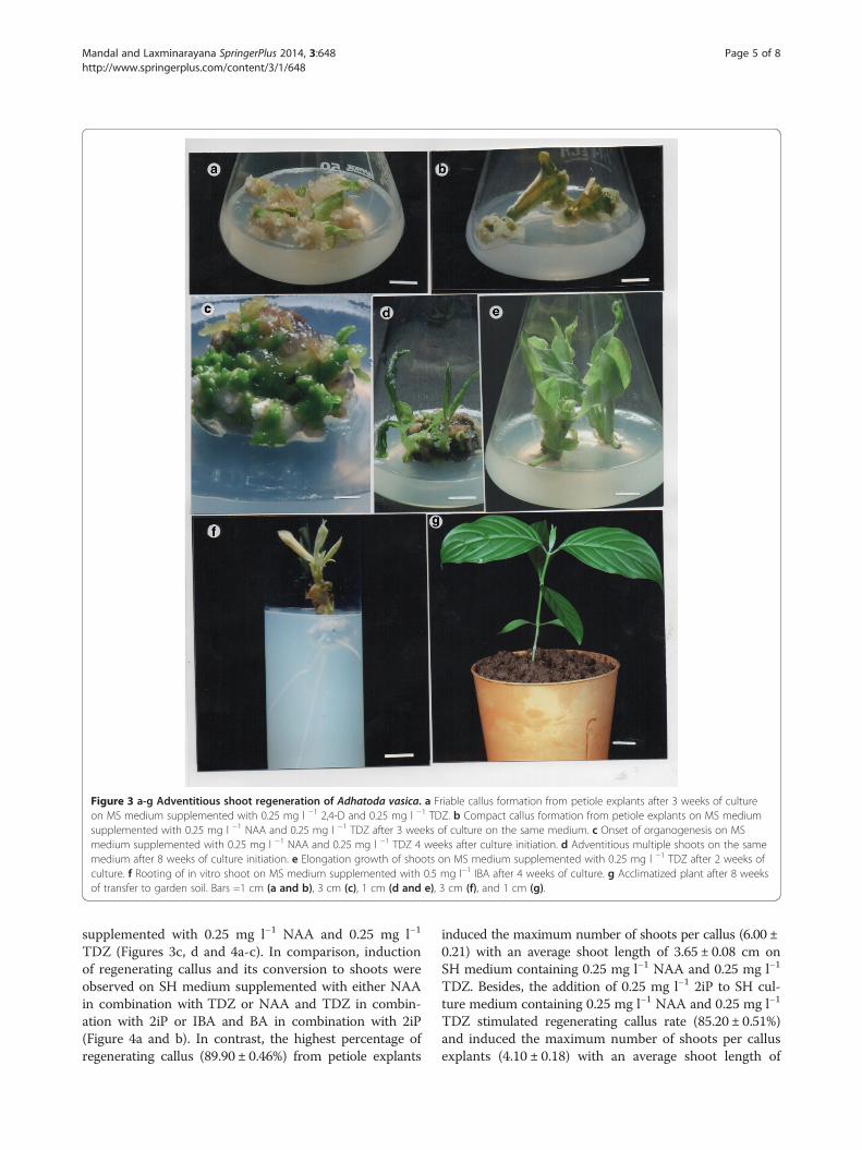
Figure 3 a-g Adventitious shoot regeneration of Adhatoda vasica. a Friable callus formation from petiole explants after 3 weeks of cultureon MS medium supplemented with 0.25 mg l −1 2,4-D and 0.25 mg l −1 TDZ. b Compact callus formation from petiole explants on MS mediumsupplemented with 0.25 mg l −1 NAA and 0.25 mg l −1 TDZ after 3 weeks of culture on the same medium. c Onset of organogenesis on MSmedium supplemented with 0.25 mg l −1 NAA and 0.25 mg l −1 TDZ 4 weeks after culture initiation. d Adventitious multiple shoots on the samemedium after 8 weeks of culture initiation. e Elongation growth of shoots on MS medium supplemented with 0.25 mg l −1 TDZ after 2 weeks ofculture. f Rooting of in vitro shoot on MS medium supplemented with 0.5 mg l−1 IBA after 4 weeks of culture. g Acclimatized plant after 8 weeksof transfer to garden soil. Bars =1 cm (a and b), 3 cm (c), 1 cm (d and e), 3 cm (f), and 1 cm (g).
Mandal and Laxminarayana SpringerPlus 2014, 3:648 Page 5 of 8http://www.springerplus.com/content/3/1/648
supplemented with 0.25 mg l−1 NAA and 0.25 mg l−1
TDZ (Figures 3c, d and 4a-c). In comparison, inductionof regenerating callus and its conversion to shoots wereobserved on SH medium supplemented with either NAAin combination with TDZ or NAA and TDZ in combin-ation with 2iP or IBA and BA in combination with 2iP(Figure 4a and b). In contrast, the highest percentage ofregenerating callus (89.90 ± 0.46%) from petiole explants
induced the maximum number of shoots per callus (6.00 ±0.21) with an average shoot length of 3.65 ± 0.08 cm onSH medium containing 0.25 mg l−1 NAA and 0.25 mg l−1
TDZ. Besides, the addition of 0.25 mg l−1 2iP to SH cul-ture medium containing 0.25 mg l−1 NAA and 0.25 mg l−1
TDZ stimulated regenerating callus rate (85.20 ± 0.51%)and induced the maximum number of shoots per callusexplants (4.10 ± 0.18) with an average shoot length of
a c
b
Figure 4 a-c Organogenic responses of petiole explants of Adhatoda vasica on MS and SH medium supplemented with plant growthregulators after 4 weeks. a) Mean % regenerating calli. b) Mean number of shoots per callus. c) Mean shoot length (cm). Different letter(s)indicate a significant difference between treatments at P ≤0.05 according to Tukey test.
Mandal and Laxminarayana SpringerPlus 2014, 3:648 Page 6 of 8http://www.springerplus.com/content/3/1/648
3.09 ± 0.05 cm within 4 weeks of culture. The addition ofhigher concentration of 2iP (0.5 mg l−1) in plant re-generating SH medium containing 0.5 mg l−1 BA and0.25 mg l−1 IBA induced conversion of regeneratingcallus (83.70 ± 0.50%) into the maximum number ofshoots per callus explant (7.3 ± 1.05) with an averageshoot length of 3.27 ± 0.04 cm (Figure 4a-c). The adventi-tious shoots produced via indirect organogenesis wereisolated from callus explants and incubated on MSmedium supplemented with 0.25 mg l−1 TDZ for elong-ation growth (Figure 3e). The shoots with well developedleaves were subcultured on this elongation growth mediumin every three weeks.
Rooting and acclimatizationAfter 4 weeks of incubation on the SH medium supple-mented with 0.5 mg l−1 IBA regenerated shoots obtainedfrom petiole explants produced 9–10 healthy roots pershoot with 75% rooting (Figure 3f ). Total nine rooted
plantlets were transferred from in vitro to in vivo condi-tions and eight plants were successfully acclimatized for12 weeks in a garden soil under green house conditions.The rooted plantlet had high survival rate (90%) (datanot shown). The plantlet grew successfully into normalplant (Figure 3g).
DiscussionCalli were obtained from all the explants types: petioleexplants produced the highest callusing percentage com-pared to leaf segment explants. This suggests that thesource of explants with varying endogenous auxin con-centrations or their hormone responsivity might play arole in determining callus induction ability (Lane 1978).Higher callusing response on MS medium than SHmedium was possibly attributable to the differences innitrogen source which may play a critical role in callusinduction and morphogenesis (George 1993). Prelimin-ary investigation of callus induction, organogenic callus
Mandal and Laxminarayana SpringerPlus 2014, 3:648 Page 7 of 8http://www.springerplus.com/content/3/1/648
formation and shoot regeneration potential of both leafsegments and petiole explants revealed that all explantstypes induced callus but only petiole explants derivedcallus was responsive to organogenic callus formationand shoot regeneration. These results are consistent withprevious reports that callus derived from explants ofvarying potentials results in different morphogenic re-sponses (Erisen et al. 2010; Ghimire et al. 2010; Piovanet al. 2010; Sun and Hong 2010; Wadl et al. 2011; Shaiket al. 2011; Lin et al. 2011; Kumar and Thomas 2012). Inthe present study, the highest number of shoot regener-ation (8.10 ± 0.28) in Adhatoda vasica was obtainedfrom petiole explants on MS medium supplementedwith 0.25 mg l−1 NAA and 0.25 mg l−1 TDZ within4 weeks of incubation of culture. Similarly, the positiveeffect of BA or Kin or TDZ or 2iP or Zn in combinationwith IAA, or NAA on in vitro shoot regenerationthrough organogenic differentiation has been reportedby several researchers (Bhagya et al. 2013; Erisen et al.2010; Unda et al. 2007; Lin et al. 2011; Mandal andLaxminarayana 2012; Perez-Jimenez et al. 2012; Wadlet al. 2011; Ghimire et al. 2010; Shen et al. 2007). Incomparison, though shoot organogenesis was observedon SH medium supplemented with NAA and TDZ com-bination, optimum induction of shoot regeneration inthe petiole derived callus required the addition of 2iP ei-ther on SH medium containing NAA and TDZ or 2iP incombination with IBA and BA for maximum shoot re-generation frequency (83.70 ± 0.50%) and adventitiousin vitro shoot regeneration (7.3 ± 1.05) whereas shoot or-ganogenesis was not observed on MS medium withthese combinations of plant growth regulators. Thehighest shoot production (8.10 ± 0.28), in the presentstudy, on MS medium containing NAA and TDZ wasconsistent with the previous reports of Ghimire et al.(2012), Korban et al. (1992) and Nabors et al. (1983)where TDZ and NAA have been more widely used forregeneration of adventitious shoots than other cytoki-nins and auxins respectively. Similarly, adventitiousshoot buds were induced from young leaf explants ofJatropha curcas on MS medium supplemented withTDZ, BA and IBA (Deore and Johnson 2008) and frommature leaf tissues of Pelargonium capitatum on MSmedium supplemented with TDZ, BA and NAA (Arshadet al. 2012). Likewise in many shoot multiplication stud-ies, 2iP was crucial as potential cytokinin for shoot for-mation, shoot multiplication and plant regeneration(Voyiatzi and Voyiatzis 1989; Shen et al. 2007; Mallonet al. 2011). It may be highlighted that cytokinins notonly contribute to maintaining a small population ofstem cells that continuously generates organs and tissuesbut also mediates light responses of stem cells that leadto initiation of primordia, the progenitors of new organs.Besides, a high local auxin concentration as a major
signal redirects the cytokinin stimulated growth to neworgan initiation and positioning (Murray et al. 2012). Inthe present study, rooting of in vitro shoots of Adhatodavasica developed readily on SH medium containing IBAwhich was similar to the reports of Erisen et al. (2010)who achieved optimum number roots of in vitro shootcuttings of Austragalus cariensis on MS medium supple-mented with IBA.In Adhatoda vasica, callus derived from mature ex-
plants of petiole produced adventitious buds both onMS medium supplemented with NAA and TDZ and SHmedium either supplemented with NAA and TDZ orwith 2iP, NAA and TDZ or with 2iP, IBA and BA. Al-though, it was reported that callus derived from matureexplants was not amenable to regeneration (Amin et al.1997; Azad et al. 1999), adventitious shoots were in-duced from callus derived from mature internode ex-plants of this species on medium containing BA andNAA (Azad and Amin 1998).In conclusion, this research study presents the first re-
port of plant regeneration via organogenesis of Adhatodavasica from petiole explants. Although both petiole andleaf segment explants produced callus, higher potential fororganogenic differentiation from callus derived frompetiole explants was observed on MS medium than on SHmedium supplemented with 0.25 mg l−1 NAA and0.25 mg l−1 TDZ. The protocol presented in this studymay provide a high efficiency regeneration system for suc-cessful regeneration of adventitious shoots for ex situpreservation of Adhatoda vasica as well as genetic im-provement studies for pharmaceutical uses and future re-search investigations.
AbbreviationsBA: 6-benzyladenine; IBA: Indole-3-butyric acid; Kin: kinetin;2, 4-D: 2, 4-dichlorophenoxyacetic acid; 2iP: 6-(γ-γ, dimethylallyamino purine);NAA: α-naphthaleneacetic acid; TDZ: Thidiazuron; Zn: Zeatin.
Competing interestsThe authors declare that they have no competing interests.
Authors’ contributionsJM carried out the research work; UL carried out statistical analysis andgraphics design. Both authors read and approved the final manuscript.
AcknowledgmentsThe author expresses grateful thanks to Director, National Council ofEducational Research and Training (NCERT), New Delhi-16, India and Principal,RIE, Bhopal for providing support and facility for carrying out research studiesin Regional Institute of Education, Shyamla Hills, Bhopal, India.
Author details1Department of Education in Science and Mathematics, Regional Institute ofEducation, National Council of Educational Research and Training, ShyamlaHills, Bhopal 462013, India. 2Department of Education, Regional Institute ofEducation, National Council of Educational Research and Training, Mysore570006, India.
Received: 27 May 2014 Accepted: 24 October 2014Published: 3 November 2014
Mandal and Laxminarayana SpringerPlus 2014, 3:648 Page 8 of 8http://www.springerplus.com/content/3/1/648
ReferencesAbhyankar G, Reddy VD (2007) Rapid micropropagation via axillary bud
propliferation of Adhatoda vasica Nees from nodal segments. Indian J Exp Biol45:268–271
Amin MN, Azad MAK, Begum F (1997) In vitro plant regeneration from leaf derivedcallus cultures of Adhatoda vasica Nees. Plant Tissue Cult 7(2):109–115
Anand Y, Bansal YK (2002) Synthetic seeds: a novel approach of in vitro plantformation in vasaka (Adhatoda vasica Nees.). Plant Biotechnol 19(3):159–162
Arshad M, Silvestre J, Merlina G, Dumat C, Pinelli E, Kallerhoff J (2012) Thidiazuron-induced shoot organogenesis from mature leaf explants of scented Pelargoniumcapitatum cultivars. Plant Cell Tissue Organ Cult 108:315–322
Azad MAK, Amin MN (1998) In vitro regeneration of plantlets from internodeexplants of Adhatoda vasica Nees. Plant Tissue Cult 8:27
Azad MAK, Amin MN, Begum F (1999) In vitro rapid regeneration of planlets fromcotyledon explants of Adhatoda vasica Nees. Plant Tissue Cult 9:121
Bhagya N, Chandrashekar KR, Karun A, Bhavyashree U (2013) Plantletregeneration through indirect shoot organogenesis and somaticembryogenesis in Justicia gendarussa Burm. f., a medicinal plant. J PlantBiochem Biotechnol 22(4):474–482
Corredoira E, Ballester A, Vieitez AM (2008) Thidiazuron-induced high-frequencyplant regeneration from leaf explants of Paulownia tomentosa mature trees.Plant Cell Tissue Organ Cult 95:197–208
Deore AC, Johnson TS (2008) High-frequency plant regeneration from leaf-disccultures of Jatropha curcas L.: an important biodiesel plant. Plant BiotechnolRep 2:7–11
Dinesh KS, Parameswaran S (2009) Micropropagation and organogenesis inAdhatoda vasica for the estimation of vaccine. Pharmacog Mag 5:359–363
Erisen S, Yorgancilar M, Atalay E, Babaoglu M (2010) Prolific shoot regeneration ofAstragalus cariensis Boiss. Plant Cell Tissue Organ Cult 100:229–233
George EF (1993) Plant Propagation by Tissue Culture, Part 1 and 2. In: Practice,2nd edition. Exegetics Ltd, Edington, England. The Technology, pp. 641
Ghimire BK, Seong ES, Goh EJ, Kim NY, Kang WH, Kim EH, Yu CY, Chung IM(2010) High- frequency direct shoot regeneration from Drymaria cordataWilld. leaves. Plant Cell Tissue Organ Cult 100:209–217
Ghimire BK, Yu CY, Chung IM (2012) Direct shoot organogenesis and assessmentof genetic stability in regenerants of Solanum aculeatissimum Jacq. Plant CellTissue Organ Cult 108:455–464
Gomez-Galera S, Pelacho AM, Gene A, Capell T, Christou P (2007) The geneticmanipulation of medicinal and aromatic plants. Plant Cell Rep 26:1689–1715
Grange JM, Snell NJ (1996) Activity of bromehexine and ambroxol, semisyntheticderivatives of vasicine from the Indian shrub Adhatoda vasica againstMycobacterium tuberculosis in vitro. J Ethnopharmacol 50(1):49–53
Khalekuzzaman M, Rahman MS, Rashid MH, Hossain MS (2008) High frequencyin vitro propagation of Adhatoda vasica Nees through shoot tip and nodalexplants culture. J Bio Sci 16:35–39
Korban SS, O’Connor PA, Elcheidy A (1992) Effects of thidiazuron,naphthaleneacetic acid, dark incubation and genotype on shootorganogenesis from Malus leaves. J Hort Sci 67:341–349
Kumar GK, Thomas TD (2012) High frequency somatic embrogenesis andsynthetic seed production in Clitorea ternatea Linn. Plant Cell Tissue Organ Cult110:141–151
Lane WD (1978) Regeneration of apple plants from shoot meristem tips. Plant Sci Lett13:281–285
Lin GZ, Zhao XM, Hong SK, Lian YJ (2011) Somatic embryogenesis and shootorganogenesis in the medicinal plant Pulsatilla koreana Nakai. Plant CellTissue Organ Cult 106:93–103
Mallon R, Rodriguez-Oubina J, Gonzalez ML (2011) Shoot regeneration fromin vitro-derived leaf and root explants of Centaurea ultreiae. Plant Cell TissueOrgan Cult 106:523–530
Mandal J, Laxminarayana U (2012) In vitro organogenesis of Quisqualis indicaLinn.- an ornamental creeper. Am J Plant Sciences 3(9):1272–1282.doi:10.4236/ajps.2012.39154
Maurya S, Singh D (2010) In vitro callus culture of Adhatoda vasica: a medicinalplant. Ann Biol Res 1(4):57–60
Murashige T, Skoog F (1962) A revised medium for rapid growth and bioassayswith tobacco tissue cultures. Physiol Plant 15:473–497
Murch SJ, KrishnaRaj S, Saxena PK (2000) Phytopharmaceuticals: Mass-production,standardization and conservation. Sci Rev Alt Med 4:39–43
Murray JAH, Jones A, Godin C, Traas J (2012) Systems analysis of shoot apicalmeristem growth and development: integrating hormonal and mechanicalsignaling. Plant Cell 24:3907–3919
Nabors MW, Heyser JW, Dykes TA, DeMott KJ (1983) Long-duration, highfrequency plant regeneration from cereal tissue cultures. Planta 157:385–391
Nath S, Buragohain AK (2005) Micropropagation of Adhatoda vasica Nees- Awoody medicinal plant by shoot tip culture. Indian J Biotechnol 4:396–399
Perez-Jimenez M, Carrillo-Navarro A, Cos-Terrer J (2012) Regeneration of peach(Prunus persica L. Batsch) cultivars and Prunus persica × Prunus dulcisrootstocks via organogenesis. Plant Cell Tissue Organ Cult 108:55–62
Piovan A, Caniato R, Cappelletti EM, Filippini R (2010) Organogenesis from shootsegments and via callus of endangered Kosteletzkya pentacarpos (L.) Ledeb.Plant Cell Tissue Organ Cult 100:309–315
Schenk RU, Hildebrandt AC (1972) Medium and techniques for the induction andgrowth of monocotyledonous and dicotyledonous plant cell cultures. Can JBot 50:199–204
Shaik S, Singh N, Nicholas A (2011) Cytokinin-induced organogenesis in Lessertia(Sutherlandia) frutescens L. using hypocotyl and cotyledon explants affectsyields of L-canavanine in shoots. Plant Cell Tissue Organ Cult 105:439–446
Shameer MC, Saeeda VP, Madhusoodanan PV, Benjamin S (2009) Directorganogenesis and somatic embryogenesis in Beloperone plumbaginifolia(Jacq.) Nees. Indian J Biotechnol 8(1):132–135
Shen X, Chen J, Kane ME (2007) Indirect shoot organogenesis from leaves ofDieffenbachia cv. Camouflage. Plant Cell Tissue Organ Cult 89:83–90
Singh RP, Padmavathi B, Rao AR (2000) Modulatory influence of Adhatoda vesica(Justicia adhatoda) leaf extract on the enzymes of xenobiotic metabolism,antioxidant status and lipid peroxidation in mice. Mol Cell Biochem213(1–2):99–109
Singh TP, Singh OM, Singh HB (2011) Adhatoda vasica Nees: Phytochemical andPharmacological Profile. J Nat Prod 1:29–39
Sudha CG, Seeni S (1994) In vitro multiplication and field establishment ofAdhatoda beddomei C B Clarke. Plant Cell Rep 13(3–4):203–207. doi:10.1007/BF00239893
Sun YL, Hong SK (2010) Effects of plant growth regulators and L- glutamic acidon shoot organogenesis in the halophyte Leymus chinensis (Trin.). Plant CellTissue Organ Cult 100:317–328
Unda F, Kalynyak P, Riseman A (2007) Organogenesis plant regeneration fromleaf explants of Exacum Styer Group. Plant Cell Tissue Organ Cult 89:105–111
Vasil IK (2008) A history of plant biotechnology: from the Cell Theory ofSchleiden and Schwann to biotech crops. Plant Cell Rep 27:1423–1440
Voyiatzi C, Voyiatzis (1989) In vitro shoot proliferation rate of Dieffenbachia exoticacultivar ‘Marianna’ as affected by cytokinins, the number of recultures andthe temperature. Sci Hort 40:163–169
Wadl PA, Dattilo AJ, Vito LM, Trigiano RN (2011) Shoot organogenesis and plantregeneration in Pityopsis ruthii. Plant Cell Tissue Organ Cult 106:513–516
doi:10.1186/2193-1801-3-648Cite this article as: Mandal and Laxminarayana: Indirect shootorganogenesis from leaf explants of Adhatoda vasica Nees. SpringerPlus2014 3:648.
Submit your manuscript to a journal and benefi t from:
7 Convenient online submission
7 Rigorous peer review
7 Immediate publication on acceptance
7 Open access: articles freely available online
7 High visibility within the fi eld
7 Retaining the copyright to your article
Submit your next manuscript at 7 springeropen.com