Isolation, Identification and Antibiotic Susceptibility of...
13
IOSR Journal of Pharmacy and Biological Sciences (IOSR-JPBS) e-ISSN:2278-3008, p-ISSN:2319-7676. Volume 11, Issue 4 Ver. II (Jul. - Aug.2016), PP 27-39 www.iosrjournals.org DOI: 10.9790/3008-1104022739 www.iosrjournals.org 27 | Page Isolation, Identification and Antibiotic Susceptibility of pathogenic Bacteria Isolated from Clinical Samples Ehsan F. Hussein* *University of Summer / College of Basic Education / Department of Science Abstract: 150 Samples have been collected, through the period from January/2016 to June/2016 . This for isolation and identification of pathogenic gram negative bacteria. The samples were involve 50 burns samples for isolate Pseudomonas aeruginosa, 50 stool samples for isolate Escherichia coli and 50 urine samples for isolate Enterobacter cloacae and Proteus mirabilis. The isolates were identified by ten biochemical tests as well as the sensitivity and resistant were tested by thirteen type of antibiotics. These antibiotics explained different mode of action in their activity, were resistant, moderate and sensitive, some of them have significant differences when comparison together and some have no. The Trimethoprim, Amikacin, Aztreonam and Novobiocin have high activity against Ps. aeruginosa, E. coli, E. cloacae and P. mirabilis respectively, as well as the Aztreonam was more active when comparison the activity of these antibiotics together. Keyword: Pathogenic gram negative bacteria, Resistant, Antibiotic, biochemical tests. I. Introduction Gram negative bacteria play important role in infectious diseases, but the Pseudomonas aeruginosa, Escherichia coli, Enterobacter cloacae and Proteus mirabilis now become the broad spectrum species involved in nosocomial infections (Anuj Nautiyal et al., 2015).Antibiotics identified, action modes and bacterial resistance mechanisms have been researched topics in academia until recently in the pharmaceutical applications (Davies and Davies., 2010). Bacteria can be resistant one or more type of antibiotics, the mechanisms by which bacteria resist antibiotics include, alter the receptor for the antibiotic, decrease antibiotic amount that reaches the receptor by preventing entry or removal, antibiotic alteration or destroying and develop resistant metabolic pathways (Fisher and Mobashery., 2010). The Pseudomonas aeruginosa are aerobic, motile gram negative rod bacteria worldwide distributed and found as skin flora and in the majority of environmental sources (Jabalameli et al., 2011). It is can survive in different environments such as aquatic and terrestrial and contributes strongly to nosocomial infections and infected of immunocompromised peoples (Eguchi et al., 2013). This bacteria has widespread antibiotic resistance among their clinical isolates, as well as considered as one of the most important bacterial agents associated with nosocomial infections (Mirsalehian et al., 2010), also known as the most common bacterial agents in the burn patients (Azimi et al., 2015). This bacteria causes invasive infections in burn and this lead to sepsis from invasive burn wound infection (Alhazmi., 2015). The inherent antibiotics resistance mechanisms for Pseudomonas aeruginosa involve lower outer membrane permeability, increased expression of efflux pumps for different specificity and presence beta- lactamase give this bacteria resistance mechanisms to commonly used antibiotics (Strateva and Yordanov 2009). This is true for several opportunistic pathogenic bacteria like Ps. aeruginosa which causes number of chronic infections due to resistant to modern antimicrobial therapies (Gellatly and Hancock 2013). Whether in the clinic or in the natural environment this bacteria encounters multiple classes of traditional antibiotics, antiseptics and disinfectants (Murray et al., 2015). The Escherichia coli are a microorganism that commonly live in the large intestine of humans and other warm blooded animals, this is the most prevalent facultative gram negative bacteria in the human fecal flora and usually inhabit the colon as commensal microorganism (Manikandan and Amsath., 2014). Some strains of this bacteria can cause disease inside and outside of the gastrointestinal tract (Manikandan and Amsath., 2014). This bacteria is used in the variety of fields in both the industrial and medical, also the most used bacterium in the technology of recombinant DNA (Yoo et al., 2009). E. coli is has resistant to therapeutic levels of the first β-lactam that act on cell wall, and this because of its outer membrane is barrier to this type of antibiotics (Allocati et al., 2013). Also, this bacteria have resistant to several different types of antibiotics with distinct mechanisms of action (Johnson et al., 2012). The emergence and spread of resistant against multidrug for strains of Escherichia coli is complicating and become difficult in the treatment of several serious infections caused by this bacteria (Allocati et al., 2013). Family of Enterobacteriaceae, specillay of Escherichia coli. are the most frequent cause of hospital and this lead to community acquired infections (Pitout., 2012).
Transcript of Isolation, Identification and Antibiotic Susceptibility of...
IOSR Journal of Pharmacy and Biological Sciences (IOSR-JPBS)
e-ISSN:2278-3008, p-ISSN:2319-7676. Volume 11, Issue 4 Ver. II (Jul. - Aug.2016), PP 27-39
www.iosrjournals.org
DOI: 10.9790/3008-1104022739 www.iosrjournals.org 27 | Page
Isolation, Identification and Antibiotic Susceptibility of
pathogenic Bacteria Isolated from Clinical Samples
Ehsan F. Hussein* *University of Summer / College of Basic Education / Department of Science
Abstract: 150 Samples have been collected, through the period from January/2016 to June/2016 . This for
isolation and identification of pathogenic gram negative bacteria. The samples were involve 50 burns samples
for isolate Pseudomonas aeruginosa, 50 stool samples for isolate Escherichia coli and 50 urine samples for
isolate Enterobacter cloacae and Proteus mirabilis. The isolates were identified by ten biochemical tests as well
as the sensitivity and resistant were tested by thirteen type of antibiotics. These antibiotics explained different
mode of action in their activity, were resistant, moderate and sensitive, some of them have significant
differences when comparison together and some have no. The Trimethoprim, Amikacin, Aztreonam and
Novobiocin have high activity against Ps. aeruginosa, E. coli, E. cloacae and P. mirabilis respectively, as well
as the Aztreonam was more active when comparison the activity of these antibiotics together.
Keyword: Pathogenic gram negative bacteria, Resistant, Antibiotic, biochemical tests.
I. Introduction Gram negative bacteria play important role in infectious diseases, but the Pseudomonas aeruginosa,
Escherichia coli, Enterobacter cloacae and Proteus mirabilis now become the broad spectrum species involved
in nosocomial infections (Anuj Nautiyal et al., 2015).Antibiotics identified, action modes and bacterial
resistance mechanisms have been researched topics in academia until recently in the pharmaceutical applications
(Davies and Davies., 2010). Bacteria can be resistant one or more type of antibiotics, the mechanisms by which
bacteria resist antibiotics include, alter the receptor for the antibiotic, decrease antibiotic amount that reaches the
receptor by preventing entry or removal, antibiotic alteration or destroying and develop resistant metabolic
pathways (Fisher and Mobashery., 2010).
The Pseudomonas aeruginosa are aerobic, motile gram negative rod bacteria worldwide distributed and
found as skin flora and in the majority of environmental sources (Jabalameli et al., 2011). It is can survive in
different environments such as aquatic and terrestrial and contributes strongly to nosocomial infections and
infected of immunocompromised peoples (Eguchi et al., 2013). This bacteria has widespread antibiotic
resistance among their clinical isolates, as well as considered as one of the most important bacterial agents
associated with nosocomial infections (Mirsalehian et al., 2010), also known as the most common bacterial
agents in the burn patients (Azimi et al., 2015). This bacteria causes invasive infections in burn and this lead to
sepsis from invasive burn wound infection (Alhazmi., 2015).
The inherent antibiotics resistance mechanisms for Pseudomonas aeruginosa involve lower outer
membrane permeability, increased expression of efflux pumps for different specificity and presence beta-
lactamase give this bacteria resistance mechanisms to commonly used antibiotics (Strateva and Yordanov 2009).
This is true for several opportunistic pathogenic bacteria like Ps. aeruginosa which causes number of chronic
infections due to resistant to modern antimicrobial therapies (Gellatly and Hancock 2013). Whether in the clinic
or in the natural environment this bacteria encounters multiple classes of traditional antibiotics, antiseptics and
disinfectants (Murray et al., 2015).
The Escherichia coli are a microorganism that commonly live in the large intestine of humans and
other warm blooded animals, this is the most prevalent facultative gram negative bacteria in the human fecal
flora and usually inhabit the colon as commensal microorganism (Manikandan and Amsath., 2014). Some
strains of this bacteria can cause disease inside and outside of the gastrointestinal tract (Manikandan and
Amsath., 2014). This bacteria is used in the variety of fields in both the industrial and medical, also the most
used bacterium in the technology of recombinant DNA (Yoo et al., 2009).
E. coli is has resistant to therapeutic levels of the first β-lactam that act on cell wall, and this because of
its outer membrane is barrier to this type of antibiotics (Allocati et al., 2013). Also, this bacteria have resistant to
several different types of antibiotics with distinct mechanisms of action (Johnson et al., 2012). The emergence
and spread of resistant against multidrug for strains of Escherichia coli is complicating and become difficult in
the treatment of several serious infections caused by this bacteria (Allocati et al., 2013). Family of
Enterobacteriaceae, specillay of Escherichia coli. are the most frequent cause of hospital and this lead to
community acquired infections (Pitout., 2012).
Isolation, Identification and Antibiotic Susceptibility of pathogenic Bacteria Isolated from Clinical..
DOI: 10.9790/3008-1104022739 www.iosrjournals.org 28 | Page
The use of antimicrobial agents in wrong ways for animals and humans treatment lead to the
emergence and diffusion of the several antibiotic resistant strains of Escherichia coli that also capable to infect
other humans through either direct contact with animals or through consumption of contaminated food with this
bacteria (Ewers at al., 2012). Escherichia coli have ability to survive and adapt in different extra intestinal
locations and to produce resistances between humans, animals and their products through environment by
several transmission pathways (Ewers at al., 2012).
The Enterobacter belonging to the family of Enterobacteriaceae which is common Gram negative, rod
shaped and non spore forming bacteria, live as facultative anaerobic and have been reported as important
opportunistic and multi resistant bacterial pathogens and causes infection for humans (Davin-Regli and Pages.,
2015). There are two main known species for this genus, which are E. aerogenes and E. cloacae have take non
clinical significance as opportunistic bacteria and have emerged as nosocomial pathogens (Mezzatesta et al.,
2012). Enterobacter cloacae now is the most frequently observed in clinical isolate among Enterobacter species
but, the pathogenicity and factors contributing in the disease caused by this bacteria still not understood (Davin-
Regli and Pages., 2015). This bacteria has been associations with clinical samples particular in urines with
property of antibiotics resistant (Pestourie et al.,2014). E. cloacae has several mechanisms for resistant of wide
range of antibiotics (Davin-Regli and Pages., 2015). Various studies on antibiotic resistance E .cloacae which
associated with low level susceptibility to several antibiotics by inducing the over expression of the efflux pump
(Perez et al., 2012).
The Proteus is genus belonging to the family of Enterobactericeae and of gram negative bacteria, this
is widespread in the environment and makes third as the cause of hospital acquired infections it frequently
causes nosocomial infections of the urinary tract (Bahashwan., 2013). This genus consists of four species:
Proteus vulgaris, Proteus penneri, Proteus myxofaciens and Proteus mirabilis (Penner et.al., 1984). The latter
is the most causative agent of majority Proteus infections (Feglo et al., 2010) and the most one of bacteria that
responsible for urinary tract infections (Nabeela et al., 2004).
Proteus mirabilis is generally susceptible to most antibiotics, including beta-lactams, aminoglycosides
and imipenem but it has intrinsic resistance to nitrofurantoin, tetracycline and colistin (Hara et al., 2000).
However, the multidrug resistant strains of Proteus species has also been reported worldwide (Singla et al.,
2015). As well as has ability to resistant to several different types of antibiotics and called multi antibiotics
resistant (Dadheech et al., 2015). In addition to including recent evidences of multidrug resistance P. mirabilis
(Wong et al., 2013).
II. Materials and Methods Isolation of pathogenic gram negative bacterial:.
Table:1. Percentage of all isolated Bacterial type Site of Collection Number No. of isolates Samples (%)
Ps. aeruginosa Burns 50 22 30.137
E. coli Stool 50 18 24.657
E. cloacae
Urine
50
15 20.548
P. mirabilis 18 24.657
Total All isolates 48.667 % 150 73 99.999
This table showed the percent of all isolated was 48.667, as well as Ps. aeruginosa isolated from burn,
E. coli from Stool, E. cloacae from Urine and P. mirabilis from Urine were 30.137, 24.657, 20.548 and 24.657
respectively.
Identification of isolates:. Table:2. Identification of pathogenic bacteria by ten biochemical tests
Bacterial Gram stain Indol Urease Oxidase Catalase Citrate MR VP H2S Gas
Ps. aeruginosa* - - - + + + - - - -
E. coli** - + - - + - + - - +
E. cloacae*** - - - - + + - + - +
P. mirabilis**** - - + - + + + + + +
This table explained the results of biochemical tests for identified isolates after growing on three plates,
were Nutrient agar, Mac Conkey’s agar and Blood agar. These bacteria were identified by biochemical tests
according to the Society of American Bacteriologists (Pelczar et al., 1957). The (+) refer to positive and (–) to
negtive (*Cheesbrough., 2006 ; **Otoikhian & Tanimowo 2016 ; ***Stiles & LAI., 1981 and ****Holt et al.,
1994)
Isolation, Identification and Antibiotic Susceptibility of pathogenic Bacteria Isolated from Clinical..
DOI: 10.9790/3008-1104022739 www.iosrjournals.org 29 | Page
Identification of antibiotics sensitivity and resistant for bacterial samples:.
Antibiotic susceptibility test:
Antibiotic sensitivity tests for these bacteria were carried out by disc diffusion technique on Muller
Hinton agar plates (Bauer et al., 1966).
Statistical analysis: The statistical analysis was performed with the Statistical Package for Social Sciences (SPSS) version
20 for calculate Mean, Medium Std. Deviation and Std. Error of Mean as well as the significance between
antibiotics according to One-Way ANOVA by descriptive exclude cases analysis by analysis with LSD at 95%
confidence and significant level (P=0.05)
III. Results One hundred and fifty Samples have been collected, through the period from January/2016 to
June/2016. This for isolation and identification of pathogenic gram negative bacteria. The samples were involve
50 burns samples for isolate Pseudomonas aeruginosa, 50 stool samples for isolate Escherichia coli and 50
urine samples for isolate Enterobacter cloacae and Proteus mirabilis.
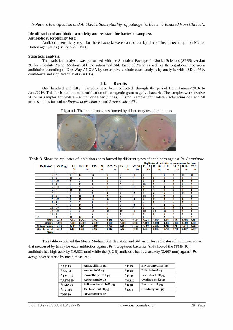
Figure:1. The inhibition zones formed by different types of antibiotics
Table:3. Show the replicates of inhibition zones formed by different types of antibiotics against Ps. Aeruginosa
This table explained the Mean, Median, Std. deviation and Std. error for replicates of inhibition zones
that measured by (mm) for each antibiotics against Ps. aeruginosa bacteria. And showed the (TMP 10)
antibiotic has high activity (10.533 mm) while the (CC 5) antibiotic has low activity (3.667 mm) against Ps.
aeruginosa bacteria by mean measured.
*AX 15 Amoxicillin15 μg *E 15 Erythromycin15 μg
*AK 30 Amikacin30 μg *R 40 Rifaximin40 μg
*TMP 10 Trimethoprim10 μg *P 10 Penicillin-G10 μg
*ATM 30 Aztreonam30 μg *OA 2 Oxolinic acid2 μg
*SMZ 25 Sulfamethoxazole25 μg *B 10 Bacitracin10 μg
*PY 100 Carbnicillin100 μg *CC 5 Clindamycin5 μg
*NV 30 Novobiocin30 μg
Isolation, Identification and Antibiotic Susceptibility of pathogenic Bacteria Isolated from Clinical..
DOI: 10.9790/3008-1104022739 www.iosrjournals.org 30 | Page
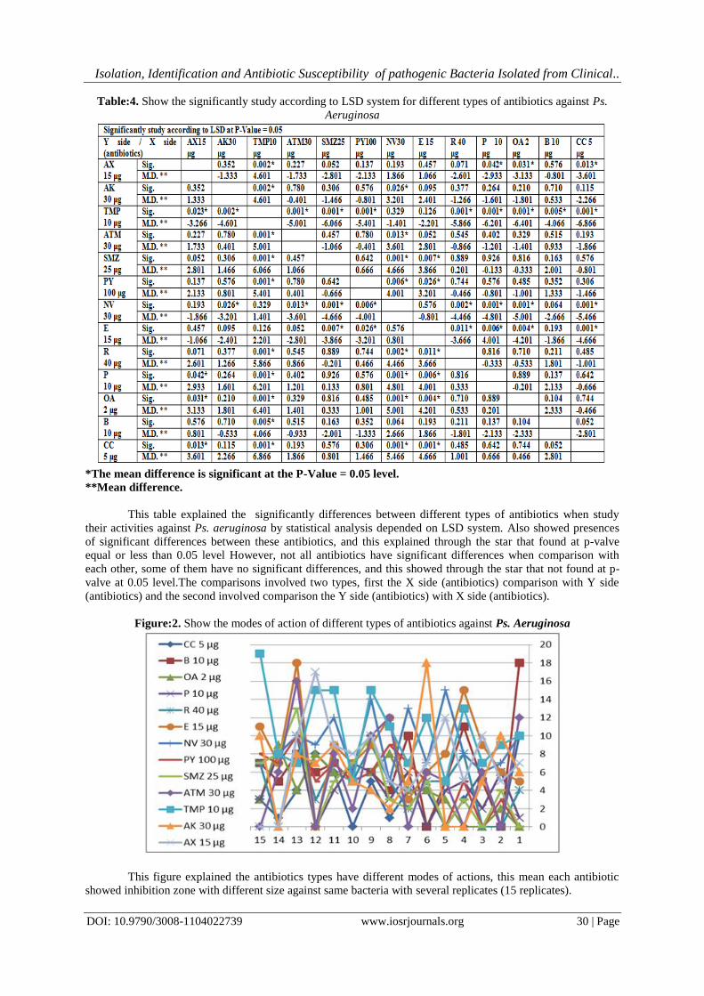
Table:4. Show the significantly study according to LSD system for different types of antibiotics against Ps.
Aeruginosa
*The mean difference is significant at the P-Value = 0.05 level. **Mean difference.
This table explained the significantly differences between different types of antibiotics when study
their activities against Ps. aeruginosa by statistical analysis depended on LSD system. Also showed presences
of significant differences between these antibiotics, and this explained through the star that found at p-valve
equal or less than 0.05 level However, not all antibiotics have significant differences when comparison with
each other, some of them have no significant differences, and this showed through the star that not found at p-
valve at 0.05 level.The comparisons involved two types, first the X side (antibiotics) comparison with Y side
(antibiotics) and the second involved comparison the Y side (antibiotics) with X side (antibiotics).
Figure:2. Show the modes of action of different types of antibiotics against Ps. Aeruginosa
This figure explained the antibiotics types have different modes of actions, this mean each antibiotic
showed inhibition zone with different size against same bacteria with several replicates (15 replicates).
Isolation, Identification and Antibiotic Susceptibility of pathogenic Bacteria Isolated from Clinical..
DOI: 10.9790/3008-1104022739 www.iosrjournals.org 31 | Page
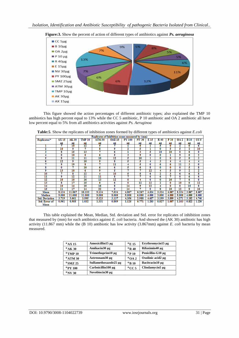
Figure:3. Show the percent of action of different types of antibiotics against Ps. aeruginosa
This figure showed the action percentages of different antibiotic types; also explained the TMP 10
antibiotics has high percent equal to 13% while the CC 5 antibiotic, P 10 antibiotic and OA 2 antibiotic all have
low percent equal to 5% from all antibiotics activities against Ps. Aeruginoa
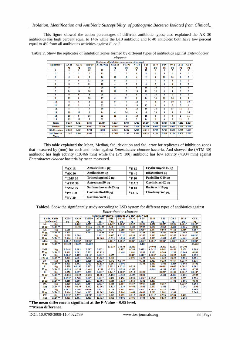
Table:5. Show the replicates of inhibition zones formed by different types of antibiotics against E.coli
This table explained the Mean, Median, Std. deviation and Std. error for replicates of inhibition zones
that measured by (mm) for each antibiotics against E. coli bacteria. And showed the (AK 30) antibiotic has high
activity (11.867 mm) while the (B 10) antibiotic has low activity (3.867mm) against E. coli bacteria by mean
measured.
*AX 15 Amoxicillin15 μg *E 15 Erythromycin15 μg
*AK 30 Amikacin30 μg *R 40 Rifaximin40 μg
*TMP 10 Trimethoprim10 μg *P 10 Penicillin-G10 μg
*ATM 30 Aztreonam30 μg *OA 2 Oxolinic acid2 μg
*SMZ 25 Sulfamethoxazole25 μg *B 10 Bacitracin10 μg
*PY 100 Carbnicillin100 μg *CC 5 Clindamycin5 μg
*NV 30 Novobiocin30 μg

Isolation, Identification and Antibiotic Susceptibility of pathogenic Bacteria Isolated from Clinical..
DOI: 10.9790/3008-1104022739 www.iosrjournals.org 32 | Page
Table:6. Show the significantly study according to LSD system for different types of antibiotics against E. coli
*The mean difference is significant at the P-Value = 0.05 level.
**Mean difference.
This table explained the significantly differences between different types of antibiotics when study
their activities against E. coli by statistical analysis depended on LSD system. Also showed presences of
significant differences between these antibiotics, and this explained through the star that found at p-valve equal
or less than 0.05 level.However, not all antibiotics have significant differences when comparison with each
other, some of them have no significant differences, and this showed through the star that not found at p-valve at
0.05 level. The comparisons involved two types, first the X side (antibiotics) comparison with Y side
(antibiotics) and the second involved comparison the Y side (antibiotics) with X side (antibiotics).
Figure:4. Show the modes of action of different types of antibiotics against E. coli
This figure explained the antibiotics types have different modes of actions, this mean each antibiotic
showed inhibition zone with different size against same bacteria with several replicates (15 replicates).
Figure:5. Show the percent of action of different types of antibiotics against E. coli
Isolation, Identification and Antibiotic Susceptibility of pathogenic Bacteria Isolated from Clinical..
DOI: 10.9790/3008-1104022739 www.iosrjournals.org 33 | Page
This figure showed the action percentages of different antibiotic types; also explained the AK 30
antibiotics has high percent equal to 14% while the B10 antibiotic and R 40 antibiotic both have low percent
equal to 4% from all antibiotics activities against E. coli.
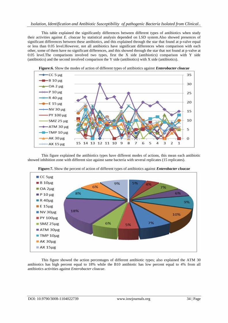
Table:7. Show the replicates of inhibition zones formed by different types of antibiotics against Enterobacter
cloacae
This table explained the Mean, Median, Std. deviation and Std. error for replicates of inhibition zones
that measured by (mm) for each antibiotics against Enterobacter cloacae bacteria. And showed the (ATM 30)
antibiotic has high activity (19.466 mm) while the (PY 100) antibiotic has low activity (4.934 mm) against
Enterobacter cloacae bacteria by mean measured.
*AX 15 Amoxicillin15 μg *E 15 Erythromycin15 μg
*AK 30 Amikacin30 μg *R 40 Rifaximin40 μg
*TMP 10 Trimethoprim10 μg *P 10 Penicillin-G10 μg
*ATM 30 Aztreonam30 μg *OA 2 Oxolinic acid2 μg
*SMZ 25 Sulfamethoxazole25 μg *B 10 Bacitracin10 μg
*PY 100 Carbnicillin100 μg *CC 5 Clindamycin5 μg
*NV 30 Novobiocin30 μg
Table:8. Show the significantly study according to LSD system for different types of antibiotics against
Enterobacter cloacae
*The mean difference is significant at the P-Value = 0.05 level.
**Mean difference.
Isolation, Identification and Antibiotic Susceptibility of pathogenic Bacteria Isolated from Clinical..
DOI: 10.9790/3008-1104022739 www.iosrjournals.org 34 | Page
This table explained the significantly differences between different types of antibiotics when study
their activities against E. cloacae by statistical analysis depended on LSD system.Also showed presences of
significant differences between these antibiotics, and this explained through the star that found at p-valve equal
or less than 0.05 level.However, not all antibiotics have significant differences when comparison with each
other, some of them have no significant differences, and this showed through the star that not found at p-valve at
0.05 level.The comparisons involved two types, first the X side (antibiotics) comparison with Y side
(antibiotics) and the second involved comparison the Y side (antibiotics) with X side (antibiotics).
Figure:6. Show the modes of action of different types of antibiotics against Enterobacter cloacae
This figure explained the antibiotics types have different modes of actions, this mean each antibiotic
showed inhibition zone with different size against same bacteria with several replicates (15 replicates).
Figure:7. Show the percent of action of different types of antibiotics against Enterobacter cloacae
This figure showed the action percentages of different antibiotic types; also explained the ATM 30
antibiotics has high percent equal to 18% while the B10 antibiotic has low percent equal to 4% from all
antibiotics activities against Enterobacter cloacae.
Isolation, Identification and Antibiotic Susceptibility of pathogenic Bacteria Isolated from Clinical..
DOI: 10.9790/3008-1104022739 www.iosrjournals.org 35 | Page
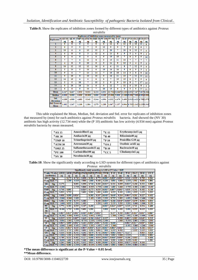
Table:9. Show the replicates of inhibition zones formed by different types of antibiotics against Proteus
mirabilis
This table explained the Mean, Median, Std. deviation and Std. error for replicates of inhibition zones
that measured by (mm) for each antibiotics against Proteus mirabilis bacteria. And showed the (NV 30)
antibiotic has high activity (12.734 mm) while the (P 10) antibiotic has low activity (4.934 mm) against Proteus
mirabilis bacteria by mean measured.
*AX 15 Amoxicillin15 μg *E 15 Erythromycin15 μg
*AK 30 Amikacin30 μg *R 40 Rifaximin40 μg
*TMP 10 Trimethoprim10 μg *P 10 Penicillin-G10 μg
*ATM 30 Aztreonam30 μg *OA 2 Oxolinic acid2 μg
*SMZ 25 Sulfamethoxazole25 μg *B 10 Bacitracin10 μg
*PY 100 Carbnicillin100 μg *CC 5 Clindamycin5 μg
*NV 30 Novobiocin30 μg
Table:10. Show the significantly study according to LSD system for different types of antibiotics against
Proteus mirabilis
*The mean difference is significant at the P-Value = 0.05 level.
**Mean difference.
Isolation, Identification and Antibiotic Susceptibility of pathogenic Bacteria Isolated from Clinical..
DOI: 10.9790/3008-1104022739 www.iosrjournals.org 36 | Page
This table explained the significantly differences between different types of antibiotics when study
their activities against P. mirabilis by statistical analysis depended on LSD system.
Also showed presences of significant differences between these antibiotics, and this explained through the star
that found at p-valve equal or less than 0.05 level.
However, not all antibiotics have significant differences when comparison with each other, some of
them have no significant differences, and this showed through the star that not found at p-valve at 0.05 level.
The comparisons involved two types, first the X side (antibiotics) comparison with Y side (antibiotics) and the
second involved comparison the Y side (antibiotics) with X side (antibiotics).
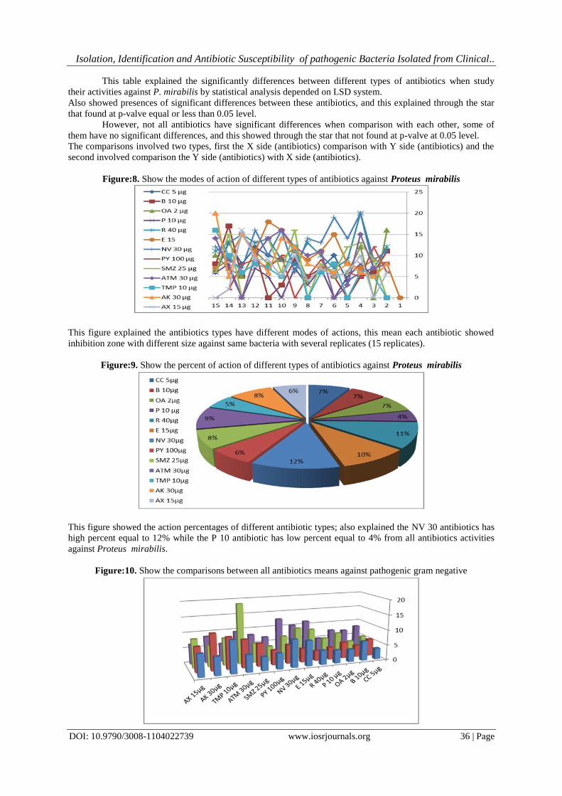
Figure:8. Show the modes of action of different types of antibiotics against Proteus mirabilis
This figure explained the antibiotics types have different modes of actions, this mean each antibiotic showed
inhibition zone with different size against same bacteria with several replicates (15 replicates).
Figure:9. Show the percent of action of different types of antibiotics against Proteus mirabilis
This figure showed the action percentages of different antibiotic types; also explained the NV 30 antibiotics has
high percent equal to 12% while the P 10 antibiotic has low percent equal to 4% from all antibiotics activities
against Proteus mirabilis.
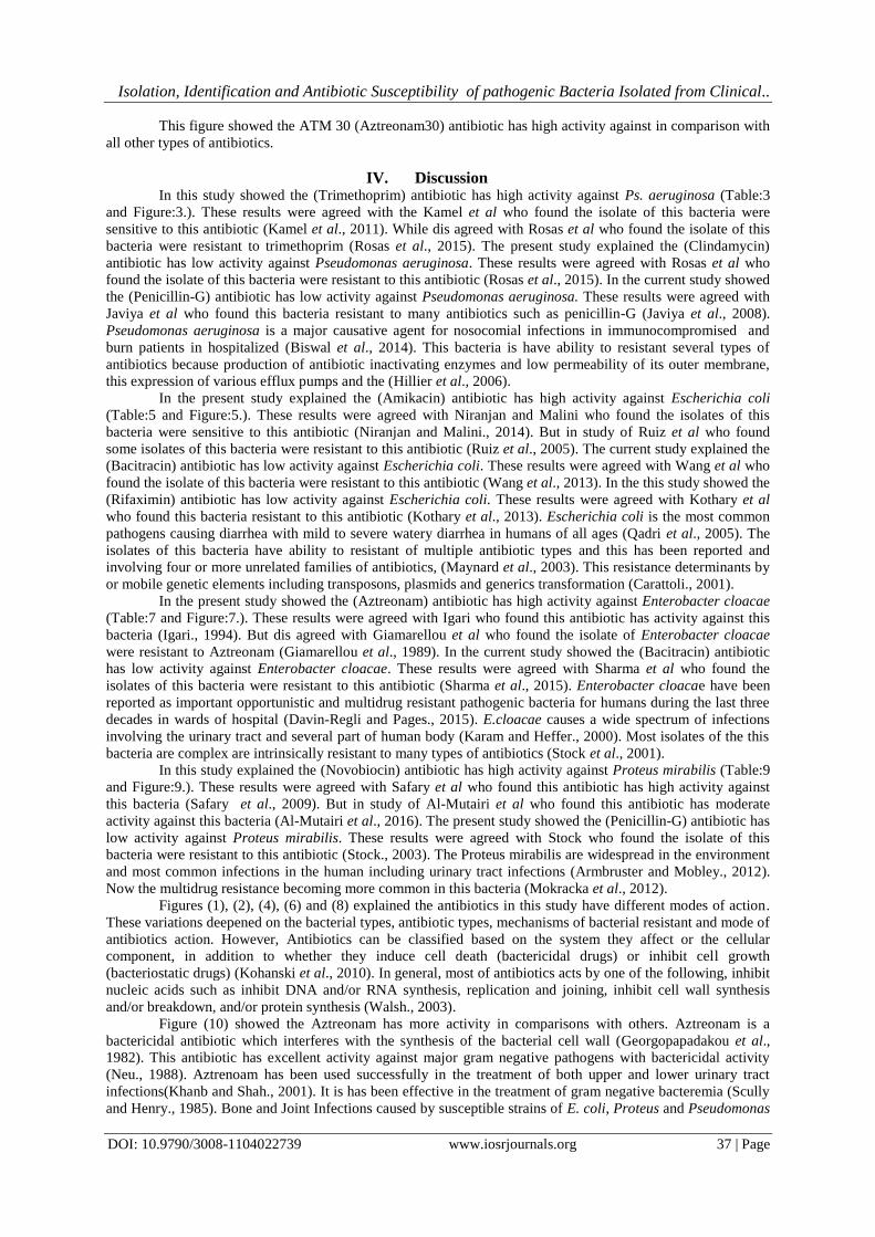
Figure:10. Show the comparisons between all antibiotics means against pathogenic gram negative
Isolation, Identification and Antibiotic Susceptibility of pathogenic Bacteria Isolated from Clinical..
DOI: 10.9790/3008-1104022739 www.iosrjournals.org 37 | Page
This figure showed the ATM 30 (Aztreonam30) antibiotic has high activity against in comparison with
all other types of antibiotics.
IV. Discussion In this study showed the (Trimethoprim) antibiotic has high activity against Ps. aeruginosa (Table:3
and Figure:3.). These results were agreed with the Kamel et al who found the isolate of this bacteria were
sensitive to this antibiotic (Kamel et al., 2011). While dis agreed with Rosas et al who found the isolate of this
bacteria were resistant to trimethoprim (Rosas et al., 2015). The present study explained the (Clindamycin)
antibiotic has low activity against Pseudomonas aeruginosa. These results were agreed with Rosas et al who
found the isolate of this bacteria were resistant to this antibiotic (Rosas et al., 2015). In the current study showed
the (Penicillin-G) antibiotic has low activity against Pseudomonas aeruginosa. These results were agreed with
Javiya et al who found this bacteria resistant to many antibiotics such as penicillin-G (Javiya et al., 2008).
Pseudomonas aeruginosa is a major causative agent for nosocomial infections in immunocompromised and
burn patients in hospitalized (Biswal et al., 2014). This bacteria is have ability to resistant several types of
antibiotics because production of antibiotic inactivating enzymes and low permeability of its outer membrane,
this expression of various efflux pumps and the (Hillier et al., 2006).
In the present study explained the (Amikacin) antibiotic has high activity against Escherichia coli
(Table:5 and Figure:5.). These results were agreed with Niranjan and Malini who found the isolates of this
bacteria were sensitive to this antibiotic (Niranjan and Malini., 2014). But in study of Ruiz et al who found
some isolates of this bacteria were resistant to this antibiotic (Ruiz et al., 2005). The current study explained the
(Bacitracin) antibiotic has low activity against Escherichia coli. These results were agreed with Wang et al who
found the isolate of this bacteria were resistant to this antibiotic (Wang et al., 2013). In the this study showed the
(Rifaximin) antibiotic has low activity against Escherichia coli. These results were agreed with Kothary et al
who found this bacteria resistant to this antibiotic (Kothary et al., 2013). Escherichia coli is the most common
pathogens causing diarrhea with mild to severe watery diarrhea in humans of all ages (Qadri et al., 2005). The
isolates of this bacteria have ability to resistant of multiple antibiotic types and this has been reported and
involving four or more unrelated families of antibiotics, (Maynard et al., 2003). This resistance determinants by
or mobile genetic elements including transposons, plasmids and generics transformation (Carattoli., 2001).
In the present study showed the (Aztreonam) antibiotic has high activity against Enterobacter cloacae
(Table:7 and Figure:7.). These results were agreed with Igari who found this antibiotic has activity against this
bacteria (Igari., 1994). But dis agreed with Giamarellou et al who found the isolate of Enterobacter cloacae
were resistant to Aztreonam (Giamarellou et al., 1989). In the current study showed the (Bacitracin) antibiotic
has low activity against Enterobacter cloacae. These results were agreed with Sharma et al who found the
isolates of this bacteria were resistant to this antibiotic (Sharma et al., 2015). Enterobacter cloacae have been
reported as important opportunistic and multidrug resistant pathogenic bacteria for humans during the last three
decades in wards of hospital (Davin-Regli and Pages., 2015). E.cloacae causes a wide spectrum of infections
involving the urinary tract and several part of human body (Karam and Heffer., 2000). Most isolates of the this
bacteria are complex are intrinsically resistant to many types of antibiotics (Stock et al., 2001).
In this study explained the (Novobiocin) antibiotic has high activity against Proteus mirabilis (Table:9
and Figure:9.). These results were agreed with Safary et al who found this antibiotic has high activity against
this bacteria (Safary et al., 2009). But in study of Al-Mutairi et al who found this antibiotic has moderate
activity against this bacteria (Al-Mutairi et al., 2016). The present study showed the (Penicillin-G) antibiotic has
low activity against Proteus mirabilis. These results were agreed with Stock who found the isolate of this
bacteria were resistant to this antibiotic (Stock., 2003). The Proteus mirabilis are widespread in the environment
and most common infections in the human including urinary tract infections (Armbruster and Mobley., 2012).
Now the multidrug resistance becoming more common in this bacteria (Mokracka et al., 2012).
Figures (1), (2), (4), (6) and (8) explained the antibiotics in this study have different modes of action.
These variations deepened on the bacterial types, antibiotic types, mechanisms of bacterial resistant and mode of
antibiotics action. However, Antibiotics can be classified based on the system they affect or the cellular
component, in addition to whether they induce cell death (bactericidal drugs) or inhibit cell growth
(bacteriostatic drugs) (Kohanski et al., 2010). In general, most of antibiotics acts by one of the following, inhibit
nucleic acids such as inhibit DNA and/or RNA synthesis, replication and joining, inhibit cell wall synthesis
and/or breakdown, and/or protein synthesis (Walsh., 2003).
Figure (10) showed the Aztreonam has more activity in comparisons with others. Aztreonam is a
bactericidal antibiotic which interferes with the synthesis of the bacterial cell wall (Georgopapadakou et al.,
1982). This antibiotic has excellent activity against major gram negative pathogens with bactericidal activity
(Neu., 1988). Aztrenoam has been used successfully in the treatment of both upper and lower urinary tract
infections(Khanb and Shah., 2001). It is has been effective in the treatment of gram negative bacteremia (Scully
and Henry., 1985). Bone and Joint Infections caused by susceptible strains of E. coli, Proteus and Pseudomonas

Isolation, Identification and Antibiotic Susceptibility of pathogenic Bacteria Isolated from Clinical..
DOI: 10.9790/3008-1104022739 www.iosrjournals.org 38 | Page
have been treated with Aztreonam (Simons and Lee., 1985). It is useful in treatment of multi drug resistant
Shigella and Salmonella as well as in therapy of gastro intestinal gram negative organisms (Wasfy et al., 2000).
Aztreonam has been shown to penetrate inflamed meninges, and reach therapeutic levels in the central nervous
system (Kapoor and Gathwala., 2004).
References [1]. Alhazmi. A. (2015). Pseudomonas aeruginosa Pathogenesis and Pathogenic Mechanisms. International Journal of Biology. 7(2):
44-67. [2]. Allocati, N. Masulli, M. Alexeyev, M.F. and Carmine, B.D. (2013). Escherichia coli in Europe: An Overview. Int. J. Environ. Res.
Public Health. 10: 6235-6254.
[3]. Al-Mutairi, M.H. Ali, S. Aly, S.M. and Aldebasi, Y. (2016). Antimicrobial activity of Sider (Ziziphus Spina- Christi), Leaves extract against selected pathogenic bacteria. Ejpmr. 3(5): 138-144.
[4]. Anuj Nautiyal, N.V. Madhav, S. Sharma R.K. Ojha, A. and Bhargava, S. (2015). Review on Nosocomial Infections. Carib. J .Sci
Tech. 3: 781-788. [5]. Armbruster, C.E. and Mobley, H.L.T. (2012). Merging mythology and morphology: the multifaceted lifestyle of Proteus
mirabilis. Nat Rev Micro. 10: 743-754.
[6]. Azimi, A. Naserpour., T. Bazmi., F. Peymani., A. Aslanimehr., M. Saadat, S. (2015). Evaluation of oprD Gene Expression in Carbapenem-Resistant Pseudomonas aeruginosa Strains Isolated From Severe Burn Patients With Secondary Infection. Biotech
Health Sci. 2(3): 43- 47.
[7]. Bahashwan, S.A. (2013). Antimicrobial Resistance Patterns of Proteus isolate from clinical specimens. European Scientific Journal September. 9(27): 1857-7881.
[8]. Bauer, A.W. Kirby, W.M.M. Sherris, J.C. and Turck, M. (1966). Antibiotic susceptibility testing by a standardized single disk
method. Am. J. Clin. Pathol., 45: 493 496. [9]. Biswal, I. Arora, B.S. Kasana, D. (2014). Incidence of multidrug resistant pseudomonas aeruginosa isolated from burn patients and
environment of teaching institution. J Clin Diagn Res. 8(5): 26-29. [10]. Carattoli, A. (2001). Importance of integrons in the diffusion of resistance. Vet. Res. 32: 243-259.
[11]. Cheesbrough, M. (2006). District Laboratory Practice in Tropical Countries. Part 2. Cambridge University Press, Cambridge, 434.
[12]. Dadheech, T. Vyas, R. and Rastogi, V. (2015). Antibiotics Resistance of aerobic bacterial isolates of Proteus mirabilis from sick layer Chickens infected with septicemia and salinities in Ajmer region of Rajasthan. World Journal of Pharmacy and
Pharmaceutical Sciences. 4(7): 2002-2011.
[13]. Davies, J. and Davies., D. (2010). Origins and Evolution of Antibiotic Resistance. Microbiology and Molecular Biology Rev. (74)3: 417-433.
[14]. Davin-Regli, A. and Pages, J. M. (2015). Enterobacte raerogenes and Enterobacter cloacae; versatile bacterial pathogens
confronting antibiotic treatment. FrontiersinMicrobiology. 6: 1-10. [15]. Eguchi, H. Miyamoto, T. Kuwahara, T. Mitamura, S. and Mitamura, Y. (2013). Infectious conjunctivitis caused by Pseudomonas
aeruginosa isolated from a bathroom. BMC Res Notes. 6:245. doi:10.1186/1756-0500-6-245.
[16]. Ewers, C. Bethe, A. Semmler, T.Guenther, S. and Wieler, L.H. (2012). Extended-spectrum beta-lactamase-producing and AmpC-producing Escherichia coli from livestock and companion animals, and their putative impact on public health: A global perspective.
Clin. Microbiol. Infect. 18: 646–655.
[17]. Feglo, P.K. Gbedema, SY. Quay, S.N.A. Adu-Sarcode, Y.O. poku, O. and krah,C. (2010). Occurrence species distribution and antibiotic resistance of Proteus isolates: A case study at the Komfo Anoke Teaching Hospital(KATH) in Ghana. J.International
Journal of Pharma science and research.1(9): 347-352.
[18]. Fisher, J.F. and Mobashery., S. (2010). Enzymology of Bacterial Resistance. Comprehensive Natural Products II. In: Enzymes and Enzyme Mechanisms. Elsevier. 8: 443-487.
[19]. Gellatly, S.L. and Hancock, R.E.W. (2013). Pseudomonas aeruginosa: new insights into pathogenesis and host defenses. Pathog
Dis. 67:159-173. [20]. Georgopapadakou, N.H. Smit, S.A. and Sykes, R.B. (1982). Modes of action of Aztreonam. Antimicrobial Agents Chemother. 21:
950-956.
[21]. Giamarellou, H. Evagelopoulou, V. and Lelekis, M. (1989). Enhanced in-vitro bactericidal activity of amikacin combined with
latamoxef, cefotaxime and aztreonam against multi-resistant Enterobacter cloacae. Journal of Antimicrobial Chemotherapy. 23(4):
537-45.
[22]. Hara, C.M. Brenner, F.W. and Miller, J.M. (2000). Classification, identification, and clinical significance of Proteus, Providencia, and Morganella . Clin Microbiol Rev.13(4): 534-546.
[23]. Hillier, A. Alcorn, J.R. Cole, L.K. Kowalski, J.J. (2006) Pyoderma caused by Pseudomonas aeruginosa infection in dogs: 20 cases.
Vet Dermatol. 17(6): 432-439. [24]. Holt, J.G. N.R. Krieg, P.H.A. Sneath, J.T. Staley, and S.T. Williams. (1994). Bergy's manual of determinative bacteriology. 9th
ed. Williams and Wilkins, Baltimore, USA.
[25]. Igari, J. (1994). Antibacterial activity of aztreonam against clinical isolates. Jpn J Antibiot. 47(5):502-520. [26]. Jabalameli, F. Mirsalehian, A. Sotoudeh, N. Jabalameli, L. Aligholi, M. and Khoramian., B. (2011). Multiple-locus variable
number of tandem repeats (VNTR) fingerprinting (MLVF) and antibacterial resistance profiles of extended spectrum beta lactamase
(ESBL) producing Pseudomonas aeruginosa among burnt patients in Tehran. Burns. 37(7): 1202-1207. [27]. Javiya, V.A. Ghatak, S.B. Patel, K.R. & Patel, J.A. (2008). Antibiotic susceptibility patterns of Pseudomonas aeruginosa at a
tertiary care hospital in Gujarat, India. Indian Jourl of Pharma. 40(5): 230-234.
[28]. Johnson, T.J. Logue, C.M. Johnson, J.R. Kuskowski, M.A. Sherwood, J.S. Barnes, H.J. DebRoy, C. Wannemuehler, Y.M. Obata-Yasuoka, M. and Spanjaard, L. (2012). Associations between multidrug resistance, plasmid content, and virulence potential among
extra intestinal pathogenic and commensal Escherichia coli from humans and poultry. Foodborne Pathog. rrDis. 9: 37-46.
[29]. Kamel, G.M. Ezz eldeen, N.A. El-Mishad, M.Y. and Ezzat,R.F. (2011). Susceptibility Pattern of Pseudomonas aeruginosa Against Antimicrobial Agents and Some Plant Extracts with Focus on its Prevalence in Different Sources. Global Veterinaria. 6
(1): 61-72.
[30]. Kapoor,S. and Gathwala, G. (2004). Aztreonam. Drug Therapy. 41(17): 359-364. [31]. Karam, G.H. and Heffer. J.E. (2000). Emerging issue in Antibiotic Resistance in Blood - borne Infections. Am. J. Respir. Crit.
Care. Med. 162(5): 1610-1616.
Isolation, Identification and Antibiotic Susceptibility of pathogenic Bacteria Isolated from Clinical..
DOI: 10.9790/3008-1104022739 www.iosrjournals.org 39 | Page
[32]. Khan, M.T. and Shah, S.H. (2001). Comparison of aztreonam against other antibiotics used in urinary tract infections. J Ayub Med
Coll Abbottabad. 13: 22-24.
[33]. Kohanski, M.A. Dwyer, D.J. and Collins, J.J (2010). How antibiotics kill bacteria: from targets to networks. Nat Rev Microbiol. 8(6): 423–435.
[34]. Kothary, V. Scherl, E.J. Bosworth, B. Jiang, Z.D. DuPont, H.L. Harel, J. W. Simpson, K.W and Dogane, B. (2013). Rifaximin
Resistance in Escherichia coli Associated with Inflammatory Bowel Disease Correlates with Prior Rifaximin Use, Mutations in rpoB, and Activity of Phe-Arg-β-Naphthylamide-Inhibitable Efflux Pumps. Antimicrobial Agents and Chemotherapy.57(2) : 811-
817.
[35]. Manikandan, C. and Amsath, A. (2014). Antibiotic susceptibility pattern of Escherichia coli isolated from urine samples in Pattukkottai, Tamilnadu. Int.J.Curr.Microbiol.App.Sci. 3(10): 449-457.
[36]. Maynard, C., Fairbrother, J.M. Bekal, S. Sanschagrin, F. Levesque, R.C. Brousseau, R. Masson, L. Lariviere, S. and Harel,
J. (2003). Antimicrobial resistance genes in enterotoxigenic Escherichia coli O149:K91 isolates obtained over a 23-year period from pigs. Antimicrob. Agents Chemother. 47:3214-3221.
[37]. Mezzatesta, M.L. Gona, F. and Stefani, S. (2012). Enterobactercloacae complex: clinical impact and emerging antibiotic
resistance. Future Microbiol. 7: 887-902. [38]. Mirsalehian, A. Feizabadi., M. Nakhjavani., F.A. Jabalameli., F. Goli, H. and Kalantari., N (2010). Detection of VEB-1, OXA-10
and PER-1 genotypes in extended-spectrum beta-lactamase-producing Pseudomonas aeruginosa strains isolated from burn patients.
Burns. 36(1): 70–4. [39]. Mokracka, J. Gruszczynska, B. and Kaznowski, A. (2012). Integrons, β-lactamase and qnr genes in multidrug resistant clinical
isolates of Proteus mirabilis and P. vulgaris. APMIS 2012;120: 950-958.
[40]. Murray, J.L. Kwon, T. Marcotte, E.M. and Whiteleya, M. (2015). Intrinsic Antimicrobial Resistance Determinants in the Superbug Pseudomonas aeruginosa. mbio.asm.org. 6(6): 1-10.
[41]. Nabeela, N. Munazza, A. Shekh., A.R. and Zaid, A.P. (2004). Urinary tract infections associated with multidrug resistant enteric
bacilli: characterization and genetical studies. Journal of Pakistan Pharma. Scien. 17(2): 115-123. [42]. Neu H.C. (1988). Aztreonam: The first monobactam. Med Clin North Am. 72: 556-566.
[43]. Niranjan, V. and Malini, A. (2014). Antimicrobial resistance pattern in Escherichia coli causing urinary tract infection among
inpatients. Indian J Med Res. 139: 945-948. [44]. Otoikhian, C.S.O. and Tanimowo, W.O. (2016) Identification of intestinal microbes of pig sold in local market in Niger delta
environment. Nigerian Journal of Agriculture, Food and Environment. 12(1): 111-115.
[45]. Pelczar, M. Reid, R. and Chan, E. (1957). Microbiology. T.M.H. Edition, Tata McGraw-Hill Publishing Company Ltd.,Noida. [46]. Penner, J.L. (1984). Genus XI.Proteus , Bergey's manual of systemic bacteriology. In N.R. Krieg and J.G. Holt(ed.),The William
and Wilkins Co., Baltimore, Md. .1: 491-494.
[47]. Perez, A. Poza, M. Aranda, J. Latasa, C. Medrano, F.J. and Tomas, M. (2012). Effect of transcriptional activators SoxS, RobA, and
RamA on expression of multidrug efflux pump AcrAB-TolC in Enterobacter cloacae. Antimicrob. Agents Chemother. 56, 6256–
6266.
[48]. Pestourie, N. Garnier, F. Barraud,O. Bedu, A. Ploy, M. C. and Mounier, M.(2014). Outbreak of AmpC β-lactamase hyper producing Enterobacter cloacae in a neonatal intensive care unit in a French teaching hospital. Am. J. Infect. Control. 42: 456–458.
[49]. Pitout, J. D. (2012). Extraintestinal pathogenic Escherichia coli: An update on antimicrobial resistance, laboratory diagnosis and treatment. Expert Rev. Anti Infect. Ther. 10: 1165-1176.
[50]. Qadri, F. Svennerholm, A.M. Faruque, A.S. and Sack, R.B.(2005). Enterotoxigenic Escherichia coli in developing countries:
Epidemiology, microbiology, clinical features, treatment, and prevention. Clin. Microbiol. Rev. 18: 465–483. [51]. Rosas, Y.B. Munoz, K.O. and Garcia O.T. (2015). Pseudomonas aeruginosa: an emerging nosocomial trouble in veterinary;
Pseudomonas aeruginosa: un problema nosocomial emergente en veterinaria. Rev.MVZ Cordoba. 20 :4937-4946.
[52]. Ruiz, J. Bertran, S. Sauca, G. Julia, A. Vila, X. Gomez, E. Jimenez de Anta, M. T and Vila, J. (2005). Isolation of an amikacin resistant Escherichia coli strain after tobramycin treatment of previous recurrent episodes of respiratory tract infections caused by
Pseudomonas aeruginosa. Clin Microbiol Infect. 11: 71-73.
[53]. Safary, A. Motamedi, H. Maleki, S. and Seyyednejad, S.M. (2009). A Preliminary Study on the Antibacterial Activity of Quercus brantii Against Bacterial Pathogens, Particularly Enteric Pathogens. IJB, 5: 176-180.
[54]. Scully, B.E. and Henry, S.A. (1985). Clinical experience with Aztreonam in the treatment of gram negative bacterimia. Rev Infect
Dis. 7: S 789-793.
[55]. Sharma, S.K. Rahul Yadav, R. and Nathawat, P. (2015). Multidrug Resistance Pattern of Enterobacter Spp. Isolated from Acute
Respiratory Tract Infected Camels (Camelus dromedarius). Adv. Anim. Vet. Sci. 3(2): 128-132.
[56]. Simons W.J. and Lee, T.J. (1985). Aztreonam in the treatment of bone and joint infections caused by gram negative bacilli. Rev Infect Dis. 7: 783-788.
[57]. Singla, P. Sangwan, J. Garg, S. and Chaudhary, U. (2015). Prevalence and Antibiogram of Multidrug resistant Uropathogenic
Isolates of Proteus mirabilis in a Teaching Tertiary Care Hospital. Int.J.Curr.Microbiol.App.Sci. 4(12): 675-682. [58]. Stiles, M and LAI, K.N.G. (1981). Biochemical Characteristics and Identification of Enterobacteriaceae Isolated from Meats. Appl.
and Envir. Microbio. Mar. Vol. (41) 3: 639-645.
[59]. Stock, I. Gruger, T. Wiedemann, B. (2001). Natural antibiotic susceptibility of strains of the Enterobacter cloacae complex. Int. J. Antimicrob. Agents.18 (6): 537–545.
[60]. Stock, I. (2003). Natural antibiotic susceptibility of Proteus spp., with special reference to P. mirabilis and P. penneri strains. J
Chemo ther. 15(1): 12-26. [61]. Strateva, T. and Yordanov, D. (2009). Pseudomonas aeruginosa-a phenomenon of bacterial resistance. J Med Microbiol. 58(9):
1133-1148.
[62]. Walsh. C. (2003). Antibiotics: actions, origins, resistance. ASM Press; Washington, D.C. [63]. Wang, D. Hu, E. Chen, J. Tao, X. Gutierrez, K. and Qi, Y. (2013). Characterization of novel ybjG and dacC variants in Escherichia
coli. Journal of Medical Microbiology. 62: 1728–1734.
[64]. Wasfy, M.O. Oyofo, B.A. David, J.C. Ismail, T.F. El-Gendy, A.M. and Mohran, Z.S. (2000). Isolation and antibiotic susceptibility of Salmonella, Shigella and Campylobacter from acute enteric infections in Egypt. J Health Popul Nutr. 18: 33-38.
[65]. Wong, M.H. Wan, H.Y. and Chen, S. (2013). Characterization of multidrug-resistant Proteus mirabilis isolated from chicken
carcasses. Foodborne Pathog Dis. 10: 177-181. [66]. Yoo, S. H. Jeong, H. Kwon, S.K. and Kim, J.F. (2009). Genomics, Biological Features, and Biotechnological Applications of
Escherichia coli B: Is B for better; Springer: Berlin, Germany.