An interaction switch predicts the nested architecture of mutualistic networks

JOURNAL OF BACTERIOLOGY, May 2003, p. 3147–3154 Vol. 185, No. 100021-9193/03/$08.00�0 DOI: 10.1128/JB.185.10.3147–3154.2003Copyright © 2003, American Society for Microbiology. All Rights Reserved.
Early Colonization Events in the Mutualistic Associationbetween Steinernema carpocapsae Nematodes and
Xenorhabdus nematophila BacteriaEric C. Martens, Kurt Heungens,† and Heidi Goodrich-Blair*
Department of Bacteriology, University of Wisconsin, Madison, Wisconsin 53706
Received 16 December 2002/Accepted 4 February 2003
The bacterium Xenorhabdus nematophila is a mutualist of the entomopathogenic nematode Steinernemacarpocapsae. During its life cycle, the bacterium exists both separately from the nematode and as an intestinalresident of a nonfeeding nematode form, the infective juvenile (IJ). The progression of X. nematophila from anex vivo existence to a specific and persistent colonization of IJs is a model to understand the mechanismsmediating the initiation and maintenance of benign host-microbe interactions. To help characterize thisprocess, we constructed an X. nematophila strain that constitutively expresses green fluorescent protein, whichallowed its presence to be monitored within IJs. Using this strain, we showed that few bacterial cells initiatecolonization of an individual IJ and that these grow inside the lumen of the IJ intestine in a reproduciblepolyphasic pattern during colonization. In accordance with these two observations, we demonstrated that thefinal population of bacteria in a nematode is of predominantly monoclonal origin, suggesting that only one ortwo bacterial clones initiate or persist during colonization of an individual nematode. These data suggest thatX. nematophila initiates IJ colonization by competing for limited colonization sites or resources within thenematode intestine. This report represents the first description of the biological interactions occurring betweenX. nematophila and S. carpocapsae during the early stages of the colonization process, provides insights into thephysiology of X. nematophila in its host niche, and will facilitate interpretation of future data regarding themolecular events mediating this process.
The mutualistic, monospecific partnership between thegram-negative enterobacterium Xenorhabdus nematophila andits entomopathogenic nematode host, Steinernema carpocap-sae, is one of several emerging models to study benign host-microbe interactions (9, 13, 27). The natural life cycle of S.carpocapsae nematodes includes reproductive stages that occurexclusively within larval-stage insects and that are not colo-nized by X. nematophila and also includes a nonreproductive,nonfeeding, soil-dwelling stage known as the infective juvenile(IJ), which is colonized, at a discrete intestinal location termedthe vesicle, by a monoculture of X. nematophila bacteria (9).
Colonized IJs exist in the soil until they invade a susceptibleinsect host, seeking the blood system or hemolymph. Oncethere, the IJs release their X. nematophila symbionts by defe-cation (19, 31; E. C. Martens and H. Goodrich-Blair, unpub-lished data). At this point in their life cycles, bacteria andnematodes exist separately although in close proximity to oneanother. The released X. nematophila bacteria contribute tothe killing of the insect host and grow to high density in theresulting cadaver. These specific bacteria are essential for nem-atode growth and development presumably both by serving asa direct food source and by supplying nutrients through deg-radation of the insect carcass (2, 22).
When nematode numbers become high and nutrients be-come limiting in the insect cadaver, S. carpocapsae nematodeprogeny reassociate with X. nematophila bacteria and differen-
tiate into the colonized, nonfeeding IJ form that emerges intothe soil to forage for a new host (23). IJ formation, and con-comitant colonization with X. nematophila, is a model of theprocess by which a specific and intimate association between amicrobe and a eukaryotic host in a benign relationship devel-ops (9, 27). A deeper understanding of X. nematophila-nema-tode interactions will provide insights into the general mech-anisms mediating the development and maintenance of benignmicrobe-host interactions and also allow a comparison of be-nign interactions with those that are pathogenic (27).
We chose to characterize the early stages of nematode col-onization by X. nematophila to better understand the processby which this mutualistic interaction is initiated and main-tained. A characteristic of the IJ stage of the nematode, whichdevelops within the spent insect cadaver prior to emergenceinto the soil, is its sealed mouth and anus (18). The IJ thereforedoes not ingest resources or excrete intestinal waste during itsexistence outside an insect and must be colonized by X. nema-tophila prior to development into this stage. Until now, exper-iments exploring the initiation of X. nematophila colonizationof the pre-IJ nematode have not been reported. We consideredtwo possible models to explain how mature IJs (i.e., those thathave emerged from an insect host) obtain a full complement ofcolonizing bacteria (13, 19). In the simplest model, we postu-lated that a developing IJ ingests a large number of bacteriafrom its environment before it ceases feeding and that thesebacteria, instead of being digested, are retained in the intesti-nal vesicle. A more complex model supposes that a pre-IJnematode retains only one or a few bacteria in a selectivemanner and, after the IJ ceases ingesting external bacteria,
* Corresponding author. Mailing address: Department of Bacteriol-ogy, University of Wisconsin—Madison, Madison, WI 53706. Phone:(608) 265-4537. Fax: (608) 262-9865. E-mail: [email protected].
† Present address: DGB-CLO, B-9820 Merelbeke, Belgium.
3147
Dow
nloa
ded
from
http
s://j
ourn
als.
asm
.org
/jour
nal/j
b on
21
Oct
ober
202
1 by
106
.163
.185
.210
.
these few bacteria grow until they reach the full density foundin a mature IJ.
To test these models, we conducted experiments utilizing anin vitro culturing technique in which S. carpocapsae nematodesare grown on a lawn of X. nematophila bacteria (31). Thistechnique allows monitoring of nematodes throughout theirreproductive life cycle, and progeny IJs emerging from theselawns are colonized at levels that are comparable to those ofIJs produced from insect infection (13). In addition to thistechnique, we developed and/or implemented several newtools that facilitated the examination of early stages in theassociation between nematodes and bacteria. We present datathat document the process by which nematodes achieve thefully colonized state, differentiate between the hypotheticalcolonization models described above, and provide insight intomechanisms mediating early colonization events.
MATERIALS AND METHODS
Strains, plasmids, and culture conditions. X. nematophila strains and plasmidsused in this study are listed in Table 1. Permanent stocks of bacterial strains weremaintained at �80°C in Luria-Bertani (LB) broth (14) supplemented with 25%glycerol (for Escherichia coli) or 10% dimethyl sulfoxide (for X. nematophila).Unless stated otherwise, X. nematophila and E. coli were grown at 30°C in LBbroth or agar that either had not been exposed to light or to which 0.1% pyruvatehad been added (32). When appropriate, media were supplemented with chlor-amphenicol (10 �g ml�1 for X. nematophila or 30 �g ml�1 for E. coli) andgentamicin (30 �g ml�1). Solid, lipid-agar medium for coculture of S. carpocap-sae on lawns of X. nematophila was prepared as described elsewhere (27). E. coliS17-1 � pir (25) was used to conjugally transfer plasmids into X. nematophilastrains as previously described (10). S. carpocapsae (strain All) was propagated bypassage through Galleria mellonella larvae (Vanderhorst Wholesale Inc., St.Marys, Ohio) and harvested in White traps as previously described (27, 30). Asneeded, axenic, J1 stage S. carpocapsae organisms were isolated by harvestingnematode eggs with bleach from gravid adult females as previously described(28), except that the eggs were rinsed with sterile LB broth instead of water orbuffered salts solution.
The enhanced isoform of green fluorescent protein (GFP) used in this workwas Super-Glo GFP (Qbiogene, Carlsbad, Calif.) and is hereafter referred to asGFP. To obtain constitutive expression of the GFP gene, it was fused to a 126-bpfragment containing the aphA promoter (PaphA) amplified from E. coli transpo-son Tn903 by using pNK2859 (29) as a template and the primers 5�-GGGGGG
AGATCTCGTTGTGTCTCAAAATCTCTG-3� and 5�-GGGTCTAGATGAATATGGCTCATAACACCC-3�. The resulting PaphA fragment was cut with BglIIand XbaI and ligated into pQBI63 (Qbiogene) cut with BglII and XbaI, resultingin the plasmid pECM9. For subsequent transfer of this fusion into X. nemato-phila, a BglII-EcoRV PaphA-GFP gene fragment was subcloned from pECM9 intopKR100B to create the plasmid pECM2. Plasmid pKR100B is a suicide vectorused for delivering constructs into X. nematophila and was constructed by cuttingpKR100 (a derivative of pGP704 [pJM703.1] [15], kindly provided by KarenVisick, Loyola University of Chicago, Chicago, Ill.) with SalI and XbaI andinserting a linker comprised of the sequence 5�-GTCGACAGATCTAGA-3�,thereby generating a unique BglII site. To direct the PaphA-GFP gene fragmentto the chromosome of X. nematophila, Sau3AI genomic fragments (average size,0.4 to1.6 kb) were ligated into pECM2 cut with BglII. To generate an X. nema-tophila strain expressing the GFP gene, this library was electroporated as previ-ously described (4) into E. coli S17-1 � pir and conjugated en masse into X.nematophila ATCC 19061 (25). Exconjugants were selected on chloramphenicol.Since pECM2 replicates using the origin oriR6K, which is not functional in X.nematophila, these exconjugants must contain integrations, directed by homolo-gous recombination with the cloned Sau3AI fragments, of pECM2 into thebacterial chromosome. Four exconjugants were selected based on their highfluorescence levels detected by flow cytometry (conducted at the University ofWisconsin—Madison CSC Flow Cytometry Facility). Of these, strain HGB340was the brightest and was used for all subsequent studies. To determine thelocation of the plasmid insertion in strain HGB340, the original library fragmentthat was cloned in pECM2 and that directed integration into the X. nematophilachromosome was isolated and sequenced (24). The cloned fragment (GenBankaccession number AY194223) is 614 bp and shares predicted amino acid se-quence similarity with a putative potassium efflux system from Yersinia pestis. Wehave found that HGB340 colonizes S. carpocapsae similarly to HGB007 (the wildtype) under all conditions tested (data not shown), indicating that integration ofpECM20 and constitutive expression of the GFP gene do not exert an observableeffect on the process of bacterial colonization of nematodes. Additionally, the E.coli S17-1 � pir strain harboring pECM20 has proven suitable for labeling otherX. nematophila strains and mutants with the GFP gene (data not shown).
Mini-Tn7 constructs containing the ECFP and DsRed genes (Clontech, PaloAlto, Calif.) were kind gifts from Lotte Lambertsen (Technical University ofDenmark, Lyngby). These constructs were conjugated into HGB007 from E. coliS17-1 � pir and delivered to the X. nematophila att Tn7 site (Martens andGoodrich-Blair, unpublished data) by triparental mating using the helper plas-mid pUX-BF13 (5).
In vitro coculture of nematodes on bacterial lawns. In vitro coculture ofnematodes on bacterial lawns was performed as described previously (27) in6-cm-diameter petri plates (Fisher Scientific, Pittsburgh, Pa.) containing lipid-agar medium, except that cocultures for preparation of immature IJs werecarried out in 500-cm2 polystyrene plates (Corning, Corning, N.Y.) and inocu-
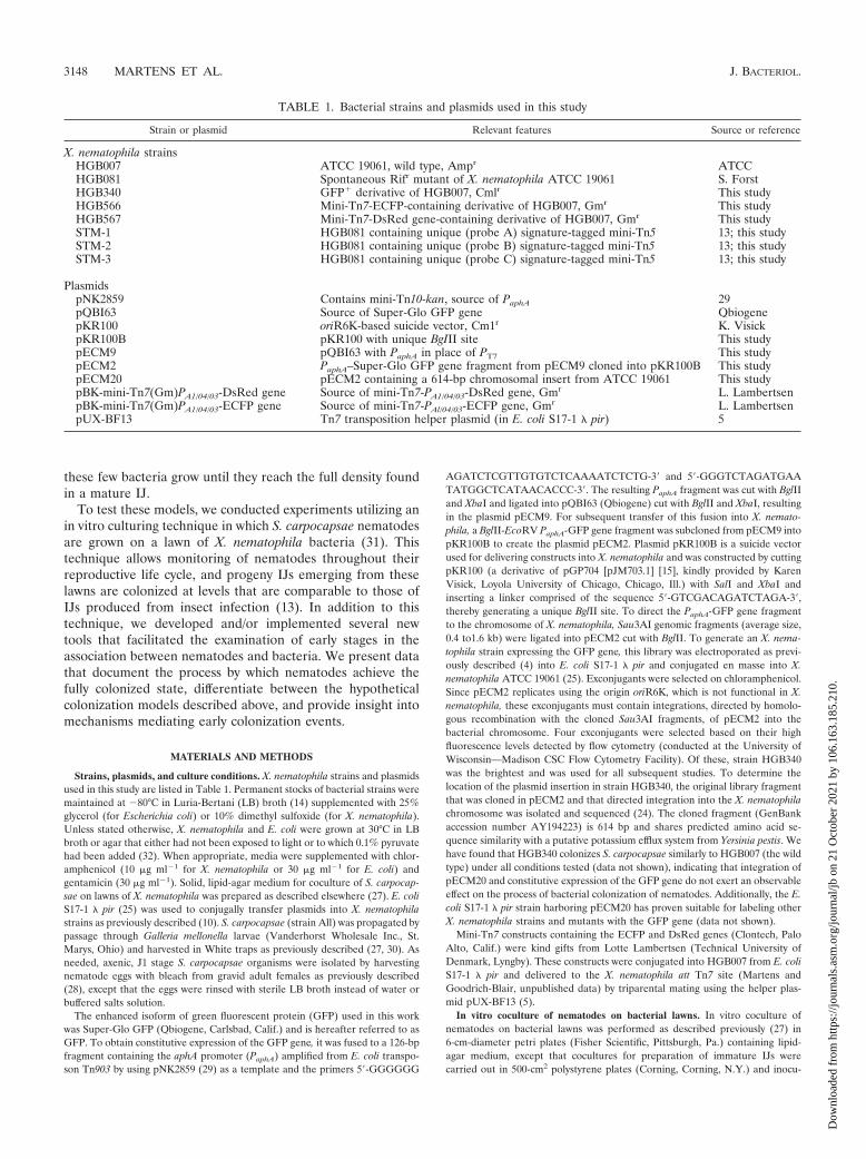
TABLE 1. Bacterial strains and plasmids used in this study
Strain or plasmid Relevant features Source or reference
X. nematophila strainsHGB007 ATCC 19061, wild type, Ampr ATCCHGB081 Spontaneous Rifr mutant of X. nematophila ATCC 19061 S. ForstHGB340 GFP� derivative of HGB007, Cmlr This studyHGB566 Mini-Tn7-ECFP-containing derivative of HGB007, Gmr This studyHGB567 Mini-Tn7-DsRed gene-containing derivative of HGB007, Gmr This studySTM-1 HGB081 containing unique (probe A) signature-tagged mini-Tn5 13; this studySTM-2 HGB081 containing unique (probe B) signature-tagged mini-Tn5 13; this studySTM-3 HGB081 containing unique (probe C) signature-tagged mini-Tn5 13; this study
PlasmidspNK2859 Contains mini-Tn10-kan, source of PaphA 29pQBI63 Source of Super-Glo GFP gene QbiogenepKR100 oriR6K-based suicide vector, Cm1r K. VisickpKR100B pKR100 with unique BgIII site This studypECM9 pQBI63 with PaphA in place of PT7 This studypECM2 PaphA–Super-Glo GFP gene fragment from pECM9 cloned into pKR100B This studypECM20 pECM2 containing a 614-bp chromosomal insert from ATCC 19061 This studypBK-mini-Tn7(Gm)PA1/04/03-DsRed gene Source of mini-Tn7-PA1/04/03-DsRed gene, Gmr L. LambertsenpBK-mini-Tn7(Gm)PA1/04/03-ECFP gene Source of mini-Tn7-PAl/04/03-ECFP gene, Gmr L. LambertsenpUX-BF13 Tn7 transposition helper plasmid (in E. coli S17-1 � pir) 5
3148 MARTENS ET AL. J. BACTERIOL.
Dow
nloa
ded
from
http
s://j
ourn
als.
asm
.org
/jour
nal/j
b on
21
Oct
ober
202
1 by
106
.163
.185
.210
.
lated with IJs instead of axenic J1 nematodes. Approximately 4 � 105 IJs wereinoculated onto each 500-cm2 plate to initiate nematode growth. Culture pro-gression was monitored by microscopic examination of nematodes (see below).Immature IJs typically formed 8 to 9 days after introduction of the IJ inoculuminto cocultures.
SDS isolation of immature S. carpocapsae IJs from mixtures of developmentalstages. Immature IJs were harvested when approximately 50% of the nematodespresent in an in vitro coculture were microscopically observed to be IJs and wereselectively isolated from the heterogeneous populations of other S. carpocapsaestages by treatment with sodium dodecyl sulfate (SDS; Sigma, St. Louis, Mo.). IJsare resistant to treatment with 1% SDS, while other stages are not. This treat-ment was based on the procedure described by Cassada and Russell (8) for thespecific isolation of Caenorhabditis elegans dauerlarvae from mixed developmen-tal stages. Nematodes were harvested by flooding the culture plates with �200 mlof distilled water (dH2O), resuspending the nematodes and remaining bacterialcells, and centrifuging the suspensions at 1,000 � g. Nematode suspensions weretreated with 1% SDS for 20 min with periodic agitation and then washed a totalof three times with dH2O (50 ml/wash). Nematodes were rinsed an additionaltwo times with dH2O in a filter apparatus equipped with an 11-�m-pore-sizenylon membrane (Fisher Scientific). Immature IJs recovered in this manner werestored in dH2O at 25°C during the period of bacterial outgrowth. Control ex-periments were conducted to determine if SDS treatment affects the number ofbacteria in IJs. In one of these experiments, 0.5% bleach was employed to surfacesterilize IJs and to remove other nematode stages from the population (seeResults). Treatment with bleach achieved the same result that treatment withSDS did; however, IJ survival was poor after this treatment, so it was not used forselective isolation of IJs used for prolonged observation.
Isolation of nematodes from bacteria without SDS. To monitor outgrowth ofHGB340 in immature IJs, nematodes were harvested from heterogeneous pop-ulations without the use of SDS treatment. These nematodes were washedrepeatedly with excess volumes of sterile dH2O to remove external bacteria andwere subsequently stored in dH2O.
Microscopic examination of S. carpocapsae. To observe S. carpocapsae micro-scopically, suspensions of nematodes were applied to the surface of a glass slideon which a 4% agarose pad containing 10 mM sodium azide (Sigma) had beenapplied. A coverslip was applied to the slide before the nematode suspension wasallowed to dry completely, and the slide was observed under a Nikon EclipseTE300 inverted microscope at �600 magnification. Fluorescence microscopy ofGFP-containing samples was performed by using fluorescein isothiocyanate,tetramethyl rhodamine isocyanate, and triple-band DAPI (4�,6�-diamidino-2-phenylindole)-FITC (fluorescein isothiocyanate)-TRITC (tetramethyl rhoda-mine isothiocyanate) filter sets (Chroma, Brattleboro, Vt.; items 31001, 31002,and 8200, respectively). GFP-expressing bacteria were differentiated from nem-atode intestinal autofluorescence by using filter set 8200 and appeared green,whereas autofluorescence appears white-yellow. Images were recorded electron-ically with a digital camera (Hamamatsu, Hamamatsu City, Japan; model C4742-95-10NR) and a personal computer equipped with MetaMorph version 4.5r6software (Universal Imaging Corporation, West Chester, Pa.). Images were pre-pared for publication by using Adobe Photoshop version 6.0.1 and DenebaCanvas version 7.0.
Quantification of colonizing bacteria. Quantification of X. nematophila CFUper IJ was performed as previously described (13). Samples of maturing IJs thathad been isolated by use of the SDS isolation procedure described above wereremoved at various times after isolation, surface sterilized, enumerated, andsonicated in a water bath sonicator (Branson Ultrasonics, Danbury, Conn.) toliberate bacteria from the intestines of the IJs. Between 9 � 103 and 1.1 � 104
IJs were sonicated at each time point, and the number of CFU per IJ wasdetermined by plating dilutions of sonicates on LB agar plus 0.1% pyruvate. Thedetection limit of this procedure was �0.003 CFU/IJ, and approximately 60CFU/IJ were normally detected in IJs cultivated on wild-type X. nematophila(13). Because microscopic examination of nematodes containing HGB340 de-tected visible bacteria in the intestinal vesicle exclusively (see below), we believethat viable counts of CFU per IJ determined through homogenization andplating reflect bacteria that are liberated from this location only. Raw data fromthe curves shown below (see Fig. 3) were analyzed by analysis of variance and at test to determine if significant reductions in recoverable CFU per IJ occurredduring the course of growth. To calculate the maximum doubling time of X.nematophila bacteria in nematodes, a linear regression analysis was performedwith data taken from four regions of the curves shown below (see Fig. 3).Regions containing three or more consecutive points of increasing value wereselected for analysis, and the slopes of best-fit lines were used directly to deter-mine the doubling time at each point.
Analysis of signature-tagged clones in single nematodes. To determine whichsignature-tagged clone(s) was present in individual nematodes, IJs produced ona mixed lawn of strains STM-1, STM-2, and STM-3 were surface sterilized andindividual IJs were ground in 100 �l of LB broth in a Duall 20 homogenizer(Kimble-Kontes, Vineland, N.J.). The entire 100-�l volume of homogenate wasplated on LB agar, and only homogenates from which �88 CFU were recoveredwere analyzed. Individual X. nematophila colonies were patched onto LB agarplates overlaid with sterilized 10-cm-diameter Hybond nylon membranes (Am-ersham-Pharmacia, Piscataway, N.J.). Colony blot and hybridization analyses todetect signature-tagged strains were performed as previously described (12, 13),with the exception that bacterial DNA was cross-linked to nylon membranes byusing a UV cross-linker (Stratagene, La Jolla, Calif.).
RESULTS
To observe interactions between X. nematophila and S. car-pocapsae during early stages of the colonization process, weconstructed a derivative of wild-type X. nematophila HGB007that constitutively expresses an enhanced isoform of GFP (seeMaterials and Methods; Table 1). This strain, HGB340, isreadily detectable inside mature IJs by epifluorescence micros-copy (Fig. 1A) and colonizes IJs to levels similar to those of theparent (data not shown).
Premigratory IJs are colonized by few bacteria. S. carpocap-sae nematodes were cultured on lawns of HGB340, and theresulting IJ progeny were microscopically examined for thepresence of bacteria in the vesicle. Two types of IJs wereexamined: those that had migrated away from the lawn onwhich they were cultured (mature IJs) and those that hadrecently formed on the lawn but had not yet migrated (imma-ture IJs). The latter were isolated from the mixed populationbased on the exclusive resistance of IJs to 1% SDS (see Ma-terials and Methods) (8). Both types of IJs were observed byusing epifluorescence microscopy to detect GFP (Fig. 1). Asexpected, mature IJs had brightly fluorescent vesicles with nu-merous individual bacterial cells distinguishable, which is in-dicative of complete colonization (Fig. 1A). In mature IJs,HGB340 bacteria were observed to be present in the anteriorregion of the intestine only and not in the posterior intestine,the pharynx, or on the surface of the nematode (data notshown). In contrast to these mature IJs, immature IJs wereusually colonized by only a few bacteria (“oligocolonization”)(Fig. 1B to D). To address the possibility that SDS treatmentdirectly affects levels of bacteria inside IJs, a heterogeneousmixture of S. carpocapsae developmental stages cultivated onlawns of HGB340 was microscopically examined without priorexposure to SDS (see below). Oligocolonization was also ob-served in immature IJs in this population. Nematode samplescontaining immature IJs were also surface sterilized withbleach (see Materials and Methods), and the number of CFUper IJ was enumerated before SDS treatment. Counts of CFUper IJ both before and after SDS treatment for identicalbatches of nematodes were found to be the same (data notshown).
Bacterial growth occurs in the vesicle of maturing IJs. Theobservation that immature IJs contain few bacteria, eventhough their mouths and anuses are already sealed, indicatesthat the fully colonized vesicle typical of a mature IJ resultsfrom bacterial growth within the intestinal vesicle. To monitorthis process, a population of nematodes was cultivated onHGB340 until immature IJs began to form and were removedfrom exogenous X. nematophila by rinsing with large volumes
VOL. 185, 2003 EARLY X. NEMATOPHILA COLONIZATION EVENTS 3149
Dow
nloa
ded
from
http
s://j
ourn
als.
asm
.org
/jour
nal/j
b on
21
Oct
ober
202
1 by
106
.163
.185
.210
.

of distilled water (see Materials and Methods). These nema-todes were intermittently observed by fluorescence microscopyfor 138 h after isolation. Individual IJs in this nematode pop-ulation were distinguished by IJ-specific morphological fea-tures (20) and scored according to the number of green fluo-rescent bacteria that each appeared to contain (Fig. 2). IJswere scored as belonging to one of three groups: (i) thosecontaining no observable fluorescent bacteria, (ii) those inwhich the vesicle was oligocolonized (Fig. 1B and C), and (iii)those with full or nearly full vesicles (approximately one-thirdor more fully colonized) (Fig. 1A). The percentages of botholigocolonized IJs and those with no observable bacteria de-creased over time, while the number of near fully or fullycolonized IJs increased over time. At each observation time,the average number of CFU per IJ was quantified (see Mate-rials and Methods) and found to increase over time, whichcorrelated with the increase in the number of IJs with brightlyfluorescing vesicles. Furthermore, oligocolonized IJs are notobserved in populations of mature IJs (i.e., �150 h postisola-tion), suggesting that the trend toward increased colonizationis not reversible (data not shown).
To examine the growth characteristics of X. nematophila inIJs in more detail, we quantified the number of CFU per IJover time in three separate populations of immature IJs de-rived from wild-type (nonfluorescent) X. nematophila lawnsand isolated by SDS treatment. After isolation, samples were
FIG. 1. Bacterial colonization of immature IJs. Vesicles of matureIJs may contain 40 to �100 X. nematophila cells (A), whereas thevesicles of immature IJs contain only a few X. nematophila cells (indi-cated by enclosure in a dashed white line) (B to D). In panel B, six toseven rod-shaped X. nematophila cells cluster in close proximity toeach other in the vesicle. The vesicle shown in panel C contains slightlymore X. nematophila cells, and that in panel D contains even morethan the IJ shown in panel B, but each has noticeably fewer than the
FIG. 2. Microscopic analysis of colonization density of GFP-la-beled X. nematophila in nematode IJs. Nematodes were cultivated onlawns of HGB340, and immature IJs were isolated by repeated rinsingwith sterile dH2O (see Materials and Methods). At various times afterisolation (indicated below each set of bars), nematodes were observedby fluorescence microscopy and rated as belonging to one of threeclasses: not visibly colonized (open bars), oligocolonized (hatchedbars), or fully colonized (filled bars). Between 170 and 303 IJs wereobserved for each time point, and at each time point a sample of IJswas used to quantify the average CFU per IJ (values shown belowgraph).
full complement of cells found in a mature IJ (A). GFP-labeled X.nematophila cells were distinguished from nematode intestinalautofluorescence by virtue of bacterial cell shape and differential spec-tral emission under appropriate fluorescence filters (see Materials andMethods). In all images, nematodes are oriented with heads off the leftside of the panel. Magnification, �600. Bar, 10 �m.
3150 MARTENS ET AL. J. BACTERIOL.
Dow
nloa
ded
from
http
s://j
ourn
als.
asm
.org
/jour
nal/j
b on
21
Oct
ober
202
1 by
106
.163
.185
.210
.

taken from the immature IJ populations at 4- or 6-h intervalsover approximately a 150-h period and the average number ofCFU per IJ was quantified (Fig. 3). The data indicate that thebacterial population within IJs fluctuates, with an overall trendtoward increasing bacterial density. Several of the decreases indetectable CFU per IJ were determined to be statistically sig-nificant relative to previous peaks in CFU per IJ. Four regionsof individual growth curves were chosen to determine the max-imum growth rate of X. nematophila in the intestinal vesicle.The doubling time was determined to be 10 0.5 h (average standard error) per doubling.
X. nematophila populations in individual IJs are oftenclonal. The results presented thus far are consistent with thehypothesis that limited colonization sites are available for X.nematophila within the forming IJ and that competition forthese sites may occur. To address this possibility, we assessed
the number of X. nematophila clones that were present insideindividual IJs by using two approaches (Fig. 4 and 5). First, wecultivated IJs on a lawn containing a mixture of three strainsthat each contain a uniquely tagged mini-Tn5 insertion (strainsSTM-1, -2, and -3). These strains were generated in a previ-ously published signature-tagged mutagenesis study conductedby our lab and were selected because they do not have acompetitive colonization defect (13). These three strains canbe distinguished by hybridization to a unique probe (A, B, orC, respectively). Four individual IJs derived from the mixedbacterial lawn were homogenized, and the homogenates wereplated to recover liberated X. nematophila bacteria. Individualbacterial colonies isolated from each nematode were grown onnylon membranes, and colony blot analyses were separatelyperformed with probes A, B, and C (Fig. 4). Of the fournematodes, three yielded bacteria that hybridized to one of thethree probes. The fourth nematode had been colonized by amixture of bacteria that hybridized to two probes. In the caseof this nematode, 49% (43 of 88) of the clones were detectedby probe B, whereas the remaining 51% (45 of 88) were de-tected by probe C.
We wished to directly observe the apparent monoclonal andbiclonal colonization phenomenon suggested by the experi-ment described above in a larger sample size. To do this, weconstructed two X. nematophila strains, HGB566 and HGB567,that are differentially detectable in vivo because they constitu-tively express the genes for enhanced cyan fluorescent protein(ECFP) and a red fluorescent protein (DsRed). IJs were cul-tured on a mixed lawn that was initially established by mixingequal amounts of HGB566 and HGB567. Resultant IJs wereanalyzed by epifluorescence microscopy to determine if theycontained only ECFP- or DsRed-labeled bacteria or a mixtureof both. We found that of 481 IJs examined, 88.5% containedonly one type of labeled bacteria (70.7% with ECFP, 17.8%with DsRed) (Fig. 5A and B), whereas the remaining 11.5% ofthe nematodes contained a mixture of both ECFP- andDsRed-labeled bacteria (Fig. 5C). In nematodes that con-tained a mixture of both ECFP- and DsRed-labeled bacteria,bacterial growth occurred in distinct sectors (e.g., Fig. 5C),suggesting that bacterial movement may be physically re-stricted during growth in the nematode intestine. The percent-age of IJs containing both DsRed- and ECFP-labeled bacteria(11.5%) can be used to estimate the actual percentage ofbiclonal colonization. Because equivalent percentages of IJsshould be biclonally colonized by two ECFP-labeled strainsalone or by two DsRed-labeled strains alone, we estimated thepercentage of biclonal colonization for this experiment to beapproximately 35% (3 � 11.5%). This estimate agrees with thepercentage of biclonal colonization from the previous experi-ment, which was 25% (one in four IJs).
DISCUSSION
Previous work from many laboratories has established thatthe IJ stage of Steinernematid nematodes is specifically colo-nized by a single species of Xenorhabdus bacteria (2, 11; C. E.Cowles and H. Goodrich-Blair, unpublished data) and that thiscolonization is limited to the intestinal vesicle (6, 21, 22).However, until the present study was done, an examination ofearly events in the colonization process had not been reported.
FIG. 3. X. nematophila growth in isolated immature IJs. Popula-tions of immature IJs were assayed at 4- or 6-h intervals to determinethe average number of X. nematophila cells associated with IJs. Tworeplicates each from two separate experiments (A and B) are shown.Each point is the average result for three individual assays standarderror (measured in CFU per IJ). Lowercase letters a to d indicate theregions of growth where data were used to determine the maximumgrowth rate (see Materials and Methods). Peaks from both curvesunder the region labeled c and d were used.
VOL. 185, 2003 EARLY X. NEMATOPHILA COLONIZATION EVENTS 3151
Dow
nloa
ded
from
http
s://j
ourn
als.
asm
.org
/jour
nal/j
b on
21
Oct
ober
202
1 by
106
.163
.185
.210
.

Our results have allowed us to differentiate between the twohypothetical colonization models that we have proposed and todemonstrate the existence of two previously undescribed earlystages in the association between S. carpocapsae and X. nema-tophila. The first is a competitive initiation event that typically
results in colonization by a monoclonal or oligoclonal popula-tion of X. nematophila. This stage was demonstrated by twoexperiments in which differentially labeled wild-type strainswere competing for colonization (Fig. 4). The observation thatX. nematophila populations are frequently of oligoclonal origin
FIG. 4. Competition of signature-tagged X. nematophila strains for nematode colonization. X. nematophila bacteria recovered from individualIJs (numbered 1 to 4) were probed to determine if they hybridized to probe A, B, or C. All 88 colonies recovered from IJ 1 hybridized to probeC. All 88 colonies recovered from IJs 2 and 3 hybridized to probe A. For IJ 4, 49% (43 of 88) of the clones hybridized to probe B, whereas theremaining 51% (45 of 88) hybridized to probe C.
FIG. 5. Competition of HGB566 and HGB567 for nematode colonization. IJs raised on a lawn inoculated with equal amounts of HGB566 andHGB567 were analyzed to determine whether they contained only strain HGB567 (A), only strain HGB566 (B), or a mixture of both fluorescentstrains (C). The frequency of each colonization type is indicated in the lower right corner of each panel. A total of 481 IJs were examined. Thereason for the disproportionately high percentage of nematodes colonized by ECFP-expressing bacteria only may be due to the unequalcompetition of these strains for growth and survival during the �10-day coculture period. This inequality in strain representation furtherexemplifies the tendency toward monoclonal colonization because the 17.8% of nematodes that contained only DsRed-expressing bacteria wouldhave been fully colonized by those bacteria despite the fact that they were a minority population.
3152 MARTENS ET AL. J. BACTERIOL.
Dow
nloa
ded
from
http
s://j
ourn
als.
asm
.org
/jour
nal/j
b on
21
Oct
ober
202
1 by
106
.163
.185
.210
.

corresponds with the direct observation that early in the colo-nization process very few GFP-labeled bacteria can be seenoccupying the vesicle (Fig. 1) and supports the hypothesis thata small number of X. nematophila bacteria initiate vesicle col-onization and subsequently grow inside the host. The secondearly colonization stage we have identified in this work is anoutgrowth phase during which bacteria reproduce inside theintestine of an IJ and ultimately achieve their maximum pop-ulation density. Evidence for this stage includes the fact thatimmature IJs derived from lawns of fluorescent X. nematophilabacteria contain noticeably fewer fluorescent bacteria (Fig. 1Bto D) than those IJs that have been allowed to mature (Fig.1A). The second line of evidence for an outgrowth stage comesfrom the fact that a pure population of nonfeeding IJs exhibitsa quantitative increase in the average number of CFU per IJover time (Fig. 2 and 3) and that this increase in bacterialnumber can be microscopically localized to the intestinal ves-icle (Fig. 2). Since the only source of bacteria within the IJpopulation is that within the intestinal vesicle, this increase inbacterial cell counts over time must result from outgrowth ofthe initial colonizing bacteria within the vesicle. A reproduc-ible feature of the in vivo growth patterns observed for X.nematophila is repeated waves of population rise and decline.This observation demonstrates the dynamic nature of the X.nematophila population within the nonfeeding IJ and suggeststhat the nematode may be controlling the bacterial populationin some way (see below).
The clarification of the processes through which uncolonizednematodes become persistently colonized by X. nematophilawill provide a useful foundation for proposing and testinghypotheses regarding the molecular mechanisms that mediatebacterium-nematode interactions. For example, the coloniza-tion competition assays (Fig. 4 and 5) and the ability to label X.nematophila mutant strains with autofluorescent proteins de-scribed in this report can help reveal the precise stage(s) ofcolonization that is blocked in recently described colonizationmutants (13, 27). Furthermore, our work provides insights intothe early events in the development of the colonized IJ stagethat will be useful in the mass production and field applicationof nematodes for biological control.
A highly efficient colonization initiation event results inoligocolonized vesicles. In a remarkably efficient process, fewinitiating X. nematophila bacteria per S. carpocapsae IJ resultsin greater than 90% of a given population of IJs (isolated fromeither insects or in vitro) being colonized (3, 27; Martens andGoodrich-Blair, unpublished data). This finding elicits the fol-lowing questions: how is colonization initiation limited to a fewbacteria and what mechanisms account for its efficiency? Onepossibility is that there is a specific, physical contact betweenthe bacteria and the nematode, such as a receptor-adhesin-mediated interaction, that is anatomically restricted to theregion at which initiation takes place. If an essential physicalinteraction between the bacteria and the nematode exists, thenwe predict that colonization could be inhibited by the presenceof competing bacteria that are capable of binding the initiationregion and perhaps also by the presence of small molecules,pure protein, or antibodies that interfere with the initial inter-action.
A second possible explanation for the predominantly clonalnature of bacterial populations in IJs is that multiple cells
initiate colonization but that, most frequently, either one celloutcompetes the others in establishing a fully colonized vesicleor the phenomenon of repetitive rounds of population rise anddecline observed during X. nematophila in vivo growth (Fig. 3)eventually eliminates all but a few clonal bacterial lineages.Future experiments that monitor the percentage of polyclonalbacterial populations (Fig. 5C) in nematodes over time willdetermine whether this percentage changes and should differ-entiate between these possibilities and the model suggestingthat few cells actually initiate nematode colonization.
Initiating bacterial colonizers grow to full density within theIJ. Once nematode colonization has been initiated, X. nema-tophila cells reproduce in the intestinal vesicle until a maxi-mum bacterial cell density is achieved (Fig. 2 and 3). Based onthis finding, we can conclude that the vesicular environment isconducive to bacterial growth and that the bacteria have accessto a nutrient source, possibly from the nematode. X. nemato-phila may have evolved mechanisms to scavenge nutrients fromthe nematode, as do pathogens from their hosts. Alternatively,the nematode might actively provide nutrients to the growingX. nematophila monoculture. In light of the possibility of eitherpassive or active transfer of nutrients from the nematode to thebacteria, it is interesting that axenic S. carpocapsae IJs survivelonger at ambient temperatures than colonized IJs do (17).This suggests that maintenance of bacterial symbionts is anenergy drain for the nematode and is consistent with the ideathat the nematode provides food for its bacterial symbionts.
It is notable that when multiple clones of X. nematophilacolonize an individual IJ, these bacteria grow in distinct sectors(Fig. 5C). The reason for this spatial limitation is unknown, butit implies that the vesicular environment may contain a matrixin which bacteria grow and which limits bacterial movement.This idea is in agreement with the findings of Bird and Akhurst(6), who observed that in electron micrographs, Xenorhabdusspp. appear to be embedded in an amorphous matrix inside theintestinal vesicle.
In the present experiments, X. nematophila reproduced inthe intestinal vesicle with an overall trend toward a maximumcapacity of approximately 45 to 70 CFU/IJ (Fig. 3). However,over the �150 h of the experiment, the detectable populationsize repeatedly increased and decreased, and during a discreterise in population, X. nematophila numbers could be observedto double within as few as 10 h. Although this doubling time islengthy compared to the 1-h doubling time of X. nematophilacells grown in LB broth at 30°C (Martens and Goodrich-Blair,unpublished data), it should be noted that immature IJ popu-lations exhibit some amount of asynchrony regarding the stateof bacterial populations within individuals (for an example, seeFig. 2). Therefore calculated rates of population growth basedon growth dynamics of an entire population may underesti-mate the actual rate of growth of X. nematophila in the intes-tinal vesicles of individuals.
X. nematophila population declines may indicate a selectivepressure imposed by the nematode to limit the size of itssymbiont colony (e.g., through the production of antimicrobialcompounds or vesicular structural rearrangements). Such amechanism occurs in the symbiosis between the Hawaiian bob-tail squid and its bacterial symbiont, Vibrio fischeri, in which thesquid daily purges its light organ of 90% of bacteria and theremaining 10% of the bacterial population then grows to re-
VOL. 185, 2003 EARLY X. NEMATOPHILA COLONIZATION EVENTS 3153
Dow
nloa
ded
from
http
s://j
ourn
als.
asm
.org
/jour
nal/j
b on
21
Oct
ober
202
1 by
106
.163
.185
.210
.

populate this niche (26). We are currently investigating themechanism(s) by which S. carpocapsae may periodically restrictgrowth or eliminate viability of X. nematophila in the intestinalvesicle.
The observation that overall X. nematophila population sizedoes not surpass a maximal level within the vesicle is importantfrom the perspective of benign host-microbe interactions be-cause it implies that the nematode efficiently limits the growthpotential of X. nematophila inside its intestine. Bacteria such asSalmonella enterica, Enterococcus faecalis, Pseudomonasaeruginosa, Streptococcus pneumoniae, Staphylococcus aureus,and Burkholderia pseudomallei can be lethal intestinal patho-gens of nematodes such as C. elegans (1). Furthermore, severalexamples exist in which ordinarily benign bacteria or fungi gainthe ability to overcome host defenses and exhibit pathogenicqualities as they exploit new niches in the host (7, 16). Un-checked growth of X. nematophila within the intestine of S.carpocapsae could lead to pathogenesis and death of the IJ, butthe complete absence of X. nematophila from the vesicle wouldblock the reproductive fitness of an IJ. Thus, S. carpocapsaenematodes must have evolved exquisitely balanced mecha-nisms to promote X. nematophila colonization while concom-itantly limiting it to a specific area, allowing these two organ-isms to coevolve as mutualists rather than as host andpathogen. By using the details of colonization initiation re-vealed by this study, our future goal is to understand themechanisms that underlie the balanced X. nematophila-S. car-pocapsae association. Such work will likely reveal generalmechanisms by which hosts attempt to limit growth of micro-bial flora as well as how bacteria circumvent such attempts.
ACKNOWLEDGMENTS
We thank Murray Clayton for assistance in statistical analysis andmembers of the H.G.-B. laboratory, as well as Steven Finkel, KatrinaForest, and Jon Woods, for comments on the manuscript.
This work was supported by NIH grant GM59776 and USDA/CRESgrant CRHF-0-6055, both awarded to H.G.-B.
REFERENCES
1. Aballay, A., and F. M. Ausubel. 2002. Caenorhabditis elegans as a host for thestudy of host-pathogen interactions. Curr. Opin. Microbiol. 5:97–101.
2. Akhurst, R. J. 1983. Neoaplectana species: specificity of association withbacteria of the genus Xenorhabdus. Exp. Parasitol. 55:258–263.
3. Akhurst, R. J., and N. Boemare. 1990. Biology and taxonomy of Xenorhab-dus, p. 75–87. In R. Gaugler and H. K. Kaya (ed.), Entomopathogenicnematodes in biological control. CRC Press, Inc., Boca Raton, Fla.
4. Ausubel, F. A., R. Brent, R. E. Kingston, D. D. Moore, J. G. Seidman, J. A.Smith, and K. Struhl. 1998. Current protocols in molecular biology. Wileyand Sons, New York, N.Y.
5. Bao, Y., D. P. Lies, H. Fu, and G. P. Roberts. 1991. An improved Tn7-basedsystem for the single-copy insertion of cloned genes into chromosomes ofgram-negative bacteria. Gene 109:167–168.
6. Bird, A. F., and R. J. Akhurst. 1983. The nature of the intestinal vesicle innematodes of the family Steinernematidae. Int. J. Parasitol. 13:599–606.
7. Casadevall, A., and L. Pirofski. 2001. Host-pathogen interactions: the at-tributes of virulence. J. Infect. Dis. 184:337–344.
8. Cassada, R. C., and R. L. Russell. 1975. The dauerlarva, a post-embryonicdevelopmental variant of the nematode Caenorhabditis elegans. Dev. Biol.46:326–342.
9. Forst, S., and D. Clarke. 2002. Bacteria-nematode symbioses, p. 57–77. In R.Gaugler (ed.), Entomopathogenic nematology. CABI Publishing, Walling-ford, United Kingdom.
10. Forst, S. A., and N. Tabatabai. 1997. Role of the histidine kinase, EnvZ, inthe production of outer membrane proteins in the symbiotic-pathogenicbacterium Xenorhabdus nematophilus. Appl. Environ. Microbiol. 63:962–968.
11. Grewal, P. S., M. Matsuura, and V. Converse. 1997. Mechanisms of speci-ficity of association between the nematode Steinernema scapterisci and itssymbiotic bacterium. Parasitology 114:483–488.
12. Grunstein, M., and D. S. Hogness. 1975. Colony hybridization: a method forthe isolation of cloned DNAs that contain a specific gene. Proc. Natl. Acad.Sci. USA 72:3961–3965.
13. Heungens, K., C. E. Cowles, and H. Goodrich-Blair. 2002. Identification ofXenorhabdus nematophila genes required for mutualistic colonization ofSteinernema carpocapsae nematodes. Mol. Microbiol. 45:1337–1353.
14. Miller, J. H. 1972. Experiments in molecular genetics. Cold Spring HarborLaboratory Press, Cold Spring Harbor, N.Y.
15. Miller, V. L., and J. J. Mekalanos. 1988. A novel suicide vector and its usein construction of insertion mutations: osmoregulation of outer membraneproteins and virulence determinants in Vibrio cholerae requires toxR. J.Bacteriol. 170:2575–2583.
16. Min, K. T., and S. Benzer. 1997. Wolbachia, normally a symbiont of Dro-sophila, can be virulent, causing degeneration and early death. Proc. Natl.Acad. Sci. USA 94:10792–10796.
17. Mitani, D. K. 2002. Master’s thesis. University of California—Davis, Davis,Calif.
18. Mracek, Z., J. Weiser, and S. Gerdin. 1981. Head and cuticular structures ofsome species in the family Steinernematidae (Nematoda). Nematologica27:443–448.
19. Poinar, G. O. 1966. The presence of Achromobacter nematophilus in theinfective stage of a Neoaplectana sp. (Steinernematidae: Nematoda). Nema-tologica 12:105–108.
20. Poinar, G. O., Jr. 1990. Taxonomy and biology of Steinernematidae andHeterorhabditidae, p. 23–61. In R. Gaugler and H. K. Kaya (ed.), Ento-mopathogenic nematodes in biological control. CRC Press, Boca Raton, Fla.
21. Poinar, G. O., Jr., and R. Leutenegger. 1968. Anatomy of the infective andnormal third-stage juvenile of Neoaplectana carpocapsae Weiser (Steinerne-matidae: Nematoda). J. Parasitol. 54:340–350.
22. Poinar, G. O., Jr., and G. M. Thomas. 1966. Significance of Achromobacternematophilus Poinar and Thomas (Achromobacteraceae: Eubacteriales) inthe development of the nematode, DD-136 (Neoaplectana sp. Steinernema-tidae). Parasitology 56:385–390.
23. Popiel, I., D. L. Grove, and M. J. Friedman. 1989. Infective juvenile forma-tion in the insect parasitic nematode Steinernema feltiae. Parasitology 99:77–81.
24. Rainey, P. B., D. M. Heithoff, and M. J. Mahan. 1997. Single-step conjugativecloning of bacterial gene fusions involved in microbe-host interactions. Mol.Gen. Genet. 256:84–87.
25. Simon, R., U. Priefer, and A. Puhler. 1983. A broad host range mobilizationsystem for in vivo genetic engineering: transposon mutagenesis in gramnegative bacteria. Biotechnology 1:784–791.
26. Visick, K. L., and M. J. McFall-Ngai. 2000. An exclusive contract: specificityin the Vibrio fischeri-Euprymna scolopes partnership. J. Bacteriol. 182:1779–1787.
27. Vivas, E. I., and H. Goodrich-Blair. 2001. Xenorhabdus nematophilus as amodel for host-bacterium interactions: rpoS is necessary for mutualism withnematodes. J. Bacteriol. 183:4687–4693.
28. Volgyi, A., A. Fodor, A. Szentirmai, and S. Forst. 1998. Phase variation inXenorhabdus nematophilus. Appl. Environ. Microbiol. 64:1188–1193.
29. Way, J. C., M. A. Davis, D. Morisato, D. E. Roberts, and N. Kleckner. 1984.New Tn10 derivatives for transposon mutagenesis and for construction oflacZ operon fusions by transposition. Gene 32:369–379.
30. Woodring, J. L., and H. K. Kaya. 1988. Steinernematid and Heterorhabditidnematodes: a handbook of biology and techniques. Southern CooperativeSeries Bulletin 331. Arkansas Agricultural Experiment Station, Fayetteville,Ark.
31. Wouts, W. M. 1984. Nematode parasites of Lepidopterans, p. 655–696. InW. R. Nickle (ed.), Plant and insect nematodes. Marcel Dekker, New York,N.Y.
32. Xu, J., and R. E. Hurlbert. 1990. Toxicity of irradiated media for Xeno-rhabdus spp. Appl. Environ. Microbiol. 56:815–818.
3154 MARTENS ET AL. J. BACTERIOL.
Dow
nloa
ded
from
http
s://j
ourn
als.
asm
.org
/jour
nal/j
b on
21
Oct
ober
202
1 by
106
.163
.185
.210
.