Ranking Species in Mutualistic Networks - Virginia Domínguez García and Miguel Á. Muñoz
The Mutualistic Fungus Piriformospora indica ColonizesArabidopsis Roots by Inducing an Endoplasmic ReticulumStress–Triggered Caspase-Dependent Cell Death C W
Xiaoyu Qiang,a Bernd Zechmann,b Marco U. Reitz,a Karl-Heinz Kogel,a and Patrick Schafera,1
a Research Centre for Biosystems, Land Use, and Nutrition, Justus Liebig University, 35392 Giessen, Germanyb Institute of Plant Sciences, University of Graz, 8010 Graz, Austria
In Arabidopsis thaliana roots, the mutualistic fungus Piriformospora indica initially colonizes living cells, which die as the
colonization proceeds. We aimed to clarify the molecular basis of this colonization-associated cell death. Our cytological
analyses revealed endoplasmic reticulum (ER) swelling and vacuolar collapse in invaded cells, indicative of ER stress and
cell death during root colonization. Consistent with this, P. indica–colonized plants were hypersensitive to the ER stress
inducer tunicamycin. By clear contrast, ER stress sensors bZIP60 and bZIP28 as well as canonical markers for the ER stress
response pathway, termed the unfolded protein response (UPR), were suppressed at the same time. Arabidopsis mutants
compromised in caspase 1–like activity, mediated by cell death–regulating vacuolar processing enzymes (VPEs), showed
reduced colonization and decreased cell death incidence. We propose a previously unreported microbial invasion strategy
during which P. indica induces ER stress but inhibits the adaptive UPR. This disturbance results in a VPE/caspase 1–like-
mediated cell death, which is required for the establishment of the symbiosis. Our results suggest the presence of an at
least partially conserved ER stress–induced caspase-dependent cell death pathway in plants as has been reported for
metazoans.
INTRODUCTION
The endoplasmic reticulum (ER) plays a vital role in the processing
of glycoproteins destined for secretion. These secretory proteins
pass the ER to obtain their proper three-dimensional structures,
which is a prerequisite for their functionality. The ER makes
essential contributions to the protein composition of the plasma
membrane, extracellularmatrix (apoplast), and vacuoles. Because
of these functions, it is intimately involved in plant developmental
processes (Padmanabhan and Dinesh-Kumar, 2010). The ER
machinery recognizes defects in nascent proteins, which results
in improper processing and their degradation, as demonstrated
for the plasma membrane–localized brassinosteroid receptor
BRI1 (Jin et al., 2007). In Arabidopsis thaliana, accurate protein
processing in the ER is termed ER quality control (ER-QC) and
regulated by at least three systems: the STROMAL-DERIVED FAC-
TOR2 (SDF2)–HEAT SHOCK PROTEIN40/ER LUMEN-LOCALIZED
DnaJ PROTEIN3b (ERdj3b)–LUMINAL BINDING PROTEIN (BIP)
complex, the CALRETICULIN/CALNEXIN (CRT/CNX) cycle, and
the protein disulfide isomerase (PDI) system (Anelli and Sitia, 2008).
After cotranslational translocation into the ER, for which the SEC61
translocon is required, nascent proteins are N-glycosylated by the
oligosaccharyl transferase complex (OST). This OST complex con-
sists of various subunits (e.g., DEFENDER AGAINST APOPTOTIC
DEATH1 [DAD1]; Kelleher andGilmore, 2006). AfterN-glycosylation,
nascent proteins bind to theSDF2-ERdj3b-BIP complex. Thereafter,
the coordinated action of CRTs and CNXs is required to mediate
protein folding. For certain glycoproteins, PDIs perform intramolec-
ular disulfide isomerization that is eventually required for correct
folding.Misfolded proteins leave the ER for degradation by cytosolic
proteasomes (Anelli and Sitia, 2008; Vitale and Boston, 2008). The
ER-QC machinery is apparently highly conserved among eukary-
otes, includingplants (Liu andHowell, 2010a). TheERworkload and,
thus, ER-QC activities vary depending on the developmental stage,
the type of tissue, and the occurrence of external stresses. In case
ER-QC does not meet the demand of protein processing, the
enrichment of misfolded proteins in the ER triggers ER stress. In
mammals, ER stress is sensed by the ER membrane–localized
receptors INOSITOL REQUIRING PROTEIN1, ACTIVATING TRAN-
SCRIPTIONFACTOR6 (ATF6), andPROTEINKINASERNA-LIKEER
KINASE, which activate the unfolded protein response (UPR)
(Schroder, 2006). The UPR functions as an adaptive process of
cells to enhance quality control and to relieve ER stress (Malhotra
and Kaufman, 2007). In mammals, the UPR consists of induction of
ER chaperones, elevated ER-associated degradation, and attenu-
ated translation of secreted proteins (Malhotra and Kaufman, 2007).
By contrast, prolonged ER stress or malfunctional UPR results in
proapoptotic signaling and programmed cell death (PCD), which is
mediated by the same set of ER stress sensors that are activating
the UPR. ER stress–induced apoptosis relies on the activation of a
set of cell death–associated Cys proteases, the so-called caspases
1Address correspondence to [email protected] author responsible for distribution of materials integral to thefindings presented in this article in accordance with the policy describedin the Instructions for Authors (www.plantcell.org) is: Patrick Schafer([email protected]).CSome figures in this article are displayed in color online but in blackand white in the print edition.WOnline version contains Web-only data.www.plantcell.org/cgi/doi/10.1105/tpc.111.093260
The Plant Cell, Vol. 24: 794–809, February 2012, www.plantcell.org ã 2012 American Society of Plant Biologists. All rights reserved.

(Szegezdi et al., 2006; Rasheva and Domingos, 2009). By contrast,
ER-QC, ER stress sensing/signaling, and ER stress–induced cell
death are less well understood in plants, althoughmolecular studies
indicate that ERstress sensing/signalingand theUPRareconserved
in plants (Kamauchi et al., 2005; Vitale and Boston, 2008). Recent
studies identified the transcription factors BASIC REGION/LEU
ZIPPER MOTIF (bZIP) bZIP28 and bZIP60 as ER membrane–
localized ER stress sensors that are involved in the induction of
UPR (Liu et al., 2007a; Iwata et al., 2008). Both proteins are similar to
the mammalian bZIP transcription factor ATF6 (Urade, 2009).
Moreover, the ER is known to participate in plant PCD pathways
as itmodulatesdrought stress–inducedPCD (Duanet al., 2010), and
ER dysfunction initiates PCD (Malerba et al., 2004; Watanabe and
Lam, 2006). Whereas a regulatory role of the ER in plant cell death
initiation has been demonstrated (Watanabe and Lam, 2009), the
molecular basis of PCD initiation and execution in response to ER
stress has remained elusive.
Plantssensemicrobial invaders viaplasmamembrane–localized
pattern recognition receptors (PRRs). PRRs specifically recog-
nize microbial molecules known as microbe-associated molec-
ular patterns (MAMPs). In plants, the PRRs EF-TU RECEPTOR
(EFR), FLAGELLIN-SENSITIVE2 (FLS2), and CHITIN ELICITOR
RECEPTOR KINASE1 (CERK1) recognize the MAMPs bacterial
flagellin, elongation factor TU, and fungal chitin, respectively,
and induce a highly effective immune response (Gomez-Gomez
and Boller, 2000; Zipfel et al., 2006; Petutschnig et al., 2010). The
ER is essential for the function of the plant immune system. A
considerable number of antimicrobial proteins traverse the ER to
their final destination, such as the apoplast, vacuole, or plasma
membrane (Jelitto-Van Dooren et al., 1999; Wang et al., 2005;
Lipka et al., 2007; Kwon et al., 2008). Disturbance of ER-QC
results in improper processing of immunity-related proteins and
impairs plant immunity, as recently reported for EFR (Nekrasov
et al., 2009; Saijo et al., 2009). Mutants lacking components of
the ER-QC were specifically impaired in EFR processing and its
delivery to the plasma membrane. Consequently, plants were
more susceptible to bacterial pathogens. Interestingly, the in-
duction of genes encoding ER-QC proteins accompanies path-
ogen recognition, thereby priming the ER for higher processing
capacities for defense components (Wang et al., 2005). Mutants
lacking SEC61a (a subunit of ER membrane translocon), DAD1
(a subunit of the OST complex), or BIP2 (a chaperone of the
SDF2-ERdj3b-BIP complex) exhibit reduced immune activation
by the salicylic acid analog benzothiadiazole (Wang et al., 2005).
In this study, we investigated the colonization strategy of the
mutualistic fungus Piriformospora indica in Arabidopsis roots. P.
indica was discovered in the 1990s and thereafter found to
confer various beneficial effects to host plants, such as growth
promotion in different plants, seed yield increase, abiotic stress
tolerance, and biotic stress resistance (Verma et al., 1998; Varma
et al., 1999;Waller et al., 2005). The fungus has obviously evolved
efficient invasion strategies as it colonizes a broad array of
plants, and nonhosts have not been identified (Qiang et al.,
2011). Recent studies indicated that its colonization success
relies on the broad suppression of root immunity (Schafer et al.,
2009; Jacobs et al., 2011). Elucidation of the P. indica genome
sequence and the fungal transcriptome during colonization of
living and dead barley (Hordeum vulgare) roots identified traits
commonly associated with biotrophy and saprotrophy (Zuccaro
et al., 2011). Consistent with this, our recent studies revealed a
biphasic colonization strategy inArabidopsis roots (Jacobs et al.,
2011). P. indica initially colonizes living cells hallmarked by
plasma membrane invagination and by the intactness of all cell
organelles. This biotrophic phase is observed until 3 d after
inoculation (DAI) and is followed by a cell death–associated
colonization phase (>3 DAI), which is consistent with previous
reports on interactions of P. indica with barley (Deshmukh et al.,
2006). In barley, the latter phase involved transcriptional regula-
tion of the ER membrane–localized cell death inhibitor BAX
INHIBITOR-1 (BI-1), raising the possibility that the fungus recruits
a host cell death program to establish a successful interaction.
P. indica shows a patchy colonization pattern and preferentially
colonizes the rootmaturation zone, while it is hardly detectable at
the root meristematic and elongation zones. In addition, the
fungus is obviously unable to pass the root endodermis and enter
the central cylinder (Deshmukh et al., 2006; Jacobs et al., 2011).
In this study, we analyzed themolecular basis of those processes
involved in the initiation and execution of cell death during P.
indica colonization. Based on cytological and molecular studies,
we suggest that P. indica successfully colonizes Arabidopsis
roots by triggering ER stress and simultaneously suppressing the
adaptive UPR. Furthermore, we hypothesize that the inability of
colonized root cells to relieve ER stress results in the induction of
a vacuolar processing enzyme (VPE)/caspase 1–like activity-
dependent vacuolar cell death program. We propose a model in
which fungal colonization success is dependent on ER stress–
induced cell death.
RESULTS
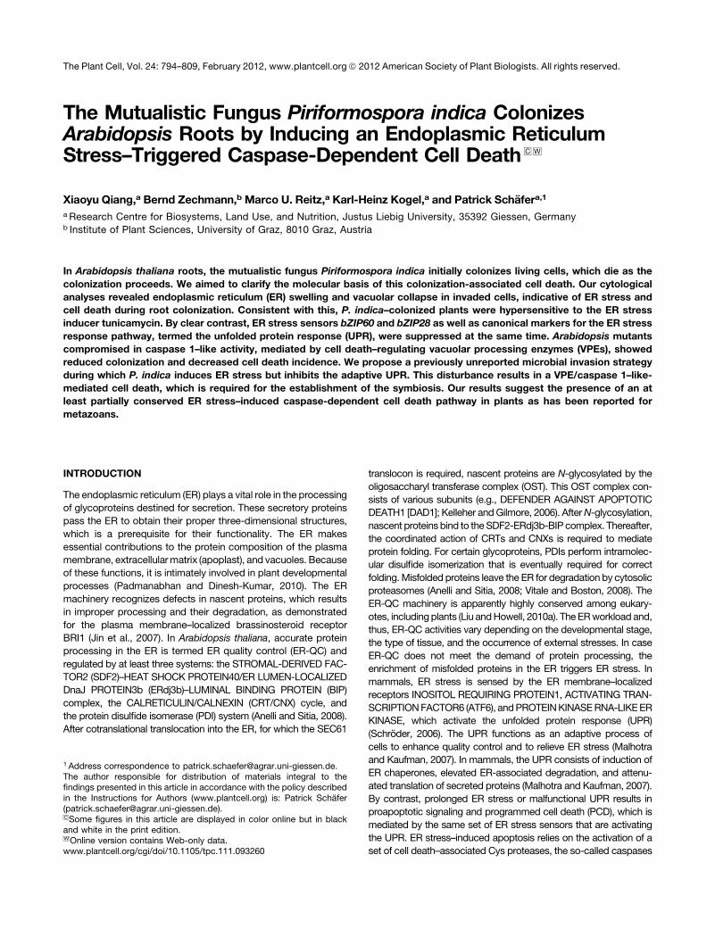
P. indica Impairs the Integrity of the ER, Thereby Enhancing
Mutualistic Root Colonization
By analyzing P. indica colonization of roots of Arabidopsis line
green fluorescent protein (GFP)-tmKKXX, in which the ER is GFP
tagged (Benghezal et al., 2000), we observed a loss of the
continuous ER network and the local appearance of globular ER
structures around intracellular hyphae during biotrophic root
colonization, suggesting an disintegration of the ER (see asterisk
in Figure 1A). In comparison, surrounding cells showed contin-
uous ER networks, reflecting the structural intactness of the ER
(Figure 1A). To assess subcellular changes occurring during
fungal colonization of Arabidopsis roots, we performed trans-
mission electronmicroscopy. Transmission electronmicroscopy
studies confirmed that ER swelling accompanies biotrophic root
colonization (Figure 1B). Since other organelles were ultrastruc-
turally unaltered and we did not observe lysis of the cytoplasm,
these colonized cells were considered alive. As colonization
proceeded (>3 DAI), we observed ER swelling that was followed
by tonoplast rupture and lysis of the cytoplasm (Figures 1C and
1D), which are hallmarks of PCD (van Doorn et al., 2011).
Interestingly, even at this stage, plastids and mitochondria
remained ultrastructurally unaltered (Figures 1C and 1D). These
observations prompted us to hypothesize that P. indica disturbs
ER function and that the resulting ER stress might induce a
ER Stress–Induced PCD in Root Symbiosis 795
vacuole-mediated cell death. ER swelling thus represents an
ultrastructural hallmark for the termination of biotrophic and the
initiation of cell death–associated colonization by P. indica.
Next, we examined to what extent the disturbed ER function
affects colonization success. To this end, the Arabidopsis mu-
tants bip2 (deficient in the chaperone BIP2), dad1 (deficient in a
subunit of the oligosaccharyl transferase DAD1), and sec61a
(deficient in a component of the SEC61 translocon complex), all
of which lack components of the ER-QC, were selected and
analyzed for altered fungal colonization at 3 DAI (biotrophic
phase) and 7 DAI (cell death phase). Quantitative real-time PCR
(qRT-PCR)–based quantification of fungal biomass showed en-
hanced fungal colonization rates at 7 DAI in all mutants com-
pared with wild-type Columbia-0 (Col-0) (Figure 1E), suggesting
Figure 1. P. indica Impairs ER Integrity and Induces Vacuole Collapse in Colonized Root Cells.
(A) Confocal microscopy of P. indica–colonized GFP-tmKKXX (ER marker)–expressing Arabidopsis roots at 3 DAI. The fungus penetrated (arrows) two
cells and intracellular hyphae are visible (arrowheads). The ER of the upper colonized cell is still intact, while ER disintegration is associated with
colonization of the lower cell (asterisk). Note the ER of surrounding, noncolonized cells is intact. P. indica was stained with chitin-specific WGA-AF488.
Intracellular hyphae are faintly stained due to limited dye diffusion. Bar = 20 mm.
(B) Micrograph of a biotrophic colonization phase. Intracellular hypha invaginated the host plasma membrane. The ER of this cell is partially swollen
(arrows). CW, cell wall; H, hypha; M, mitochondria; V, vacuole. Bar = 2 mm.
(C) ER swelling and lysis of the cytoplasm during cell death–associated root colonization. Arrows indicate ER swelling in a cell harboring an intracellular
hypha (H). Cell lysis is restricted to the colonized cell, while noncolonized neighboring cells are unaffected, as indicated by the intact tonoplasts
(asterisks). CW, cell wall; H, hypha; M, mitochondria; V, vacuole. Bar = 2 mm.
(D) ER swelling and vacuolar collapse during cell death–associated colonization. Intracellularly colonized cell shows ER swelling (arrows), vacuolar
collapse, as indicated by tonoplast rupture (arrowheads), and lysis of the cytoplasm. The micrograph shows intracellular (top H) and intercellular
(bottom H) hyphae. CW, cell wall; H, hyphae; M, mitochondria; V, vacuole. Bar = 2 mm.
(E) ER dysfunction improves cell death–associated colonization of Arabidopsis roots by P. indica. Arabidopsis Col-0 and the mutants sec61a, dad1, and
bip2, which are impaired in ER function, were inoculated with P. indica and fungal biomass was determined at biotrophic (3 DAI) and cell death–
associated colonization stages (7 DAI) by qRT-PCR. Fungal colonization levels in all mutants were normalized with wild-type Col-0 colonization (set to
1). Results shown are means 6 SE of three independent experiments. For each experiment, ;200 plants were analyzed per line at each time point.
Asterisks indicate significant differences in the colonization of mutants compared with Col-0 at 7 DAI at P < 0.05 (*) or P < 0.01 (**) as analyzed by two-
way analysis of variance (ANOVA).
[See online article for color version of this figure.]
796 The Plant Cell
that impaired ER-QC supports fungal development during cell
death–associated colonization. Alternatively, malfunctional im-
munity in these ER-QC mutants might explain the altered colo-
nization. Therefore, we examined their responsiveness to
MAMPs as well as their colonization by two pathogens following
two different lifestyles, the biotrophic powdery mildew fungus
Erysiphe cruciferarum and necrotrophic Botrytis cinerea. In the
first assay, we analyzed the flg22-induced seedling growth
inhibition in wild-type Col-0 and the ER-QC mutants. flg22
treatment reduced the biomass of wild-type Col-0 and all mutant
plants, as opposed to the flg22-insensitive fls2c mutant (see
Supplemental Figure 1A online). Second, we analyzed the oc-
currence of flg22- or chitin-induced oxidative burst in roots of the
ER-QC mutants. Consistently, we observed transient root oxi-
dative bursts upon treatment with flg22 or chitin in bip2, dad1,
and sec61amutants like in wild-type Col-0, but not in flg22- and
chitin-insensitive mutants fls2c and cerk1-2, respectively (see
Supplemental Figures 1B and 1C online), indicating intactness of
MAMP-triggered immunity in the mutants. In addition, bip2,
dad1, and sec61a showed no significant alterations in coloniza-
tion by biotrophic E. cruciferarum and necrotrophic B. cinerea
compared with wild-type Col-0 (see Supplemental Figures 2A
and 2B online).
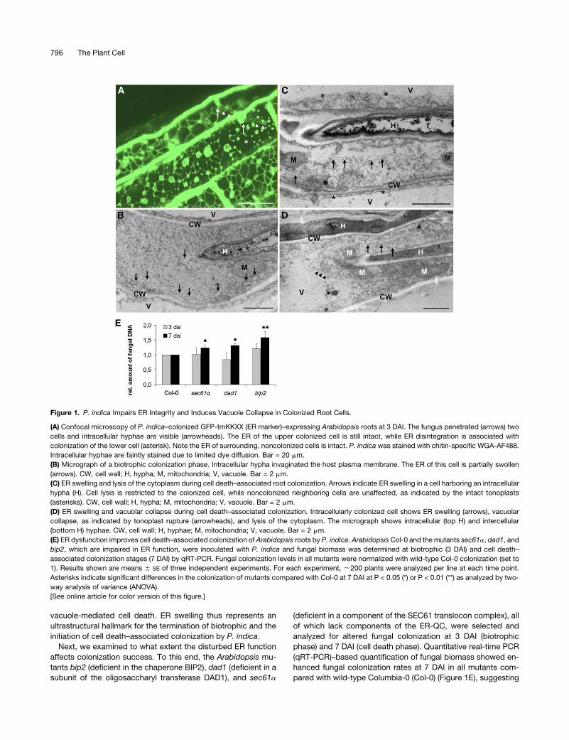
P. indica–Colonized Plants Are Hypersensitive to ER Stress
but Disturbed in the UPR
Since analysis of fungal growth in roots indicated improved colo-
nization of mutants lacking crucial components of the ER-QC, we
investigated whether P. indica affected tolerance of colonized
plants to ER stress.We applied the ER stress inducer tunicamycin
(TM), which specifically blocks UDP-N-ACETYLGLUCOSAMINE:
DOLICHOL PHOSPHATE N-ACETYGLUCOSAMINE-1-P
TRANSFERASE and thereby inhibits protein N-glycosylation
in the ER (Pattison and Amtmann, 2009). P. indica–colonized
(3 DAI, biotrophic stage) and noncolonized (mock-treated) Col-0
Figure 2. P. indica–Colonized Plants Are Hypersensitive to ER Stress, but ER Stress Signaling Is Not Activated in Colonized Roots.
(A) Arabidopsis Col-0 plants colonized by P. indica (Pi) are more sensitive to the ER stress inducer TM than are noncolonized plants, as evidenced by a
reduced biomass. Plant biomass was determined at 7 DAT. Data presented show means of three independent experiments6 SE. For each experiment,
10 plants were analyzed per treatment. Asterisks indicate significance at P < 0.05 (*) and P < 0.01 (**) as analyzed by two-way ANOVA.
(B) Expression of ER stress sensors (bZIP17, bZIP28, and bZIP60) and markers for the UPR (sPDI, BIP3, and CNX2) was measured by qRT-PCR.
Arabidopsis plants were inoculated with P. indica or mock treated. Data shown represent fold changes of genes and display the ratio of candidate gene
expression to housekeeping gene UBIQUITIN5 in colonized roots relative to mock-treated roots. Data presented show means of three independent
experiments 6 SE.
(C) BIP protein accumulation is reduced during P. indica colonization. Arabidopsis roots were inoculated with P. indica or mock treated and harvested
for protein extraction. The staining with Coomassie blue (CBB) indicates equal loading of all samples. Numbers on top of the immunoblot indicate
relative BIP protein band intensities (0 DAI was set to 1) as determined by ImageJ.
ER Stress–Induced PCD in Root Symbiosis 797

plants were treated with TM or DMSO (control). Plant fresh
weights were determined at 7 d after treatment (DAT). In non-
colonized plants, TM treatment resulted in an;20% reduction in
fresh weight compared with DMSO-treated plants. In P. indica–
colonized plants, TM significantly reduced plant biomass by
;60% compared with untreated controls (Figure 2A). This sup-
ports the view that P. indica disturbs ER function already during
biotrophic colonization and further suggests that P. indica–colo-
nized plants are hypersensitive to ER stress.
Next, we examined whether colonization-associated ER
stress resulted in both activation of ER stress sensing and
subsequent UPR. P. indica predominantly colonizes the matu-
ration zone of roots, while the fungus is hardly detectable in the
juvenile root sections, the elongation and meristematic zones.
Thus, to avoid dilution effects of noncolonized root tissues, we
harvested the maturation zone of P. indica–colonized and non-
colonized roots at 1, 3, and 7DAI andmonitored gene expression
levels of putative ER stress sensors (bZIP17, bZIP28, and
bZIP60) and markers of the UPR (sPDI, BIP3, and CNX2) by
qRT-PCR. Unexpectedly, none of the tested genes was ex-
pressed at higher levels during colonization (Figure 2B). Instead,
expression of bZIP28, BIP3, and CNX2 was suppressed in
colonized roots at some of the investigated time points (Figure
2B). Consistent with this, BIP protein levels were reduced in
colonized roots at 3 and 7 DAI by 35 and 85%, respectively,
compared with noncolonized roots (Figure 2C). To elucidate
whether the selected ER stress markers were induced by ER
stress in roots and whether P. indica might suppress ER stress
signaling, we treated noncolonized andP. indica–colonized roots
(3 DAI, biotrophic stage) with TM or DMSO (control). The root
samples were harvested at 1 and 3 d after TM treatment, and the
expression levels of ER stress sensors and UPR markers were
analyzed by qRT-PCR. TM treatment induced the expression of
all these genes (except bZIP28) in noncolonized roots (Figure 3A,
gray columns). By contrast, induction of bZIP17, bZIP60, BIP3,
and sPDI expression was much weaker in P. indica–colonized
Figure 3. P. indica Colonization Results in the Suppression of the UPR.
(A) Arabidopsis roots were inoculated with P. indica or mock treated. Inoculated and mock-treated plants were treated with TM (5 mg mL�1) or DMSO
(control). Root samples from different treatments (Pi + TM, Pi + DMSO; mock + TM, mock + DMSO) were harvested at 1 and 3 DAT. Data shown
represent fold changes of genes and display the ratio of candidate gene expression to housekeeping gene UBIQUITIN5 using the DDCT method
(Schmittgen and Livak, 2008). DDCT values obtained from Pi + TM samples were divided by DDCT values of Pi + DMSO to obtain the displayed fold
changes. Similarly, DDCT values of samples mock + TM were divided by DDCT values of mock + DMSO. Fold changes >1 or <1 indicate induction or
suppression of genes, respectively. Data are means of three independent experiments 6 SE.
(B) BIP protein accumulation after TM treatment and P. indica inoculation. Samples were run on the same blot, but the lanes were arranged for
presentation. Arabidopsis roots were inoculated with P. indica or mock treated prior to treatment with TM or DMSO (control) and harvested 2 d later. The
staining with Coomassie blue (CBB) indicates equal loading of all samples. Numbers on top of the immunoblots represent relative BIP protein band
intensities (mock treatment was set to 1) as determined by ImageJ. M, mock.
798 The Plant Cell
roots, while CNX2 and BI-1 showed only slightly reduced ex-
pression levels (Figure 3A). These data indicated impaired ER
stress signaling at the transcript level.
In addition to sPDI, BIP3, and CNX2, gene products of ERDj3A
and GRP94 participate in ER-localized protein folding and are
induced duringERstress (Iwata et al., 2008). Notably, TM-induced
expression of ERDj3A and GRP94 was also suppressed in P.
indica–colonized roots (Figure 3A). We further tested the expres-
sion of ER stress–induced genes, whose products function in
protein degradation (Derlin-like 1 and AAA-type ATPase), glyco-
sylation (putative galactinol synthase and UDP-galactose/UDP-
glucose transporter [UDP-transp.]), and the secretory pathway
(ADP-ribosylation factor [ADP-RF], SAR1B, and SEC61g). These
genes are induced in leaves by TM (Urade, 2007; Iwata et al.,
2008). Except for UDP-transp., TM induced the expression of
these genes in roots at 1 and/or 3 DAT. By clear contrast,
expression of all genes (except CNX2) was suppressed in P.
indica–colonized roots at 1 and/or 3 d after TM treatment (Figure
3A). We further examined whether root colonization by P. indica
also affected BIP accumulation in response to TM. For this, P.
indica–colonized (3 DAI) and noncolonized roots were treatedwith
TM or DMSO (control), and roots were harvested 2 d later. BIP
accumulated in response to TM treatment in noncolonized roots
(increase of 47%) (Figure 3B). By contrast, BIP protein synthesis
was suppressed in P. indica–colonized mock (decrease of 67%)
and TM-treated roots (decrease of 95%). Taken together, the
analyses suggested that the fungus disturbed ER stress signaling
as well as UPR-associated processes such as protein folding,
glycosylation, protein degradation, and secretion.
Vacuole-Mediated Cell Death Is Downstream of ER Stress
Induction and Affects Mutualistic Root Colonization
Our electron microscopy studies indicated that colonization-
associated ER swelling preceded vacuolar collapse during cell
death–associated colonization (Figure 1). Therefore, we wanted
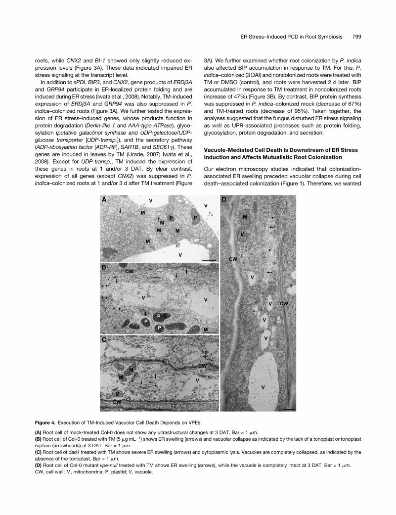
Figure 4. Execution of TM-Induced Vacuolar Cell Death Depends on VPEs.
(A) Root cell of mock-treated Col-0 does not show any ultrastructural changes at 3 DAT. Bar = 1 mm.
(B) Root cell of Col-0 treated with TM (5 mg mL�1) shows ER swelling (arrows) and vacuolar collapse as indicated by the lack of a tonoplast or tonoplast
rupture (arrowheads) at 3 DAT. Bar = 1 mm.
(C) Root cell of dad1 treated with TM shows severe ER swelling (arrows) and cytoplasmic lysis. Vacuoles are completely collapsed, as indicated by the
absence of the tonoplast. Bar = 1 mm.
(D) Root cell of Col-0 mutant vpe-null treated with TM shows ER swelling (arrows), while the vacuole is completely intact at 3 DAT. Bar = 1 mm.
CW, cell wall; M, mitochondria; P, plastid; V, vacuole.
ER Stress–Induced PCD in Root Symbiosis 799
to know which proteins were essential for vacuole collapse
following ER stress. VACUOLAR PROCESSING ENZYMES
(VPEs)mediate vacuolar collapse and execution of virus-induced
cell death (hypersensitive response) in tobacco (Nicotiana taba-
cum; Hatsugai et al., 2004). VPEs form a small gene family
consisting of four members (aVPE, bVPE, gVPE, and dVPE)
(Hatsugai et al., 2006). AllVPEs but dVPEwere expressed in roots
as indicated by RT-PCR (see Supplemental Figure 3 online). In a
first assay, we treated roots of wild-type Col-0 and the quadruple
vpe-null mutant, which is deficient in the four VPEs, with TM or
DMSO (control) and examined root cells for ultrastructural
changes (Figure 4). In addition, we included dad1, in which the
impairment in ER functionmight result in TM hypersensitivity. We
did not observe any changes in wild-type Col-0 or mutant root
cells after mock treatment or at 1 DAT with TM (Figure 4A; data
not shown). At 3 DAT, we observed ER swelling, vacuolar
collapse, and lysis of the cytoplasm in root cells of wild-type
Col-0 and dad1 (Figures 4B and 4C). dad1 showed the most
severe effects, as lysis of the cytoplasm was most pronounced
and detected in all cells at 3 DAT. By contrast, vacuolar collapse
was not detected in root cells of vpe-null, although ER swelling
occurred (Figure 4D). These results indicated that the genetic
impairment of ER homeostasis (in dad1) accelerated the ER
stress–induced cell death. Moreover, VPEs are required for ER
stress–induced collapse of the vacuole. Notably, ultrastructural
changes, associated with ER stress–induced vacuolar cell death
after TM treatment, were highly similar to those observed in root
cells during P. indica colonization (Figure 1). In both cases, ER
stress seems to lie upstream of vacuolar collapse, and ER
swelling most likely is not the consequence of cell death.
The results prompted us to test whether vacuolar collapse is
essential for root colonization. We quantified P. indica coloniza-
tion of avpe, bvpe, gvpe, dvpe, and vpe-null roots at 3 (biotrophic
phase) and 7DAI (cell death phase) by qRT-PCR. vpe-nullmutant
showed higher fungal colonization at 3 DAI, consistent with
earlier studies demonstrating an immune-related function of
VPEs (Hatsugai et al., 2004; Rojo et al., 2004). We observed
significantly reduced colonization of avpe, gvpe, and vpe-null
mutants at 7 DAI, while bvpe and dvpe mutants showed little if
any enhancement in colonization (Figure 5), indicating that VPE-
related activities contribute to cell death–associated coloniza-
tion. To confirm that VPE-related activities act downstream of P.
indica–induced ER stress, we generated gvpe dad1 double
mutants and quantified P. indica colonization by qRT-PCR.
gvpe dad1 displayed reduced colonization at 7 DAI, which was
highly similar to the colonization phenotype of gvpe (Figure 5).
Together, these data suggest that VPEs participate in the exe-
cution of ER stress–induced cell death and that gVPE plays a
critical role in cell death–associated root colonization by P.
indica.
VPE- and Caspase 1–Like Activities Are Enhanced in
TM-Treated and P. indica–Colonized Roots
In addition to VPE activity, VPEs have caspase 1–like protease
activity. It is suggested that both enzyme activities are required
for vacuole-mediated plant cell death execution (Hatsugai et al.,
2004; Kuroyanagi et al., 2005), although the enzyme targets are
unknown. To examine the occurrence of these protease activ-
ities during root colonization, we measured VPE- and caspase
1–like activities in wild-type Col-0 roots during biotrophic (3 DAI)
and cell death–associated colonization (7 DAI). To this end, we
set up an assay to measure VPE- and caspase 1–like activities in
root extracts from P. indica–colonized and noncolonized roots.
Upon addition of either 1 mM VPE specific substrate Ac-ESEN-
MCA or caspase 1–specific substrate Ac-YVAD-MCA to root
extracts, VPE-mediated cleavage of MCA was spectrometrically
determined. P. indica itself was unable to cleave these sub-
strates (data not shown). The analyses did not reveal P. indica–
dependent changes in enzyme activities at 3 DAI (biotrophic
phase) but significantly enhanced VPE- and caspase 1–like
activities at 7 DAI (cell death phase) with P. indica (Figure 6A).
To relate theenhancedenzymeactivities tocolonization-associated
cell death, we performed a fluorescein diacetate (FDA)-based
cell viability assay. Esterases cleave off fluorescein in living cells,
and the degree of cleavage can be quantified spectrometrically.
We found a strong correlation between the length of analyzed
root segments and the measured absolute fluorescence indicat-
ing FDA cleavage (see Supplemental Figure 4 online). The FDA
assay revealed unaltered cell viability at 3 DAI but significantly
reduced cell viability at 7 DAI with P. indica (Figure 6B). Thus, the
experiments indicate a clear correlation of enhanced enzyme
activities with the occurrence of enhanced cell death at 7 DAI.
We next examined whether the altered P. indica colonization of
the ER-QC mutants bip2 and dad1 as well as gvpe, vpe-null, and
gvpe dad1 mutants during biotrophic or cell death–associated
interaction stages (Figures 1C, 1D, and 5) was associated with
alteredVPE- andcaspase 1–like activities (Figures 6Cand 6D). In a
complementary experiment, we determined whether TM-induced
cell death observed in our cytological studies (Figure 4) correlated
with changed VPE- and caspase 1–like activities (Figures 6E and
Figure 5. Colonization of Arabidopsis Roots by P. indica Is Dependent
on VPEs.
Arabidopsis wild type (WT) and mutants avpe, bvpe, gvpe, dvpe, and
vpe-null, as well as double mutant gvpe dad1 were inoculated with P.
indica. Fungal biomass was determined at biotrophic (3 DAI) and cell
death–associated colonization stages (7 DAI) by qRT-PCR. Fungal
colonization levels in all mutants were normalized with wild-type colo-
nization (set to 1). Results shown are means 6 SE of three independent
experiments. For each experiment, around 200 plants were analyzed per
line at each time point. Asterisks indicate significant differences in the
colonization of mutants compared with Col-0 at 3 or 7 DAI at P < 0.05 (*)
as analyzed by two-way ANOVA.
800 The Plant Cell
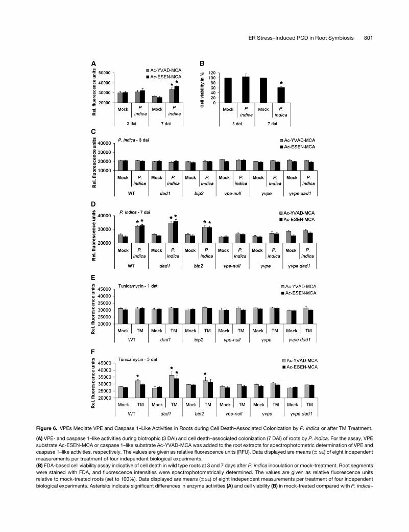
Figure 6. VPEs Mediate VPE and Caspase 1–Like Activities in Roots during Cell Death–Associated Colonization by P. indica or after TM Treatment.
(A) VPE- and caspase 1–like activities during biotrophic (3 DAI) and cell death–associated colonization (7 DAI) of roots by P. indica. For the assay, VPE
substrate Ac-ESEN-MCA or caspase 1–like substrate Ac-YVAD-MCA was added to the root extracts for spectrophotometric determination of VPE and
caspase 1–like activities, respectively. The values are given as relative fluorescence units (RFU). Data displayed are means (6 SE) of eight independent
measurements per treatment of four independent biological experiments.
(B) FDA-based cell viability assay indicative of cell death in wild type roots at 3 and 7 days after P. indica inoculation or mock-treatment. Root segments
were stained with FDA, and fluorescence intensities were spectrophotometrically determined. The values are given as relative fluorescence units
relative to mock-treated roots (set to 100%). Data displayed are means (6SE) of eight independent measurements per treatment of four independent
biological experiments. Asterisks indicate significant differences in enzyme activities (A) and cell viability (B) in mock-treated compared with P. indica–
ER Stress–Induced PCD in Root Symbiosis 801

6F). Since colonization data indicated a central role of gVPE during
cell death–associated colonization, we performed both sets of
experiments with gvpe and used vpe-null as control. To unravel
whether improved colonization of ER-QCmutants (Figure 1E) and
the hypersensitivity of dad1 to TM (Figure 4C) were linked to
altered VPE and caspase 1–like activities, we examined bip2
besides the dad1 mutant. Finally, we included the double mutant
gvpe dad1 in this enzyme activity studies because of its reduced
colonization phenotype (Figure 5). We did not detect altered VPE
and caspase 1–like activities in all mutants at 3 DAI (Figure 6C).
However, we detected changes in VPE and caspase 1–like
activities in a mutant-specific manner at 7 DAI. Enzyme activities
were elevated inwild-type,dad1, andbip2 roots at a similar level at
7 DAI. By contrast, neither VPE nor caspase 1–like activities were
observed in vpe-null and in gvpe at 7 DAI (Figure 6D). These
findings suggested that the P. indica–induced activation of both
enzyme activities relied on gVPE. Interestingly, elevated VPE and
caspase 1–like activities inP. indica–colonizeddad1 roots at 7 DAI
was not detected in colonized gvpe dad1 roots at 7 DAI. In fact,
enzyme activities in gvpe dad1 roots were highly similar to those in
gvpe mutants (Figure 6D). TM treatment of all mutant roots
resulted in changes of VPE and caspase 1–like activities that
were similar to our analyseswithP. indica–colonized roots.We did
not observe altered enzyme activities at 1 DAT (Figure 6E).
Caspase 1–like activity increased in dad1, bip2, and wild-type
roots,while VPE activitieswere enhanced indad1 roots at 3 d after
TM treatment (Figure 6F). TM-induced VPE and caspase 1–like
activities were not detectable in vpe-null and gvpe. Again, gvpe
dad1 did not show TM-induced elevation of enzyme activities as
detected in dad1 at 3 DAT, and the enzyme activity phenotypes
strongly resembled those of gvpe. In summary, the results of the
enzyme activity assays were consistent with the colonization and
TM-induced cell death phenotypes (Figures 1E, 4, and 5).
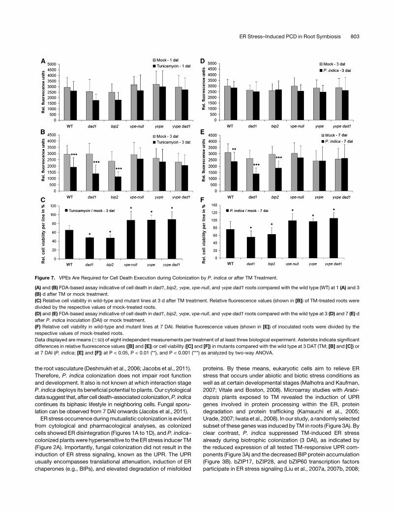
ER Dysfunction Enhances P. indica– and TM-Induced Cell
Death in a VPE-Dependent Manner
Finally, we were interested to know whether the variation in VPE
and caspase 1–like enzyme activities in the various mutants after
TM treatment or during cell death–associated root colonization
(Figures 6Dand 6F)was associatedwith an altered occurrence of
cell death. In addition, these analyses should reveal whether P.
indica and TM-induced ER stress initiated cell death andwhether
this was a VPE/caspase 1–like-mediated cell death. Therefore,
we applied the FDA-based cell viability assay as described
above. First, we treated dad1, bip2, vpe-null, gvpe, and gvpe
dad1 mutants and the respective wild type with TM and stained
root segments with FDA at 1 and 3 DAT. Whereas all mutants
exhibited unaltered cell viability compared with the wild type at
1 DAT (Figure 7A), wild-type dad1 and bip2mutants displayed a
reduced cell viability at 3 DAT, which was not observed in vpe-
null, gvpe, and gvpe dad1mutants (Figures 7B and 7C). Notably,
we detected higher cell death ratios in dad1 and bip2 as well as
higher cell viability ratios in all vpe mutants compared with the
wild type at 3DAT (Figure 7C).We next determined FDAcleavage
in the same mutants during biotrophic (3 DAI) and cell death–
associated (7 DAI) colonization by P. indica. Consistent with our
enzyme assays, none of the mutants showed an altered viability
phenotype at 3 DAI (Figure 7D). Similar to the results obtained in
the TM assay, the wild type, dad1, and bip2 exhibited more cell
death at 7 DAI with P. indica. Again, cell viability was unaltered in
vpe-null, gvpe, and gvpe dad1 mutants upon P. indica coloniza-
tion at 7 DAI (Figures 7E and 7F). Unaltered cell viability in gvpe
dad1was in clear contrast with the reduced cell viability in dad1.
Again, cell death ratios were enhanced in dad1 and bip2, while
cell viability ratios were enhanced in all vpe mutants compared
with the wild type at 7 DAI (Figure 7F). These results were
consistent with the enzyme activity assays in that enhanced cell
death in wild-type, dad1, and bip2 roots coincided with an
enhanced caspase 1–like activity at 3 DAT with TM and en-
hanced VPE and caspase 1–like activities at 7 DAI with P. indica
in these mutants. Accordingly, at those time points (1 DAT [TM]
and 3 DAI [P. indica]) and in those mutants (vpe-null, gvpe, and
gvpe dad1) wherewe did not detect altered enzyme activities, we
did not observe reduced cell viability. Together, these data
confirm enhanced ER stress (TM)- and P. indica–induced cell
death in mutants impaired in ER-QC. Moreover, our data iden-
tified gVPE as a key factor in the execution of ER stress–induced
cell death triggered by TM or by fungal colonization in the
mutualistic interaction of P. indica and Arabidopsis.
DISCUSSION
In this study, we examine the molecular basis of cell death–
associated root colonization of Arabidopsis by the mutualistic
fungus P. indica. Our study suggests that P. indica suppresses
ER stress signaling as initial step, which eventually results in a
vacuole-mediated cell death that is dependent on VPE/caspase
1–like activities (Figure 8). Cell death–dependent colonization
and mutualism is counterintuitive, but, notably, P. indica only
colonizes parts of the root maturation zone and does not enter
Figure 6. (continued).
colonized roots at P < 0.05 (*) analyzed by two-way ANOVA.
(C) and (D) VPE- and caspase 1–like activities during root colonization of various mutants by P. indica. The experiments were performed as described in
(A). Root samples were harvested at 3 (C) and 7 DAI (D). Data displayed are means with (6SE) of four independent measurements per treatment of four
biological experiments. Asterisks indicate significant differences in respective enzyme activities between P. indica–colonized andmock-treated roots at
P < 0.05 (*) as analyzed by two-way ANOVA.
(E) and (F) VPE- and caspase 1–like activities in roots of various mutants after TM or mock treatment. The experiments were performed as described in
(A). Root samples were harvested at 1 (E) and 3 DAT (F). Data displayed are means (6SE) of four independent measurements per treatment of four
biological experiments. Asterisks indicate significant difference in respective enzyme activities between TM- and mock-treated roots at P < 0.05 (*) as
analyzed by two-way ANOVA. WT, wild type.
802 The Plant Cell
the root vasculature (Deshmukh et al., 2006; Jacobs et al., 2011).
Therefore, P. indica colonization does not impair root function
and development. It also is not known at which interaction stage
P. indica deploys its beneficial potential to plants. Our cytological
data suggest that, after cell death–associatedcolonization,P. indica
continues its biphasic lifestyle in neighboring cells. Fungal sporu-
lation can be observed from 7 DAI onwards (Jacobs et al., 2011).
ER stress occurrence duringmutualistic colonization is evident
from cytological and pharmacological analyses, as colonized
cells showed ER disintegration (Figures 1A to 1D), and P. indica–
colonized plantswere hypersensitive to the ER stress inducer TM
(Figure 2A). Importantly, fungal colonization did not result in the
induction of ER stress signaling, known as the UPR. The UPR
usually encompasses translational attenuation, induction of ER
chaperones (e.g., BIPs), and elevated degradation of misfolded
proteins. By these means, eukaryotic cells aim to relieve ER
stress that occurs under abiotic and biotic stress conditions as
well as at certain developmental stages (Malhotra and Kaufman,
2007; Vitale and Boston, 2008). Microarray studies with Arabi-
dopsis plants exposed to TM revealed the induction of UPR
genes involved in protein processing within the ER, protein
degradation and protein trafficking (Kamauchi et al., 2005;
Urade, 2007; Iwata et al., 2008). In our study, a randomly selected
subset of these geneswas induced by TM in roots (Figure 3A). By
clear contrast, P. indica suppressed TM-induced ER stress
already during biotrophic colonization (3 DAI), as indicated by
the reduced expression of all tested TM-responsive UPR com-
ponents (Figure 3A) and the decreased BIP protein accumulation
(Figure 3B). bZIP17, bZIP28, and bZIP60 transcription factors
participate in ER stress signaling (Liu et al., 2007a, 2007b, 2008;
Figure 7. VPEs Are Required for Cell Death Execution during Colonization by P. indica or after TM Treatment.
(A) and (B) FDA-based assay indicative of cell death in dad1, bip2, gvpe, vpe-null, and gvpe dad1 roots compared with the wild type (WT) at 1 (A) and 3
(B) d after TM or mock treatment.
(C) Relative cell viability in wild-type and mutant lines at 3 d after TM treatment. Relative fluorescence values (shown in [B]) of TM-treated roots were
divided by the respective values of mock-treated roots.
(D) and (E) FDA-based assay indicative of cell death in dad1, bip2, gvpe, vpe-null, and gvpe dad1 roots compared with the wild type at 3 (D) and 7 (E) d
after P. indica inoculation (DAI) or mock treatment.
(F) Relative cell viability in wild-type and mutant lines at 7 DAI. Relative fluorescence values (shown in [E]) of inoculated roots were divided by the
respective values of mock-treated roots.
Data displayed are means (6SD) of eight independent measurements per treatment of at least three biological experiment. Asterisks indicate significant
differences in relative fluorescence values ([B] and [E]) or cell viability ([C] and [F]) in mutants compared with the wild type at 3 DAT (TM; [B] and [C]) or
at 7 DAI (P. indica; [E] and [F]) at P < 0.05, P < 0.01 (**), and P < 0.001 (***) as analyzed by two-way ANOVA.
ER Stress–Induced PCD in Root Symbiosis 803
Iwata et al., 2008; Liu and Howell, 2010a). Although bZIP17 and
bZIP60 were induced by TM in roots (Figure 3A), none of these
genes were induced during P. indica colonization (Figure 2B).
This suggests that the fungus apparently inhibits the initiation of
ER stress signaling. Consistent with this, TM treatment could not
induce the expression of bZIP60- and bZIP28-regulated UPR
genes (Iwata et al., 2008; Liu and Howell, 2010b) in P. indica–
colonized roots (Figure 3A).
Most probably, fungal suppression of the UPR (Figures 3A and
3B) is required for cell death initiation. Failed ER stress adapta-
tion or severe ER stress can result in the activation of cell death
(Szegezdi et al., 2006). In mammals, the principles of ER stress–
induced proapoptotic signaling have been intensively studied
(Szegezdi et al., 2006; Rasheva and Domingos, 2009). The same
plasma membrane–localized ER stress sensors that induce the
UPR also initiate apoptotic signaling under severe ER stress
by activating the bZIP transcription factor ATF4, the c-Jun
N-terminal kinase pathway, and a caspase cascade. Central to
this proapoptotic state is the activation of Bcl2-ASSOCIATED X
PROTEIN (BAX) and B-CELL LYMPHOMA 2 INTERACTING
MEDIATOR OF CELL DEATH (BIM), which contribute to the
execution of apoptosis by enhancing Ca2+ release from ER and
mitochondria. In addition, BAX and BIM mediate cytochrome c
release from mitochondria, thereby activating the apoptosome
(Szegezdi et al., 2006). Several studies suggest a conservation of
ER stress signaling in plants and mammals. BI-1 is a negative
regulator of cell death in mammals that antagonizes BAX-
induced lethality (Xu and Reed, 1998). Although BAX homologs
are not present in plants, barley BI-1 and other plant BI-1 proteins
suppress BAX-induced cell death in planta (Huckelhoven, 2004;
Eichmann et al., 2006;Watanabe and Lam, 2009). BI-1 is thought
to control Ca2+ release from the ER under stress conditions in
plants and mammals (Chae et al., 2004; Watanabe and Lam,
2009). Arabidopsis plants overexpressing BI-1 exhibit enhanced
TM tolerance, which indicates its negative regulatory function in
ER-PCD (Watanabe and Lam, 2008). In barley, P. indica sup-
presses BI-1 transcription and BI-1 overexpression resulted in
reduced P. indica colonization (Deshmukh et al., 2006). Consis-
tent with this, we observed a suppression of BI-1 by P. indica
after TM application (Figure 3A). However, 3-week-old Arabi-
dopsismutants lacking BI-1 (atbi1-2) (Watanabe and Lam, 2006)
did not show an alteredP. indica colonization at 3 and 7DAI (data
not shown) using the qRT-PCR–based fungal colonization assay
(see Methods).
Similar to mammalian ER-PCD (Szegezdi et al., 2006), we
present several lines of evidence that ER stress is an initiator of
PCD in Arabidopsis roots and that this PCD is dependent on
VPEs. Application of TM to Col-0 resulted in ER swelling followed
by vacuolar collapse and lysis of the cytoplasm (Figure 4B).
Whereas TM triggered ER swelling in vpe-nullmutants, vacuolar
collapse and lysis of the cytoplasm did not occur (Figure 4D).
Consistent with this, TM treatment did not induce VPE and
caspase 1–like activities in vpe-null and in gvpe at 3 DAT. This
coincided with the inability of TM to induce cell death in both
mutants (Figures 6F, 7B, and 7C). By contrast, TM hypersensi-
tivity of dad1 was associated with an increased cell death
occurrence (Figures 4C, 7B, and 7C). Caspase 1–like activity
might be most important to transduce cell death after TM
treatment. Despite the lack of VPE activity, cell death occurrence
was similar in bip2 compared with dad1 (Figures 6F, 7B, and 7C).
Notably, the gvpe dad1 double mutant behaved almost identi-
cally to gvpe in terms of enzyme activities and cell viability at 3
DAT (Figures 6F, 7B, and 7C). Therefore, TM-induced ER-PCD
most probably strongly relies on the activity of gVPE for the
execution of cell death even in those cases where ER-QC is
impaired, as indicated by the double mutant gvpe dad1.
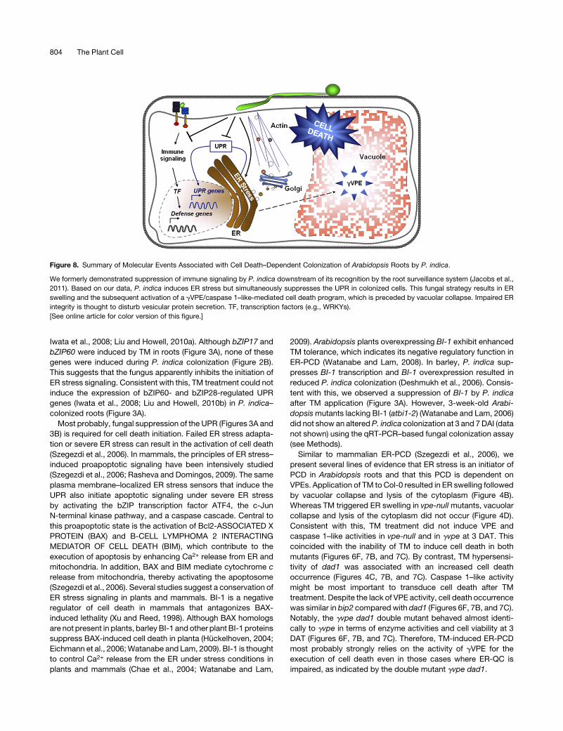
Figure 8. Summary of Molecular Events Associated with Cell Death–Dependent Colonization of Arabidopsis Roots by P. indica.
We formerly demonstrated suppression of immune signaling by P. indica downstream of its recognition by the root surveillance system (Jacobs et al.,
2011). Based on our data, P. indica induces ER stress but simultaneously suppresses the UPR in colonized cells. This fungal strategy results in ER
swelling and the subsequent activation of a gVPE/caspase 1–like-mediated cell death program, which is preceded by vacuolar collapse. Impaired ER
integrity is thought to disturb vesicular protein secretion. TF, transcription factors (e.g., WRKYs).
[See online article for color version of this figure.]
804 The Plant Cell
Interestingly, P. indica activated an ER-PCD that was similar to
TM-induced ER-PCD.P. indica–induced ER-PCDwas associated
with enhanced VPE/caspase 1–like activities and reduced cell
viability at 7 DAI (Figures 6A, 6B, and 7E). Enzyme activities and
cell death occurrence were unaltered in vpe-null, gvpe, and gvpe
dad1 at 7 DAI (Figures 6D, 7E, and 7F), and this was linked to a
reduced colonization of these mutants (Figure 5). Moreover, P.
indica colonization resulted in enhanced VPE and caspase 1–like
activities and cell death occurrence in roots of dad1 and bip2
mutants at 7 DAI, and this was associated with improved coloni-
zation (Figures 1E). This means that the fungus depends on this
ER-PCD for successful colonization of Arabidopsis roots. Appar-
ently, gVPE mediates most of the P. indica–induced VPE and
caspase 1–like activities (Figures 6D, 7E, and 7F), while participa-
tion of aVPE and bVPE in ER-PCD remains elusive. Based on the
colonization experiments (Figure 5), aVPE might function in the
same signaling pathway as gVPE. It is tempting to speculate that
bvpe might antagonize cell death signaling since it showed a
tendency to higher colonization by P. indica. In contrast with
previous studies (Kinoshita et al., 1995;Gruis et al., 2004), our PCR
analyses indicate expression of all VPEs but dVPE in roots (see
Supplemental Figure 3 online). The reduction in VPE and caspase
1–like activities in roots lacking gVPE further confirmed its pres-
ence in roots. Since we did not observe enhanced VPE and
caspase 1–like activities or the occurrence of cell death in wild-
type or vpe mutants at 3 DAI (biotrophic stage) (Figures 6C and
7D), the enhanced colonization of gvpe at 3 DAI (Figure 5) might
indicate anadditional immunity-related function of gVPE.Although
gvpe dad1 and gvpe showed a similar degree of P. indica colo-
nization at 7 DAI (Figure 5), gvpe dad1 displayed a colonization
phenotype comparable to dad1 at 3 DAI. Onemight argue that ER
stress induction may not be upstream of gVPE-mediated PCD.
However, we monitored virtually identical VPE/caspase 1–like
activities andcell viability ingvpeand gvpedad1at 7DAI (P. indica)
and at 3 DAT (TM) (Figures 6D, 6F, 7B, and 7E). Therefore, we
speculate that, during biotrophic root colonization (3 DAI), dad1
might counteract a yet unknownprocess ingvpe that is unlinked to
ER-PCD.
Impairment of ER integrity might not only serve to induce PCD.
The ER plays a crucial role in plant innate immunity by processing
antimicrobial proteins (Wang et al., 2005),whose delivery to the site
of microbial attack relies on vesicle-mediated transport processes
(Lipka et al., 2005; Huckelhoven, 2007). In addition, specific com-
ponents of the ERmachinerywere identified tomediate processing
of the PRR EFR (Nekrasov et al., 2009; Saijo et al., 2009), which
activates innate immunity after recognition of the bacterial MAMP
elongation factor TU (Zipfel et al., 2006). Recently, we found that P.
indica suppresses early root immune signaling and that this is
essential for biotrophic root colonization (Jacobs et al., 2011). In
this study, we recorded a disturbance in ER homeostasis as early
as 3 DAI (Figures 1A, 1B, 3A, and 3B). Although improved root
colonization of sec61a, dad1, and bip2 mutants might reflect
disturbance in immune signaling, several indications contradict this
assumption: (1) The improved colonization of these mutants oc-
curred only during cell death–dependent but not biotrophic colo-
nization. By contrast, colonization of cerk1 mutants lacking a
functional chitin receptor showed improved colonization at 3 DAI
(Jacobs et al., 2011). (2) flg22- and chitin-triggered root oxidative
burst and flg22-induced seedling growth inhibition, representing
early and lateMAMP responses of plants, are not impaired in these
mutants (see Supplemental Figure 1 online). (3) All three mutants
show an unaltered susceptibility to the biotrophic powdery mildew
fungusE. cruciferarum and the necrotrophic fungusB. cinerea (see
Supplemental Figure 2online).Notably, thesemutants are impaired
in systemic immune signaling induced by the salicylic acid analog
benzothiadiazole (Wang et al., 2005). This indicates an impairment
of thesemutants inadistinct immunepathway rather thanageneral
defect in various immune pathways. (4) The gvpe dad1 double
mutant showed a colonization phenotype similar to gvpe at 7 DAI
(Figure 5). Therefore, the improved root colonization ofdad1 (Figure
1E) rather indicates an altered ER stress–induced cell death
threshold as supported by our enzyme and cell viability assays
(Figures 6 and 7).
Our data support the existence of a previously unknown
pathway in plants, in which ER stress induces a vacuolar cell
death dependent on VPE/caspase 1–like activities, which oc-
curs after TM treatment and during microbial colonization. It
further indicates a sophisticated strategy for the mutualistic
fungus P. indica to colonize Arabidopsis roots successfully. It
will be interesting to see in future studies to what extent
P. indica–triggered ER dysfunction in roots impairs generation
and secretion of MTI components (e.g., antimicrobial proteins
and PRRs). Furthermore, it is tempting to speculate that im-
paired ER integrity also affects the function of the ER in vacuole
loading with antimicrobial proteins. This would explain why
VPE-mediated vacuolar collapse supports rather than stops
fungal growth as was reported for bacterial pathogens (Hatsugai
et al., 2009). We speculate that the impairment of ER function is a
more common strategy of microbes to colonize (and kill) eukar-
yotic host cells.
METHODS
Plant, Fungal Material, Plant Inoculation, and TM Treatment
bip2, dad1, and sec61a mutants were provided by X. Dong (Wang et al.,
2005); avpe, bvpe, gvpe, dvpe, and vpe-null were provided by I. Hara-
Nishimura (Kuroyanagi et al., 2005); the cerk1-2 mutant was provided by
V. Lipka (Petutschnig et al., 2010); the fls2c mutant was provided by C.
Zipfel (Zipfel et al., 2004); the atbi1-2 mutant was provided by E. Lam
(Watanabe and Lam, 2006); and GFP-tmKKXX was provided by D.A.
Jones (Benghezal et al., 2000). gvpe dad1 mutant was generated within
this study by crossing dad1 and gvpe. The isolate ofPiriformospora indica
DSM11827 was obtained from the German collection of microorganisms
and cell cultures in Braunschweig, Germany. For inoculation, all Arabi-
dopsis thaliana seeds were sterilized in 3% sodium hypochlorite and
grown in squared Petri dishes on half-strength Murashige and Skoog
(MS) under 8 h light (180 mmol m22 s21 photon flux density)/16 h night,
228C/188C, and 60% relative humidity. If not stated otherwise, 3-week-
old plant roots were inoculated with P. indica chlamydospores at a
concentration of 500,000 spores mL21 or treated with TM (5 mg mL21).
One milliliter of spore suspension was used per squared Petri dish
containing;40 plants. For all gene expression and protein analyses, we
removed the elongation and meristematic zones and only harvested the
maturation zones of roots. For TM assays, control treatment was
performed by applying DMSO. For Erysiphe cruciferarum inoculation
(see Supplemental Figure 2A online), Arabidopsis plants were grown at
228C and 65% relative humidity in a 10 h photoperiod with 120 mmol
ER Stress–Induced PCD in Root Symbiosis 805

m22 s21 light in a 2:1 soil sand mixture (Fruhstofer Erde, Type P;
Quarzsand, granulation: 0.1 to 0.5 mm; Sakret Trockenbaustoffe Eu-
ropa). E. cruciferarum was grown on Col-0, to maintain constant
aggressiveness, and on susceptible phytoalexin deficient 4 (pad4)
mutants (provided by the European Arabidopsis Stock Centre; Zhou
et al., 1998), for strong conidia production. For Botrytis cinerea inoc-
ulation (see Supplemental Figure 2B online), Arabidopsis plants were
placed under an inoculation box covered with a polyamide net
(0.2 mm2). Conidia were brushed off from pad4 plants through the net
until a density of three to four conidia mm22 was reached. Inoculated
plants were kept in a growth chamber under the above conditions.
Leaves were photographed 11 d after inoculation. B. cinerea strain
B05.10 was grown on HA agar (1% malt extract, 0.4% Glc, 0.4% yeast
extract, and 1.5% agar, pH 5.5) as described previously (Doehlemann
et al., 2006). Detached rosette leaves from 6- to 7-week-old soil grown
Arabidopsis plants were placed in Petri dishes containing 1% agar and
inoculated with 5 mL spore suspension (23 105 spores mL21 in 12 g L21
potato dextrose broth; Duchefa Biochemie). To maintain high humidity,
Petri dishes were covered with a transparent lid. Digital photographs
were taken at different time points and analyzed using ImageJ to
measure lesion size (Abramoff et al., 2004).
Quantification of Fungal Colonization by qRT-PCR
Genomic DNA was isolated from 100 mg root material with the Plant
DNeasykit (Qiagen). In qRT-PCRanalysis, 40ngof genomicDNAservedas
template. The amplification reaction was performed using 10 mL of SYBR
green JumpStart Taq ReadyMix (Sigma-Aldrich) and 350 nM of oligonu-
cleotides specifically amplifyingP. indica internal transcribed spacer (Pi ITS)
andArabidopsis ubiquitin 5 (AtUbi5) (see Supplemental Table 1 online). The
standard quantification program from the 7500 fast thermal cycler (Applied
Biosystems) was applied. To determine the relative amount of P. indica in
plant roots, the 22Dcycle threshold (CT) method (Schmittgen and Livak, 2008)
wasused. Therefore, the rawCTofP. indica ITSwere subtracted from those
obtained for At UBI5. Thereafter, the 22DCT of wild types were divided by
22DCT of mutants. In all cases, colonization of the respectivemutant is given
relative to wild-type colonization (set to one).
Gene Expression Analysis
For qRT-PCR–based gene expression studies, 3-week-old plants were
inoculatedwithP. indica ormock treated and harvested at 1, 3, and 7DAI.
For TM treatment, inoculated or mock-treated plants were treated with
TM (5 mg mL21) or DMSO (control) at 3 DAI. Roots were harvested at 12,
24, 48, and 72 h after TM treatment. For RT-PCR–based studies, roots of
3- and 5-week-old plants were harvested (see Supplemental Figure 3
online). In all cases, RNA was extracted from homogenized root material
using TRIzol (Invitrogen). For cDNA synthesis, 500 ng of RNAwas DNase-
I digested and transcribed into cDNA using a qScript cDNA synthesis kit
(Quanta Biosciences). For qRT-PCR analysis, 10 ng of cDNAwas used as
template for determining the amplification of candidate genes. As de-
scribed above the 22DCT methodwas applied to evaluate the level of gene
expression. For RT-PCR, 10 ng of cDNA served as template. Primers are
used as listed in Supplemental Table 1 online.
Protein Extraction and Immunodetection of BIP
Roots were inoculated with P. indica or mock treated and harvested at 1,
3, and 7 DAI. For TM assays, P. indica or mock-treated roots were treated
with TM 5 mg mL21 or DMSO (control) at 3 DAI and harvested at 2 DAT.
Total protein was extracted with a buffer containing 250 mM Suc, 50 mM
HEPES-KOH, 5% glycerin, 1 mM Na2MoO4 3 2H2O, 25 mM NaF, and 10
mMEDTA. Subsequently, 20mgof each protein samplewas separated by
SDS-PAGE and transferred to Roti-polyvinylidene fluoride membrane
(Roth). The proteins were probed with an Arabidopsis anti-BIP antibody
(Santa Cruz Biotechnology), followed by an anti-rabbit IgG-alkaline
phosphate antibody (Sigma-Aldrich). The anti-BIP antibody was raised
against the conserved C-terminal region of BIP and most probably
detects BIP and BIP2. The gels were stained in a solution containing 20%
(v/v) Coomassie Brilliant Blue R 250 (Roth) and 20% methanol and were
later destained with a solution of 40% methanol/10% glacial acid/50%
water (v/v/v). Protein bands were quantified using ImageJ.
Cell Death Assay
Root segments (1.5 cm) of the maturation zone from 2-week-old plants
inoculated with P. indica or treated with TM (control DMSO) were
transferred to half-strength MS containing FDA. After 10 min of incuba-
tion, root segments were washed five times and the fluorescence inten-
sities were measured at 535 nm after excitation at 485 nm using a
fluorescence microplate reader (TECAN infinite 200). See Supplemental
Figure 4 online for further details on the linearity in the quantification of cell
death using FDA.
Caspase and VPE Activity Assays
Three-week-old roots were inoculatedwithP. indica ormock treated, and
the roots were harvested at 3 and 7 DAI. For TM assays, roots were
treated with TM 5 mg mL21 or DMSO and harvested at 1 and 3 DAT. A
buffer containing 100 mM sodium acetate, pH 5.5, 100 mM NaCl, 1 mM
EDTA, and 1mM phenylmethylsulfonyl fluoride was applied to get root
extracts. To measure caspase 1–like and VPE activity, 1 mM fluorogenic
caspase 1 substrate (Ac-YVAD-MCA) and VPE substrate (Ac-ESEN-
MCA) (Peptide Institute) were added to the root extracts. Fluorescence
intensities were measured at 465 nm after excitation at 360 nm using a
fluorescence microplate reader (TECAN infinite 200).
Cytological Analyses
Root samples were either fixed or directly stained with chitin-specific
WGA-AF488 (Molecular Probes) as described (Deshmukh et al., 2006).
Confocal imageswere recorded on a TCSSP2microscope (Leica). WGA-
AF488 andGFPwere excitedwith a 488-nm laser line and detected at 505
to 540 nm. For ultrastructural studies, roots were embedded as described
(Zechmann et al., 2007), and ultrathin sections (80 nm) were investigated
after staining with uranyl acetate and lead citrate with a Philips CM10
transmission electron microscope.
Growth Retardation Assays
For the growth retardation assay with TM, Arabidopsis seedlings were
grown on half-strength MS medium containing 1% Suc for 14 d. Plants
were inoculated with P. indica or mock treated and transferred to liquid
half-strength MS medium containing 1% Suc and TM (25 ng mL21) or
DMSO (control). Seedling fresh weight was determined 7 d after TM/
DMSO treatment. For the growth retardation assaywith flg22, plantswere
grown on half-strength MS medium containing 1% Suc for 14 d and
thereafter transferred to liquid half-strength MS medium containing 1%
Suc and 10 mM flg22. Plant fresh weight was determined 10 d after
treatment. flg22 peptide sequence was used as described (Gomez-
Gomez et al., 1999).
MAMP-Induced Root Oxidative Burst
Two-week-old plant roots were treated with either 1 mM flg22 or
1 mM N-acetylchitooctaose (see Supplemental Figure 1 online). For
806 The Plant Cell
determination of the oxidative burst, roots were cut in 1-cm-long pieces
(10 mg per assay) and subjected to a luminol-based assay as described
(Gomez-Gomez et al., 1999).
Statistical Analyses
Results are expressed as means 6 SD or 6 SE as indicated in the figure
legends and represent at least three similar experiments. Data analysis
was performed by R software (www.R-project.org) applying variance
analysis using the lm procedure. Data were analyzed as a randomized
block designwith experiment as blocking factor and the factors treatment
and type and their interactions. Comparisons of means were performed
by linear contrasts using the function estimable from the package
gmodels. P # 0.05 was considered significant.
Accession Numbers
Sequence data from this article can be found in the Arabidopsis Genome
Initiative or GenBank/EMBL databases under the following accession
numbers: Arabidopsis: UBI5 (AT3G62250 and AEE80327.1), AAA-TYPE
ATPase (AT5G40010 and AED94501.1), ADP-RF (AT1G70490 and
AAG40377.1), BI-1 (AT5G47120 and AED95473.1), BIP1 (AT5G28540 and
AED93812.1), BIP2 (AT5G42020 and AAP37765.1), BIP3 (AT1G09080 and
AEE28393.1), bZIP17 (AT2G40950 and AEC09905.1), bZIP28 (AT3G10800
and AEE74956.1), bZIP60 (AT1G42990 and AEE31935.1), CERK1
(AT3G21630 and AEE76532.1), CNX2 (AT5G07340 and AAQ56828.1),
DERLIN-LIKE1 (AT4G21810 and AEE84506.1), DAD1 (AT1G32210 and
ABD38892.1), ERdJ3A (AT3G08970 andAEE74703.1), FLS2 (AT5G46330
and AED95370.1), GRP94 (AT4G24190 and AEE84862.1), PUTATIVE
GALACTINOL SYNTHASE (AT2G47180 and AEC10811.1), SEC61a
(AT2G34250 and BAH19870.1), SEC61g (AT5G50460 and
AED95948.1), SAR1B (AT1G09180 and AEE28409.1), sPDI (AT1G77510
and AEE35987.1), UDP-TRANSPORTER (AT2G02810 and AAW28558.1),
aVPE (AT2G25940 and AEC07775.1), bVPE (AT1G62710 and
AEE33996.1) gVPE (AT4G32940 and AEE86150.1), and dVPE
(AT3G20210 and AEE76348.1); P. indica: ITS (AF019636).
Supplemental Data
The following materials are available in the online version of this article.
Supplemental Figure 1. MAMP-Induced Responses in Seedlings of
sec61a, dad1, and bip2 Mutants.
Supplemental Figure 2. bip2, dad1, and sec61a Showed an Unal-
tered Susceptibility to Biotrophic E. cruciferarum and Necrotrophic B.
cinerea.
Supplemental Figure 3. Expression of VPE Genes in Roots of
Arabidopsis Seedlings.
Supplemental Figure 4. Correlation between Esterase-Mediated
Cleavage of FDA in Vivo and the Length of Analyzed Root Segments.
Supplemental Table 1. List of Primers Used in This Study.
ACKNOWLEDGMENTS
We thank the European Arabidopsis Stock Centre, Xinnian Dong, David
A. Jones, Ikuko Hara-Nishimura, Eric Lam, Volker Lipka, and Cyril Zipfel
for providing seeds. We also thank Ralph Huckelhoven for critical
reading of the manuscript, Rebekka Fensch for technical assistance,
Jorn Pons-Kuhnemann for the statistical analyses, and Ruth Eichmann
for experiments with E. cruciferarum and critical comments on the
manuscript. The work was funded by Grant DFG-SPP1212 to K.-H.K.
and P.S. and by the Austrian Science Fund (FWF P20619-B16) to B.Z.
AUTHOR CONTRIBUTIONS
P.S. designed the research. X.Q., B.Z., and M.U.R. performed research.
X.Q., B.Z., and P.S. analyzed data. X.Q., K.-H.K., and P.S. wrote the
article with help of all coauthors.
Received October 29, 2011; revised January 16, 2012; accepted January
24, 2012; published February 14, 2012.
REFERENCES
Abramoff, M.D., Magalhaes, P.J., and Ram, S.J. (2004). Image pro-
cessing with ImageJ. Biophotonics International 11: 36–42.
Anelli, T., and Sitia, R. (2008). Protein quality control in the early
secretory pathway. EMBO J. 27: 315–327.
Benghezal, M., Wasteneys, G.O., and Jones, D.A. (2000). The
C-terminal dilysine motif confers endoplasmic reticulum localization
to type I membrane proteins in plants. Plant Cell 12: 1179–1201.
Chae, H.J., et al. (2004). BI-1 regulates an apoptosis pathway linked to
endoplasmic reticulum stress. Mol. Cell 15: 355–366.
Deshmukh, S., Huckelhoven, R., Schafer, P., Imani, J., Sharma, M.,
Weiss, M., Waller, F., and Kogel, K.H. (2006). The root endophytic
fungus Piriformospora indica requires host cell death for proliferation
during mutualistic symbiosis with barley. Proc. Natl. Acad. Sci. USA
103: 18450–18457.
Doehlemann, G., Berndt, P., and Hahn, M. (2006). Different signalling
pathways involving a Galpha protein, cAMP and a MAP kinase control
germination of Botrytis cinerea conidia. Mol. Microbiol. 59: 821–835.
Duan, Y., Zhang, W., Li, B., Wang, Y., Li, K., Sodmergen, Han, C.,
Zhang, Y., and Li, X. (2010). An endoplasmic reticulum response
pathway mediates programmed cell death of root tip induced by
water stress in Arabidopsis. New Phytol. 186: 681–695.
Eichmann, R., Dechert, C., Kogel, K.H., and Huckelhoven, R. (2006).
Transient over-expression of barley BAX Inhibitor-1 weakens oxida-
tive defence and MLA12-mediated resistance to Blumeria graminis f.
sp. hordei. Mol. Plant Pathol. 7: 543–552.
Gomez-Gomez, L., and Boller, T. (2000). FLS2: An LRR receptor-like
kinase involved in the perception of the bacterial elicitor flagellin in
Arabidopsis. Mol. Cell 5: 1003–1011.
Gomez-Gomez, L., Felix, G., and Boller, T. (1999). A single locus
determines sensitivity to bacterial flagellin in Arabidopsis thaliana.
Plant J. 18: 277–284.
Gruis, D., Schulze, J., and Jung, R. (2004). Storage protein accumu-
lation in the absence of the vacuolar processing enzyme family of
cysteine proteases. Plant Cell 16: 270–290.
Hatsugai, N., Iwasaki, S., Tamura, K., Kondo, M., Fuji, K., Ogasawara,
K., Nishimura, M., and Hara-Nishimura, I. (2009). A novel membrane
fusion-mediated plant immunity against bacterial pathogens. Genes
Dev. 23: 2496–2506.
Hatsugai, N., Kuroyanagi, M., Nishimura, M., and Hara-Nishimura, I.
(2006). A cellular suicide strategy of plants: Vacuole-mediated cell
death. Apoptosis 11: 905–911.
Hatsugai, N., Kuroyanagi, M., Yamada, K., Meshi, T., Tsuda, S.,
Kondo, M., Nishimura, M., and Hara-Nishimura, I. (2004). A plant
vacuolar protease, VPE, mediates virus-induced hypersensitive cell
death. Science 305: 855–858.
Huckelhoven, R. (2004). BAX Inhibitor-1, an ancient cell death sup-
pressor in animals and plants with prokaryotic relatives. Apoptosis 9:
299–307.
Huckelhoven, R. (2007). Cell wall-associated mechanisms of disease
resistance and susceptibility. Annu. Rev. Phytopathol. 45: 101–127.
ER Stress–Induced PCD in Root Symbiosis 807
Iwata, Y., Fedoroff, N.V., and Koizumi, N. (2008). Arabidopsis bZIP60
is a proteolysis-activated transcription factor involved in the endo-
plasmic reticulum stress response. Plant Cell 20: 3107–3121.
Jacobs, S., Zechmann, B., Molitor, A., Trujillo, M., Petutschnig, E.,
Lipka, V., Kogel, K.H., and Schafer, P. (2011). Broad-spectrum
suppression of innate immunity is required for colonization of Arabi-
dopsis roots by the fungus Piriformospora indica. Plant Physiol. 156:
726–740. Erratum. Plant Physiol. 157: 531.
Jelitto-Van Dooren, E.P., Vidal, S., and Denecke, J. (1999). Antici-
pating endoplasmic reticulum stress. A novel early response before
pathogenesis-related gene induction. Plant Cell 11: 1935–1944.
Jin, H., Yan, Z., Nam, K.H., and Li, J. (2007). Allele-specific suppres-
sion of a defective brassinosteroid receptor reveals a physiological
role of UGGT in ER quality control. Mol. Cell 26: 821–830.
Kamauchi, S., Nakatani, H., Nakano, C., and Urade, R. (2005). Gene
expression in response to endoplasmic reticulum stress in Arabidop-
sis thaliana. FEBS J. 272: 3461–3476.
Kelleher, D.J., and Gilmore, R. (2006). An evolving view of the
eukaryotic oligosaccharyltransferase. Glycobiology 16: 47R–62R.
Kinoshita, T., Nishimura, M., and Hara-Nishimura, I. (1995). The
sequence and expression of the gamma-VPE gene, one member of a
family of three genes for vacuolar processing enzymes in Arabidopsis
thaliana. Plant Cell Physiol. 36: 1555–1562.
Kuroyanagi, M., Yamada, K., Hatsugai, N., Kondo, M., Nishimura,
M., and Hara-Nishimura, I. (2005). Vacuolar processing enzyme is
essential for mycotoxin-induced cell death in Arabidopsis thaliana. J.
Biol. Chem. 280: 32914–32920.
Kwon, C., et al. (2008). Co-option of a default secretory pathway for
plant immune responses. Nature 451: 835–840.
Lipka, V., et al. (2005). Pre- and postinvasion defenses both contribute
to nonhost resistance in Arabidopsis. Science 310: 1180–1183.
Lipka, V., Kwon, C., and Panstruga, R. (2007). SNARE-ware: The role
of SNARE-domain proteins in plant biology. Annu. Rev. Cell Dev. Biol.
23: 147–174.
Liu, J.X., and Howell, S.H. (2010a). Endoplasmic reticulum protein
quality control and its relationship to environmental stress responses
in plants. Plant Cell 22: 2930–2942.
Liu, J.X., and Howell, S.H. (2010b). bZIP28 and NF-Y transcription
factors are activated by ER stress and assemble into a transcriptional
complex to regulate stress response genes in Arabidopsis. Plant Cell
22: 782–796.
Liu, J.X., Srivastava, R., Che, P., and Howell, S.H. (2007a). An
endoplasmic reticulum stress response in Arabidopsis is mediated
by proteolytic processing and nuclear relocation of a membrane-
associated transcription factor, bZIP28. Plant Cell 19: 4111–4119.
Liu, J.X., Srivastava, R., Che, P., and Howell, S.H. (2007b). Salt stress
responses in Arabidopsis utilize a signal transduction pathway related
to endoplasmic reticulum stress signaling. Plant J. 51: 897–909.
Liu, J.X., Srivastava, R., and Howell, S.H. (2008). Stress-induced
expression of an activated form of AtbZIP17 provides protection from
salt stress in Arabidopsis. Plant Cell Environ. 31: 1735–1743.
Malerba, M., Cerana, R., and Crosti, P. (2004). Comparison between
the effects of fusicoccin, Tunicamycin, and Brefeldin A on program-
med cell death of cultured sycamore (Acer pseudoplatanus L.) cells.
Protoplasma 224: 61–70.
Malhotra, J.D., and Kaufman, R.J. (2007). The endoplasmic reticulum
and the unfolded protein response. Semin. Cell Dev. Biol. 18:
716–731.
Nekrasov, V., et al. (2009). Control of the pattern-recognition receptor
EFR by an ER protein complex in plant immunity. EMBO J. 28: 3428–
3438.
Padmanabhan, M.S., and Dinesh-Kumar, S.P. (2010). All hands on
deck—The role of chloroplasts, endoplasmic reticulum, and the
nucleus in driving plant innate immunity. Mol. Plant Microbe Interact.
23: 1368–1380.
Pattison, R.J., and Amtmann, A. (2009). N-glycan production in the
endoplasmic reticulum of plants. Trends Plant Sci. 14: 92–99.
Petutschnig, E.K., Jones, A.M., Serazetdinova, L., Lipka, U., and Lipka,
V. (2010). The lysin motif receptor-like kinase (LysM-RLK) CERK1 is a
major chitin-binding protein in Arabidopsis thaliana and subject to chitin-
induced phosphorylation. J. Biol. Chem. 285: 28902–28911.
Qiang, X., Weiss, M., Kogel, K.H., and Schafer, P. (November 24,
2011). Piriformospora indica - A mutualistic basidiomycete with an
exceptionally large plant host range. Mol. Plant Pathol. http://dx.doi.
org/10.1111/J.1364-3703.2011.00764.x.
Rasheva, V.I., and Domingos, P.M. (2009). Cellular responses to endo-
plasmic reticulum stress and apoptosis. Apoptosis 14: 996–1007.
Rojo, E., Martın, R., Carter, C., Zouhar, J., Pan, S., Plotnikova, J.,
Jin, H., Paneque, M., Sanchez-Serrano, J.J., Baker, B., Ausubel, F.
M., and Raikhel, N.V. (2004). VPEgamma exhibits a caspase-like
activity that contributes to defense against pathogens. Curr. Biol. 14:
1897–1906.
Saijo, Y., Tintor, N., Lu, X., Rauf, P., Pajerowska-Mukhtar, K.,
Haweker, H., Dong, X., Robatzek, S., and Schulze-Lefert, P.
(2009). Receptor quality control in the endoplasmic reticulum for
plant innate immunity. EMBO J. 28: 3439–3449.
Schafer, P., Pfiffi, S., Voll, L.M., Zajic, D., Chandler, P.M., Waller, F.,
Scholz, U., Pons-Kuhnemann, J., Sonnewald, S., Sonnewald, U.,
and Kogel, K.H. (2009). Manipulation of plant innate immunity and
gibberellin as factor of compatibility in the mutualistic association of
barley roots with Piriformospora indica. Plant J. 59: 461–474.
Schmittgen, T.D., and Livak, K.J. (2008). Analyzing real-time PCR data
by the comparative C(T) method. Nat. Protoc. 3: 1101–1108.
Schroder, M. (2006). The unfolded protein response. Mol. Biotechnol.
34: 279–290.
Szegezdi, E., Logue, S.E., Gorman, A.M., and Samali, A. (2006).
Mediators of endoplasmic reticulum stress-induced apoptosis. EMBO
Rep. 7: 880–885.
Urade, R. (2007). Cellular response to unfolded proteins in the endo-
plasmic reticulum of plants. FEBS J. 274: 1152–1171.
Urade, R. (2009). The endoplasmic reticulum stress signaling pathways
in plants. Biofactors 35: 326–331.
van Doorn, W.G., et al. (2011). Morphological classification of plant cell
deaths. Cell Death Differ. 18: 1241–1246.
Varma, A., Savita Verma, Sudha, Sahay, N., Butehorn, B., and
Franken, P. (1999). Piriformospora indica, a cultivable plant-growth-
promoting root endophyte. Appl. Environ. Microbiol. 65: 2741–2744.
Verma, S., Varma, A., Rexer, K., Hassel, A., Kost, G., Sarbhoy, A.,
Bisen, P., Butehorn, B., and Franken, P. (1998). Piriformospora
indica, gen. et sp. nov., a new root-colonizing fungus. Mycologia 90:
896–903.
Vitale, A., and Boston, R.S. (2008). Endoplasmic reticulum quality
control and the unfolded protein response: Insights from plants.
Traffic 9: 1581–1588.
Waller, F., Achatz, B., Baltruschat, H., Fodor, J., Becker, K., Fischer,
M., Heier, T., Huckelhoven, R., Neumann, C., von Wettstein, D.,
Franken, P., and Kogel, K.H. (2005). The endophytic fungus Pirifor-
mospora indica reprograms barley to salt-stress tolerance, disease
resistance, and higher yield. Proc. Natl. Acad. Sci. USA 102: 13386–
13391.
Wang, D., Weaver, N.D., Kesarwani, M., and Dong, X. (2005). Induc-
tion of protein secretory pathway is required for systemic acquired
resistance. Science 308: 1036–1040.
Watanabe, N., and Lam, E. (2006). Arabidopsis Bax inhibitor-1 functions
as an attenuator of biotic and abiotic types of cell death. Plant J. 45:
884–894.
808 The Plant Cell
Watanabe, N., and Lam, E. (2008). BAX inhibitor-1 modulates endo-
plasmic reticulum stress-mediated programmed cell death in Arabi-
dopsis. J. Biol. Chem. 283: 3200–3210.
Watanabe, N., and Lam, E. (2009). Bax inhibitor-1, a conserved cell
death suppressor, is a key molecular switch downstream from a
variety of biotic and abiotic stress signals in plants. Int. J. Mol. Sci. 10:
3149–3167.
Xu, Q., and Reed, J.C. (1998). Bax inhibitor-1, a mammalian apoptosis
suppressor identified by functional screening in yeast. Mol. Cell 1:
337–346.
Zechmann, B., Muller, M., and Zellnig, G. (2007). Membrane associ-
ated qualitative differences in cell ultrastructure of chemically and
high pressure cryofixed plant cells. J. Struct. Biol. 158: 370–377.
Zhou, N., Tootle, T.L., Tsui, F., Klessig, D.F., and Glazebrook, J.
(1998). PAD4 functions upstream from salicylic acid to control de-
fense responses in Arabidopsis. Plant Cell 10: 1021–1030.
Zipfel, C., Kunze, G., Chinchilla, D., Caniard, A., Jones, J.D., Boller,
T., and Felix, G. (2006). Perception of the bacterial PAMP EF-Tu by
the receptor EFR restricts Agrobacterium-mediated transformation.
Cell 125: 749–760.
Zipfel, C., Robatzek, S., Navarro, L., Oakeley, E.J., Jones, J.D., Felix,
G., and Boller, T. (2004). Bacterial disease resistance in Arabidopsis
through flagellin perception. Nature 428: 764–767.
Zuccaro, A., et al. (2011). Endophytic life strategies decoded by
genome and transcriptome analyses of the mutualistic root symbiont
Piriformospora indica. PLoS Pathog. 7: e1002290.
ER Stress–Induced PCD in Root Symbiosis 809

DOI 10.1105/tpc.111.093260; originally published online February 14, 2012; 2012;24;794-809Plant Cell
Xiaoyu Qiang, Bernd Zechmann, Marco U. Reitz, Karl-Heinz Kogel and Patrick SchäferTriggered Caspase-Dependent Cell Death−Endoplasmic Reticulum Stress
Roots by Inducing an Arabidopsis Colonizes Piriformospora indicaThe Mutualistic Fungus
This information is current as of June 4, 2018
Supplemental Data /content/suppl/2012/02/13/tpc.111.093260.DC1.html
References /content/24/2/794.full.html#ref-list-1
This article cites 62 articles, 19 of which can be accessed free at:
Permissions https://www.copyright.com/ccc/openurl.do?sid=pd_hw1532298X&issn=1532298X&WT.mc_id=pd_hw1532298X
eTOCs http://www.plantcell.org/cgi/alerts/ctmain
Sign up for eTOCs at:
CiteTrack Alerts http://www.plantcell.org/cgi/alerts/ctmain
Sign up for CiteTrack Alerts at:
Subscription Information http://www.aspb.org/publications/subscriptions.cfm
is available at:Plant Physiology and The Plant CellSubscription Information for
ADVANCING THE SCIENCE OF PLANT BIOLOGY © American Society of Plant Biologists


![Mutualistic Growth of the SulfateReducer Desulfovibrio ...digital.csic.es/bitstream/10261/79980/1/Mutualistic growth of the... · Meyerhof–Parnas pathway [9, 10], the degradation](https://static.fdocuments.us/doc/165x107/5e68148dbef0cd325b1073c5/mutualistic-growth-of-the-sulfatereducer-desulfovibrio-growth-of-the-meyerhofaparnas.jpg)