Digestive System Basic Divisions – Digestive tract – Accessory organs: various exocrine glands.
153
-
Upload
edmund-hodge -
Category
Documents
-
view
229 -
download
2
Transcript of Digestive System Basic Divisions – Digestive tract – Accessory organs: various exocrine glands.

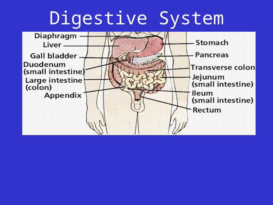
Digestive System
Digestive System
• Basic Divisions
– Digestive tract
– Accessory organs: various exocrine glands
• Digestive Processes
– Ingestion
– Mechanical Processing
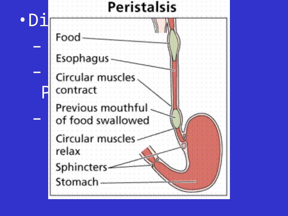
– Motility
•Peristalsis
• Digestive Processes
– Ingestion
– Mechanical Processing
– Motility
•Peristalsis
• Digestive Processes
– Ingestion
– Mechanical Processing
– Motility
•Peristalsis
•Segmentation movements
• Digestive Processes, continued
– Chemical digestion
– Secretion
– Absorption
– Excretion and defecation
• Non-Digestive Functions of Digestive Tract
– Immunity
– Storage of iron
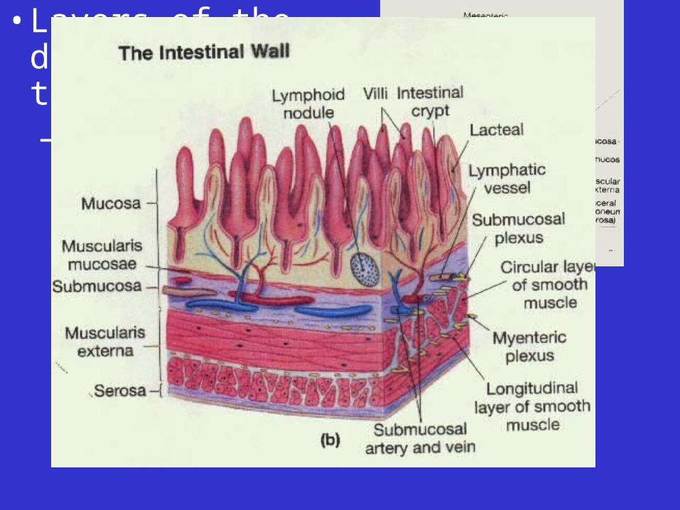
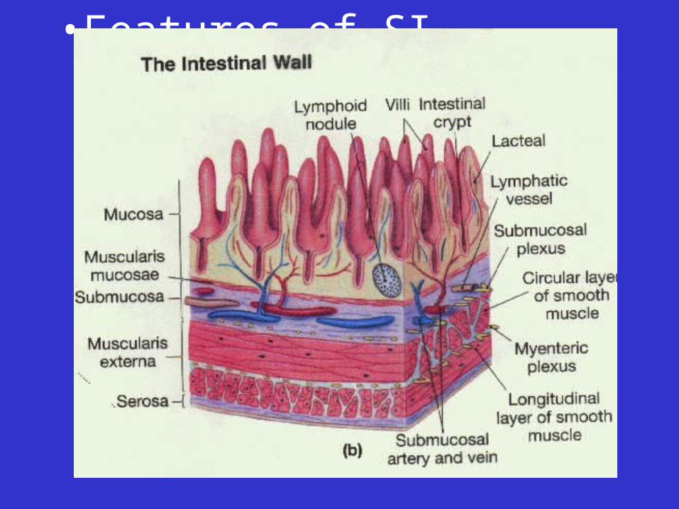
• Layers of the digestive tract– Mucosa
• Epithelium• Lamina propria (areolar CT)
• Muscularis mucosae
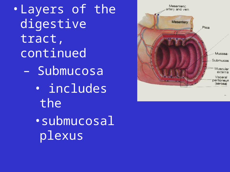
• Layers of the digestive tract, continued
– Submucosa
• includes the
•submucosal plexus
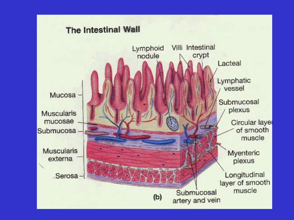

• Layers of the digestive tract, continued
– Muscularis externa:
responsible for peristalsis and segmentation movements
• longitudinal layer
• circular layer
• myenteric plexus
• Layers of the digestive tract, continued– Serosa (the visceral
peritoneum is an example)• Simple squamous epithelium
• Areolar CT Within peritoneal cavity
only
• Layers of the digestive tract, continued
– Adventitia
• Dense irregular CT Oral cavity, pharynx,
esophagus, rectum
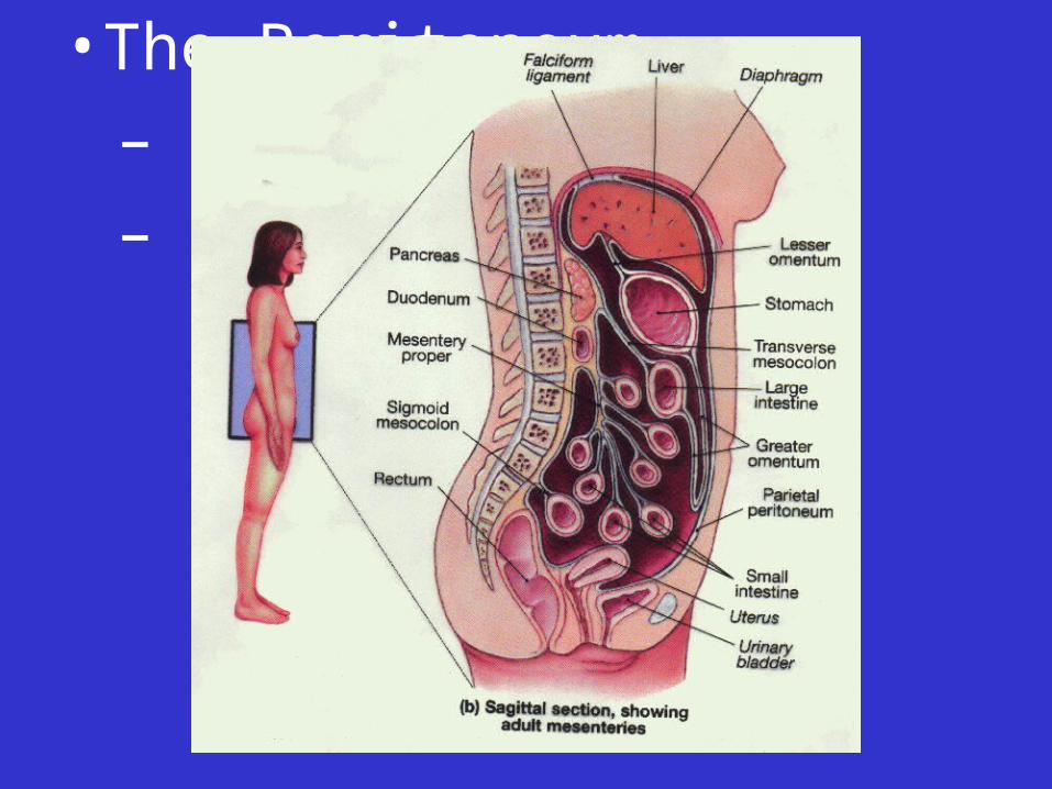
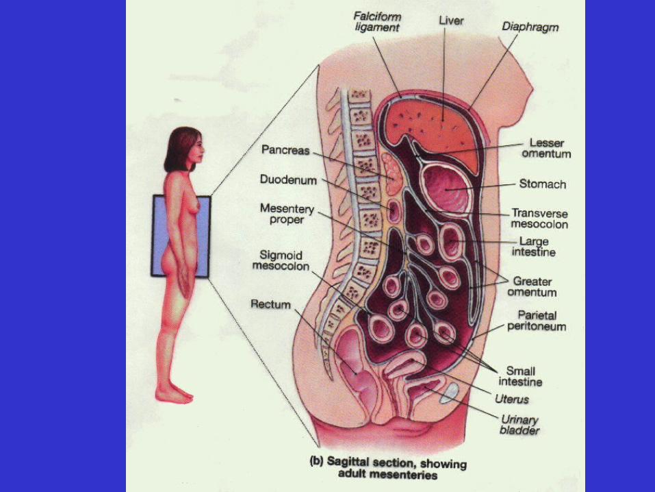
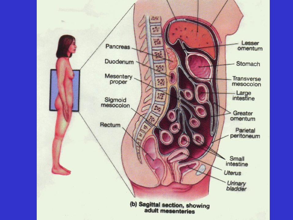
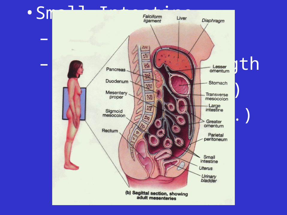
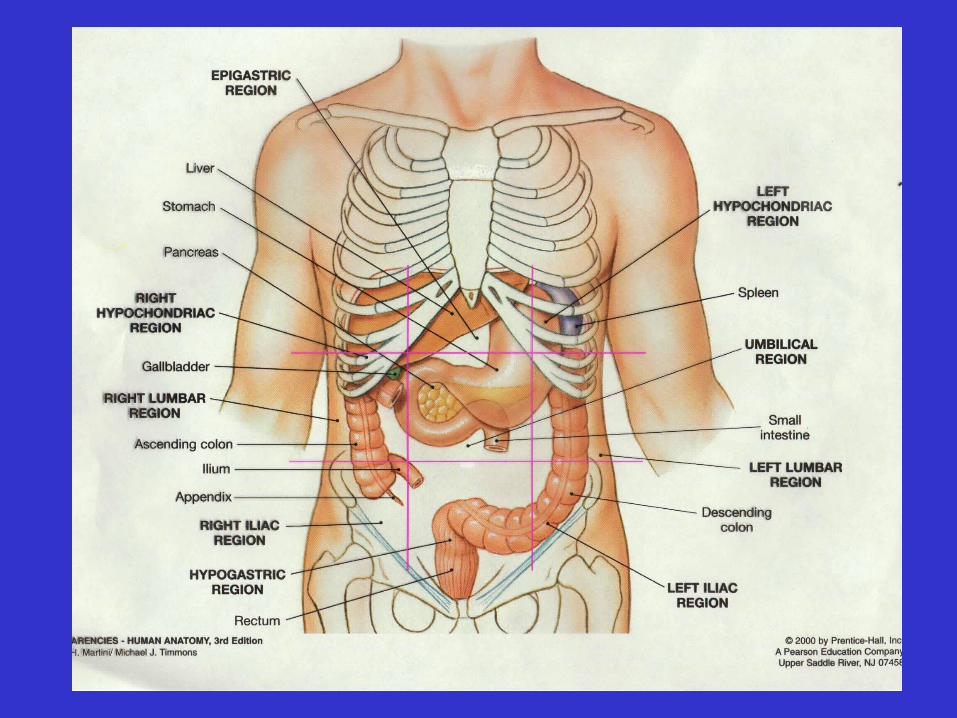
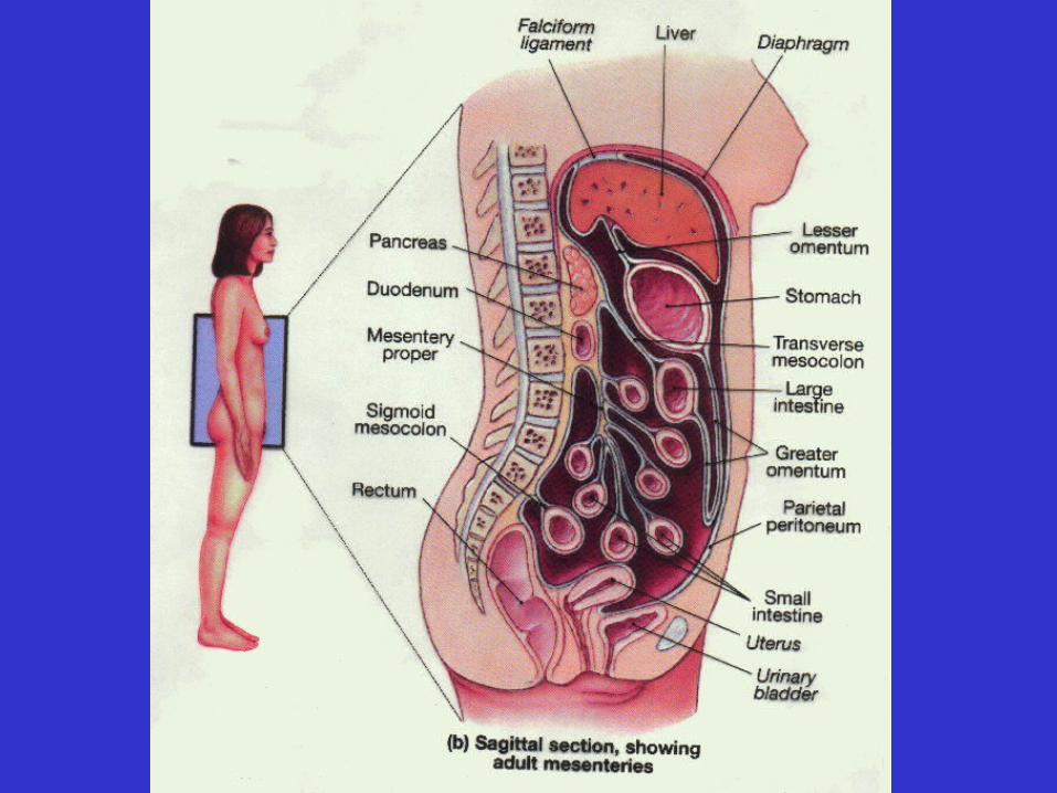
• The Peritoneum
– Parietal p.
– Visceral p.
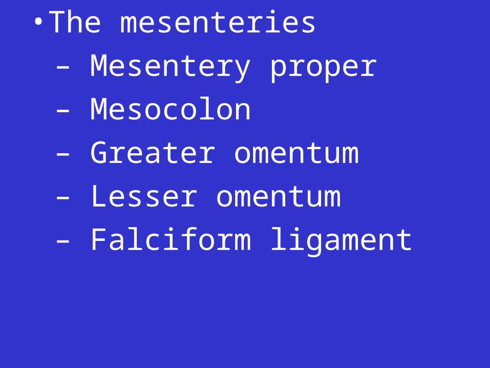
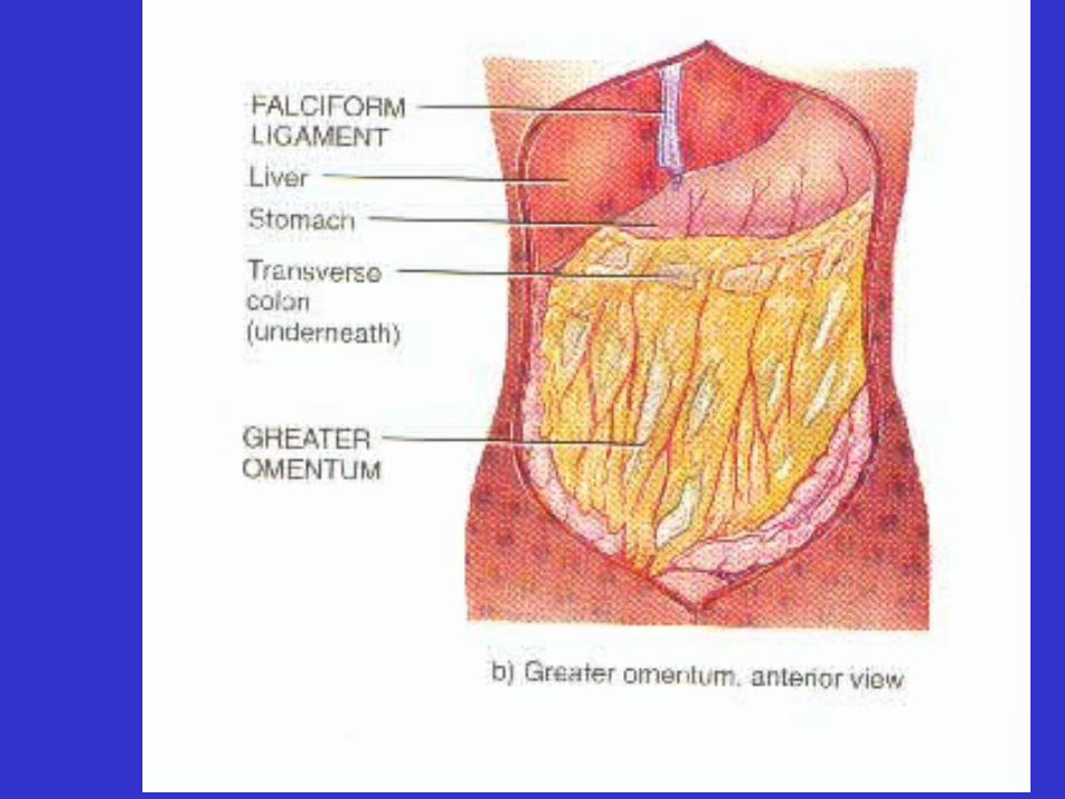
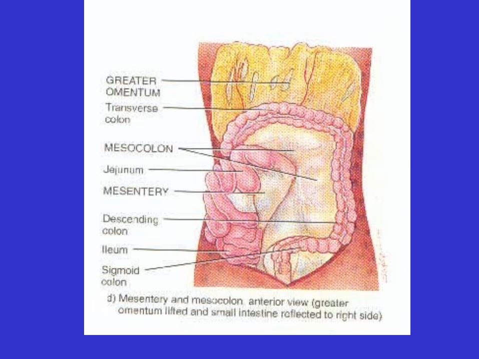
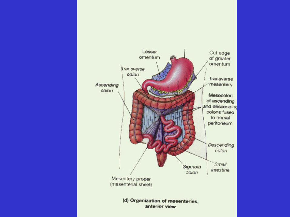
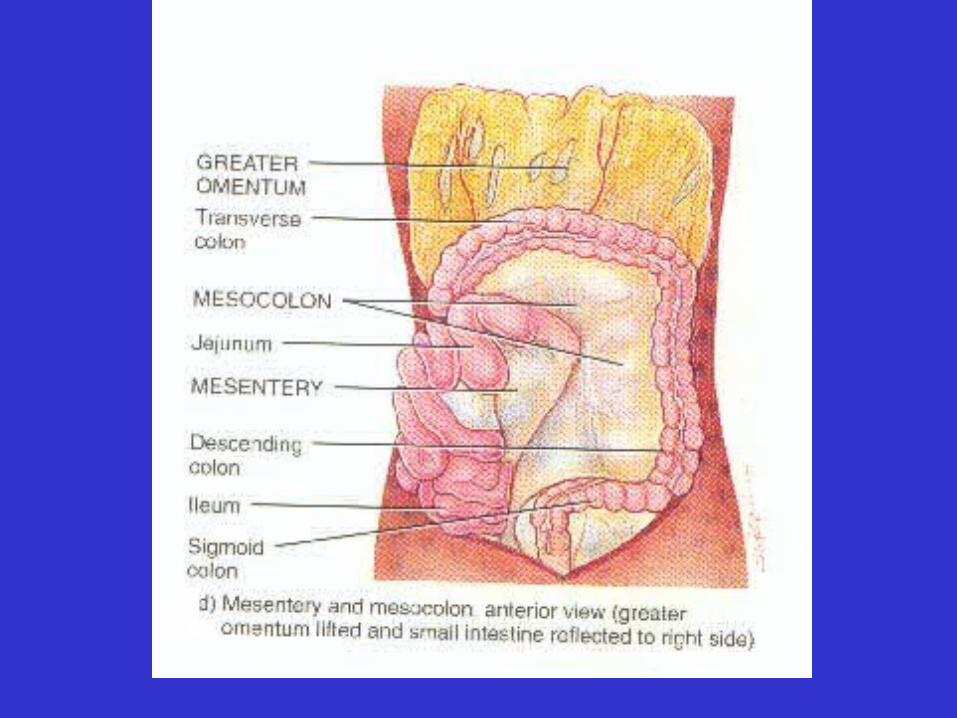
• The mesenteries
– Mesentery proper
– Mesocolon
– Greater omentum
– Lesser omentum
– Falciform ligament
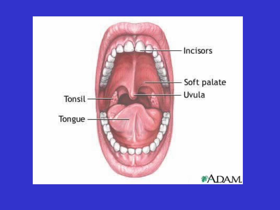
• Accessory structures of the oral (buccal) cavity– Teeth: will cover in lab– Tongue: read textbook– Salivary glands
• buccal glands• lingual glands

• Oral (buccal) cavity– Salivary Glands
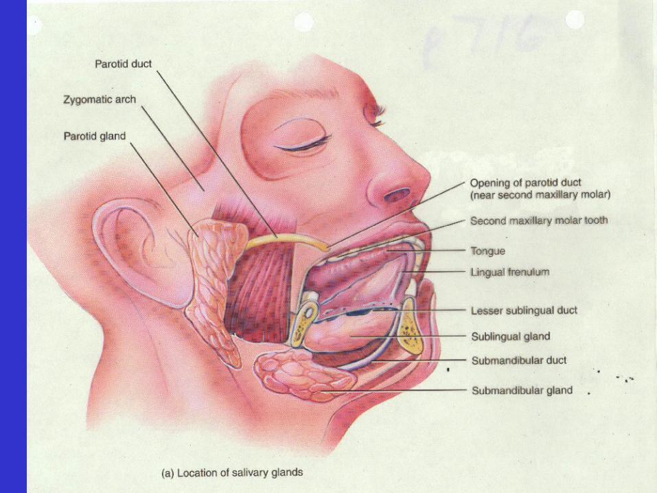
• buccal glands• lingual glands• major salivary glands
– parotid
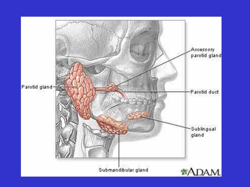
• Oral (buccal) cavity– Salivary Glands
• buccal glands• lingual glands• major salivary glands
– parotid– sublingual– submandibular

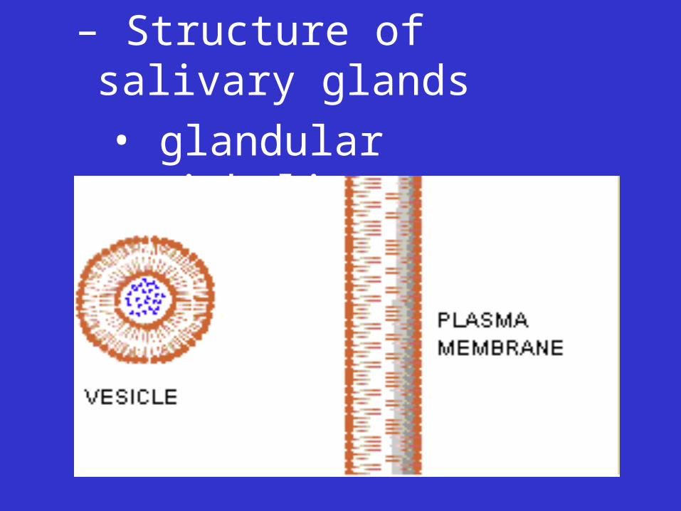
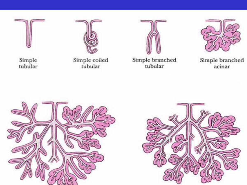
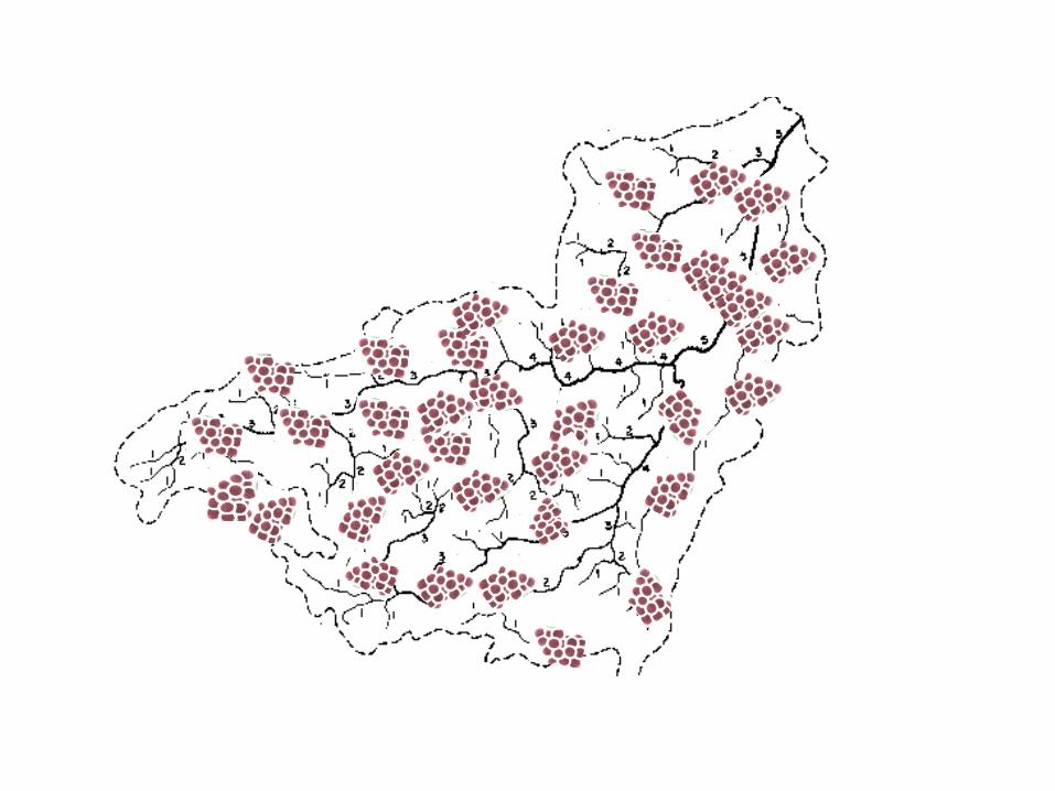
– Structure of salivary glands
• glandular epithelium
• merocrine cells
– Structure of salivary glands
• glandular epithelium
• merocrine cells
• compound tubulo-acinar
– Functions of saliva
• lubrication for swallowing, speaking
• re-mineralizes tooth enamel
• buffer
• antibodies (IgA)
• dissolves food molecules
• some chemical digestion
• Pharynx
– To be discussed with the respiratory system
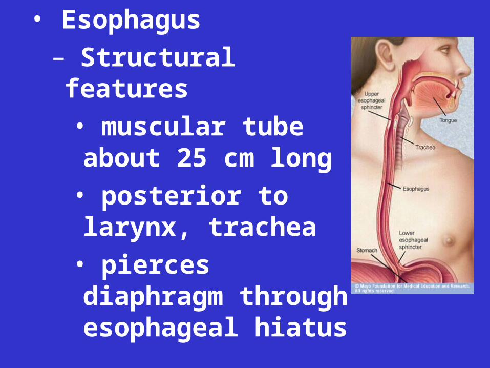
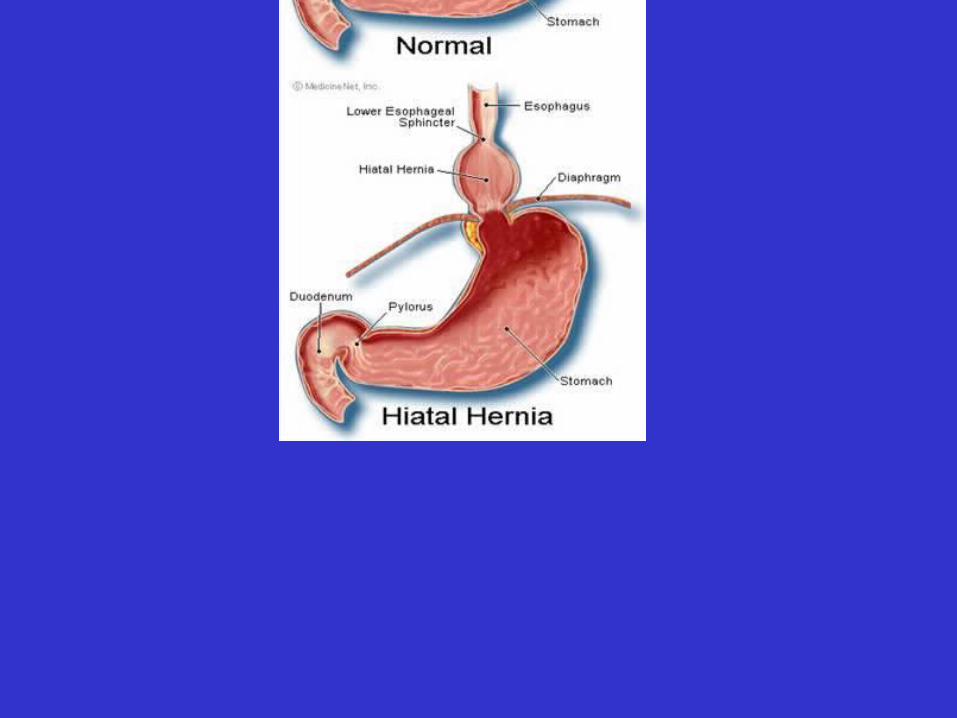
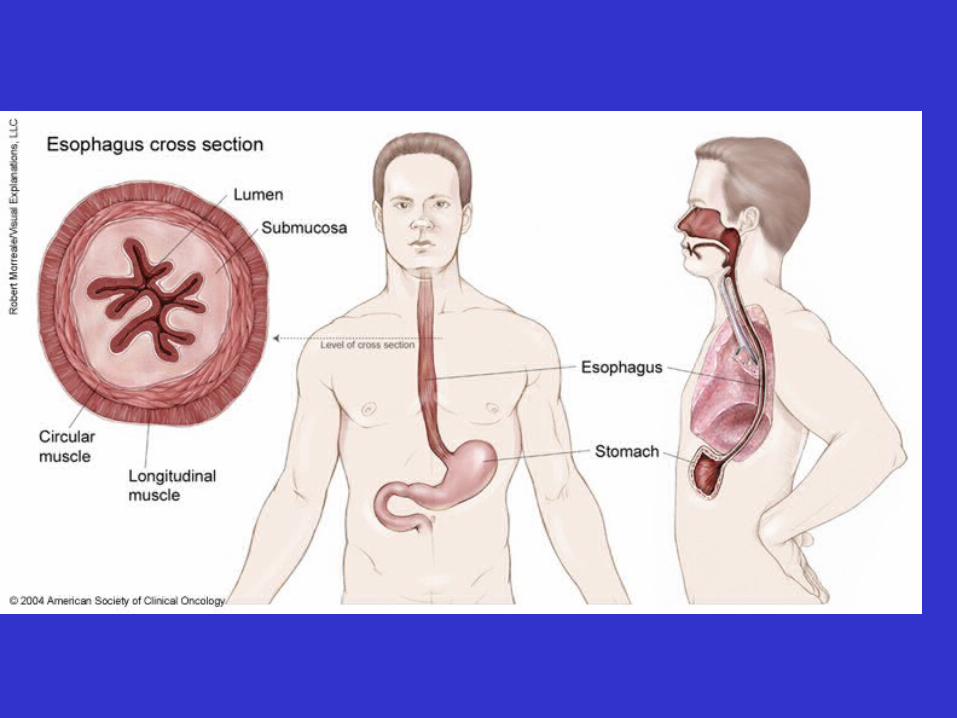
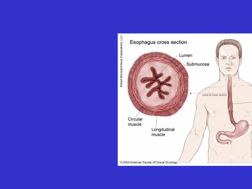
• Esophagus
– Structural features
• muscular tube about 25 cm long
• posterior to larynx, trachea
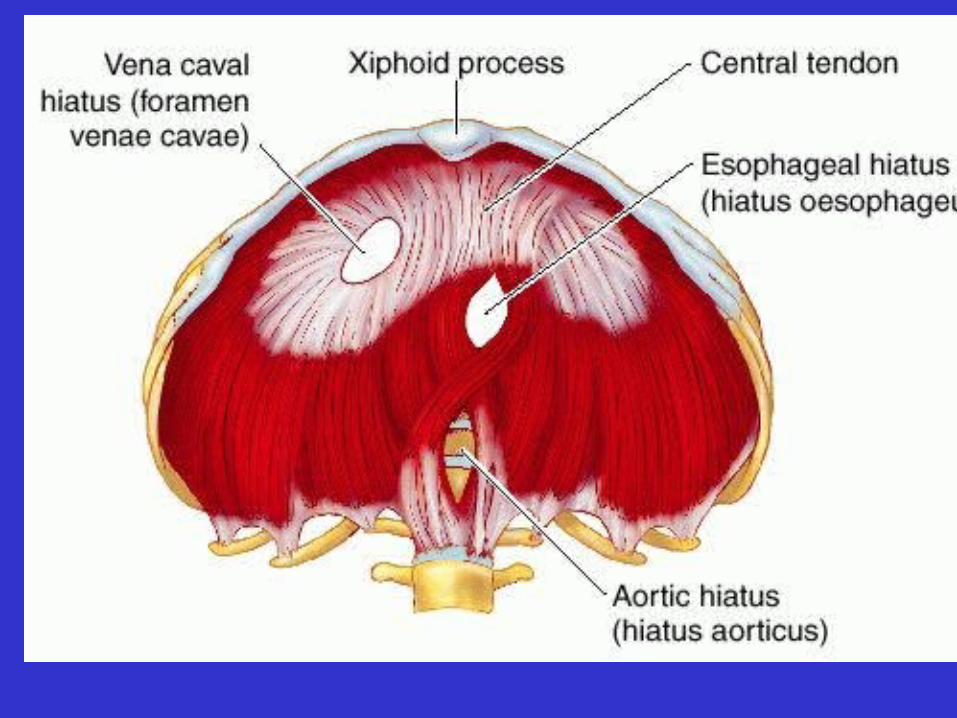
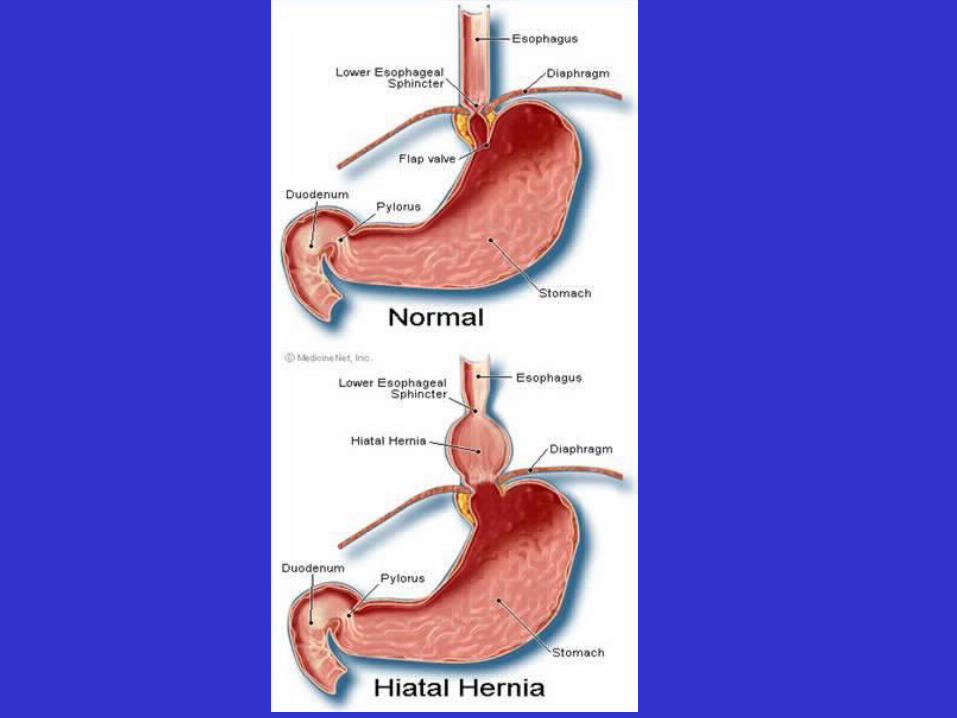
• pierces diaphragm through esophageal hiatus
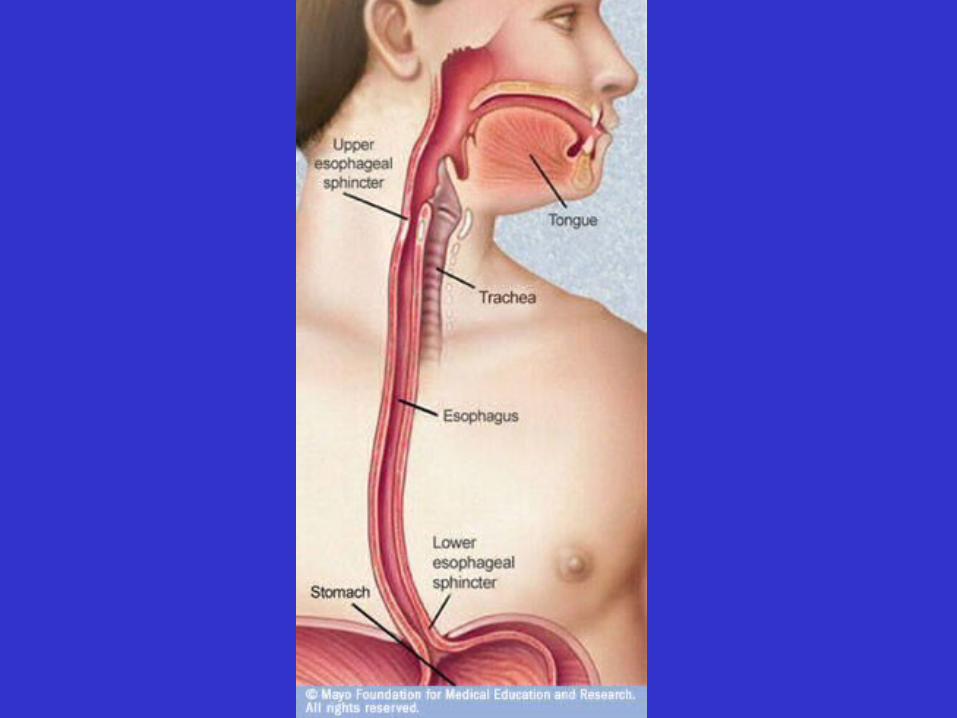
• 2 Esophogeal sphincters:
– upper esophageal sphincter
– lower esophageal sphincter
• Histology highlights
– mucosa
– submucosa: lots of mucous glands
– muscularis externa
– adventitia (no serosa)
• Gastroesophageal Reflux
• Stomach
– Location

• Stomach
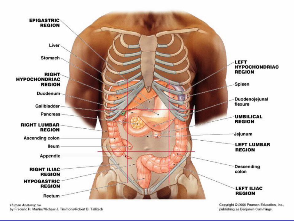
– Location: from epigastric and umbilical region
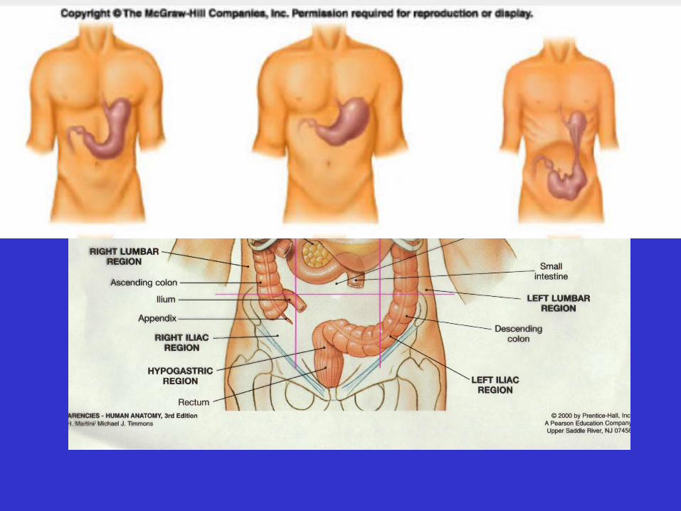
• Stomach
– Location: from epigastric and umbilical region to left hypochondriac regions
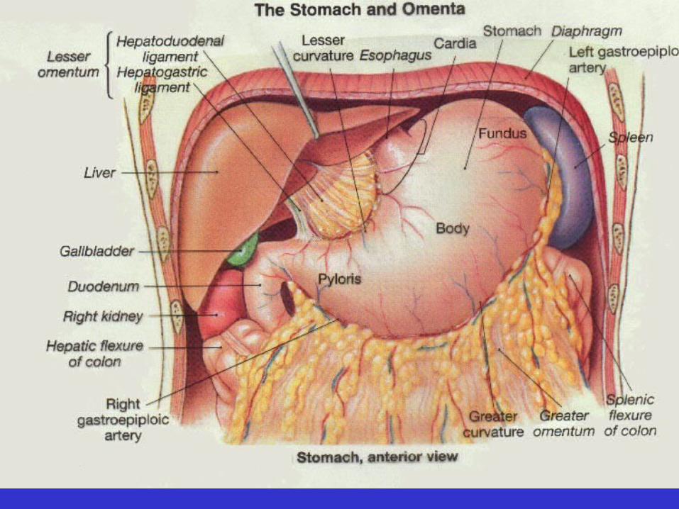
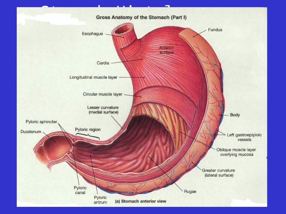
• Gross Structural Features– cardiac
region– fundus– body– pylorus– pyloric
sphincter
• Stomach motility video
• Stomach Histology
– mucosa
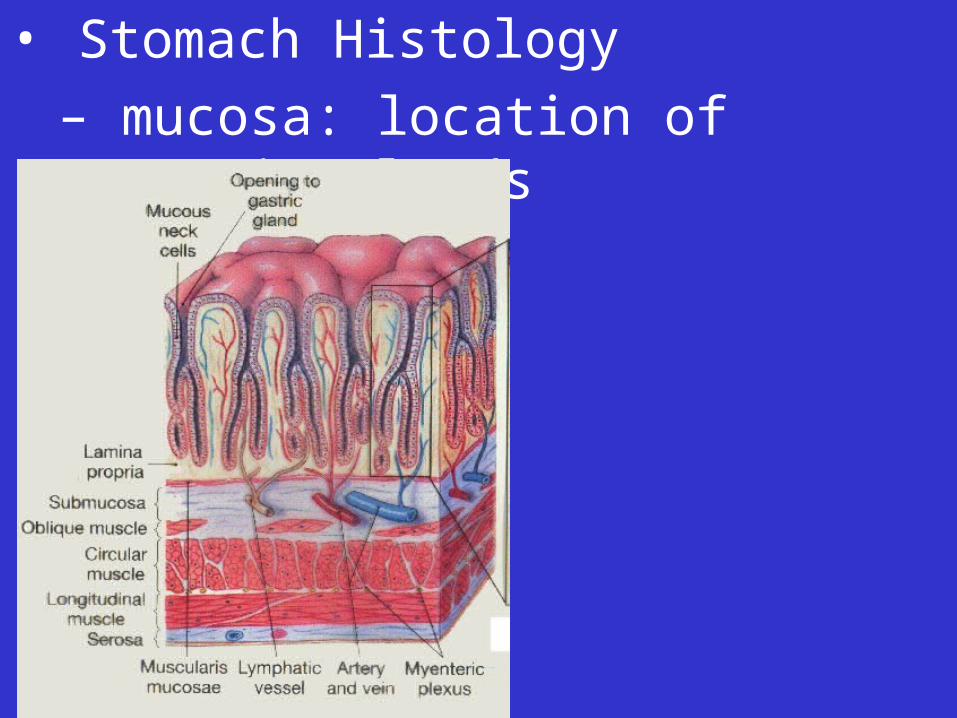
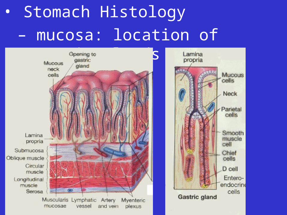
• Stomach Histology
– mucosa: location of gastric glands
• Stomach Histology
– mucosa: location of gastric glands

• Stomach Histology
– mucosa: location of gastric glands
• gastric gland cells
– mucous cells
– parietal cells
– chief cells
– endocrine cells
• Stomach Histology, continued
– muscularis: three layers
• Stomach Functions– food reservoir– formation of chyme– some chemical digestion– regulation of chyme entry into
S.I.– intrinsic factor production– some absorption
Digestive System, review
• Basic Divisions
– Digestive tract
– Accessory organs: various exocrine glands
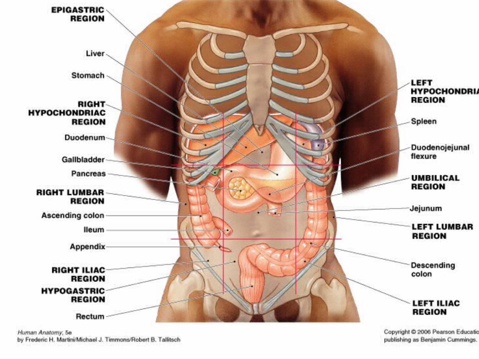
• Pancreas
– Location
• Umbilical region
• Pancreas
– Location
• Umbilical region
• Retroperitoneal
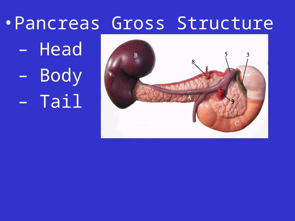
• Pancreas Gross Structure
– Head
– Body
– Tail
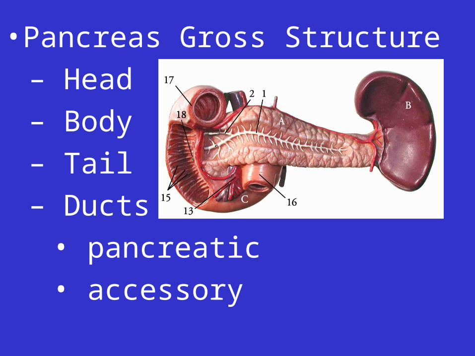
• Pancreas Gross Structure
– Head
– Body
– Tail
– Ducts
• pancreatic
• accessory
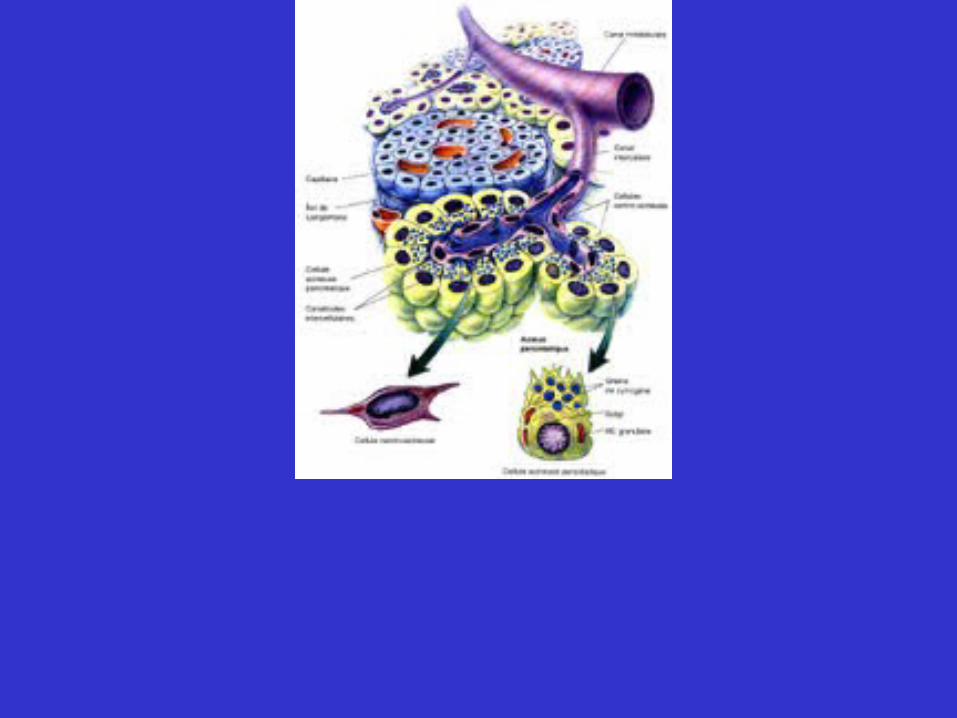
– pancreas histology
• mostly glandular epithelium
– exocrine pancreas
– endocrine pancreas
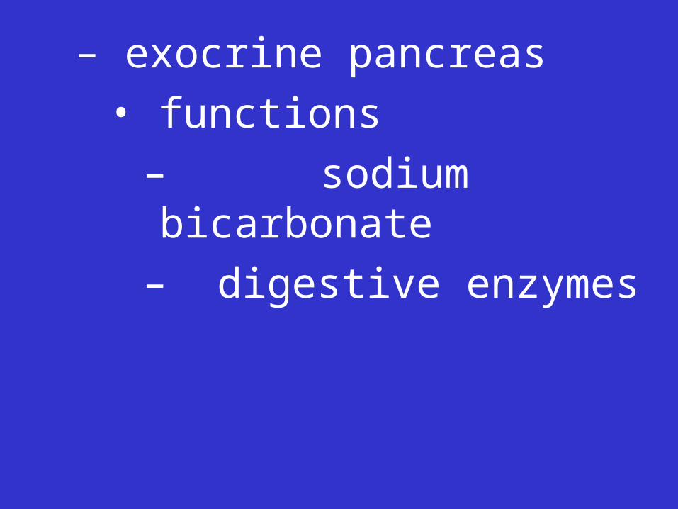
– exocrine pancreas
• functions
– sodium bicarbonate
– digestive enzymes
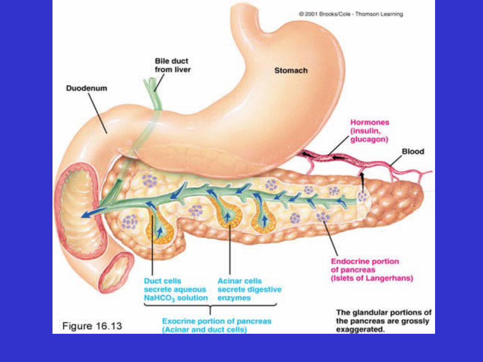
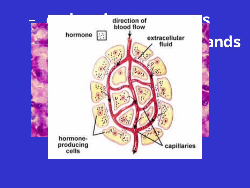
– endocrine pancreas
• structure: thousands of islets of Langerhans
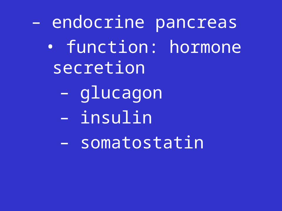
– endocrine pancreas
• function: hormone secretion
– glucagon
– insulin
– somatostatin

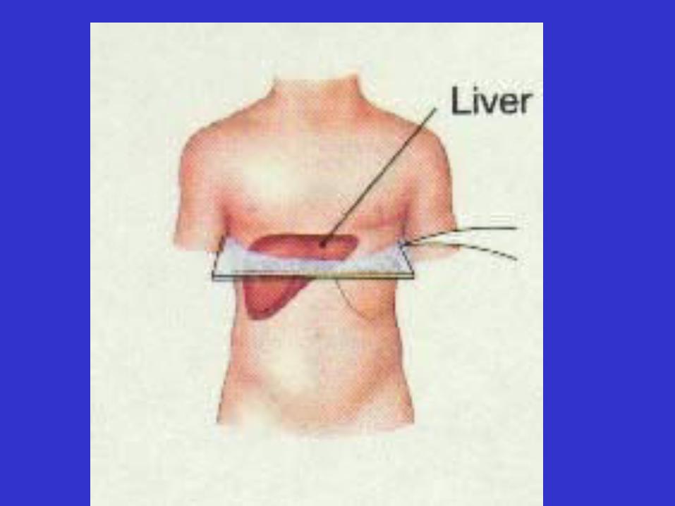
• Liver
–Location: epigastric and right hypochondriac regions
• Liver
–Location: epigastric and right hypochondriac regions

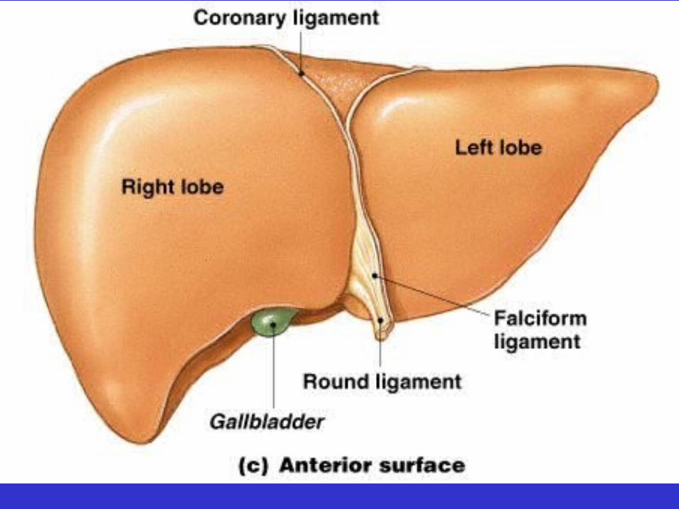
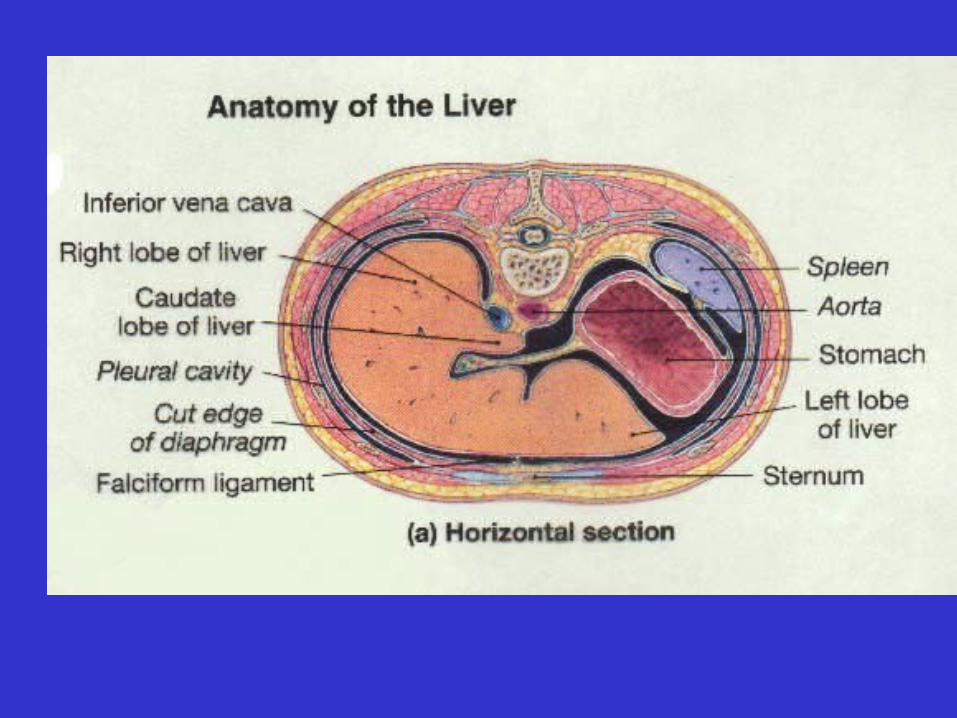
• Liver
–Gross structure: 2 major lobes separated by the falciform ligament
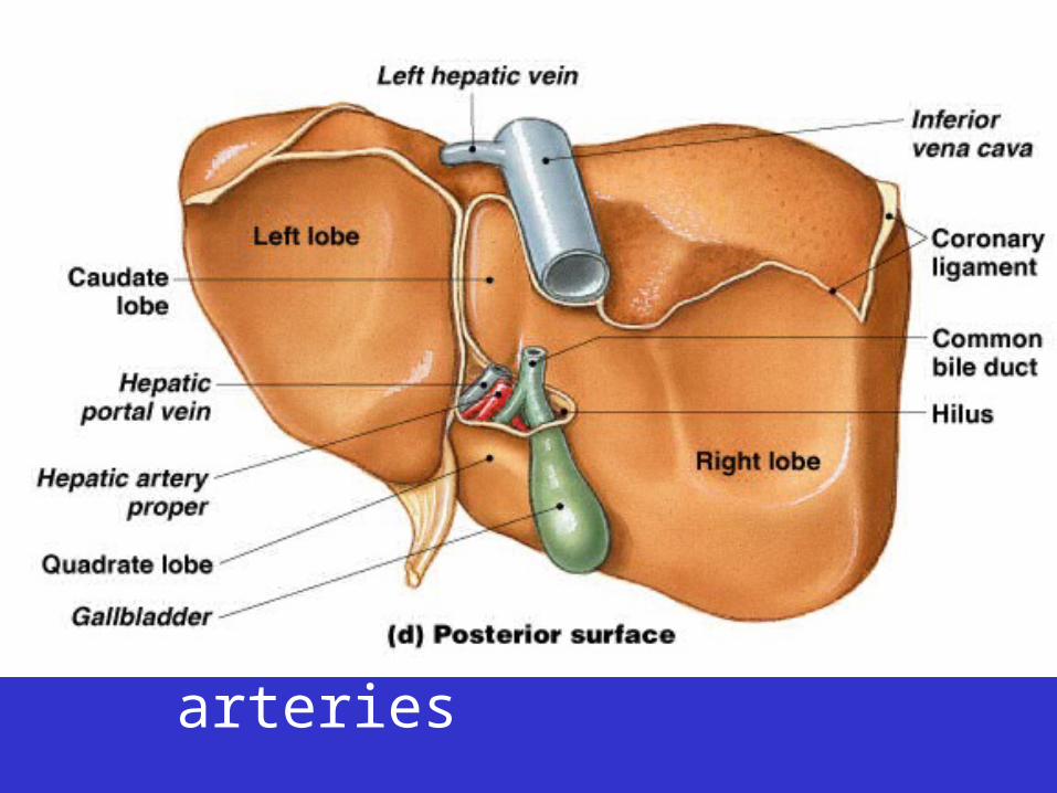
• Liver Blood Supply
– Hepatic portal vein
– Hepatic arteries
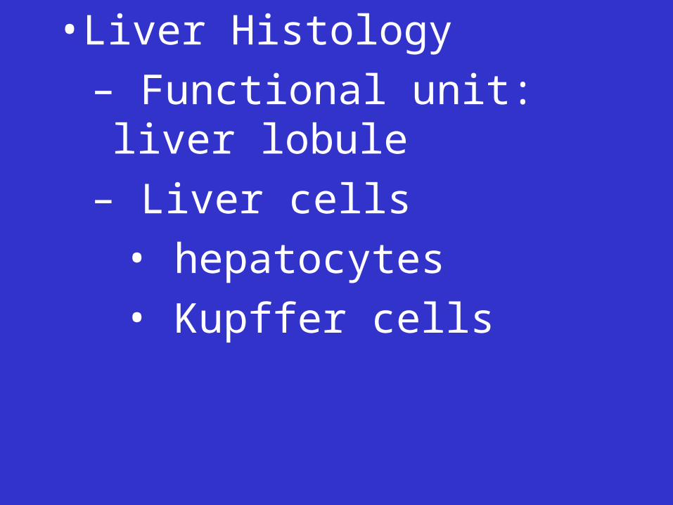
• Liver Histology
– Functional unit:
• Liver Histology
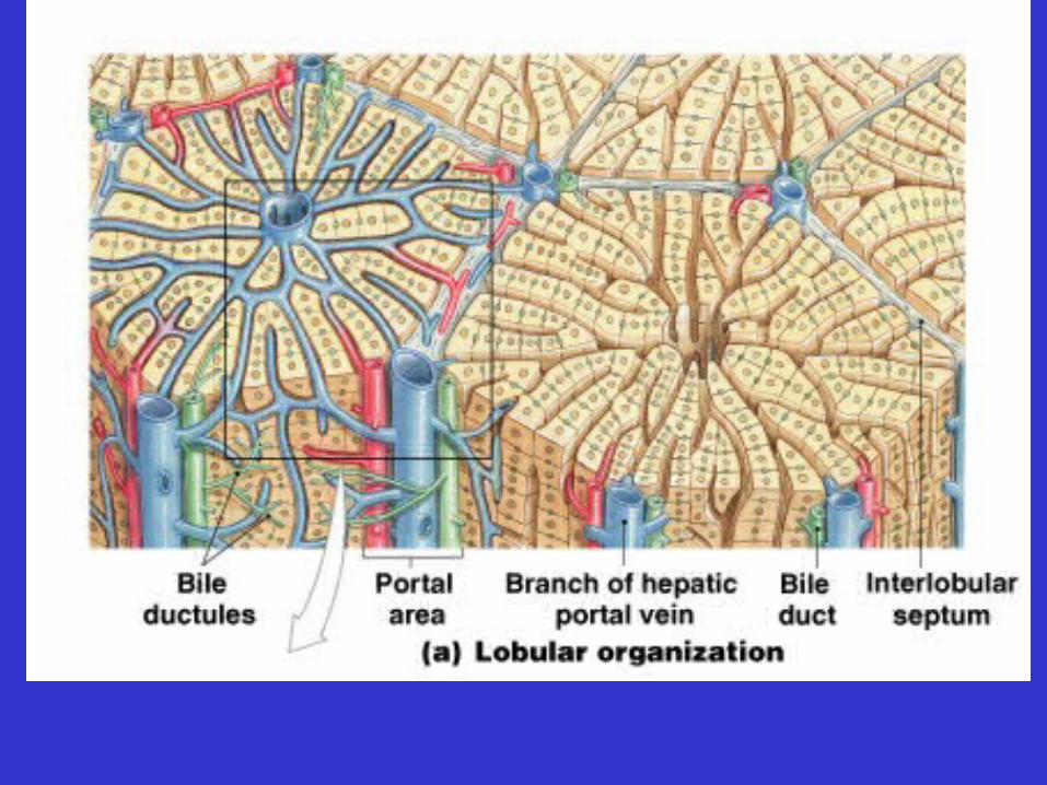
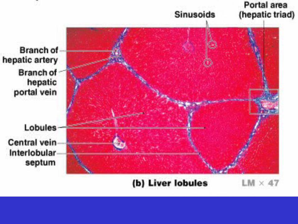
– Functional unit: liver lobule

• Liver Histology
– Functional unit: liver lobule
• Liver Histology
– Functional unit: liver lobule
– Liver cells
• Liver Histology
– Functional unit: liver lobule
– Liver cells
• hepatocytes
• Kupffer cells
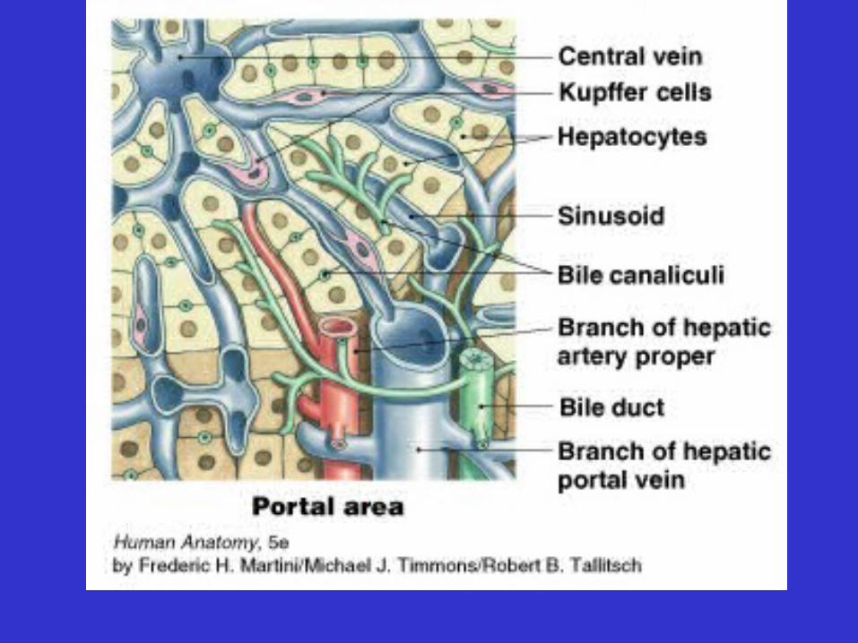
– Each liver lobule supplied by branches of:
• hepatic arteries
• hepatic portal veins

– Liver Functions
• Maintains blood glucose levels
• Cholesterol synthesis
• HDL and LDL synthesis
• Plasma protein synthesis
– Liver Functions, continued
• Hormone and drug removal
• Phagocytosis
• Vitamin storage
• Iron storage
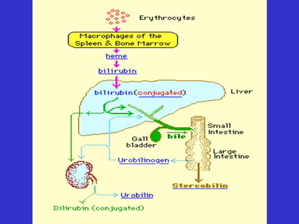
• Bilirubin excretion


– Liver Functions, continued
• Hormone and drug removal
• Phagocytosis
• Vitamin storage
• Iron storage
• Bilirubin excretion

– Liver Functions, continued
• Hormone and drug removal
• Phagocytosis
• Vitamin storage
• Iron storage
• Bilirubin excretion
• Bile salt secretion
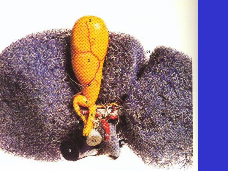
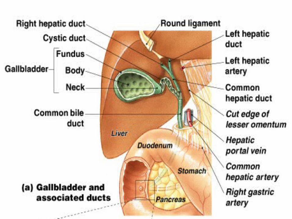
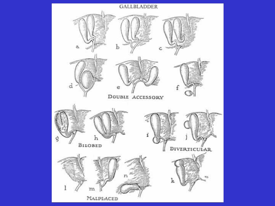
• Gall Bladder
– Location: right lumbar region

• Liver Blood Supply
– Hepatic portal vein
– Hepatic arteries
• Gross Structural Features of Gall Bladder
– Muscular sac
– Mucosa folded into rugae
– Bile enters and leaves through cystic duct

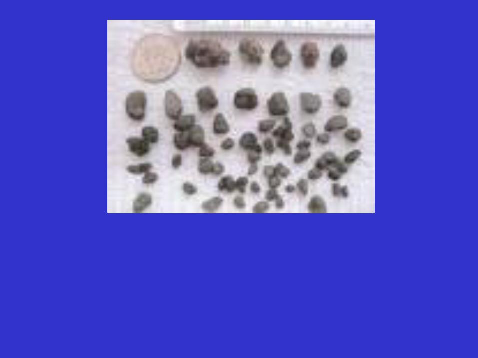
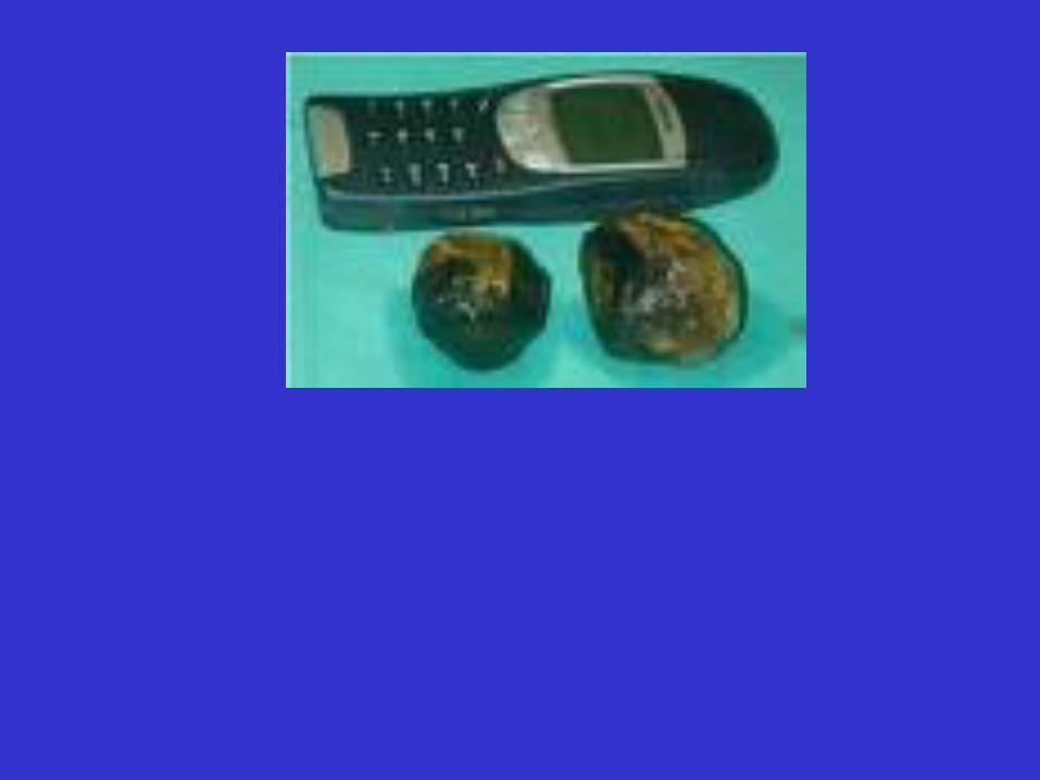
• Gall Bladder Function
– Stores and concentrates bile
– Contracts during meals to force bile into SI

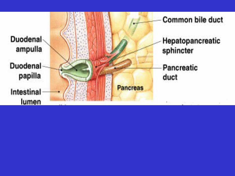
• Biliary Pathway

• Biliary Pathway: “plumbing” which drains bile

• Small Intestine
– 1 inch diameter
– 10-20 ft. in length
• duodenum (10 in)
• jejunum (3-6 ft.)
• ileum (6-12 ft)
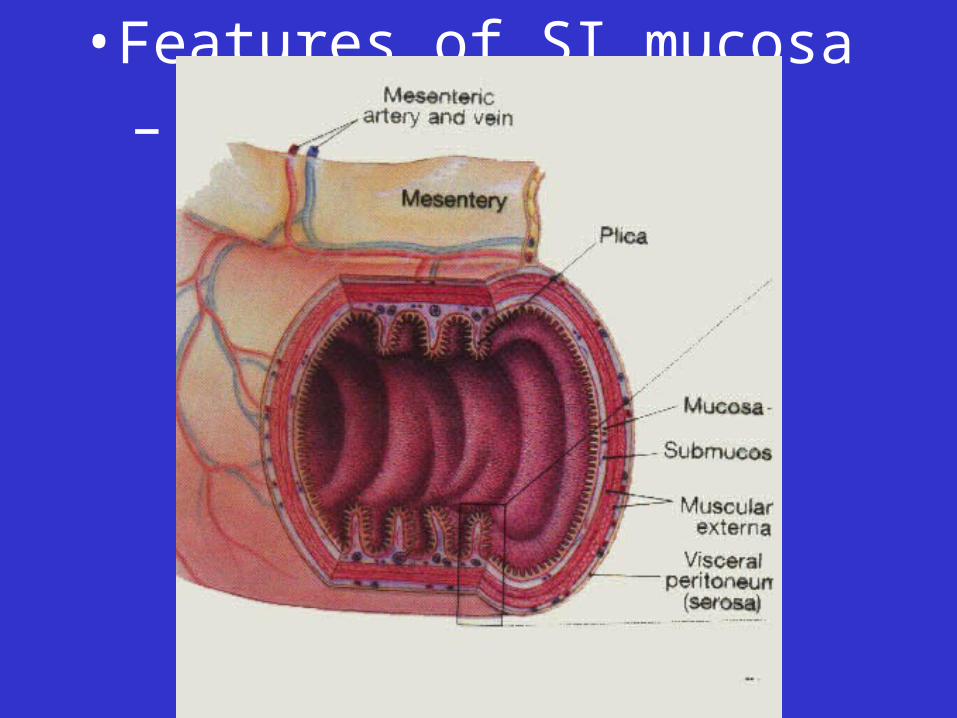
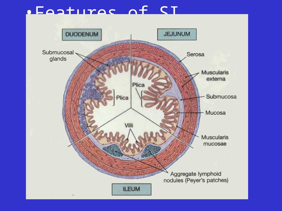
• Features of SI mucosa
– Plica circularis
• Features of SI mucosa
– Plica circularis

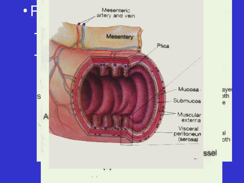
• Features of SI mucosa
–Plica circularis
–Villi
• Features of SI mucosa
–Plica circularis
–Villi
• Features of SI mucosa
–Plica circularis
–Villi
• Features of SI mucosa
–Plica circularis
–Villi
–Microvilli
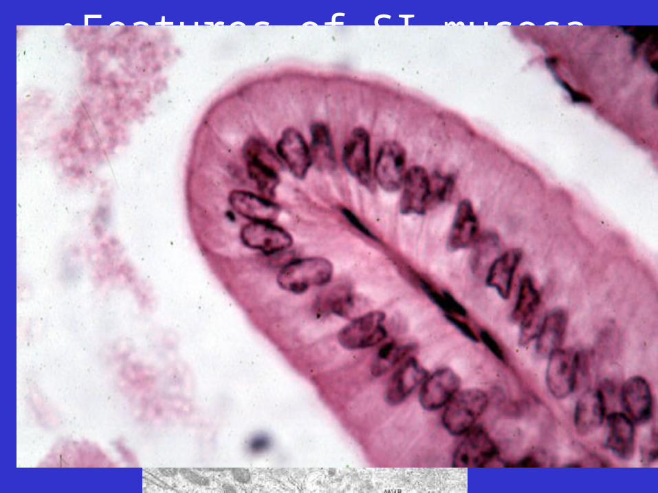
• Features of SI mucosa
–Plica circularis
–Villi
–Microvilli
• Features of SI mucosa, cont’d
–Epithelial cell types:
•absorptive cells
•Goblet cells
•Endocrine cells
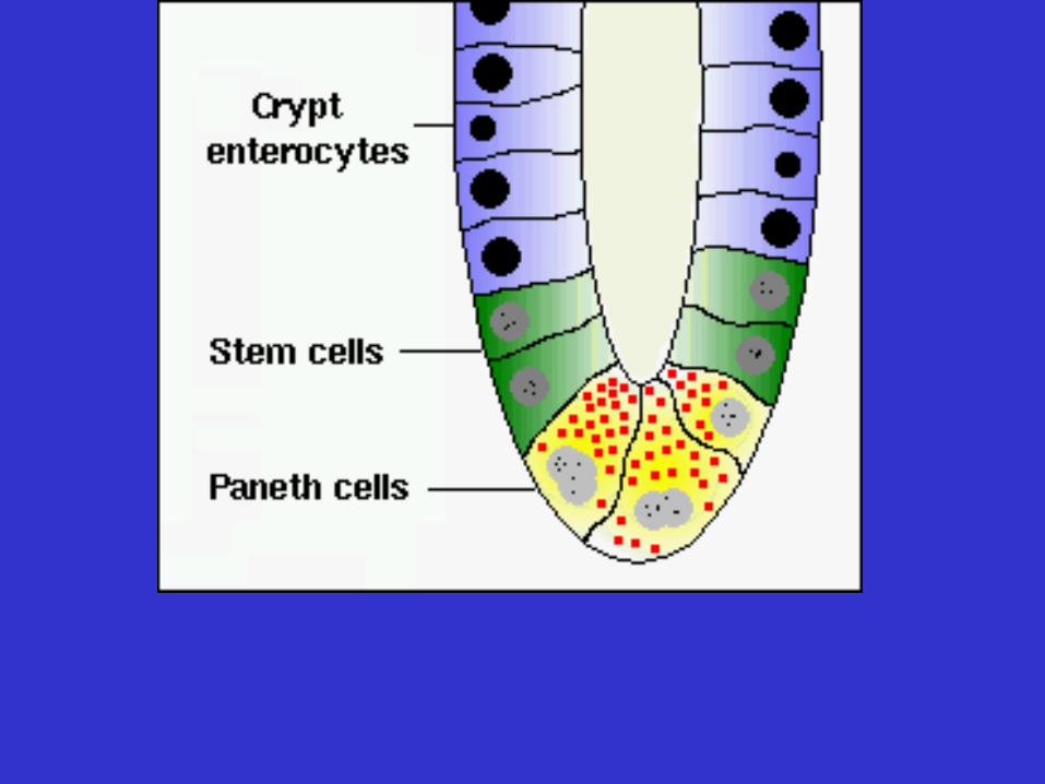
•Paneth cells
• Features of SI mucosa, cont’d
–MALT in lamina propria
• Features of SI mucosa, cont’d
–MALT in lamina propria
• Features of SI mucosa, cont’d
–MALT in lamina propria
–Intestinal glands (“crypts”)

• Features of SI submucosa
–Submucosal glands in duodenum
• Motility of SI
–Segmentation movements
–Peristalsis
• Functions of small intestine
– Completion of chemical digestion
• “brush-border” enzymes required
– Absorption
– Endocrine control of some digestive processes

Summary movie

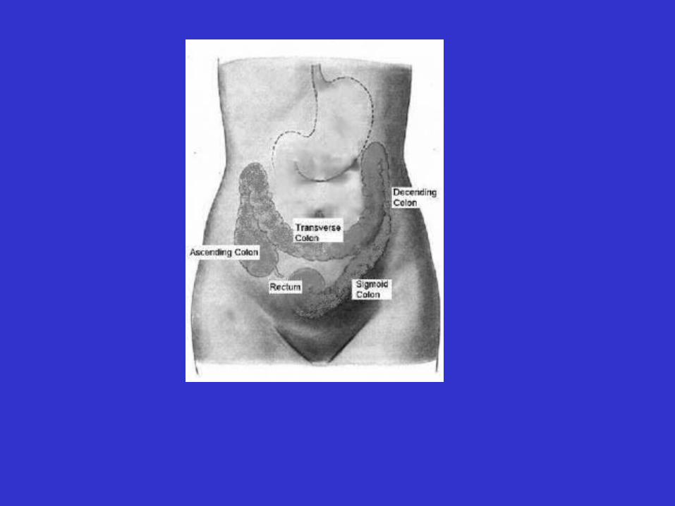
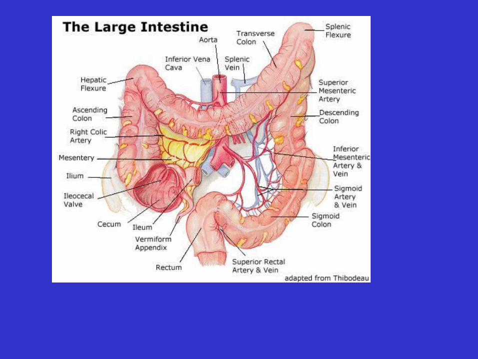
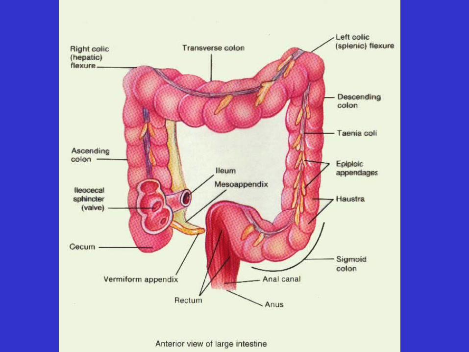
• Large Intestine

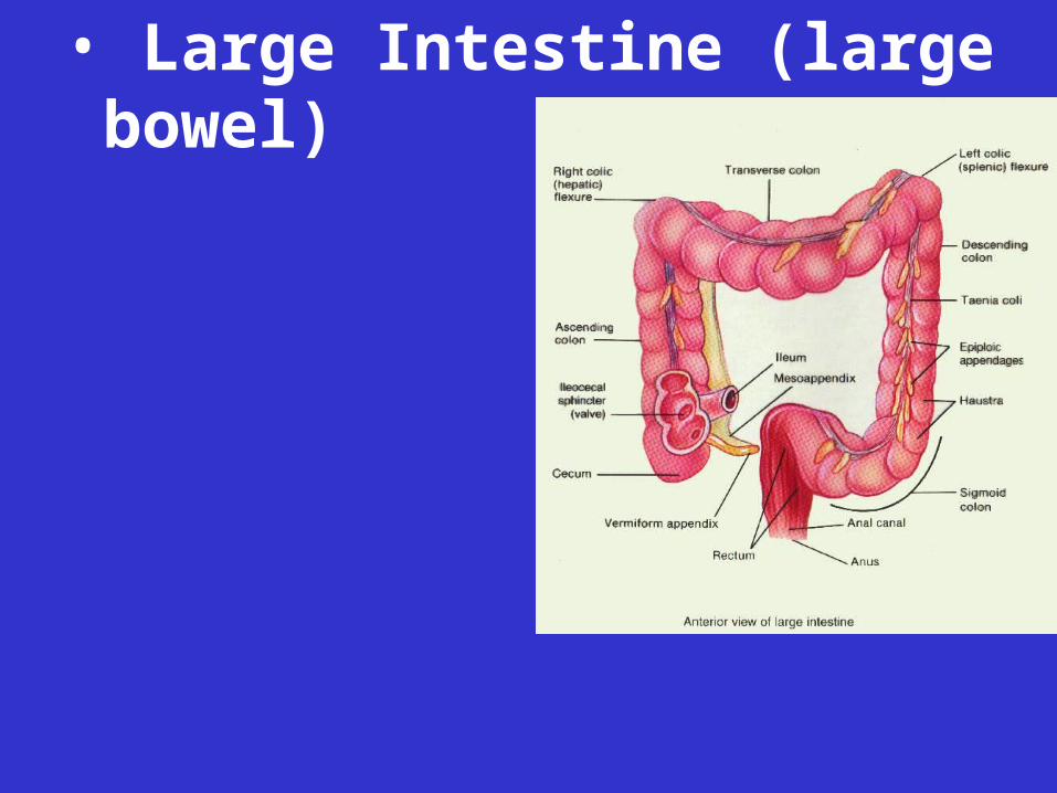
• Large Intestine (large bowel)
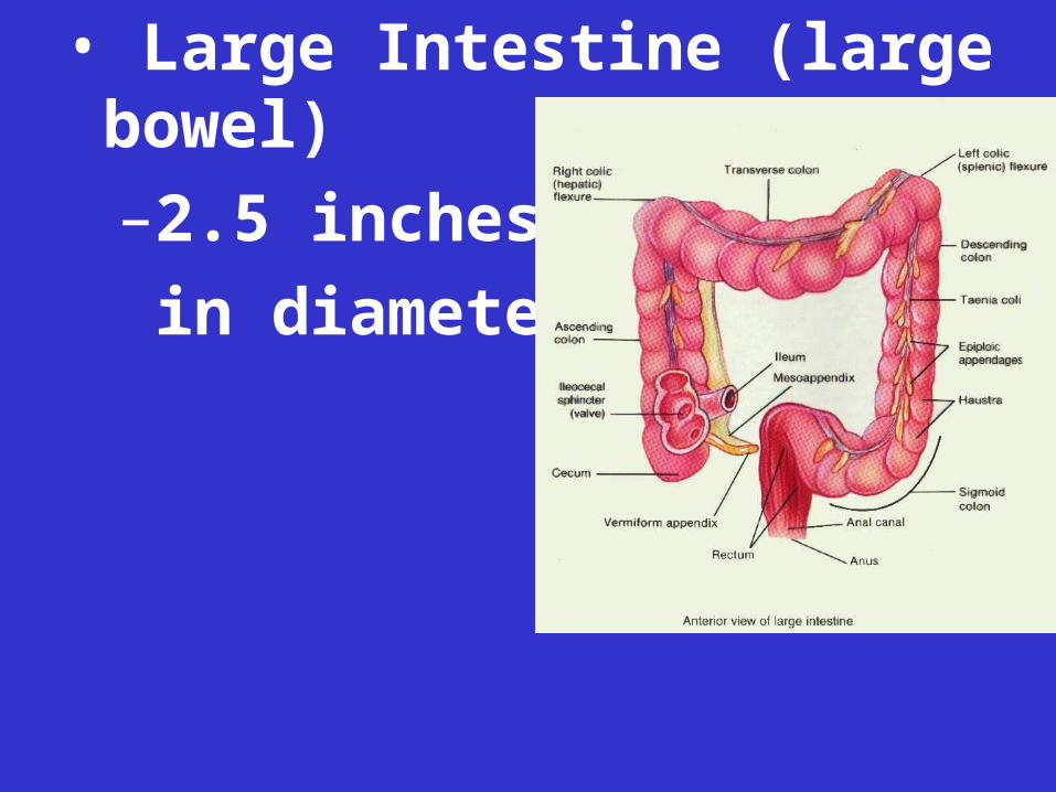
• Large Intestine (large bowel)
–2.5 inches
in diameter
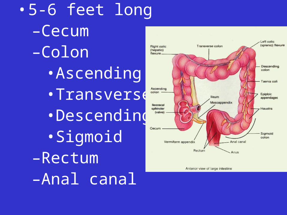
• 5-6 feet long–Cecum –Colon
•Ascending•Transverse•Descending•Sigmoid
–Rectum–Anal canal
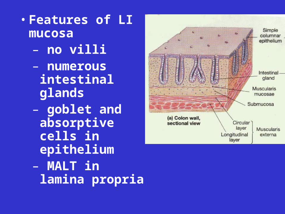
• Features of LI mucosa– no villi– numerous
intestinal glands– goblet and
absorptive cells in epithelium
– MALT in lamina propria
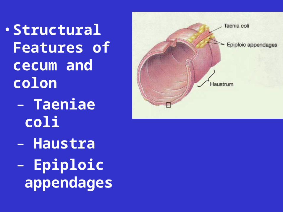
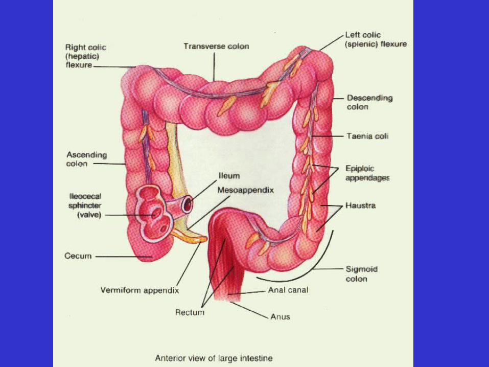
• Structural
Features of cecum and colon
– Taeniae coli
– Haustra
– Epiploic appendages

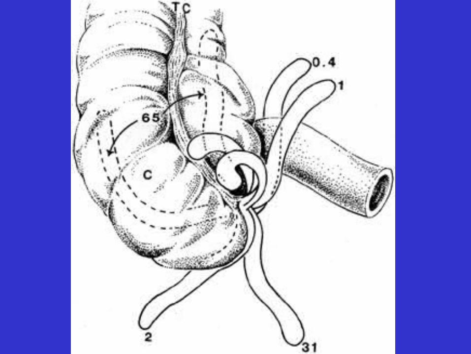
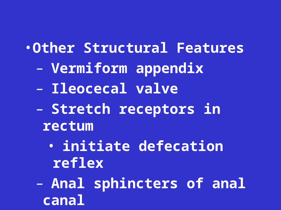
• Other Structural Features
– Vermiform appendix

• Other Structural Features
– Vermiform appendix
• Other Structural Features
– Vermiform appendix
– Ileocecal valve
• Other Structural Features
– Vermiform appendix
– Ileocecal valve
• Other Structural Features
– Vermiform appendix
– Ileocecal valve
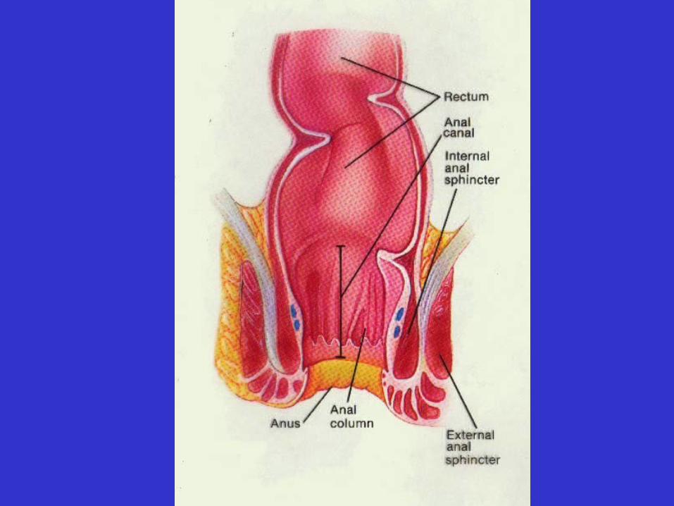
– Stretch receptors in rectum
• initiate defecation reflex
• Other Structural Features
– Vermiform appendix
– Ileocecal valve
– Stretch receptors in rectum
• initiate defecation reflex
– Anal sphincters of anal canal
• Motility of Large Intestine
– From cecum to transverse colon:
• peristalsis
– From transverse colon to rectum:
• mass movements
• Functions of Large Intestine
– Water and electrolyte absorption
– Feces formation
– Defecation
• Large Intestinal Bacteria
– Coat surface of mucosa
– Examples: E. coli
– Keep out pathogenic bacteria
48