Digestive System Digestive Glands
82
Digestive System Digestive System Digestive Glands Digestive Glands
description
Digestive System Digestive Glands. Components of Digestive Glands. ◇ small digestive glands: found in the wall of digestive tract. ◇ accessory glands (large digestive glands):. salivary glands pancreas liver. General Structure of Digestive Glands. - PowerPoint PPT Presentation
Transcript of Digestive System Digestive Glands
Digestive SystemDigestive System
Digestive GlandsDigestive Glands
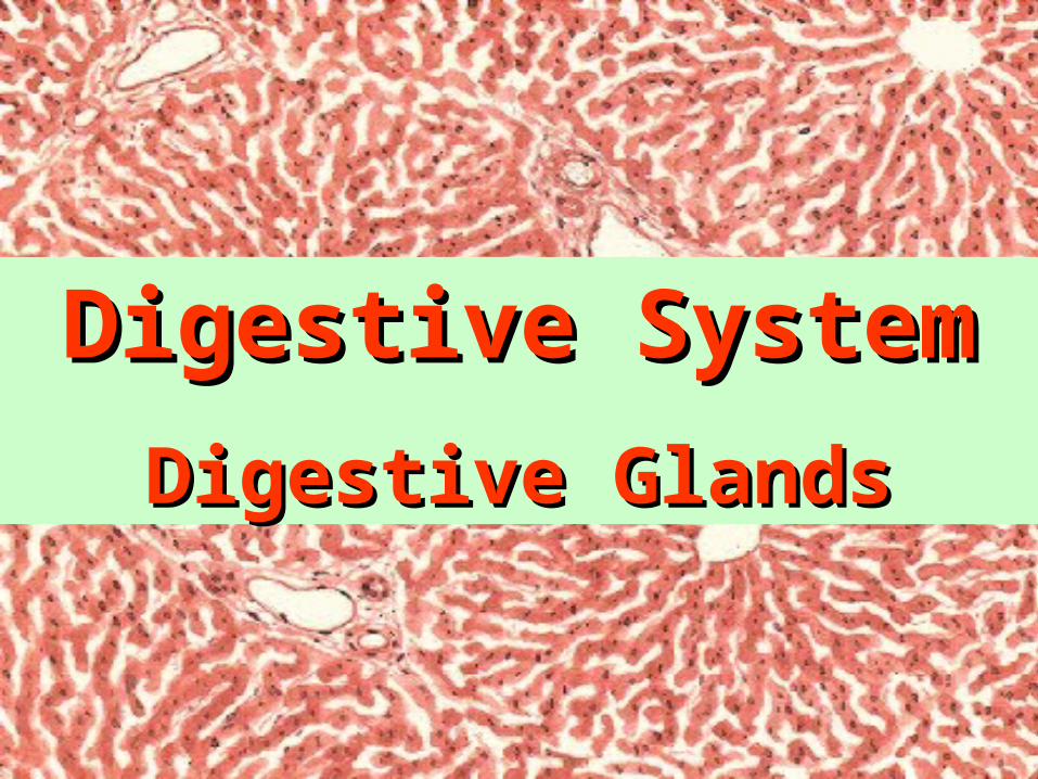
◇ small digestive glands:
found in the wall of digestive tract.
◇ accessory glands (large digestive glands):
Components of Digestive GlandsComponents of Digestive Glands
salivary glands
pancreas
liver
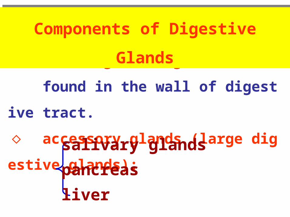
◇ Parenchyma: ( functional portion of an organ )
acini / gland cells
ducts
◇ Stroma: ( non-functional portion of an organ )
capsule
CT inside the organ.
General Structure of Digestive GlandsGeneral Structure of Digestive Glands
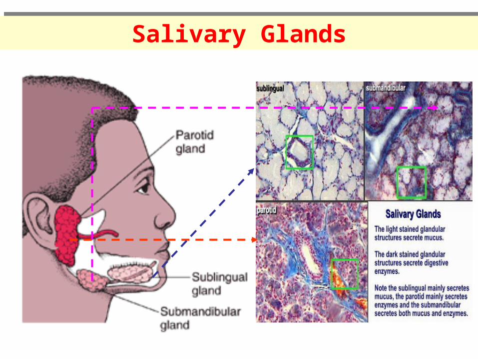
Salivary GlandsSalivary Glands
serous acinus mucous acinus seromucous / mixed acinus
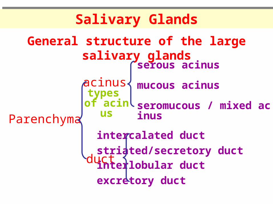
General structure of the large salivary glands
Parenchyma
acinus
duct
intercalated duct
striated/secretory ductinterlobular duct
excretory duct
types of acinus
Salivary GlandsSalivary Glands
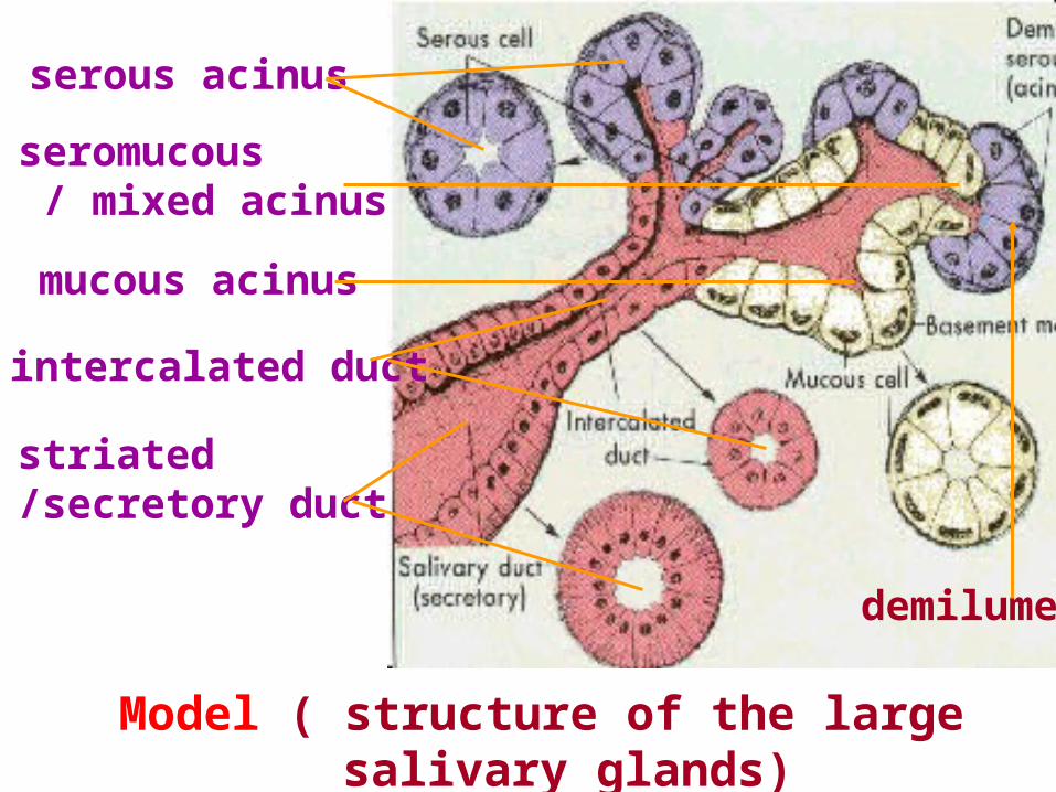
Model ( structure of the large salivary glands)
serous acinus
seromucous / mixed acinus
mucous acinus
intercalated duct
striated/secretory duct
demilume
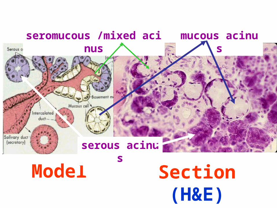
Model Section (H&E)
serous acinus
mucous acinusseromucous /mixed acinus
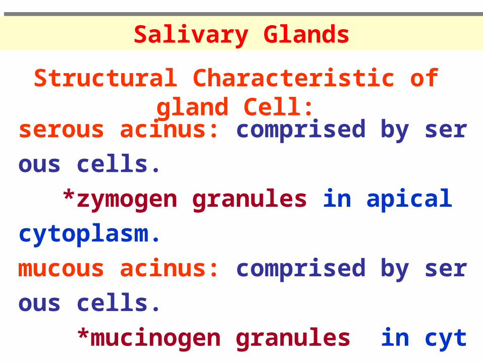
serous acinus: comprised by serous cells.
*zymogen granules in apical cytoplasm.
mucous acinus: comprised by serous cells.
*mucinogen granules in cytoplasm.
seromucous acinus: comprised by both cells.
/ mixed acinus *demilume
Structural Characteristic of gland Cell:
Salivary GlandsSalivary Glands
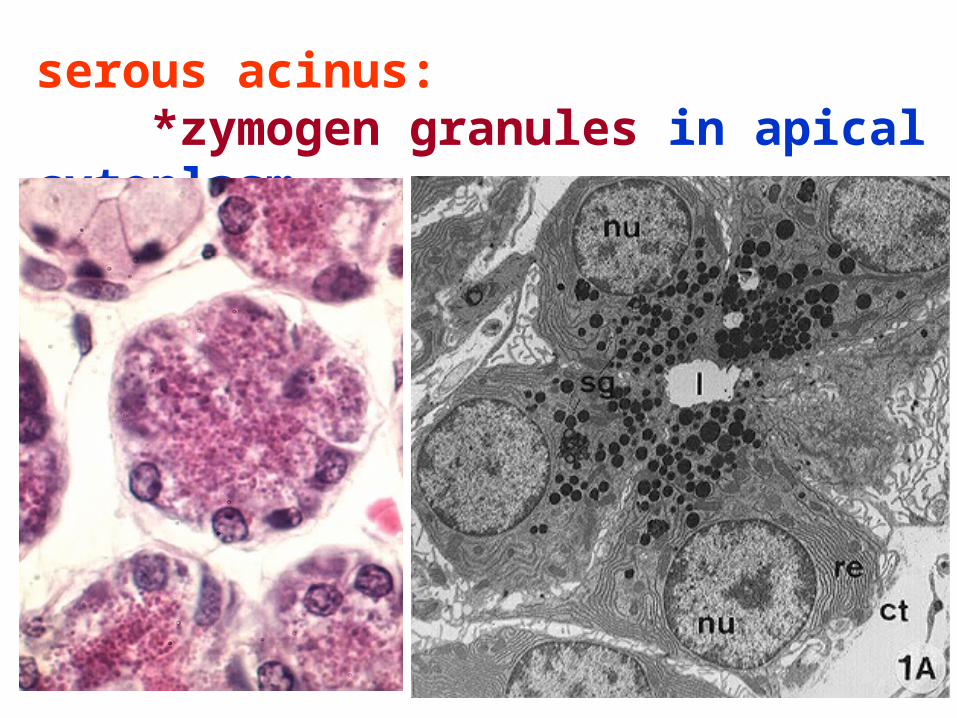
serous acinus: *zymogen granules in apical cytoplasm.
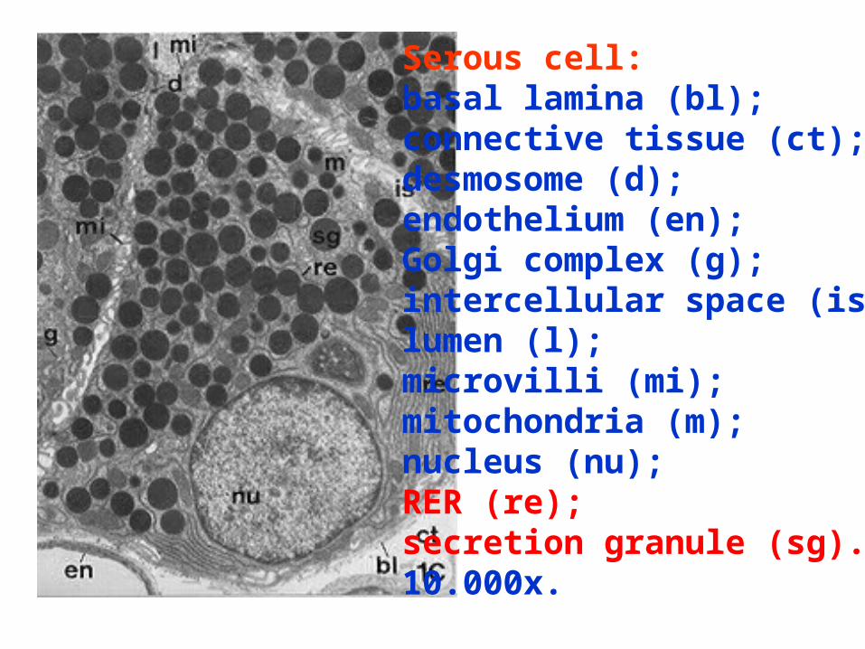
Serous cell: basal lamina (bl); connective tissue (ct); desmosome (d); endothelium (en); Golgi complex (g); intercellular space (is); lumen (l); microvilli (mi); mitochondria (m); nucleus (nu); RER (re); secretion granule (sg). 10.000x.
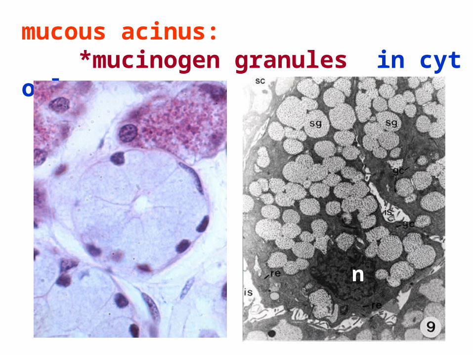
mucous acinus: *mucinogen granules in cytoplasm.
n
n
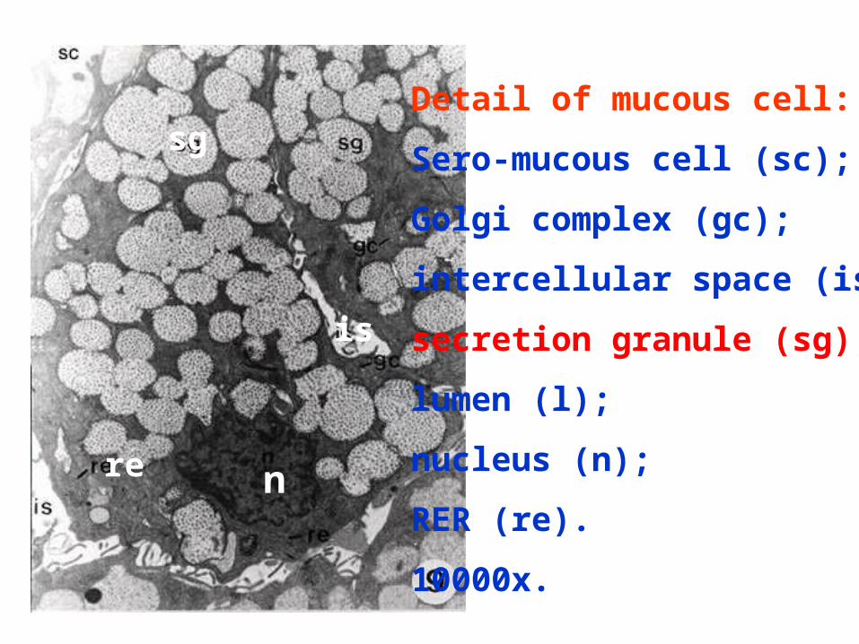
Detail of mucous cell:
Sero-mucous cell (sc);
Golgi complex (gc);
intercellular space (is);
secretion granule (sg);
lumen (l);
nucleus (n);
RER (re).
10000x.
re
is
sg
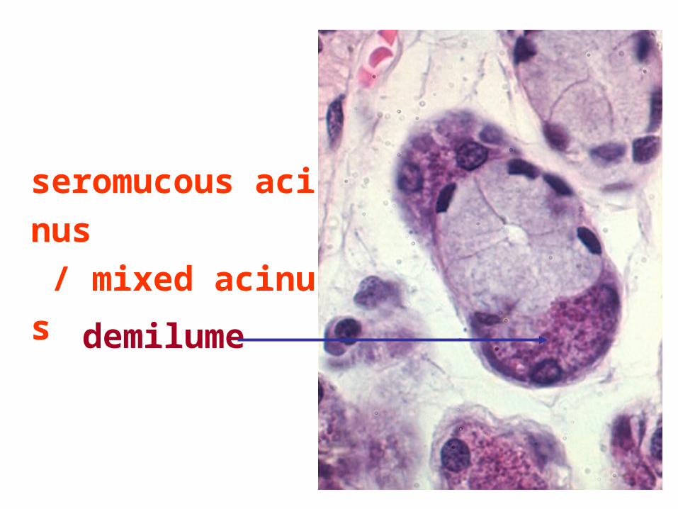
seromucous acinus
/ mixed acinus
demilume
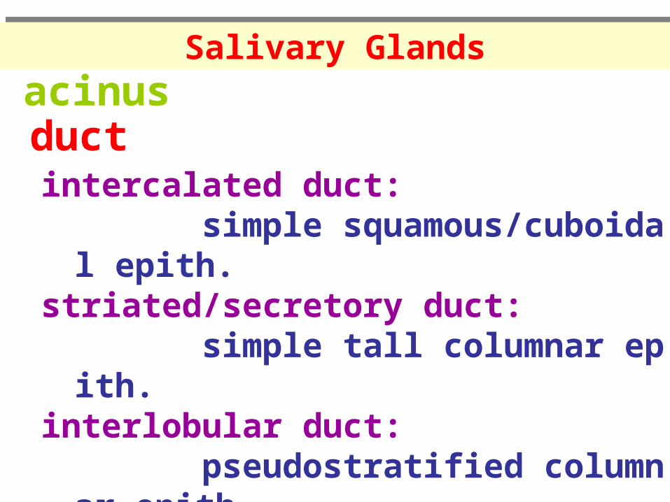
acinusductintercalated duct: simple squamous/cuboidal epith.striated/secretory duct: simple tall columnar epith.interlobular duct: pseudostratified columnar epith.excretory duct: stratified squamous epith.
Salivary GlandsSalivary Glands
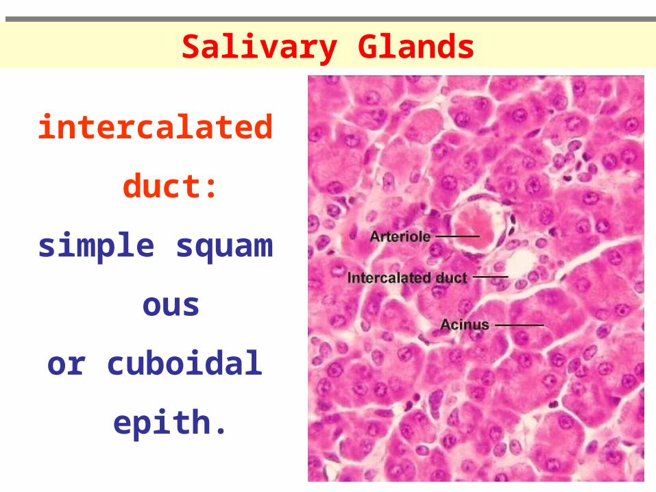
intercalated duct:
simple squamous
or cuboidal epith.
Salivary GlandsSalivary Glands
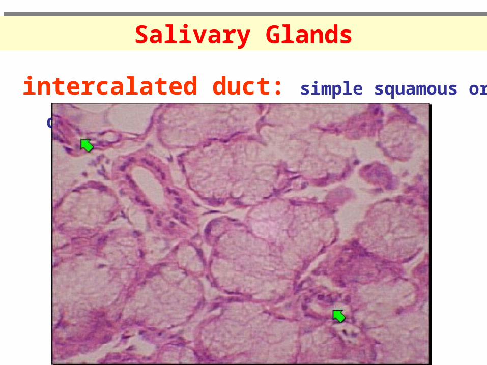
intercalated duct: simple squamous or cuboidal epith.
Salivary GlandsSalivary Glands
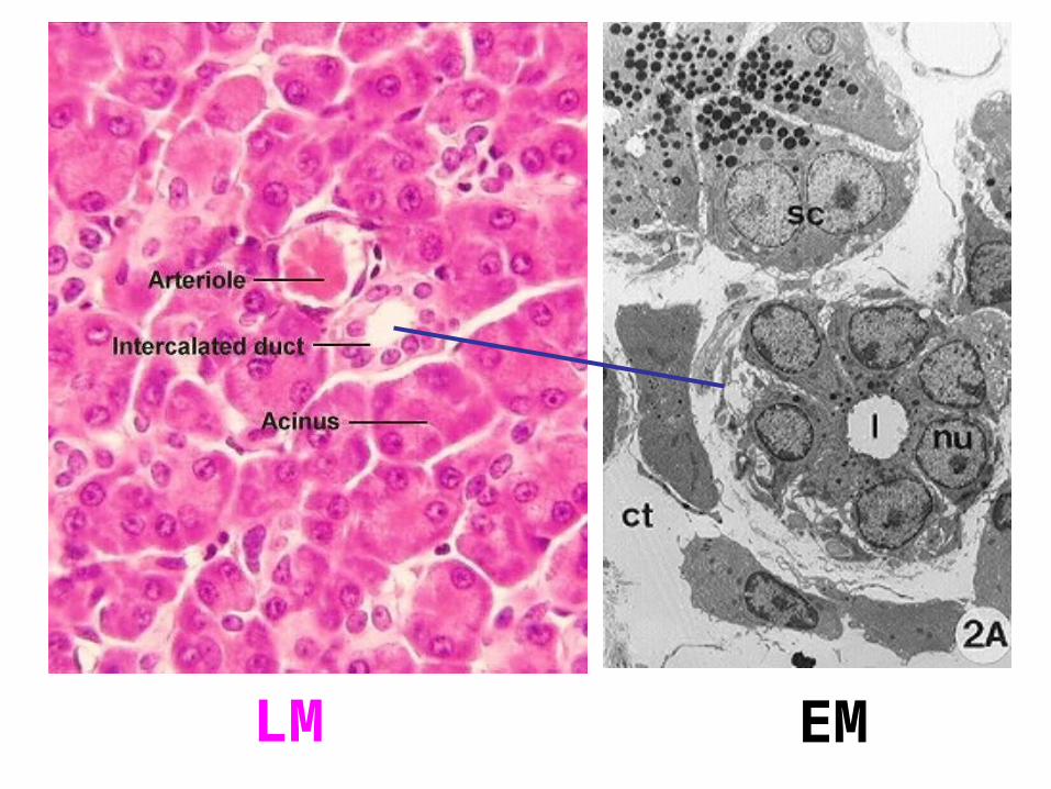
LM EM
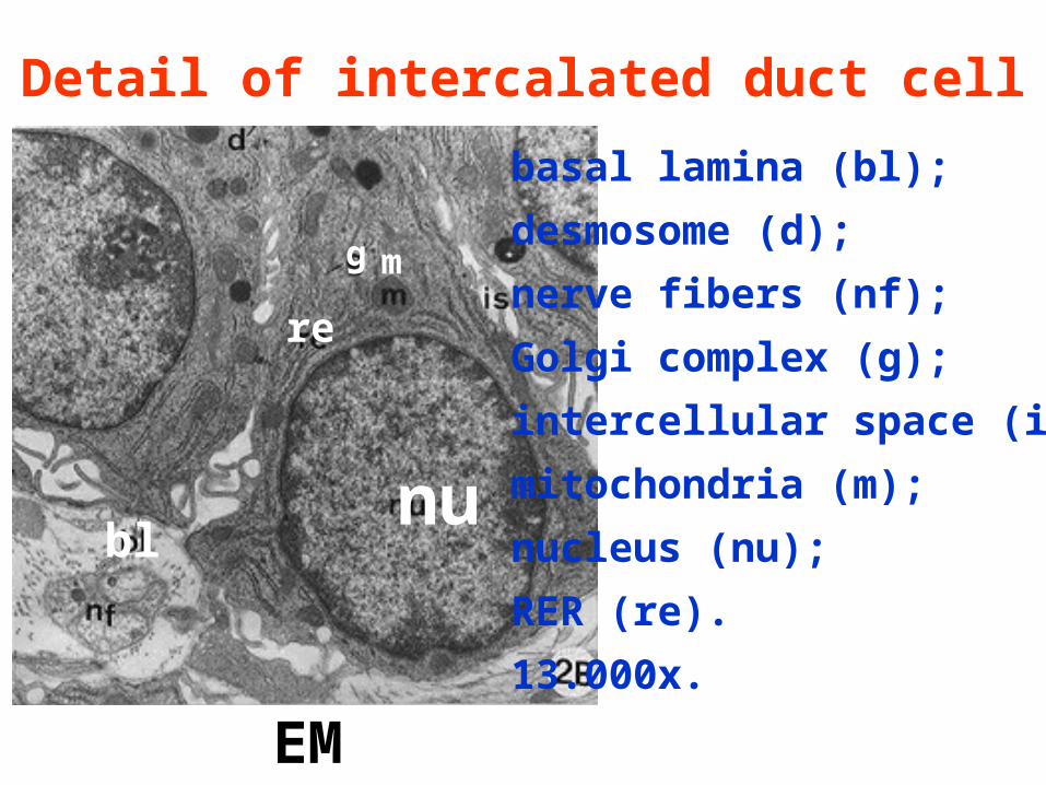
basal lamina (bl);
desmosome (d);
nerve fibers (nf);
Golgi complex (g);
intercellular space (is);
mitochondria (m);
nucleus (nu);
RER (re).
13.000x.
EM
nu
re
mg
bl
Detail of intercalated duct cell
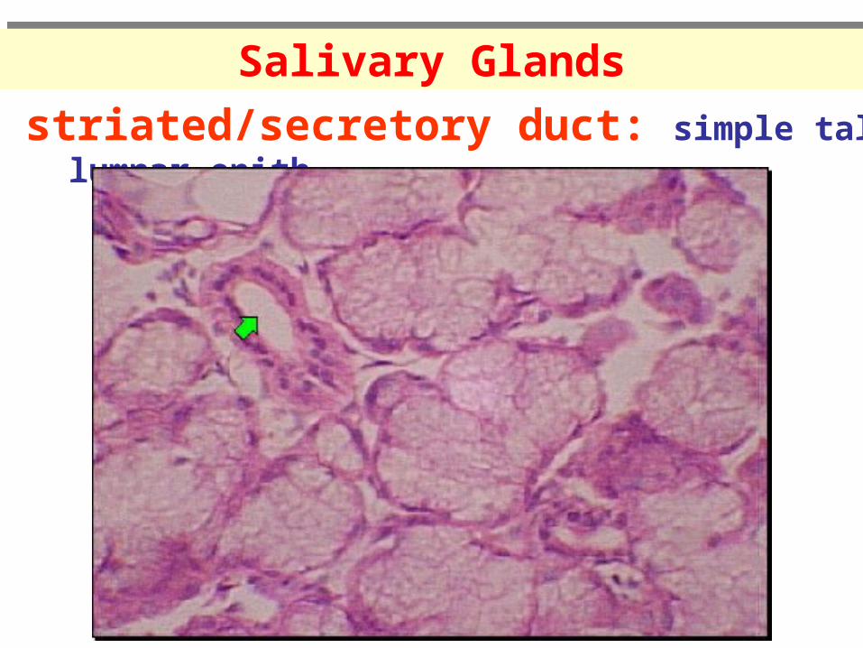
striated/secretory duct: simple tall columnar epith.
Salivary GlandsSalivary Glands
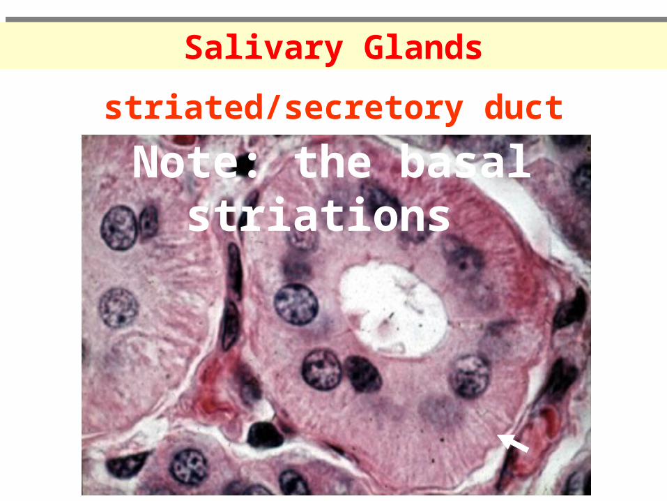
striated/secretory duct
Salivary GlandsSalivary Glands
Note: the basal striations
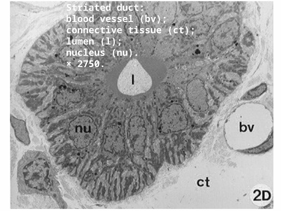
Striated duct: blood vessel (bv); connective tissue (ct); lumen (l); nucleus (nu). × 2750.
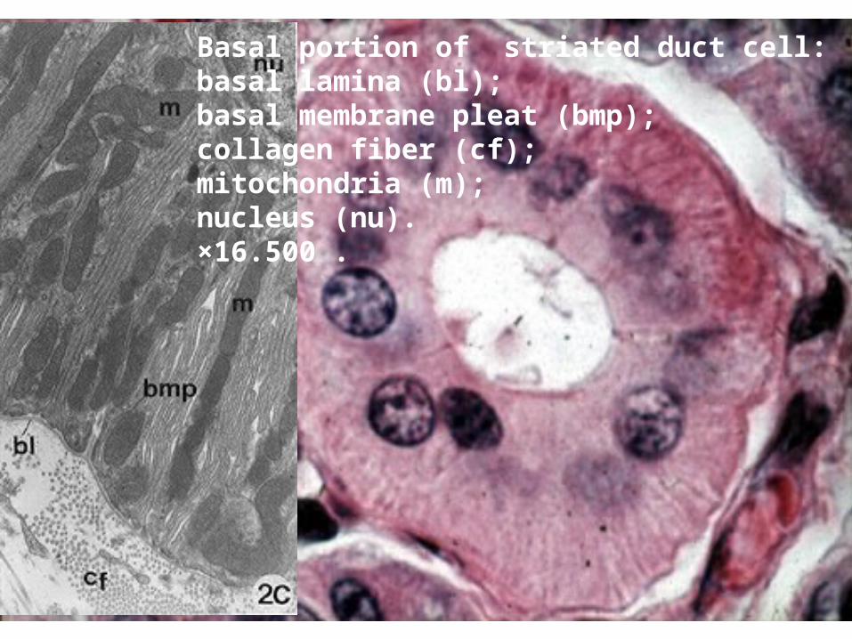
Basal portion of striated duct cell: basal lamina (bl); basal membrane pleat (bmp); collagen fiber (cf); mitochondria (m); nucleus (nu). ×16.500 .
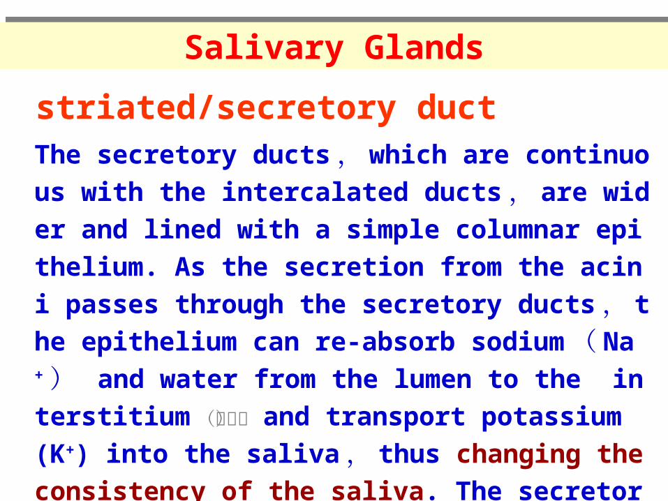
striated/secretory duct
Salivary GlandsSalivary Glands
The secretory ducts , which are continuous with the i
ntercalated ducts , are wider and lined with a simple
columnar epithelium. As the secretion from the acini p
asses through the secretory ducts , the epithelium can
re-absorb sodium ( Na+ ) and water from the lumen
to the interstitium (间质) and transport potassium (K+) into the saliva , thus changing the consistency of the
saliva. The secretory ducts drain into interlobular duct
s which run between lobules.
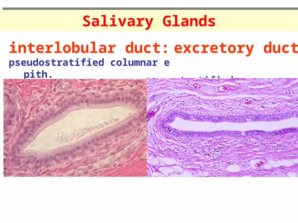
interlobular duct: pseudostratified columnar epith.
Salivary GlandsSalivary Glands
excretory duct: stratified squamous epith.
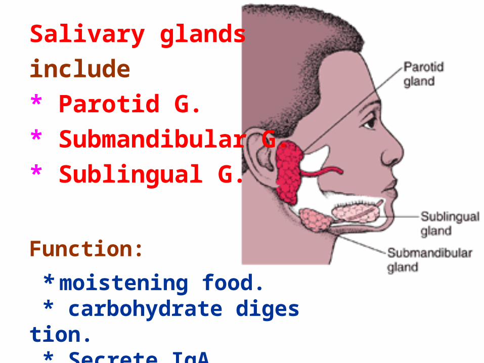
Salivary glands
include
* Parotid G.
* Submandibular G.
* Sublingual G.
Function:
* moistening food. * carbohydrate digestion. * Secrete IgA.
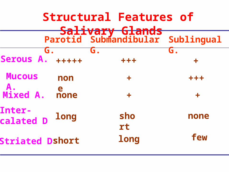
Parotid G.
Mucous A.
+++++Serous A.
Mixed A.
Inter-calated D
Submandibular G. Sublingual G.
+++
+++
short
+
Structural Features of Salivary Glands
Striated D
+
+ +
long short
long
none
few
none
none
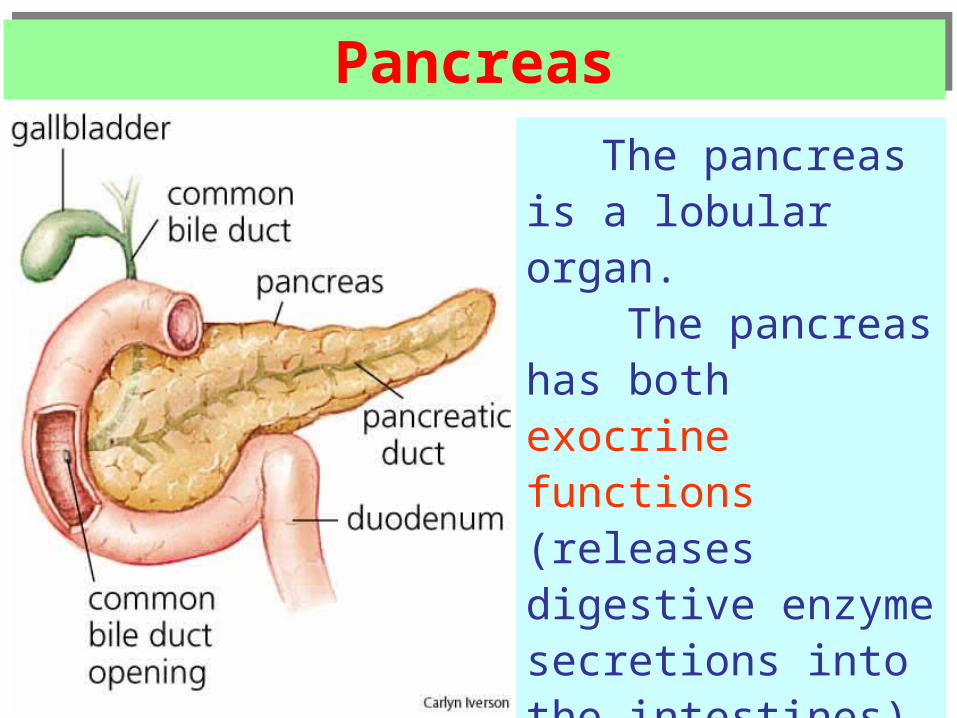
PancreasPancreas
The pancreas is a lobular organ.
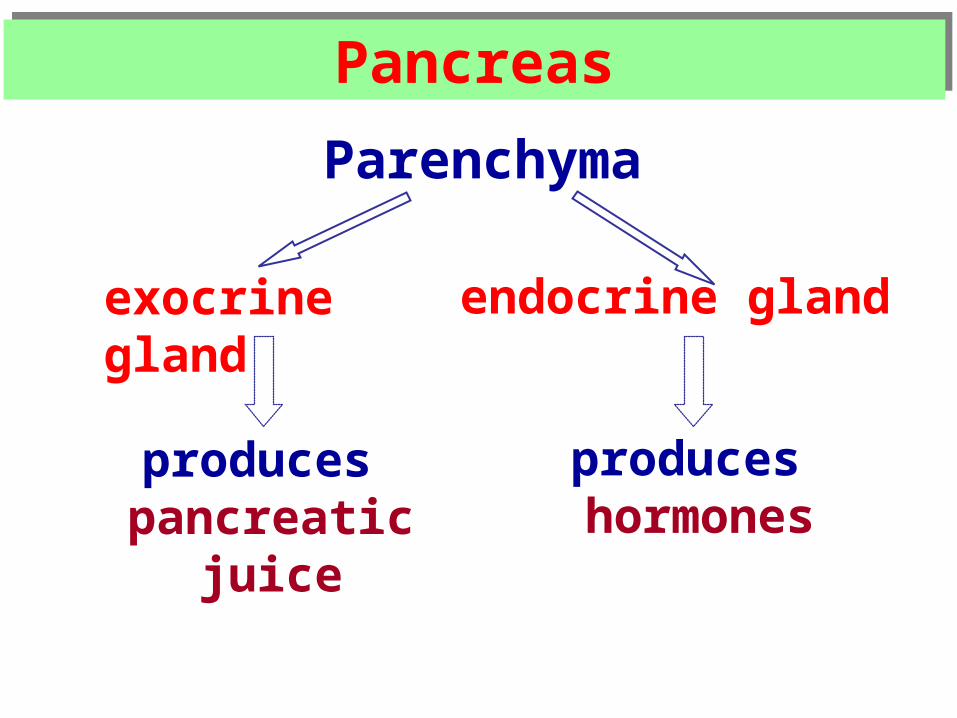
The pancreas has both exocrine functions (releases digestive enzyme secretions into the intestines) and endocrine functions (releases hormones into the blood).
PancreasPancreas
Parenchyma
exocrine gland endocrine gland
produces pancreatic juice
produces hormones
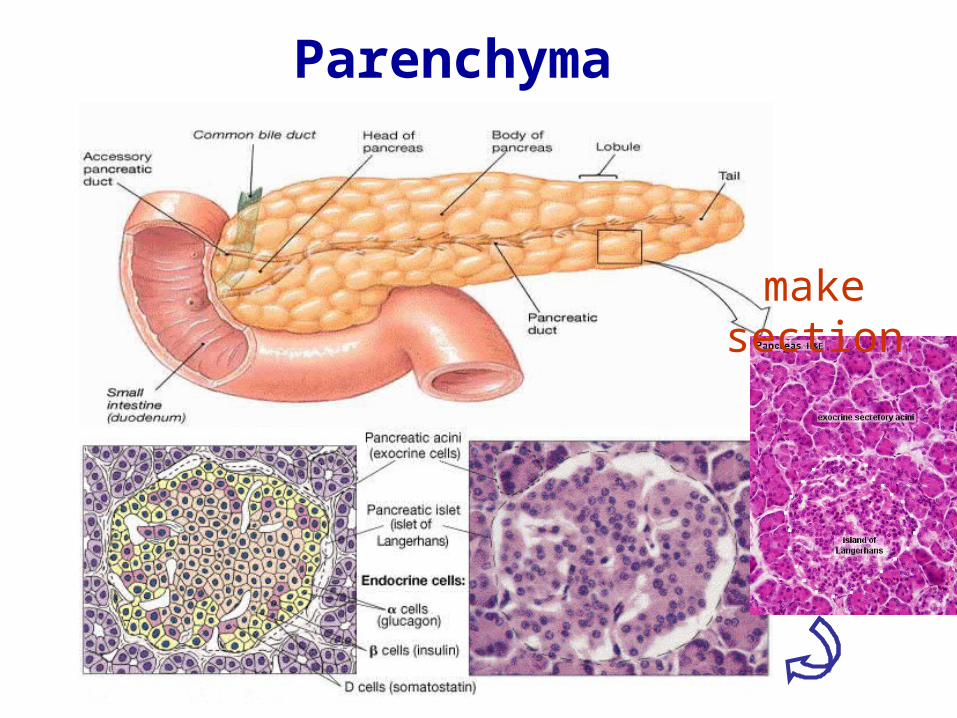
Parenchyma
make section
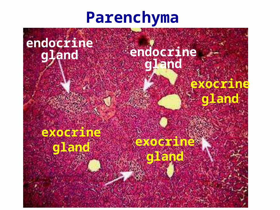
Parenchyma
exocrine gland
endocrine gland endocrine
gland
exocrine gland
exocrine gland
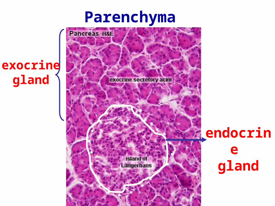
Parenchyma
exocrine gland
endocrine gland
Acini
PancreasPancreas
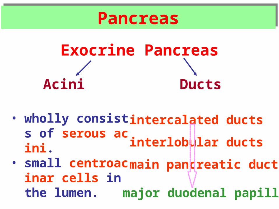
Exocrine Pancreas
Ducts
• wholly consists of serous acini.
• small centroacinar cells in the lumen.
intercalated ducts
interlobular ducts
main pancreatic ducts
major duodenal papilla
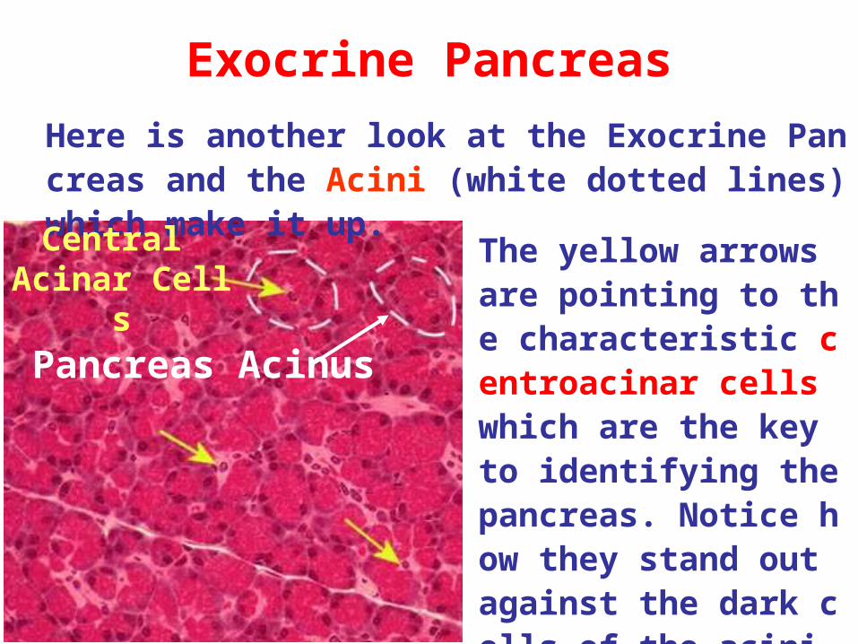
Exocrine Pancreas
The yellow arrows are pointing to the characteristic centroacinar cells which are the key to identifying the pancreas. Notice how they stand out against the dark cells of the acini.
Here is another look at the Exocrine Pancreas and the Acini (white dotted lines) which make it up.
Pancreas Acinus
Central Acinar Cells
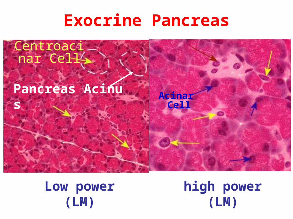
Exocrine Pancreas
Pancreas Acinus
Centroacinar Cell
Acinar Cell
Low power(LM)
high power(LM)
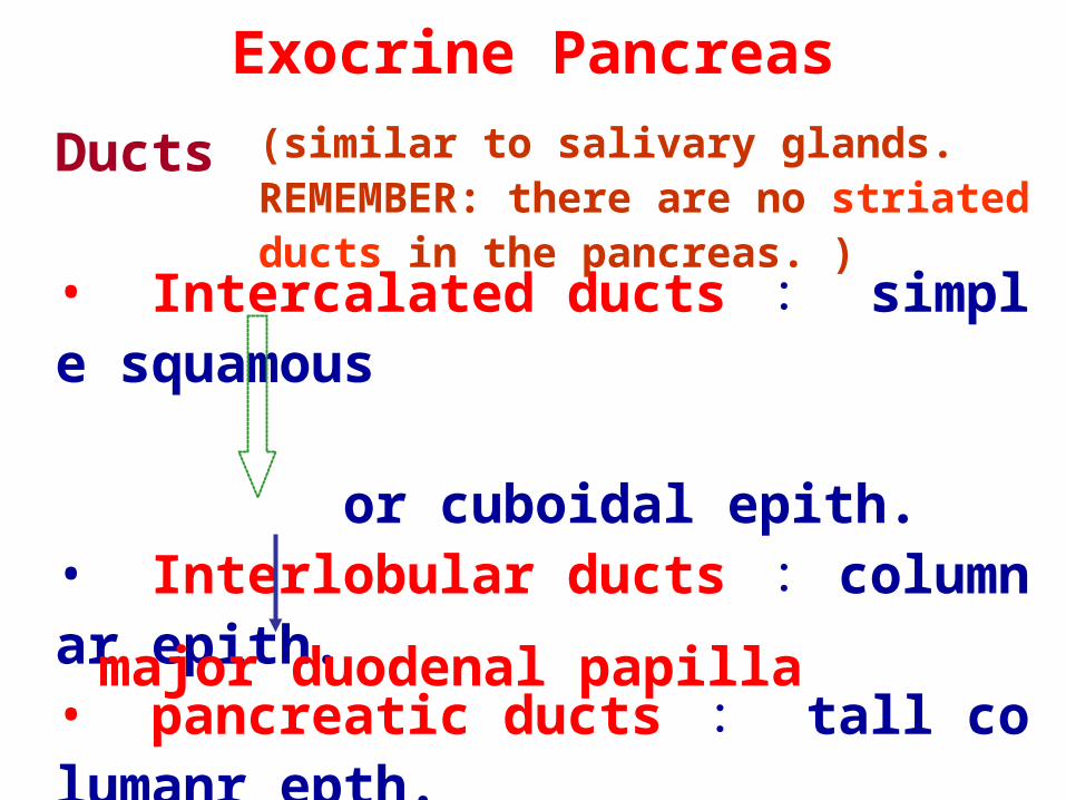
Ducts • Intercalated ducts : simple squamous or cuboidal epith.• Interlobular ducts : columnar epith.• pancreatic ducts : tall columanr epth.
Exocrine Pancreas
major duodenal papilla
(similar to salivary glands. REMEMBER: there are no striated ducts in the pancreas. )
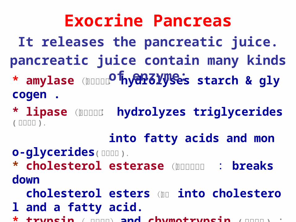
* amylase (胰淀粉酶): hydrolyses starch & glycogen .
* lipase (胰脂肪酶): hydrolyzes triglycerides( 甘油三酯 ).
into fatty acids and mono-glycerides( 甘油单酯 ).
* cholesterol esterase (胆固醇酯酶) : breaks down cholesterol esters (酯) into cholesterol and a fatty acid.* trypsin ( 胰蛋白酶) and chymotrypsin ( 糜蛋白酶 ) : hydrolyze proteins.* ribonuclease (核糖核酸酶) & deoxyribonuclease (脱氧核 糖核酸酶): split nucleic acids.
Exocrine PancreasIt releases the pancreatic juice.
pancreatic juice contain many kinds of enzyme:

PancreasPancreasEndocrine Pancreas
• Islets of Langerhans scatter throughout the exocrine pancreas. • Three types of cells :A-cells (20%) glucagon B-cells (75%) insulin D-cells ( 5% ) somatostatin• Capillaries: each islet is richly supplied with blood vessels.
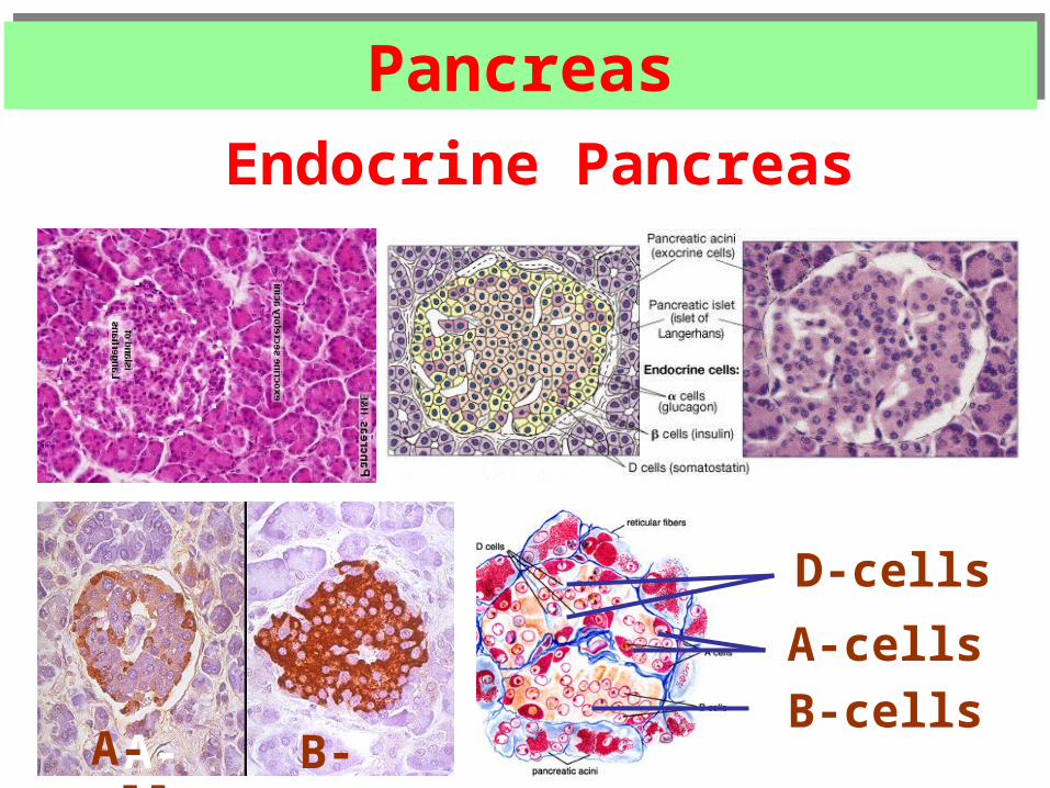
PancreasPancreas
Endocrine Pancreas
A-cells
B-cells
D-cells
A-cells B-cells A-cells
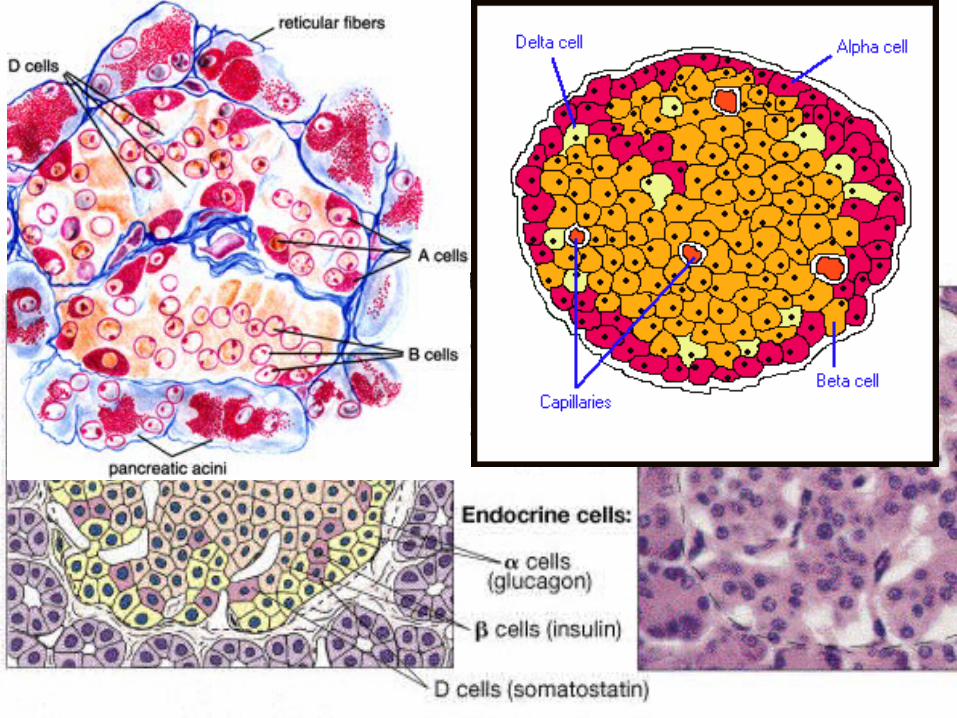
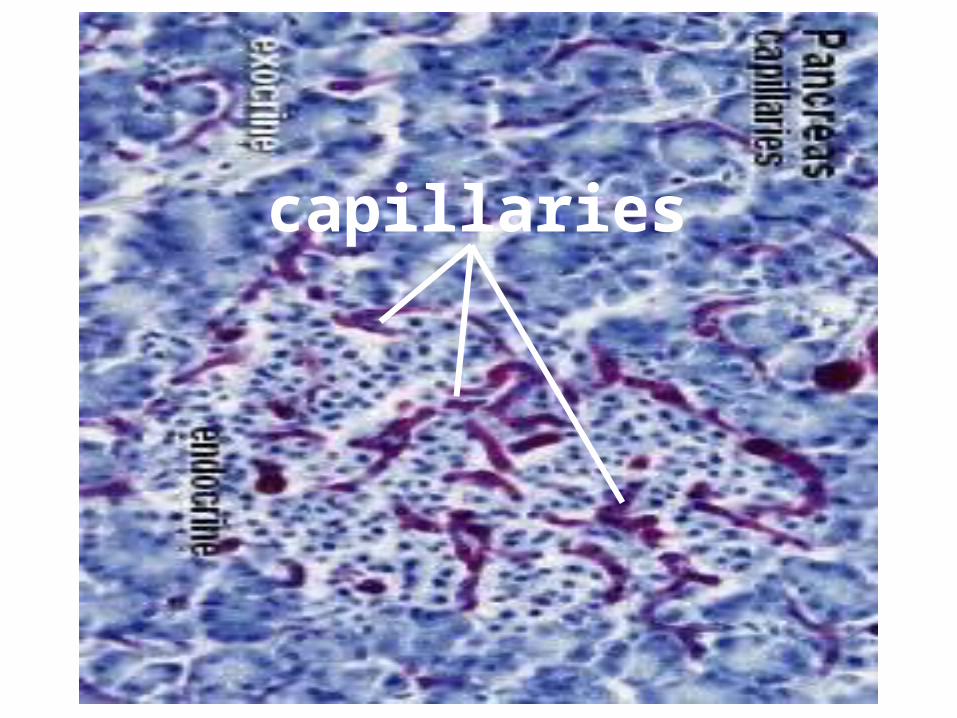
capillaries
Liver Liver
Liver Liver
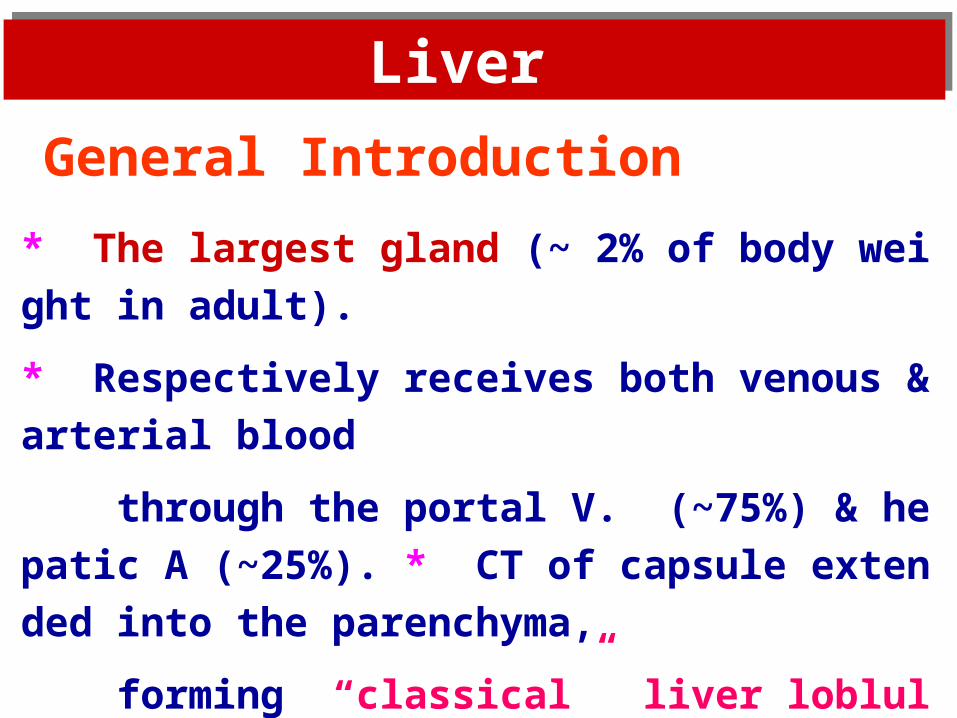
General Introduction
* The largest gland (~ 2% of body weight in adult).
* Respectively receives both venous & arterial blood
through the portal V. (~75%) & hepatic A (~25%).
* CT of capsule extended into the parenchyma,
forming “classical” liver loblules.
* Functions as an exocrine gland (secreting bile)
and other very important roles.
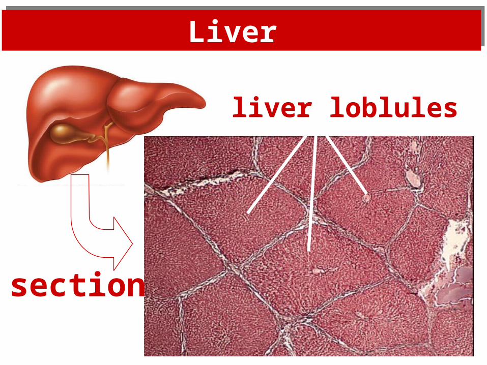
Liver Liver
section
liver loblules
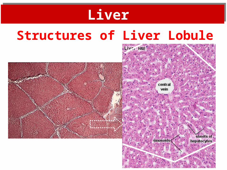
Liver Liver
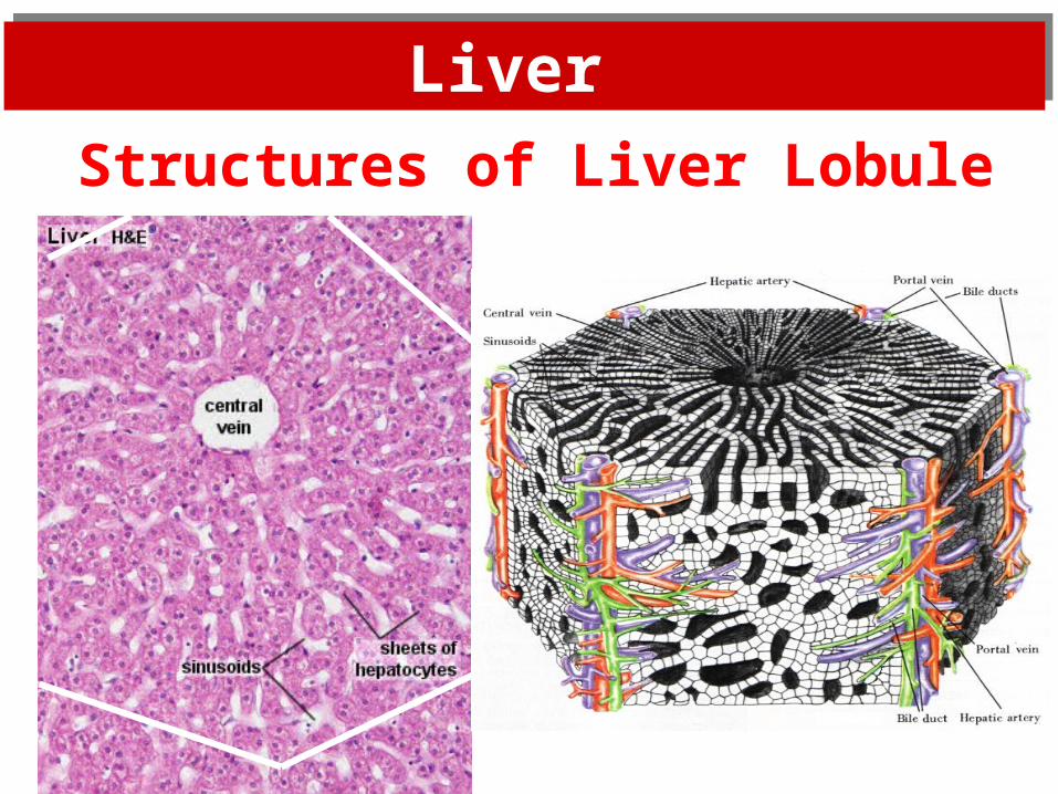
Structures of Liver Lobule
Liver Liver
Structures of Liver Lobule
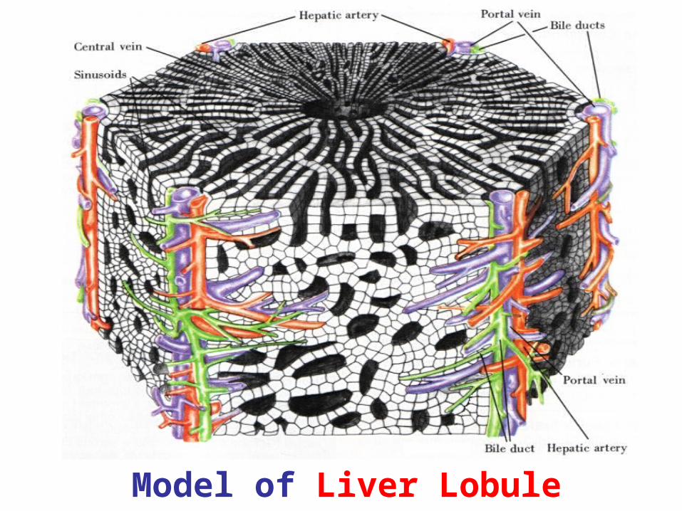
Model of Liver Lobule
Liver Liver
Structures of Liver Lobule
* six-sided prism
with a central V. at its center.
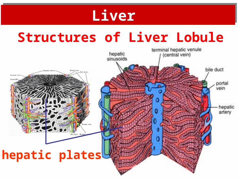
* sheets of hepatocytes ( or hepatic plates )
extend radially from the central V.
* sinusoids between hepatic plates.
portal triads ( or portal area): in the corner of lobules.
Liver Liver
Structures of Liver Lobule
hepatic plates
Liver Liver
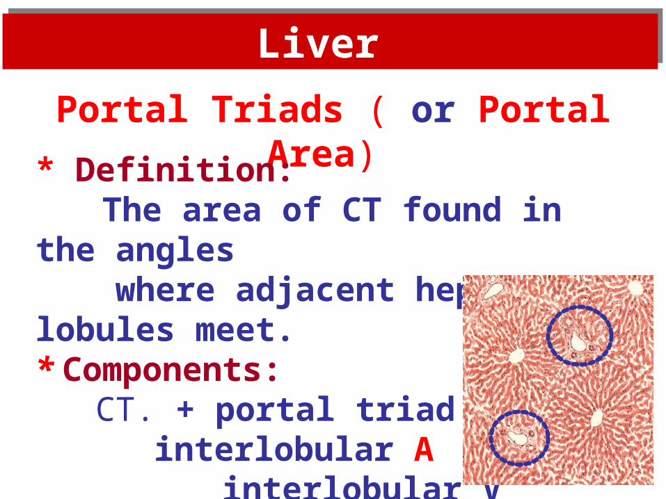
Portal Triads ( or Portal Area)
* Definition: The area of CT found in the angles
where adjacent hepatic lobules meet.* Components: CT. + portal triad
interlobular A interlobular V interlobular bile duct
Liver Liver
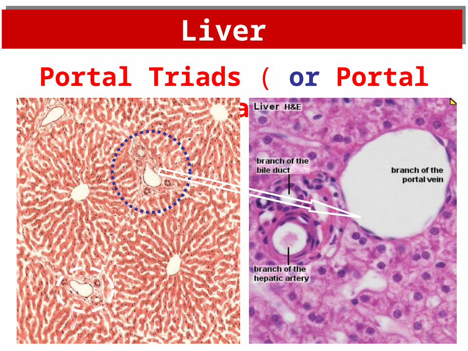
Portal Triads ( or Portal Area)
Liver Liver
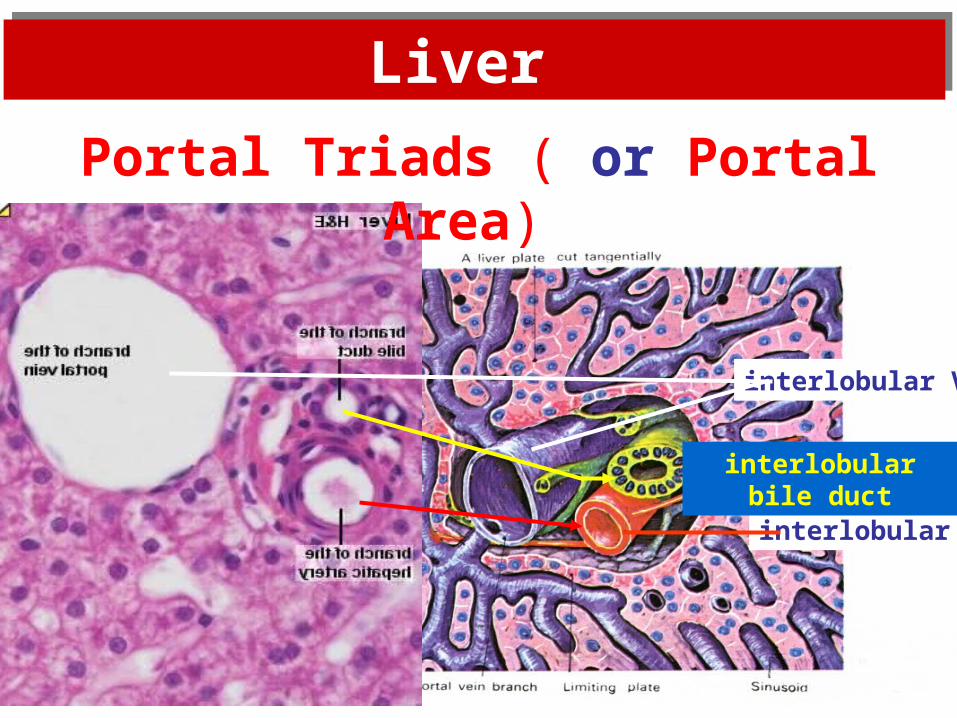
Portal Triads ( or Portal Area)
interlobular A
interlobular bile duct
interlobular V
Liver Liver
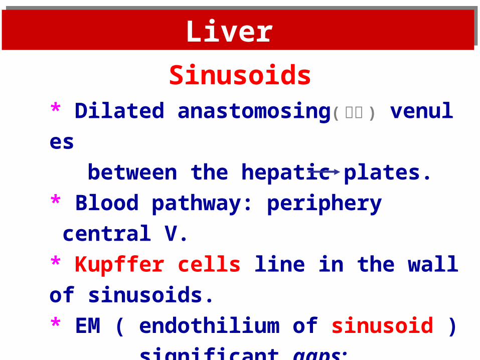
Sinusoids* Dilated anastomosing( 吻合 ) venules
between the hepatic plates.
* Blood pathway: periphery central V.
* Kupffer cells line in the wall of sinusoids.
* EM ( endothilium of sinusoid )
significant gaps;
numerous fenestration;
incomplete basal lamina.

Liver Liver
Sinusoids
Model
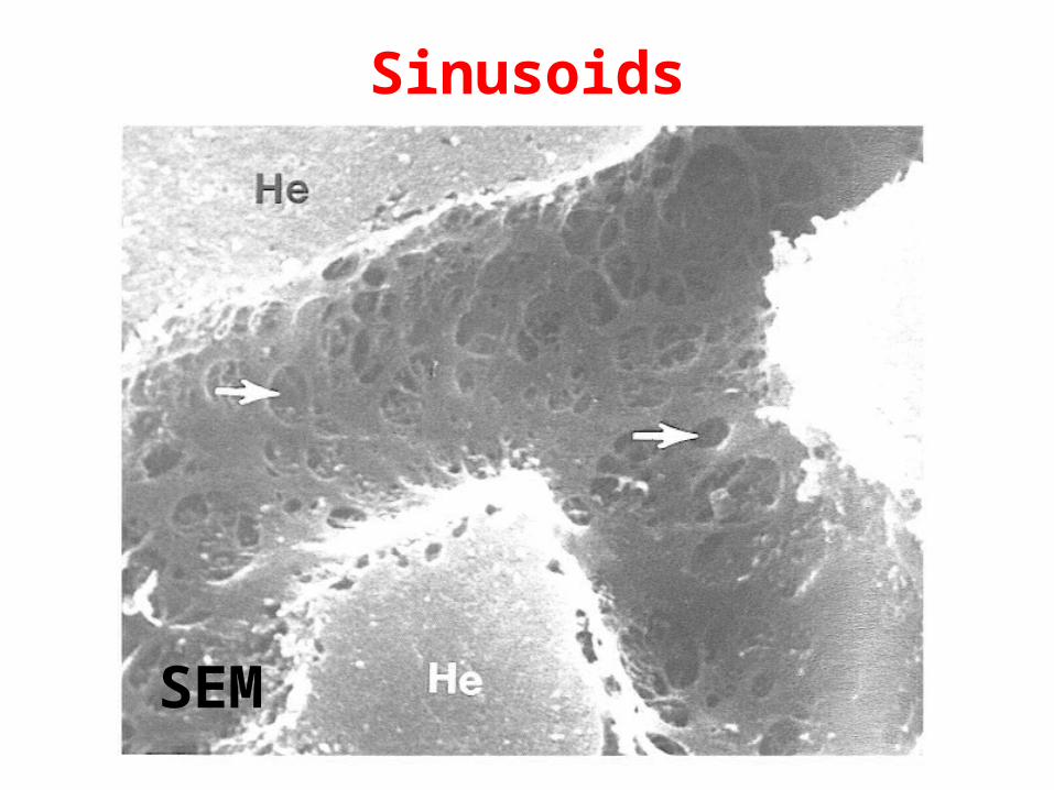
Sinusoids
SEM
Sinusoids
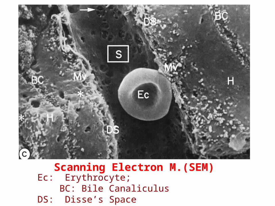
Scanning Electron M.(SEM)Ec: Erythrocyte; BC: Bile Canaliculus DS: Disse’s Space S: Sinusoid H: Hepatocyte Mv: Microvili
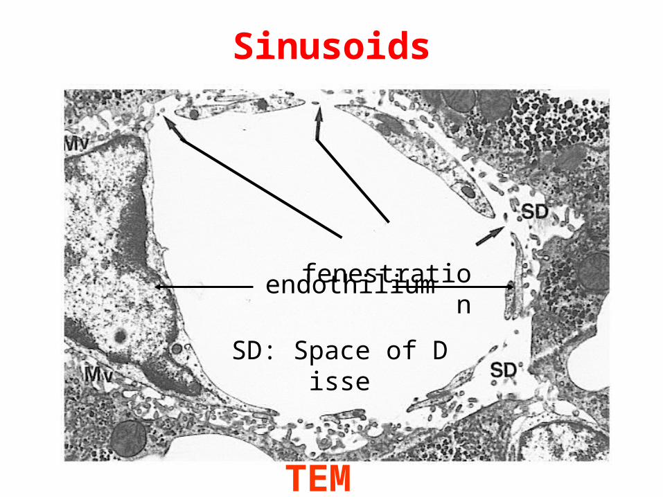
Sinusoids
TEM
SD: Space of Disse
fenestration
endothilium
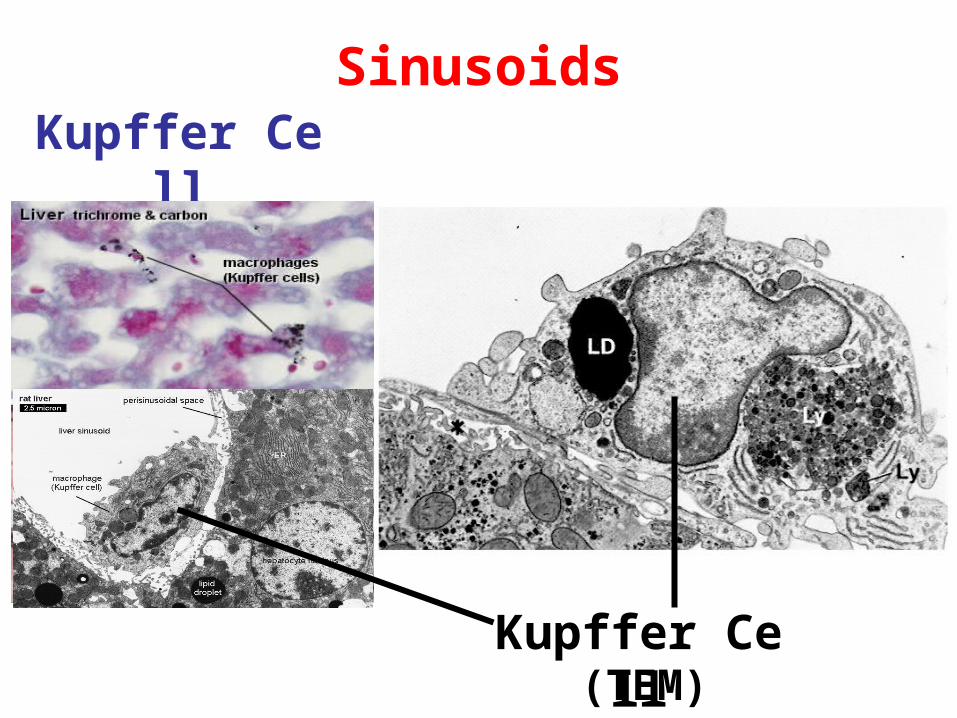
SinusoidsKupffer Cell
(LM)
(TEM)Kupffer Cell
Liver Liver
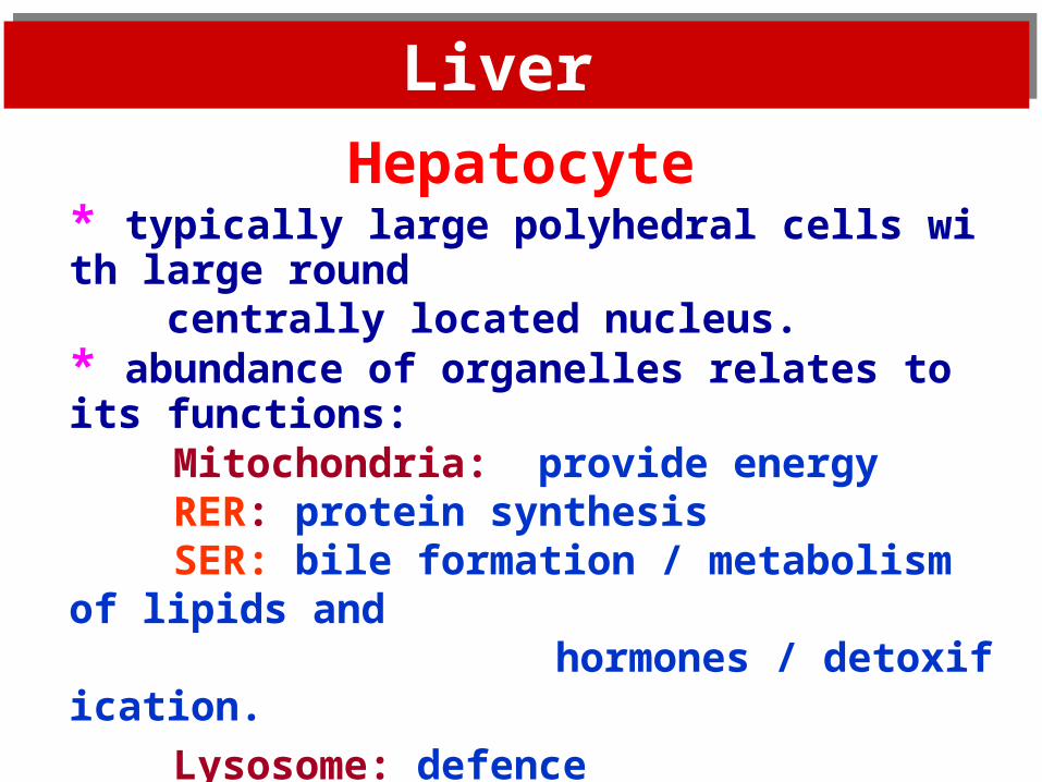
Hepatocyte* typically large polyhedral cells with large round centrally located nucleus. * abundance of organelles relates to its functions:
Mitochondria: provide energyRER: protein synthesisSER: bile formation / metabolism of lipids and
hormones / detoxification.
Lysosome: defence
Microbody: detoxification.Inclusions: glcogen particles, lipid droplets
and pigments.
Liver Liver
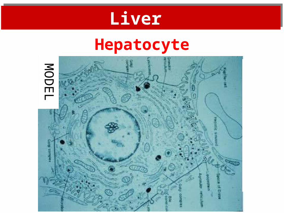
Hepatocyte
MO
DE
L
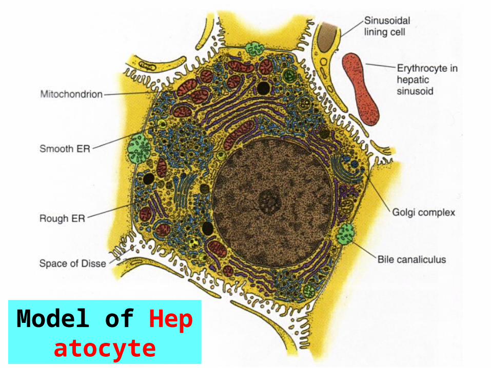
Model of Hepatocyte
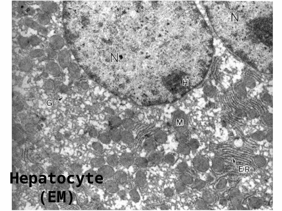
Hepatocyte(EM)
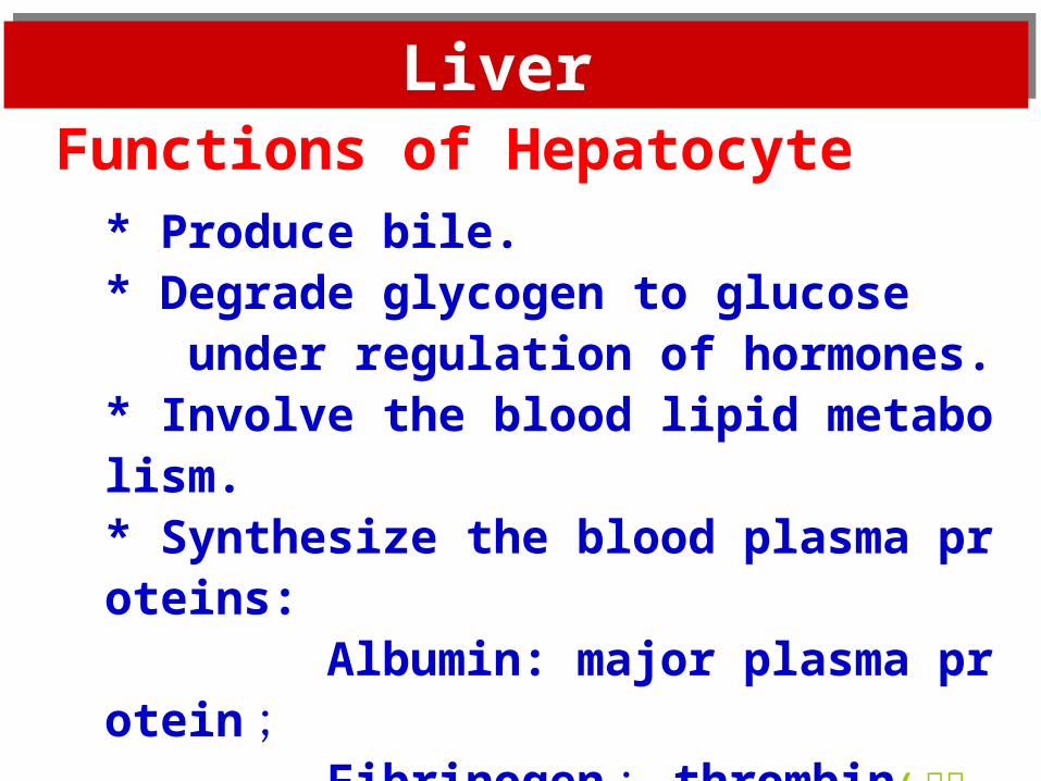
* Produce bile.* Degrade glycogen to glucose under regulation of hormones.* Involve the blood lipid metabolism.* Synthesize the blood plasma proteins: Albumin: major plasma protein ; Fibrinogen ; thrombin( 凝血酶 ) ; Clotting (凝结) factor III. * Detoxification
Functions of Hepatocyte
Liver Liver
Liver Liver
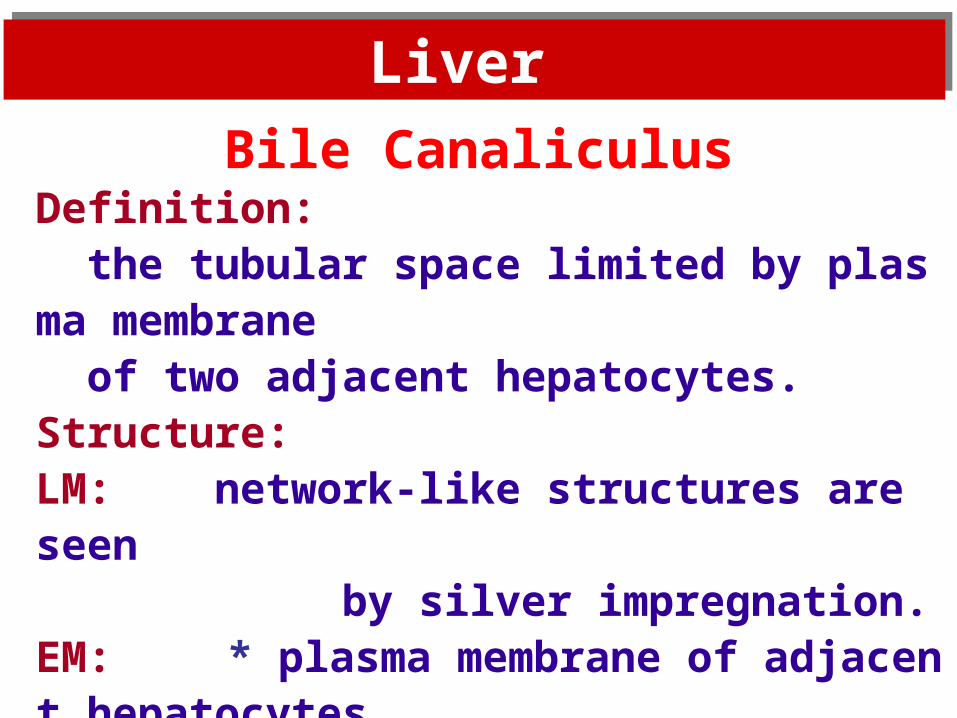
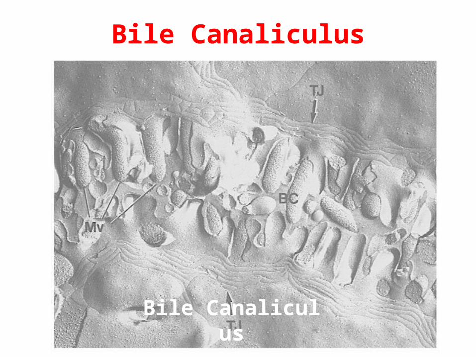
Bile CanaliculusDefinition: the tubular space limited by plasma membrane of two adjacent hepatocytes.Structure:LM: network-like structures are seen by silver impregnation.EM: * plasma membrane of adjacent hepatocytes
forms the wall of bile canaliculi.* tight junctions form seals.

Bile Canaliculus
Blie canaliculi stained by silver impregnation
(LM)
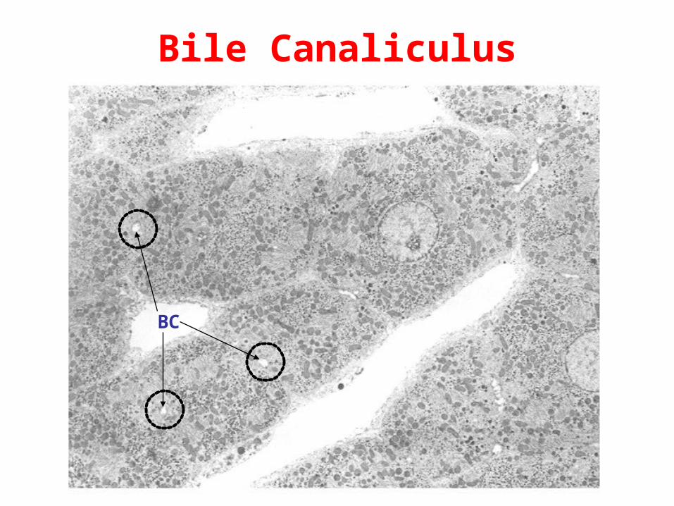
Bile Canaliculus
BC
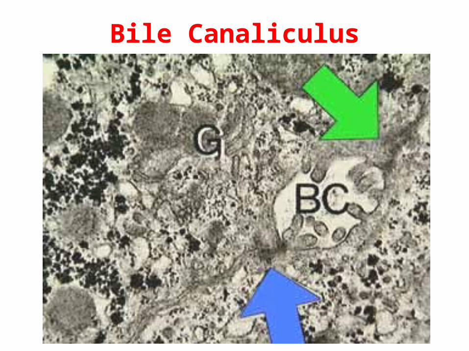
Bile Canaliculus
Bile Canaliculus
Bile Canaliculus Freeze Fracture SEM
Liver Liver
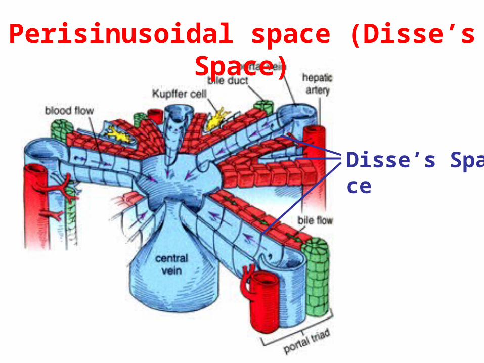
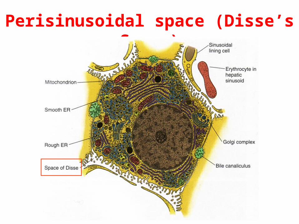
Perisinusoidal space (Disse’s Space)
A space between endothelium & hepatocyte.• Be visible under EM.• Occupy by numerous microvilli of hepatocytes.• Fill with blood plasma.• have fat-storing cells (storing fat & vitamin a)• A site for substance exchange between the blood & the hepatocytes.
Perisinusoidal space (Disse’s Space)
Disse’s Space
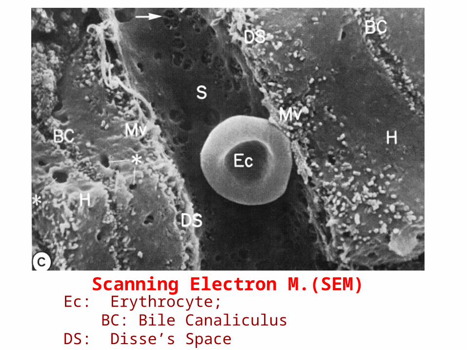
Scanning Electron M.(SEM)Ec: Erythrocyte; BC: Bile Canaliculus DS: Disse’s Space S: Sinusoid H: Hepatocyte Mv: Microvili
Perisinusoidal space (Disse’s Space)
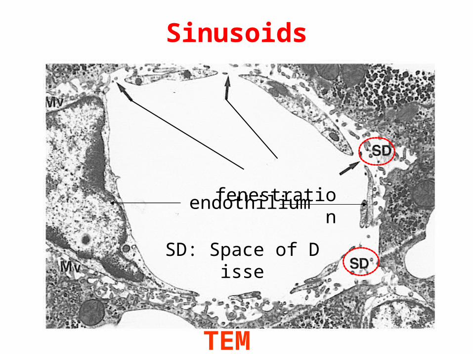
Sinusoids
TEM
SD: Space of Disse
fenestration
endothilium
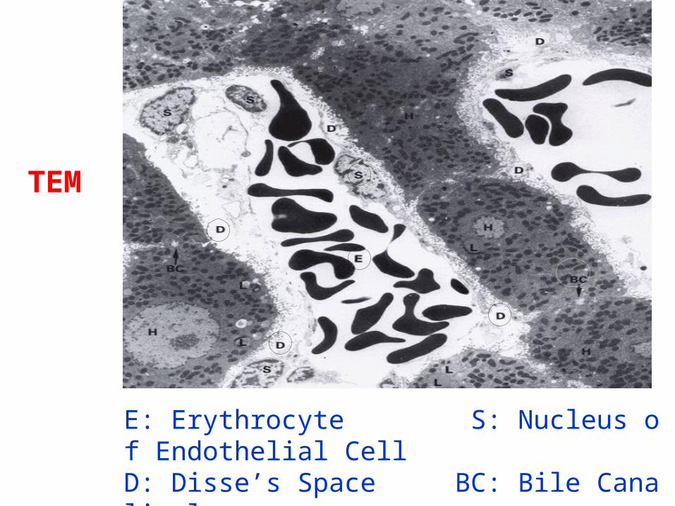
E: Erythrocyte S: Nucleus of Endothelial CellD: Disse’s Space BC: Bile Canaliculus
TEM
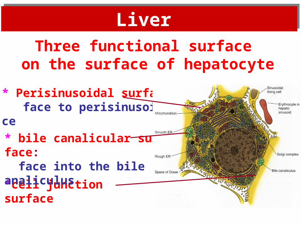
*cell junction surface
* Perisinusoidal surface: face to perisinusoidal space
* bile canalicular surface: face into the bile canaliculus
Liver Liver Three functional surface
on the surface of hepatocyte
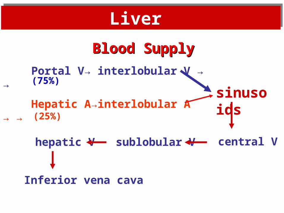
Blood SupplyBlood Supply
Portal V→ interlobular V → →
Hepatic A→interlobular A → →
sublobular V
sinusoids
central Vhepatic V
Inferior vena cava
(75%)
(25%)
Liver Liver
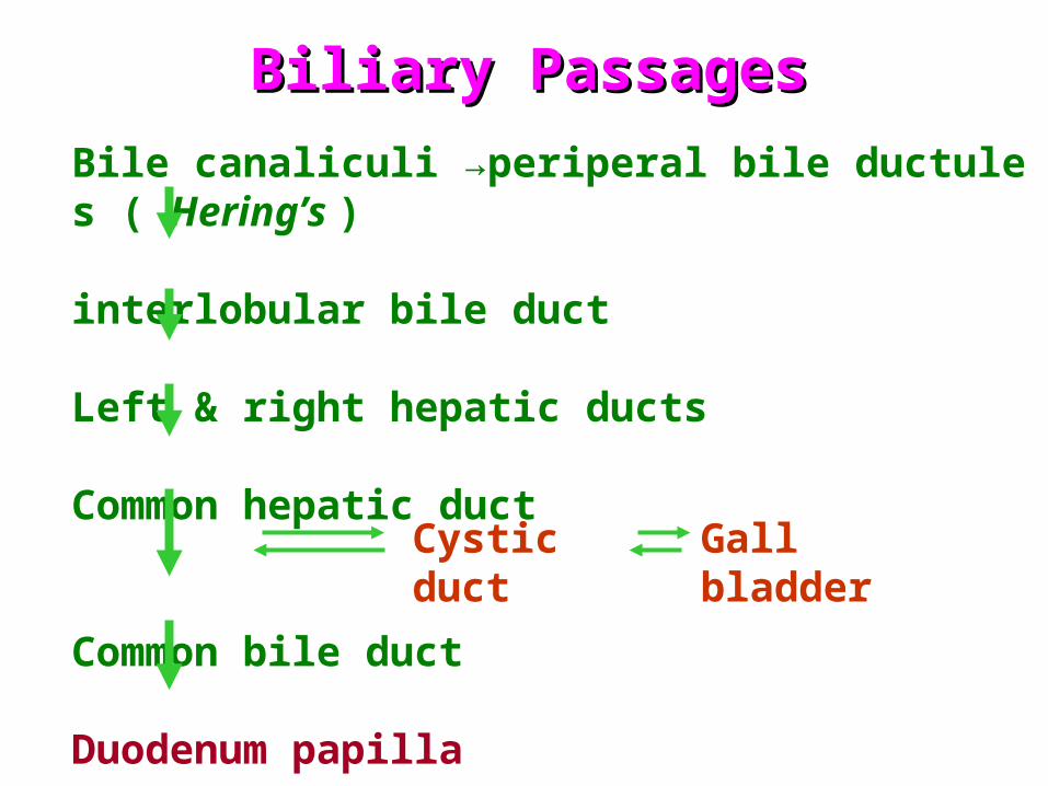
Bile canaliculi →periperal bile ductules ( Hering’s )
interlobular bile duct
Left & right hepatic ducts
Common hepatic duct
Common bile duct
Duodenum papilla
Cystic duct Gall bladder
Biliary PassagesBiliary Passages
Liver Liver
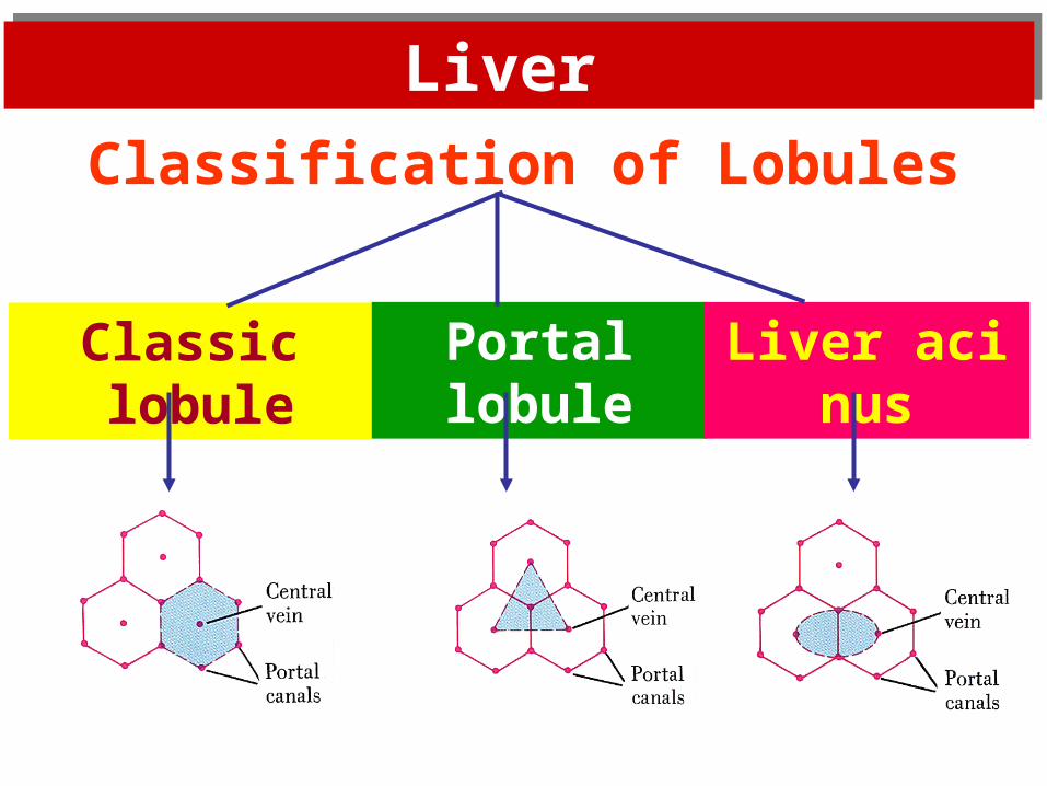
Classification of Lobules
Classic lobule Portal lobule Liver acinus
Liver Liver
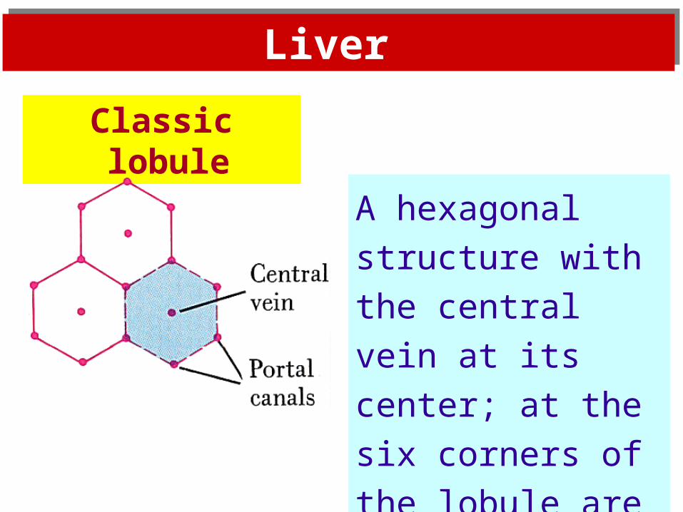
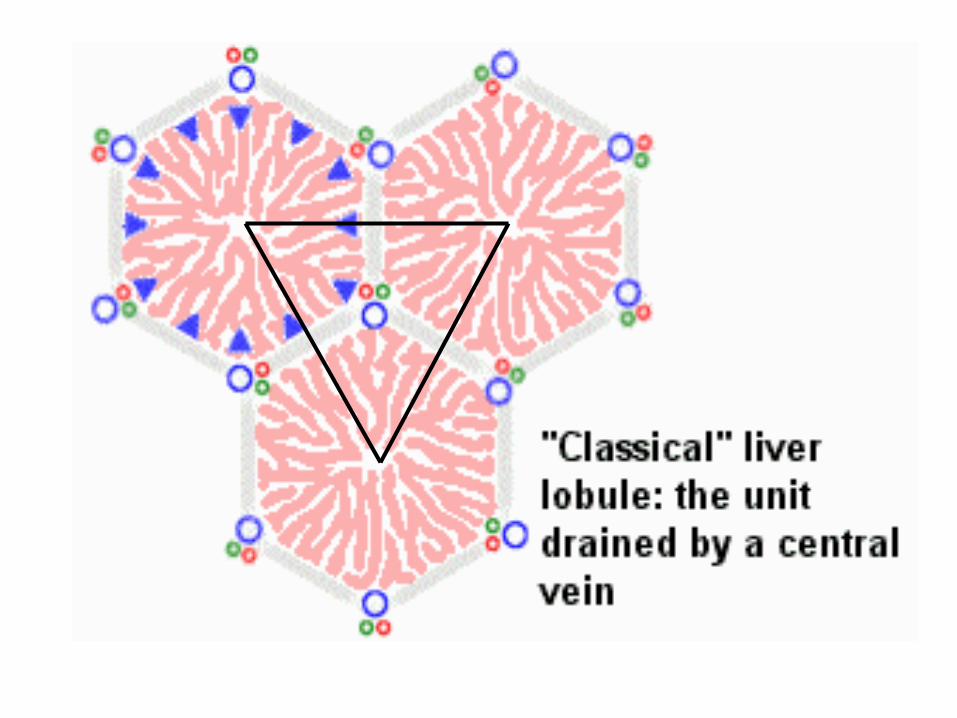
Classic lobule
A hexagonal structure
with the central vein
at its center; at the six
corners of the lobule
are hepatic triads.
Liver Liver
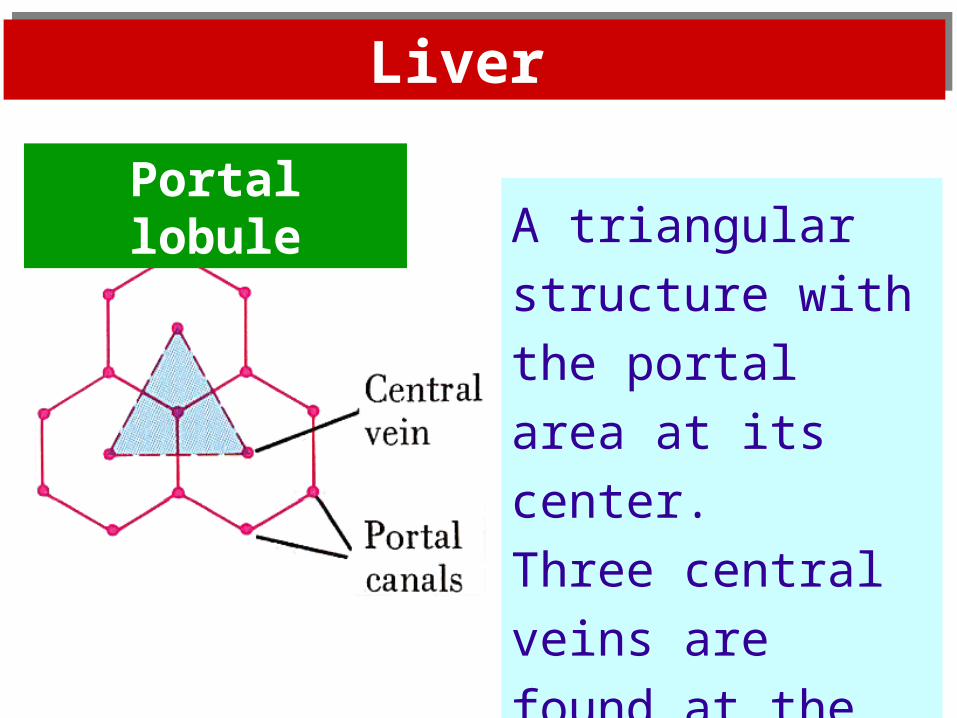
A triangular structure
with the portal area at
its center.
Three central veins
are found at the
peripheral boundary
points.
Portal lobule
Liver Liver
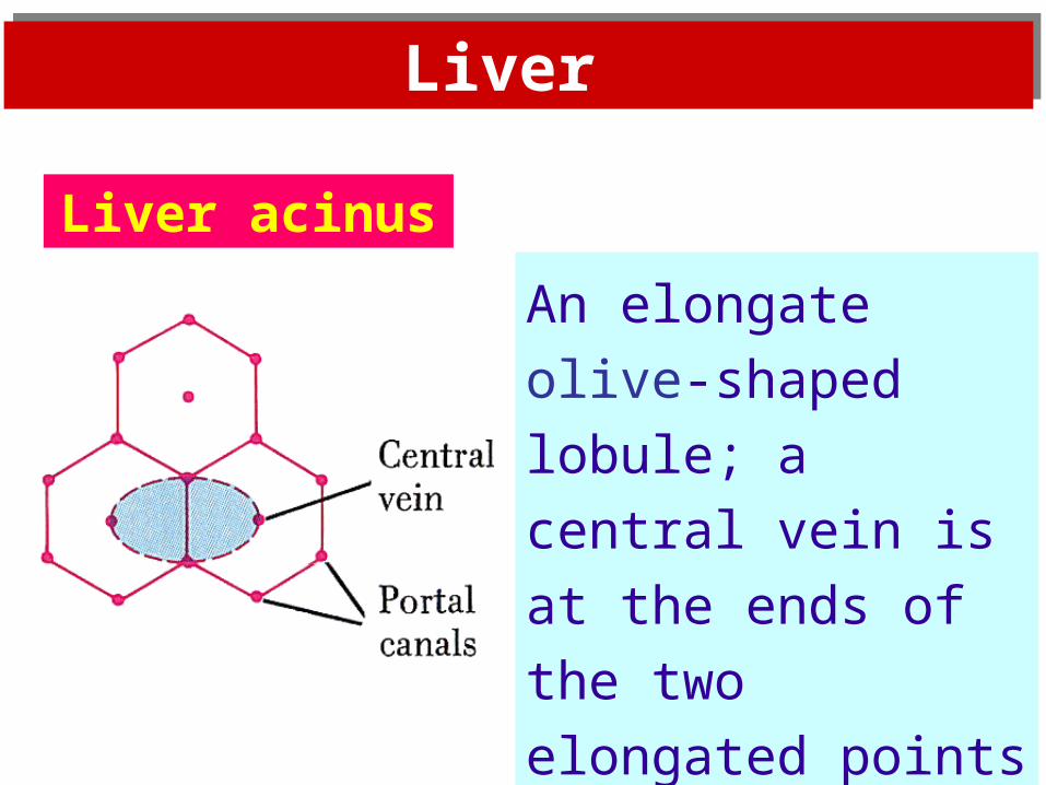
An elongate olive-
shaped lobule; a central
vein is at the ends of
the two elongated
points of the olive.
Liver acinus

OVER