Coronary Calcification; Body Mass Index (BMI) or Waist to Hip Ratio (WHR)
17
Coronary Calcification; Body Mass Index (BMI) or Waist to Hip Ratio (WHR) Siamak Sabour, MD, MSc, DSc, PhD, Postdoc Clinical Epidemiologist Persian International Epidemiology Network (PIEPNET)
-
Upload
alexandria-anagnos -
Category
Documents
-
view
29 -
download
0
description
Coronary Calcification; Body Mass Index (BMI) or Waist to Hip Ratio (WHR). Siamak Sabour, MD, MSc, DSc, PhD, Postdoc Clinical Epidemiologist Persian International Epidemiology Network (PIEPNET). SCIENTIFIC BACKGROUND. 1994: M.D , I.R. Iran - PowerPoint PPT Presentation
Transcript of Coronary Calcification; Body Mass Index (BMI) or Waist to Hip Ratio (WHR)
Coronary Calcification; Body Mass Index (BMI)
or Waist to Hip Ratio (WHR)
Siamak Sabour, MD, MSc, DSc, PhD, PostdocClinical Epidemiologist
Persian International Epidemiology Network (PIEPNET)

SCIENTIFIC BACKGROUND
• 1994: M.D, I.R. Iran
• 2004: M.Sc, Clinical Epidemiology, Erasmus MC, The Netherlands
• 2006: D.Sc, Clinical Epidemiology, Erasmus MC, The Netherlands
• 2007: Ph.D, Clinical Epidemiology, UMC Utrecht, The Netherlands
• 2008
• Post doc Cardiovascular Epidemiology
Thomas Jefferson University, Philadelphia, PA, USA
• Post doc Pharmacoepidemiology
University of Pennsylvania, Philadelphia, PA, USA
• 2008 until now
Assistant Prof of Clinical Epidemiology & Medicine
Sabous S, MD, MSc, DSc, PhD, Postdoc 2
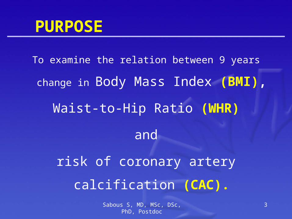
PURPOSE
To examine the relation between 9 years change in
Body Mass Index (BMI),
Waist-to-Hip Ratio (WHR)
and
risk of coronary artery calcification (CAC).
Sabous S, MD, MSc, DSc, PhD, Postdoc 3

DESIGN
Longitudinal study
Sabous S, MD, MSc, DSc, PhD, Postdoc 4

SUBJECTS
573 postmenopausal women selected from a population based cohort study.
Sabous S, MD, MSc, DSc, PhD, Postdoc 5
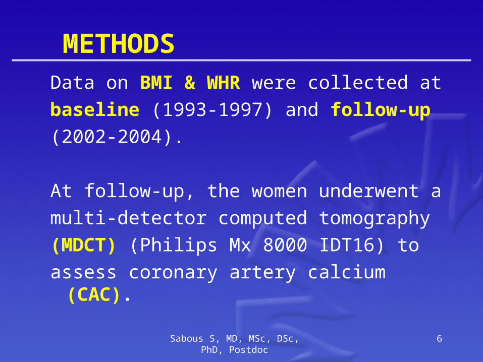
METHODS
Data on BMI & WHR were collected at
baseline (1993-1997) and follow-up
(2002-2004).
At follow-up, the women underwent a
multi-detector computed tomography
(MDCT) (Philips Mx 8000 IDT16) to
assess coronary artery calcium (CAC).
Sabous S, MD, MSc, DSc, PhD, Postdoc 6
7

8
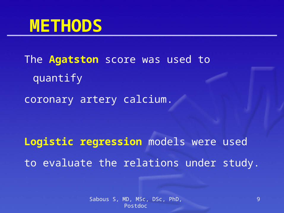
METHODS
The Agatston score was used to quantify
coronary artery calcium.
Logistic regression models were used
to evaluate the relations under study.
Sabous S, MD, MSc, DSc, PhD, Postdoc 9
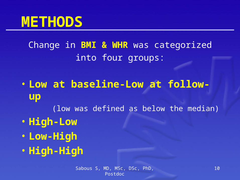
METHODS
Change in BMI & WHR was categorized
into four groups:
• Low at baseline-Low at follow-up (low was defined as below the median)
• High-Low
• Low-High
• High-High
Sabous S, MD, MSc, DSc, PhD, Postdoc 10

RESULTS
Compared to subjects whose WHR
remained below the median of the
distribution at both occasions, those with
a WHR above the median at both
occasions had a 2.8 [95% CI 1.5 - 5.7]
fold increased risk of CAC. Sabous S, MD, MSc, DSc, PhD, Postdoc 11

RESULTS
Women whose WHR rose over the 9 year
period from below the median to above
the median had a 2.6 [95%CI 1.3 - 5.2]
fold increased risk of CAC.
Sabous S, MD, MSc, DSc, PhD, Postdoc 12

RESULTS
In contrast, change in BMI was
not related to risk of CAC.
Sabous S, MD, MSc, DSc, PhD, Postdoc 13

Risk of Coronary Calcification in Categories of Change in BMI & WHR
Baseline Follow-up
Participants
OR (95% CI)
Body Mass Index Model 1 Model 2 Low Low 247 1 1 High Low 22 0.97 (0.39 - 2.46) 0.67 (0.22 – 1.99) Low High 44 1.36 (0.68 - 2.71) 0.81 (0.33 – 1.96) High High 250 1.12 (0.77 - 1.63) 0.92 (0.43 – 1.95)
Waist to Hip Ratio Low Low 224 1 1 High Low 49 1.62 (0.83 - 3.16) 1.62 (0.76 – 3.42) Low High 71 2.48 (1.38 - 4.46) 2.58 (1.29 – 5.17) High High 219 2.65 (1.76 - 3.99) 2.80 (1.52 – 5.17)
Model 1= Adjusted for Age
Model 2= Full Model (Adjusted for Age and Changes in WC, HC, WHR, BMI, SBP, DBP and Smoking)
Sabous S, MD, MSc, DSc, PhD, Postdoc 14

CONCLUSION
Change in WHR over time
relates to
an increased risk of CAC.
However, BMI has no effect on that.
Sabous S, MD, MSc, DSc, PhD, Postdoc 15

Acknowledgments
Prof. Diederick. E. Grobbee, MD, PhD
Prof. Mathias Prokop, MD, PhD
Dr. Yvonne. T. van der Schouw, PhD
Prof. Michiel. L. Bots, MD, PhD
1. Julius Centre, University Medical Centre Utrecht, The Netherlands
2. Radiology Department, University Medical Center Utrecht, The Netherlands
Sabous S, MD, MSc, DSc, PhD, Postdoc 16

CONCLUSION
Changes in Waist-to-Hip Ratio (WHR)
relates to an
increased risk of CAC.
However, Body Mass Index (BMI), has no
effect on that.
17