Copper-induced oxidative stress in three-spined ... · Copper-induced oxidative stress in...
24
Copper-induced oxidative stress in three-spined stickleback : relationship with hepatic metal levels Wilfried Sanchez, Olivier Palluel, Laurent Meunier, Marina Coquery, Jean-Marc Porcher, Selim Ait-Aissa To cite this version: Wilfried Sanchez, Olivier Palluel, Laurent Meunier, Marina Coquery, Jean-Marc Porcher, et al.. Copper-induced oxidative stress in three-spined stickleback : relationship with hepatic metal levels. Environmental Toxicology and Pharmacology, Elsevier, 2005, 19 (1), pp.177-183. <10.1016/j.etap.2004.07.003>. <ineris-00961894> HAL Id: ineris-00961894 https://hal-ineris.ccsd.cnrs.fr/ineris-00961894 Submitted on 20 Mar 2014 HAL is a multi-disciplinary open access archive for the deposit and dissemination of sci- entific research documents, whether they are pub- lished or not. The documents may come from teaching and research institutions in France or abroad, or from public or private research centers. L’archive ouverte pluridisciplinaire HAL, est destin´ ee au d´ epˆ ot et ` a la diffusion de documents scientifiques de niveau recherche, publi´ es ou non, ´ emanant des ´ etablissements d’enseignement et de recherche fran¸cais ou ´ etrangers, des laboratoires publics ou priv´ es.
Transcript of Copper-induced oxidative stress in three-spined ... · Copper-induced oxidative stress in...

Copper-induced oxidative stress in three-spined
stickleback : relationship with hepatic metal levels
Wilfried Sanchez, Olivier Palluel, Laurent Meunier, Marina Coquery,
Jean-Marc Porcher, Selim Ait-Aissa
To cite this version:
Wilfried Sanchez, Olivier Palluel, Laurent Meunier, Marina Coquery, Jean-Marc Porcher, etal.. Copper-induced oxidative stress in three-spined stickleback : relationship with hepaticmetal levels. Environmental Toxicology and Pharmacology, Elsevier, 2005, 19 (1), pp.177-183.<10.1016/j.etap.2004.07.003>. <ineris-00961894>
HAL Id: ineris-00961894
https://hal-ineris.ccsd.cnrs.fr/ineris-00961894
Submitted on 20 Mar 2014
HAL is a multi-disciplinary open accessarchive for the deposit and dissemination of sci-entific research documents, whether they are pub-lished or not. The documents may come fromteaching and research institutions in France orabroad, or from public or private research centers.
L’archive ouverte pluridisciplinaire HAL, estdestinee au depot et a la diffusion de documentsscientifiques de niveau recherche, publies ou non,emanant des etablissements d’enseignement et derecherche francais ou etrangers, des laboratoirespublics ou prives.
1
Copper-induced oxidative stress in three-spined stickleback : relationship with hepatic
metal levels
Wilfried Sancheza, Olivier Palluela, Laurent Meunierb, Marina Coqueryb, Jean-Marc Porchera
and Sélim Aït-Aïssaa*
a Unité d’évaluation des risques écotoxicologiques, Direction des risques chroniques, Institut
National de l’Environnement Industriel et des Risques (INERIS), BP 2 , F-60550 Verneuil en
Halatte, France
b Unité de chimie analytique et environnementale, Direction des risques chroniques, Institut
National de l’Environnement Industriel et des Risques (INERIS), BP 2 , F-60550 Verneuil en
Halatte, France
* Corresponding author. Fax: +33 (0)3 44 55 67 67
E-mail address: [email protected]
2
ABSTRACT
The aim of this study was to characterise biomarker responses in three-spined sticklebacks
exposed to copper. For this purpose, adult sticklebacks were exposed for 3 weeks to copper
sulphate at 0, 25, 100 and 200 µg.L-1 as Cu. At days 4, 8, 12 and 21, several parameters were
measured including liver, gonad and spleen somatic indexes, hepatic biomarkers (catalase,
superoxide dismutase, glutathione peroxidase, glutathione, glutathione-S-transferase and 7-
ethoxyresorufin-O-deethylase) and hepatic copper and zinc concentrations. Copper induced a
rapid and transient increase of antioxidant enzymes and a depletion of glutathione content
during the first 8 days of exposure. Significant copper and zinc accumulation in fish liver
were observed for the two higher exposure concentrations after 8 and 12 days respectively.
This study showed that copper induced an oxidative stress in fish liver before significant
metal accumulation in the liver could be detected, suggesting the involvement of differential
mechanisms in copper uptake and metabolism. Three-spined stickleback appears to be a
sensitive model to study oxidative stress induced by metals.
Key-words : bioaccumulation, biomarkers, copper, liver, oxidative stress, three-spined
stickleback
3
1. Introduction
Mining activity, domestic waste emission or application of fertilizers and pesticides induce
contamination of aquatic ecosystems by heavy metals. Among them, copper is a widespread
pollutant found in surface waters at concentrations up to 100 µg/L (Roy, 1997). It is also a
particular pollutant because it is an essential trace element for living organisms, used as a
cofactor for structural and catalytic properties of a variety of enzymes involved in the
biological processes of growth, development and maintenance (Turnlund, 1999). Although
this metal is a required element, high concentrations appear to be toxic to freshwater
organisms with no observed effect concentrations comprised between 4 and 120 µg.L-1 as
reported for 14 fish species (Grosell et al., 2002). The freshwater predicted no effect
concentration (PNEC) was recently evaluated for copper and the value of 1.6 µg.L-1 was
proposed (GRNC, 2002). Copper toxicity varies with water chemistry, temperature and fish
species and induces various damages that can lead to the death of the organisms.
At the cellular level, copper can interfere with several metabolic pathways and thereby induce
different cellular responses. This metal has been well described as a promoter of oxidative
stress by catalysing the formation of highly reactive oxygen species (ROS), such as HO°
radical through the Haber-Weiss reaction (Matés, 2000) and generating peroxidation of
membrane lipids (Chan et al., 1982) and DNA alterations (Ozawa et al., 1993). Copper also
binds thiol containing molecules such as glutathione (GSH) or metallothioneins (MT) where it
is trapped (Roesijadi, 1996). Direct interaction of copper with proteins can be the source of
enzyme inhibition such as cytochrome P450 associated monooxygenase activities (Kim et al.,
2002).
This study was conducted to characterise the response of several hepatic biomarkers,
including oxidative stress and xenobiotic transformation biomarkers, in three-spined
stickleback (Gasterosteus aculeatus L.) exposed to sublethal copper concentrations and to
4
determine whether these responses are related to copper accumulation in the target organ.
Three-spined stickleback is present in all aquatic ecosystems in Northern hemisphere and is
an abundant fish species in both unpolluted and polluted areas. Recently, this fish has been
used as a model organism to study bioaccumulation of organic compounds (Andersson et al.,
2001; Falandysz et al., 1998) and metals (Bervoets et al., 2001), as well as biochemical
biomarker responses to various xenobiotics such as pesticides (Sturm et al., 2000), organics
compounds (Holm et al., 1993) or environmental estrogens and androgens (Katsiadaki et al.,
2002; Pottinger et al., 2002). To our knowledge, no study has investigated the effect of metals
on the biomarker responses in three-spined stickleback. Hence, as its uses as a sentinel species
for aquatic pollution biomonitoring has been suggested (Handy et al., 2002; Pottinger et al.,
2002), there is still a need to characterise biomarker responses to widespread environmental
contaminants in this fish species.
The biomarkers that we measured were enzymatic [superoxide dismutase (SOD), catalase
(CAT) and glutathione peroxidase (GPx)] and non enzymatic (GSH) antioxidants, as well as
two biotransformation enzymes [7-ethoxyresorufin-O-deethylase (EROD) and glutathione-S-
transferase (GST)] involved in oxidative metabolism of xenobiotics. Furthermore, copper
measurement in liver was used to link metal accumulation with biochemical responses. In
addition, hepatic zinc content was measured in order to evaluate whether MT induction
occurred during our experiment.
2. Materials and methods
2.1. Exposure protocol
Male and female adult sticklebacks (3.4-6.8 cm) were collected in outdoor artificial streams
during spring 2003 and maintained in denitrated water (ionic composition : K, 3.0 mg.L-1;
Mg, 11.7 mg.L-1; Na, 10.9 mg.L-1; Si, 5.8 mg.L-1; Ca, 55.5 mg.L-1; Al<0.01 mg.L-1; Fe<0.02
mg.L-1; PO43-, <0.1 mg.L-1; NH4
+, 0.3 mg.L-1; Cl-, 58 mg.L-1; NO3-, 0.45 mg.L-1; SO4
2-, 11.7
5
mg.L-1; alkalinity, 367 mg.L-1 as CaCO3; pH, 8.35 ± 0.05) for 3 weeks prior to
experimentation. Fish were randomly distributed in four groups of 30 fish and exposed to
waterborne copper sulphate in semi-static conditions with complete water renewal and food
supply every 3 days. The concentrations of copper were chosen to be representative of
environmental concentrations encountered in polluted freshwaters (25 and 100 µg Cu.L-1)
with an additional higher concentration (200 µg Cu.L-1) that was expected to exert subtoxic
events. The control group received no added copper. Water copper concentrations in each
aquarium were controlled by inductively coupled plasma optical emission spectrometry
(Ultima 2, Jobin Yvon) and showed that actual copper concentrations were stable between
two water changes (data not shown) and throughout the experiment (table 1). The water
temperature was 13.5°C ± 0.4°C and the photoperiod was a light/dark cycle of 8/16 hours.
2.2. Sampling, biometrical and biochemical analysis
At day 0, 4, 8, 12 and 21, six fish per concentration were randomly sampled. Fish were
sacrificed, measured and weighed. Liver, gonads and spleen were dissected and weighed to
calculate the corresponding somatic index according to the following equation : organ somatic
index = (organ weight / fish weight) x 100.
Liver was homogenized in 200 µL of ice-cold phosphate buffer (100 mM, pH 7.8)
supplemented with 20% v/v glycerol and 0.2 mM phenylmethylsulfonyl fluoride as a protease
inhibitor. The homogenate was centrifuged at 10,000 g, 4°C, for 15 min and the supernatant
was used for biochemical assays. Total proteins were determined using the method of
Bradford (1976) with bovine serum albumin (Sigma) as a standard. SOD, CAT and GPx were
assessed according to the methods of Paoletti et al. (1986), Babo and Vasseur (1992) and
Paglia and Valentine (1967) respectively, using purified bovine enzymes (Sigma) as
standards. For GST activity determination, chlorodinitrobenzene was used as substrate (Habig
et al., 1974) and purified GST from equine liver (Sigma) as a standard. GSH and oxidised
6
glutathione (GSSG) concentrations were measured by the spectrophotometric method of
Vandeputte et al. (1994). All measures were carried out at room temperature in microtiter
plates, using a microplate reader (Power Wavex – Bio-Tek instruments). EROD activity was
determined by a fluorimetric method in black microplates (Flammarion et al., 1998) using a
microplate spectrofluorimeter reader (Victor2 Wallac, Perkin-Elmer).
2.3. Chemical analysis
Hepatic copper and zinc concentrations were determined after digestion of homogenized liver
using suprapure nitric acid (Carlo Erba) at 120 °C for at least 2 h (1 mL of nitric acid per 20
µL of sample). The volume was then adjusted to 20 mL with de-ionized water. Metal
concentrations were determined by inductively coupled plasma optical emission spectrometry
(Ultima 2, Jobin Yvon). Quality assurance controls based on certified reference materials of
the National Research Council of Canada (DOLT-2, dogfish liver) were carried out to validate
this procedure.
2.4. Statistics
All statistical calculations were performed with SPSS 10.1 software. Since data were not
normally distributed (Lilliefors’ test) and/or exhibited heterogeneous variances (Levene’s
test), non-parametric tests (Kruskall-Wallis ANOVA and Mann-Whitney U test) were
performed (significant for p<0.05). Data are reported as means ± standard error.
3. Results
During this experiment, low mortality was observed for fish exposed to 200 µg.L-1. Four out
of 30 fish died during the first five days and one died at day 16. This mortality prevented the
analysis of hepatic copper concentration and biomarkers at day 21. At this concentration, the
surviving fishes appeared stressed as indicated by a dark colour, a lower mobility and a loss of
appetite. Those behaviour alterations were not observed in the other groups.
7
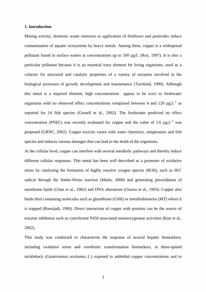
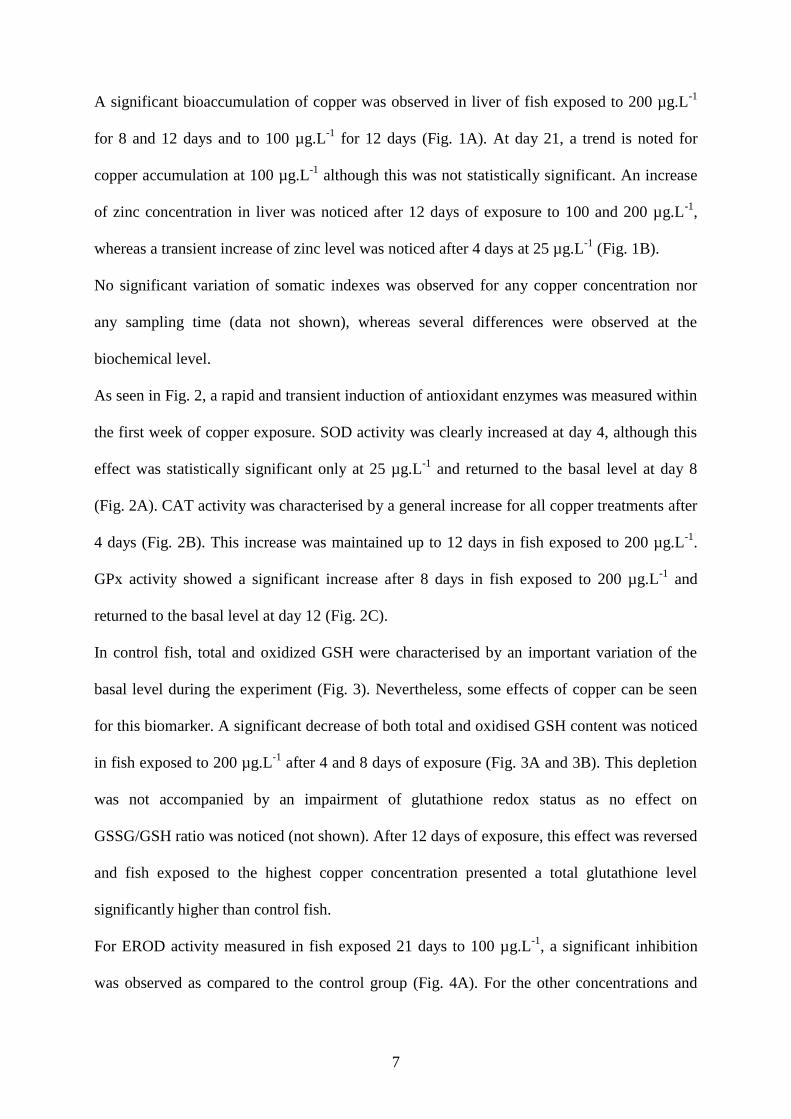
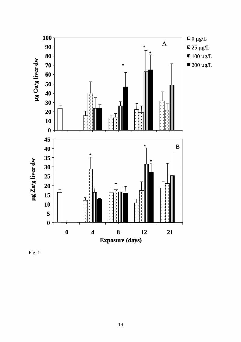
A significant bioaccumulation of copper was observed in liver of fish exposed to 200 µg.L-1
for 8 and 12 days and to 100 µg.L-1 for 12 days (Fig. 1A). At day 21, a trend is noted for
copper accumulation at 100 µg.L-1 although this was not statistically significant. An increase
of zinc concentration in liver was noticed after 12 days of exposure to 100 and 200 µg.L-1,
whereas a transient increase of zinc level was noticed after 4 days at 25 µg.L-1 (Fig. 1B).
No significant variation of somatic indexes was observed for any copper concentration nor
any sampling time (data not shown), whereas several differences were observed at the
biochemical level.
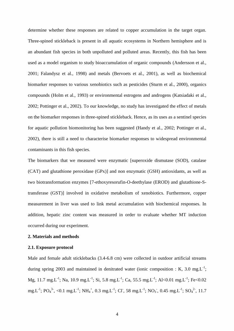
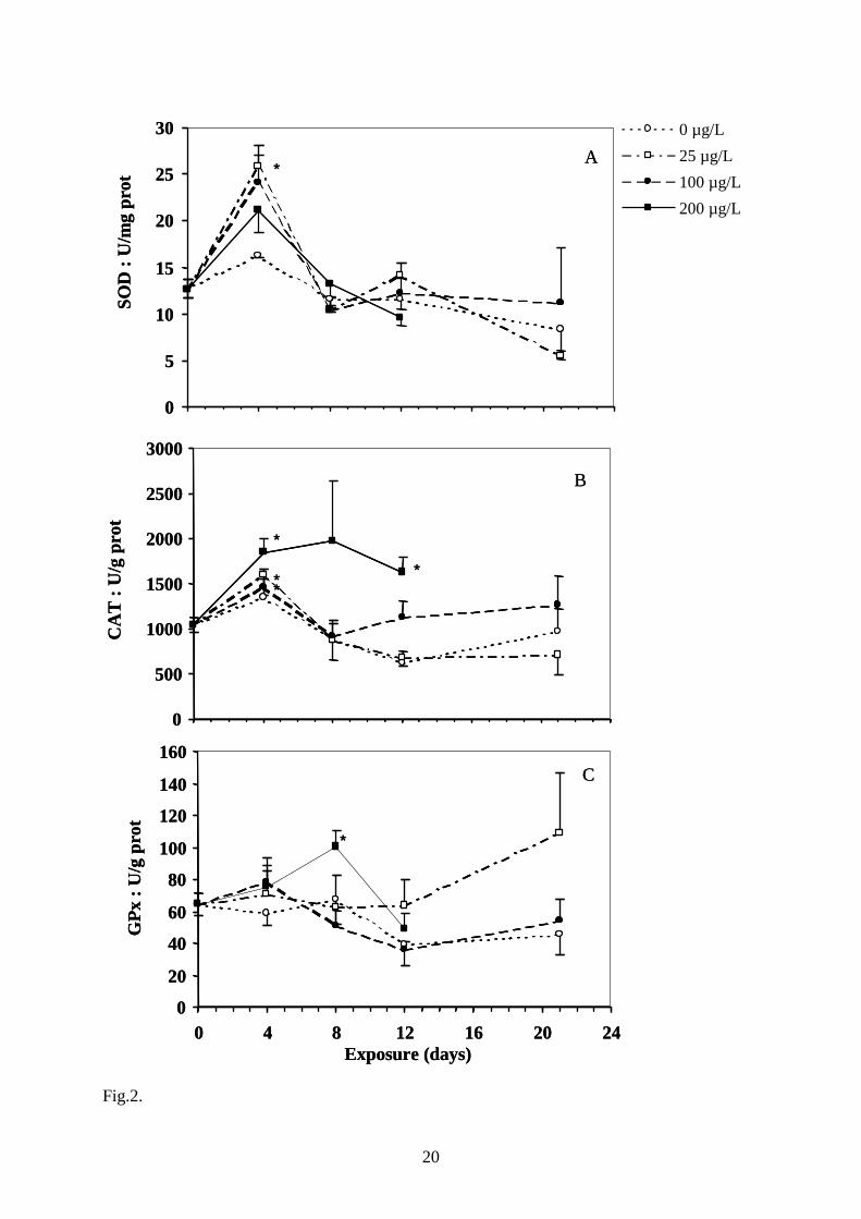
As seen in Fig. 2, a rapid and transient induction of antioxidant enzymes was measured within
the first week of copper exposure. SOD activity was clearly increased at day 4, although this
effect was statistically significant only at 25 µg.L-1 and returned to the basal level at day 8
(Fig. 2A). CAT activity was characterised by a general increase for all copper treatments after
4 days (Fig. 2B). This increase was maintained up to 12 days in fish exposed to 200 µg.L-1.
GPx activity showed a significant increase after 8 days in fish exposed to 200 µg.L-1 and
returned to the basal level at day 12 (Fig. 2C).
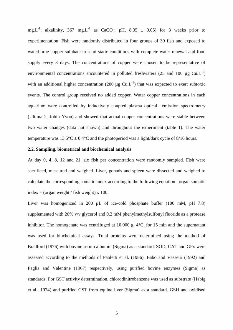
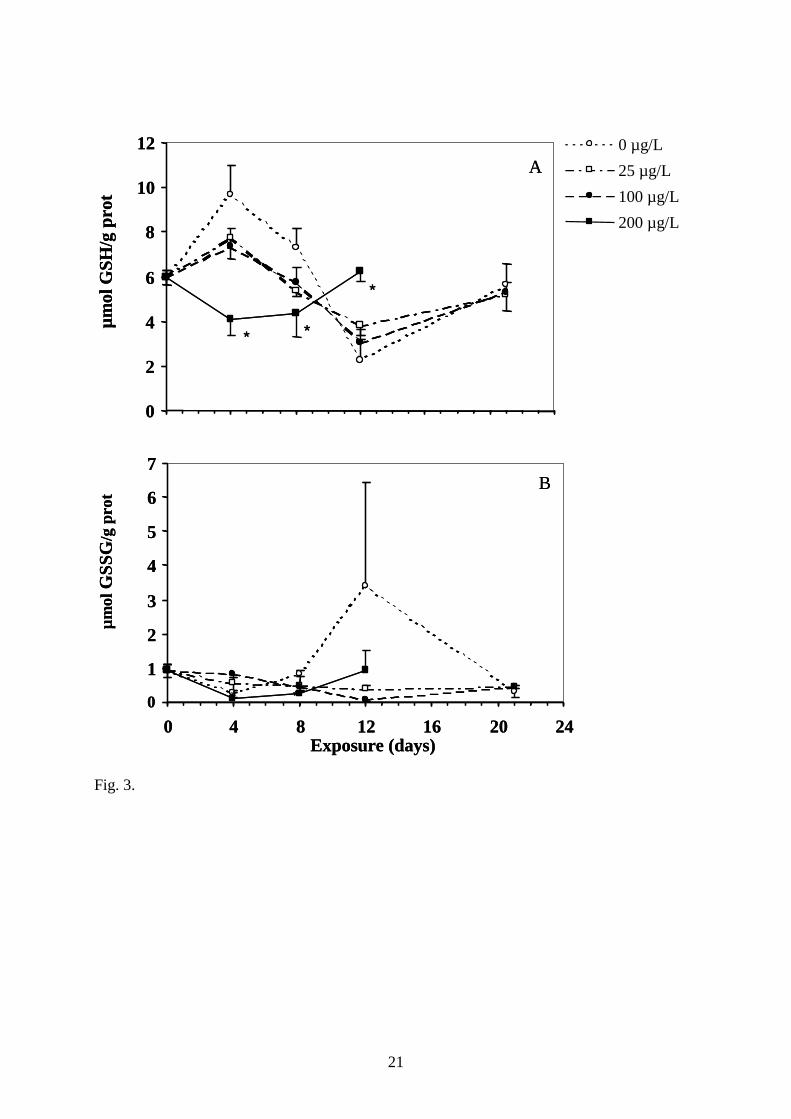
In control fish, total and oxidized GSH were characterised by an important variation of the
basal level during the experiment (Fig. 3). Nevertheless, some effects of copper can be seen
for this biomarker. A significant decrease of both total and oxidised GSH content was noticed
in fish exposed to 200 µg.L-1 after 4 and 8 days of exposure (Fig. 3A and 3B). This depletion
was not accompanied by an impairment of glutathione redox status as no effect on
GSSG/GSH ratio was noticed (not shown). After 12 days of exposure, this effect was reversed
and fish exposed to the highest copper concentration presented a total glutathione level
significantly higher than control fish.
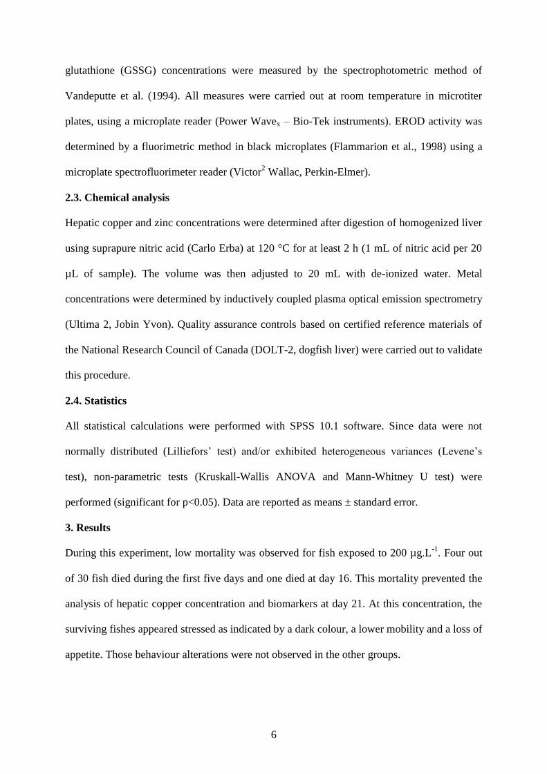
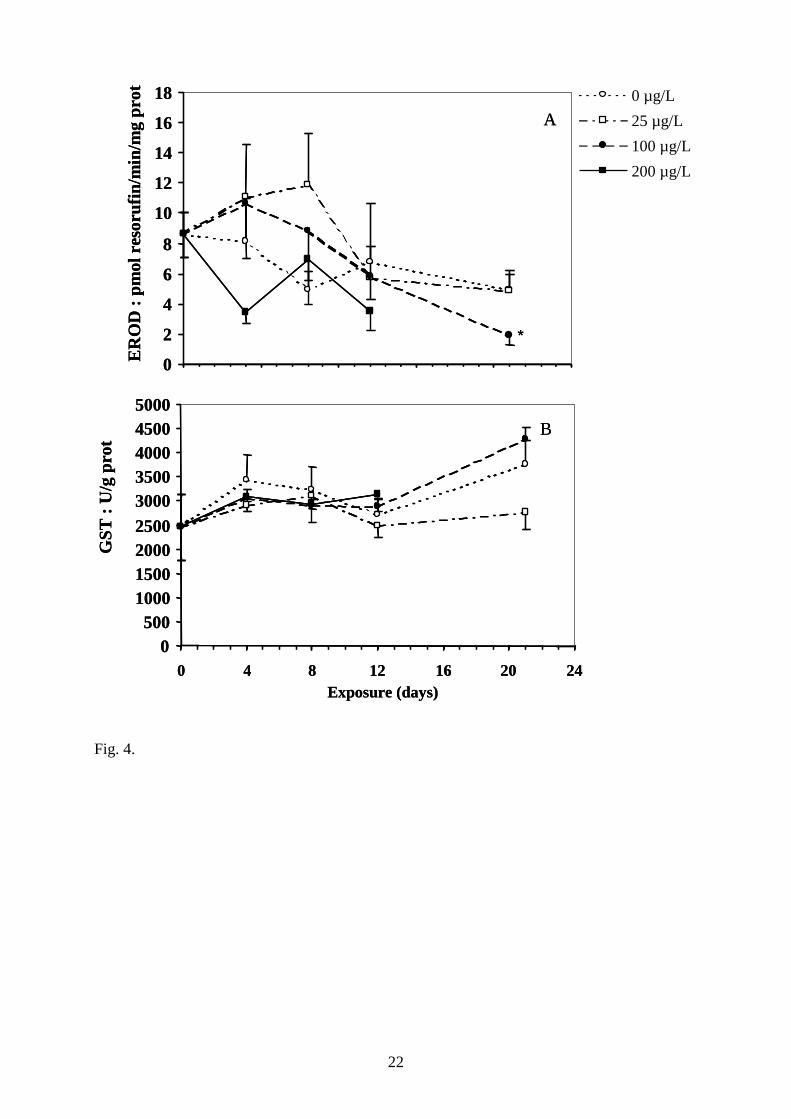
For EROD activity measured in fish exposed 21 days to 100 µg.L-1, a significant inhibition
was observed as compared to the control group (Fig. 4A). For the other concentrations and

8
sampling time, no significant variation of this enzyme was detected as compared to the
control group. The hepatic GST activity did not vary significantly with respect to either
copper concentration or time of exposure (Fig. 4B).
4. Discussion
Despite the low mortality observed after 4 days at the highest copper concentration, our
exposure conditions were clearly sublethal. For three-spined stickleback exposed to copper
sulphate, the 50 % lethal concentrations (LC50) were reported to be 2.78, 1.96 and 1.59 mg
Cu.L-1 after 24, 48 and 72 hours respectively (Svecevicius and Vosyliene, 1996), which are
much higher than the concentrations used in our study. However, comparison is rather
difficult since copper toxicity is highly dependent on several abiotic factors, such as pH, water
hardness and alkalinity that influence copper speciation. The physico-chemical forms in
which the metal is present in the aquatic environment (free ionic Cu2+, dissolved inorganic or
organic complexes, associated with particular matter) will indeed determine its bioavailability
for the organisms. In our study, the slightly basic conditions (pH 8.35) together with the
elevated water hardness (367 mg.L-1 as CaCO3) and very low dissolved organic carbon
(DOC) suggest that a major fraction of the copper in the water is present as inorganic
complexes (mainly CuCO3) and only a small fraction as free Cu2+. Nevertheless, our results
showed that these inorganic copper complexes are also bioavailable to the organisms since
significant accumulation and biochemical responses to copper were observed.
At day 12, copper did accumulate at similar levels in the liver of fish exposed either to 100 or
200 µg.L-1 (Fig. 1). Copper concentrations in liver measured during this laboratory exposure
are in accordance with the levels observed in three-spined stickleback sampled in the field
(Bervoets et al., 2001). This may indicate that saturation for copper accumulation occurred
right from 100 µg.L-1 and is in agreement with the fact that, in rainbow trout (Oncorhynchus
mykiss), the time to half saturation for copper in the liver is 6 days (McGeer et al., 2000). At

9
day 12, the bioconcentration factor (BCF = ([Cu]exposed - [Cu]control) / [Cu]water) reached 401
and 208 in the 100 and 200 µg.L-1 groups-exposed fish respectively, which are in the same
order of magnitude to those previously reported for zebrafish (Brachydanio rerio) exposed for
14 days to copper (Paris-Palacios et al., 2000). These relatively low BCF, as compared to
those reported for other metals such as Cd, may reflect the existence of homeostatic regulation
of copper levels in the liver (McGeer et al., 2000).
The biomarker responses in stickleback were in accordance with the known capacity of
copper to promote oxidative stress and to modulate enzymatic and non enzymatic antioxidant
expressions in fish tissues, as shown by a rapid and transient increase of SOD, CAT and GPx
enzymes and by a decrease of total GSH. In fish, antioxidant enzymes have been shown to be
either induced or inhibited by copper, depending on the dose, the species and/or the route of
exposure. Zebrafish submitted to 40 and 140 µg Cu.L-1 as CuSO4 presented a clear-cut
induction of CAT and GST within two weeks of waterborne copper exposure (Paris-Palacios
et al., 2000). Conversely, in carp, CAT and GST were inhibited after 96 hours of exposure to
100 and 250 µg Cu.L-1 as CuSO4 (Dautremepuits et al., 2002). Using intra-peritoneal injection
of copper, SOD inhibition was reported in carp after 48 hours (Varanka et al., 2001), while
this enzyme was rapidly induced in gilthead seabream (Sparus aurata; Pedrajas et al., 1995).
In stickleback, the rapid response of both enzymatic and non enzymatic antioxidants is in line
with the view that copper is able to act through different oxidative mechanisms, including
generation of ROS and direct interaction with intracellular thiols.
SOD are metalloenzymes that play a key role in the defence against ROS by transforming
superoxide anions into hydrogen peroxide (Yim et al., 1993). The total SOD activity that we
measured in the post-mitochondrial fraction corresponds mainly to the Cu,Zn-SOD activity,
the major isoform in the cytosol, nucleus and peroxisomes. Thus, the rapid and marked drop
in SOD activity at day 4 may result from direct binding of the metal to the enzyme, as
10
previously suggested in fish (Pedrajas et al., 1995). In addition, induction of CAT and GPx
indicated that copper induce (hydro)peroxides species in the liver. It is likely that the resulting
H2O2 produced from increased SOD activity was the source for subsequent increase of CAT
activity after the fourth day of copper exposure (Fig. 2B). Moreover, the known capacity of
copper to oxidize membrane lipids in fish (Pedrajas et al., 1995), together with the role of
GPx in the detoxification of lipoperoxidation products (Matés, 2000) may also account for the
observed increase of this enzyme at the day 8 (Fig. 2C). Unfortunately, the induction of lipid
damages by copper was not measured in this study to confirm this hypothesis.
Reduced glutathione is considered as a first line of cellular defence against metals by
chelating and detoxifying them, scavenging oxyradicals and participating in detoxification
reactions catalysed by glutathione peroxidases (Sies, 1999). The effects that we observed on
this parameter, i.e. inhibition of total GSH (both reduced and oxidised GSH) without any
impairment of GSSG/GSH ratio, is in line with the known ability of copper to interact with
GSH forming stable GS-Cu(I) binding complexes. Besides, as proposed by Canesi et al.
(1999), it is likely an inhibitory effect on GSH regenerating systems (e.g. GSH reductase or
GSH synthetase), which was not assessed in our study, that could have contributed to the
observed depletion of total GSH. The fact that we did not observe any impairment of
GSSG/GSH ratio by copper does not reach previous report in gilthead seabream fish exposed
by copper intra peritoneal injection (Rodriguez-Ariza et al., 1994). Finally, although the
mechanism by which copper inhibited GSH in fish is not fully understood and remains
complex, this rapid depletion may have contributed to the early toxicity observed at the higher
concentrations as suggested by Conners and Ringwood (2000).
Interestingly, biochemical responses were more rapid than copper accumulation, suggesting
the involvement of differential mechanisms in copper metabolism and accumulation. Cellular
copper metabolism involves copper intake by GSH and subsequent formation of GS-Cu(I)
11
complex, from which the metal is further transferred to metallothionein (MT) apoproteins
where it is stored (Freedman et al., 1989; Conners and Ringwood, 2000). The GS-Cu(I) pool
is the source for the synthesis of other metalloenzymes such as SOD or cytochrome oxidase,
as well as for ROS production through GS-Cu(I) oxidation (Freedman et al., 1989). In our
study, antioxidant responses occurred within the first week of exposure and then recovered
concomitantly with copper accumulation in the liver (Fig. 1 and 3). This indicates that at day
8 other detoxification systems, such as MTs, have been induced to replace GSH in the uptake
and sequestration of copper (Freedman et al., 1989). Moreover, the role of MTs in cellular
protection against oxidative stress (Viarengo et al., 2000) argues in the way that these proteins
could have been involved in response to copper in three-spined stickleback. This hypothesis is
strengthened by the finding that copper accumulation was accompanied by increased hepatic
zinc concentrations. In fact, a significant positive correlation was observed between these two
metals (r2 = 0.596, p<0.001, Spearman correlation). The sequestration of metals such as
cadmium and copper occurs by displacement of zinc from their binding sites, leading to a
release of free zinc that could induce de novo MT synthesis (Hollis et al., 2001; Roesijadi,
1996). Relationships between liver metal content and MT expression have been described in
aquatic organisms (Hollis et al., 2001) but further studies on MT response to copper in
stickleback will be necessary to confirm these hypotheses for this fish species.
EROD inhibition by metals has been reported in several in vitro and in vivo studies on fish
(Ghosh et al., 2001; Stien et al., 1997). In our study, we detected an inhibition of EROD
activity in fish exposed to 100 µg.L-1 for 21 days (Fig. 4). This effect, which involves direct
interaction of copper with the enzyme leading to a conformational change of the protein (Kim
et al., 2002), may reflect cellular toxicity induced by an increased content of metal in the cell
(Stien et al., 1997). Although this effect could be considered as minor regarding the responses
of the other biomarkers to copper, EROD inhibition should be taken into account when using
12
it as a biomarker of exposure in fish exposed to a mixture of chemicals, i.e. containing both
organic and metallic compounds, which mostly occurs in environmental pollution.
In summary, this dynamic study showed that waterborne copper induced several biochemical
responses in three-spined stickleback, in a transient manner, suggesting the occurrence of
adaptive processes. Rapidly after the beginning of the exposure, antioxidant biomarkers were
induced in order to compensate the oxidative stress generated by the metal. In a second phase,
copper accumulation was correlated to a recovery of GSH and a return of antioxidant
enzymes to the basal level, suggesting that other detoxification mechanisms, such as MTs,
have been involved to allow fish adaptation to copper. This study shows the suitability of
three-spined stickleback as a model fish species to study oxidative stress biomarker responses
to waterborne metal exposure. Field studies that evaluate several biomarkers, including
oxidative stress biomarkers, EROD and vitellogenin, are under progress to determine the
potential of stickleback as an indicator species of sublethal stress in multipollution contexts.
Acknowledgements
We wish to thank Jean-Pierre Blanquet for the measure of physico-chemical parameters of the
water and Dominique Hervin for technical assistance in fish maintenance. This work was
supported by the French Budget Civil de la Recherche et du Développement (BCRD 00-102).
References
Andersson, P.L., Berg, A.H., Bjerselius, R., Olsén, H., Orm, P.E., Tysklind, M., 2001.
Bioaccumulation of selected PCBs in zebrafish, three-spined stickleback and arctic
char after three different routes of exposure. Arch. Environ. Contam. Toxicol. 40, 519-
530.
Babo, S., Vasseur, P., 1992. In vitro effects of thiram on liver antioxidant enzyme activities of
rainbow trout (Oncorhynchus mykiss). Aquat. Toxicol. 22, 61-68.

13
Bervoets, L., Blust, R., Verheyen, R., 2001. Accumulation of metals in the tissues of three
spined stickelback (Gasterosteus aculeatus) from natural fresh waters. Ecotoxicol.
Environ. Saf. 48 117-127.
Bradford, M.M., 1976. A rapid sensitive method for the quantitation of microgram quantities
of protein utilizing the principle of protein-dye binding. Anal. Biochem. 72, 248-254.
Canesi, L., Viarengo, A., Leonzio, C., Filippelli, M., Gallo, G., 1999. Heavy metals and
glutathione metabolism in mussel tissues. Aquat. Toxicol. 46, 67-76.
Chan, P.C., Peller, O.G., Kesner, L., 1982. Copper (II)-catalyzed lipid peroxidation in
liposomes and erythrocyte membrane. Lipids 17, 331-337.
Conners, D.E., Ringwood, A.H., 2000. Effects of glutathione depletion on copper cytotoxicity
in oysters (Crassostrea virginica). Aquat. Toxicol. 50, 341-349.
Dautremepuits, C., Betoulle, S., Vernet, G., 2002. Antioxidant response modulated by copper
in healthy or parasitized carp (Cyprinus carpio L.) by Ptychobothrium sp. (Cestoda).
Biochim. Biophys .Acta 1573, 4-8.
Falandysz, J., Strandberg, L., Strandberg, B., Rappe, C., 1998. Polychlorinated naphthalenes
in three-spined stickleback Gasterosteus aculeatus from the Gulf of Gdansk.
Chemosphere 37, 2473-2487.
Flammarion, P., Migeon, B., Garric, J., 1998. Statistical analysis of cyprinid ethoxyresorufin-
O-deethylase data in a large french watershed. Ecotoxicol. Environ. Saf. 40, 144-153.
Freedman, J.H., Ciriolo, M.R., Peisach, J., 1989. The role of glutathione in copper metabolism
and toxicity. J. Biol. Chem. 264, 5598-5605.
Ghosh, M.C., Ghosh, R., Ray, A.K., 2001. Impact of copper on biomonitoring enzyme
ethoxyresorufin-o-deethylase in cultured catfish hepatocytes. Environ. Res. 86, 167-
173.
14
GRNC, 2002. Risque pour l'environnement - Evaluation des risques associés aux rejets
chimiques des installations nucléaires du Nord-Cotentin. Groupe de Radioécologie du
Nord Cotentin, pp. 210.
Grosell, M., Nielsen, C., Bianchini, A., 2002. Sodium turnover rate determines sensitivity to
acute copper and silver exposure in freshwater animals. Comp. Biochem. Physiol. C
133, 287-303.
Habig, W.H., Pabst, M.J., Jakoby, W.B., 1974. Glutathione S-Transferases. The first
enzymatic step in mercapturic acid formation. J. Biol. Chem. 249, 7130-7139.
Handy, R.D., Runnalls, T., Russel, P.M., 2002. Histopathologic biomarkers in three spined
sticklebacks, Gasterosteus aculeatus, from several rivers in Southern England that
meet the freshwater fisheries directive. Ecotoxicology 11, 467-479.
Hollis, L., Hogstrand, C., Wood, C.M., 2001. Tissue-specific cadmium accumulation,
metallothionein induction, and tissue zinc and copper levels during chronic sublethal
exposure in juvenile rainbow trout. Arch. Environ. Contam. Toxicol. 41, 468-474.
Holm, G., Norrgren, L., Andersson, T., Thuren, A., 1993. Effects of exposure to food
contaminated with PBDE, PCN or PCB on reproduction, liver morphology and
cytochrome P450 activity in the three-spined stickleback, Gasterosteus aculeatus.
Aquat. Toxicol. 27, 33-50.
Katsiadaki, I., Scott, A.P., Hurst, M.R., Matthiessen, P., Mayer, I., 2002. Detection of
environmental androgens: a novel method based on enzyme-linked immunosorbent
assay of spiggin, the stickleback (Gasterostus aculeatus) glue protein. Environ.
Toxicol. Chem. 21, 1946-1954.
Kim, J.S., Ahn, T., Yim, S.K., Yun, C.H., 2002. Differential effect of copper II on the
cytochrome P450 enzymes and NADPH-cytochrome P450 reductase : inhibition of
cytochrome P450-catalysed reactions by copper II ion. Biochemistry 41, 9438-9447.
15
Matés, J.M., 2000. Effects of antioxidant enzymes in the molecular control of reactive oxygen
species toxicology. Toxicology 153, 83-104.
McGeer, J.C., Szebedinszky, C., McDonald, D.G., Wood, C.M., 2000. Effects of chronic
sublethal exposure to waterborne Cu, Cd or Zn in rainbow trout 2: tissue specific
metal accumulation. Aquat. Toxicol. 50, 245-256.
Ozawa, T., Ueda, J., Shimazu, Y., 1993. DNA single strand breakage by copper (II)
complexes and hydrogen peroxide at physiological conditions. Biochem. Mol. Biol.
Int. 31, 455-461.
Paglia, D.E., Valentine, W.N., 1967. Studies on the quantitative and qualitative
characterization of erythrocyte glutathione peroxidase. J. Lab. Clinic. Med. 70, 158-
169.
Paoletti, F., Aldinucci, D., Mocali, A., Caparrini, A., 1986. A sensitive spectrophotometric
method for the determination of superoxide dismutase activity in tissue extracts. Anal.
Biochem. 154, 536-541.
Paris-Palacios, S., Biagianti-Risbourg, S., Vernet, G., 2000. Biochemical and (ultra)structural
hepatic perturbations of Brachydanio rerio (Teleostei, Cyprinidae) exposed to two
sublethal concentrations of copper sulfate. Aquat. Toxicol. 50, 109-124.
Pedrajas, J.R., Peinado, J., Lopez-Barea, J., 1995. Oxidative stress in fish exposed to model
xenobiotics. Oxidatively modified forms of Cu,Zn-superoxide dismutase as potential
biomarkers. Chem.-Biol. Interact. 98, 267-282.
Pottinger, T.G., Carrick, T.R., Yeomans, W.E., 2002. The three-spined stickleback as an
environmental sentinel: effects of stressors on whole-body physiological indices. J.
Fish Biol. 61, 207-229.
16
Rodriguez-Ariza, A., Toribio, F., Lopez-Barea, J., 1994. Rapid determination of glutathione
status in fish liver using high-performance liquid chromatography and electrochemical
detection. J. Chromatogr. B 656, 311-318.
Roesijadi, G., 1996. Metallothionein and its role in toxic metal regulation. Comp. Biochem.
Physiol. C 113, 117-123.
Roy, J., 1997. Environmental contaminants encyclopedia : copper entry. National Park
Service, Water Resources Divisions, pp. 99.
Sies, H., 1999. Glutathione and its role in cellular functions. Free Rad. Biol. Med. 27, 916-
921.
Stien, X., Risso, C., Gnassia-Barelli, M., Romeo, M., Lafaurie, M., 1997. Effect of copper
chloride in vitro and in vivo on the hepatic EROD activity in the fish Dicentrarchus
labrax. Environ. Toxicol. Chem. 16, 214-219.
Sturm, A., Wogram, J., Segner, H., Liess, M., 2000. Different sensitivity to organophosphates
of acetylcholinesterase and butyrylcholinesterase from three-spined stickleback
(Gasterosteus aculeatus): application in biomonitoring. Environ. Toxicol. Chem. 19,
1607-1615.
Svecevicius, G., Vosyliene, M.Z., 1996. Acute toxicity of copper to common freshwater
fishes of Lithuania. Ekologija 2, 17-21.
Turnlund, J.R., 1999. Copper. In: Shils, M.E., Olson, J.A., Shike, M., Ross, A.C. (Eds.),
Modern nutrition in health and disease. Lippincott Williams and Wilkins, Baltimore,
M.D.
Van der Oost, R., Beyerb, J., Vermeulen, N., 2003. Fish bioaccumulation and biomarkers in
environmental risk assessment: a review. Environ. Toxicol. Pharmacol. 13, 57-149.
Vandeputte, C., Guizon, I., Genestie-Denis, I., Vannier, B., Lorenzon, G., 1994. A microtiter
plate assay for total glutathione and glutathione disulfide contents in cultured/isolated
17
cells: performance study of a new miniaturized protocol. Cell Biol. Toxicol. 10, 415-
421.
Varanka, Z., Rojik, I., Varanka, I., Nemcsok, J., Abraham, M., 2001. Biochemical and
morphological changes in carp (Cyprinus carpio L.) liver following exposure to
copper sulfate and tannic acid. Comp. Biochem. Physiol. C 128, 467-478.
Viarengo, A., Burlando, B., Ceratto, N., Panfoli, I., 2000. Antioxidant role of
metallothioneins: A comparative overview. Cell. Mol. Biol. 46, 407-417.
Yim, M., Chock, P., Stadtman, E., 1993. Enzyme function of copper, zinc superoxide
dismutase as a free radical generator. J. Biol. Chem. 268, 4099-4105.
18
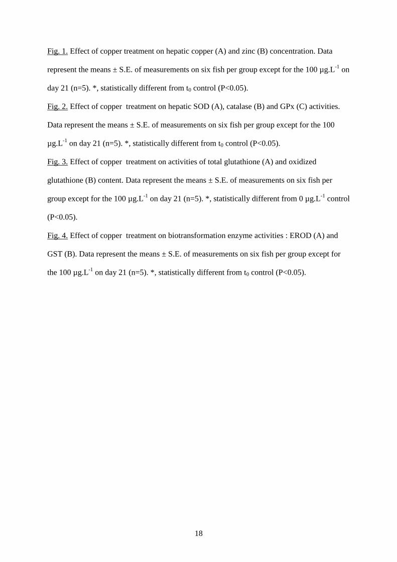
Fig. 1. Effect of copper treatment on hepatic copper (A) and zinc (B) concentration. Data
represent the means ± S.E. of measurements on six fish per group except for the 100 µg.L-1 on
day 21 (n=5). *, statistically different from t0 control (P<0.05).
Fig. 2. Effect of copper treatment on hepatic SOD (A), catalase (B) and GPx (C) activities.
Data represent the means ± S.E. of measurements on six fish per group except for the 100
µg.L-1 on day 21 (n=5). *, statistically different from t0 control (P<0.05).
Fig. 3. Effect of copper treatment on activities of total glutathione (A) and oxidized
glutathione (B) content. Data represent the means ± S.E. of measurements on six fish per
group except for the 100 µg.L-1 on day 21 (n=5). *, statistically different from 0 µg.L-1 control
(P<0.05).
Fig. 4. Effect of copper treatment on biotransformation enzyme activities : EROD (A) and
GST (B). Data represent the means ± S.E. of measurements on six fish per group except for
the 100 µg.L-1 on day 21 (n=5). *, statistically different from t0 control (P<0.05).
19
Fig. 1.
0
10
20
30
40
50
60
70
80
90
100µ
g C
u/g
liver
dw
0 µg/L
25 µg/L
100 µg/L
200 µg/L*
*
*A
0
5
10
15
20
25
30
35
40
45
0 4 8 12 21Exposure (days)
µg
Zn
/g li
ver
dw
*
*
* B
0
10
20
30
40
50
60
70
80
90
100µ
g C
u/g
liver
dw
0 µg/L
25 µg/L
100 µg/L
200 µg/L*
*
*A
0
5
10
15
20
25
30
35
40
45
0 4 8 12 21Exposure (days)
µg
Zn
/g li
ver
dw
*
*
* B
20
Fig.2.
0
5
10
15
20
25
30S
OD
: U
/mg
prot
0 µg/L
25 µg/L
100 µg/L
200 µg/L
*A
0
500
1000
1500
2000
2500
3000
CA
T :
U/g
pro
t
*
**
*
B
0
20
40
60
80
100
120
140
160
0 4 8 12 16 20 24Exposure (days)
GP
x: U
/g p
rot
*
C
0
5
10
15
20
25
30S
OD
: U
/mg
prot
0 µg/L
25 µg/L
100 µg/L
200 µg/L
*A
0
500
1000
1500
2000
2500
3000
CA
T :
U/g
pro
t
*
**
*
B
0
20
40
60
80
100
120
140
160
0 4 8 12 16 20 24Exposure (days)
GP
x: U
/g p
rot
*
C
21
Fig. 3.
0
2
4
6
8
10
12
µm
ol G
SH
/g p
rot
0 µg/L
25 µg/L
100 µg/L
200 µg/L
*
**
A
0
1
2
3
4
5
6
7
0 4 8 12 16 20 24Exposure (days)
µm
ol G
SS
G/g
prot
**
B
0
2
4
6
8
10
12
µm
ol G
SH
/g p
rot
0 µg/L
25 µg/L
100 µg/L
200 µg/L
*
**
A
0
1
2
3
4
5
6
7
0 4 8 12 16 20 24Exposure (days)
µm
ol G
SS
G/g
prot
**
B
22
Fig. 4.
0
2
4
6
8
10
12
14
16
18
ER
OD
:pm
ol r
esor
ufin
/min
/mg
prot 0 µg/L
25 µg/L
100 µg/L
200 µg/L
*
A
0500
100015002000250030003500400045005000
0 4 8 12 16 20 24Exposure (days)
GS
T :
U/g
pro
t
B
0
2
4
6
8
10
12
14
16
18
ER
OD
:pm
ol r
esor
ufin
/min
/mg
prot 0 µg/L
25 µg/L
100 µg/L
200 µg/L
*
A
0500
100015002000250030003500400045005000
0 4 8 12 16 20 24Exposure (days)
GS
T :
U/g
pro
t
B

23
Table 1 Actual copper concentrations measured in the water throughout the experimenta.
Nominal copper concentration (µg.L-1)
Measured copper concentration (µg.L-1)
0 <0.05
25 24.6 ± 1.5
100 91.6 ± 2.8
200 176.8 ± 2.3
a : measurements were performed in water sampled 2 hours after each water renewal. Data are
mean ± SE (n=7).