Effect of copper induced oxidative stress on sclerotial ...
10
ORIGINAL ARTICLE Effect of copper-induced oxidative stress on sclerotial differentiation, endogenous antioxidant contents, and antioxidative enzyme activities of Penicillium thomii PT95 Wenjing Zhao & Jianrong Han & Dandan Long Received: 30 July 2014 /Accepted: 16 September 2014 /Published online: 9 October 2014 # Springer-Verlag Berlin Heidelberg and the University of Milan 2014 Abstract Penicillium thomii PT95 strain is able to form abundant orange, sand-shaped sclerotia in which carotenoids accumulate. We have studied the effects of copper (Cu)-in- duced oxidative stress on sclerotial differentiation, biosynthe- sis of some endogenous antioxidants, and activities of a num- ber of the antioxidative enzymes of strain PT95. The associ- ation between sclerotial biomass, carotenoid, ascorbate and glutathione contents, and the activities of superoxide dismut- ase (SOD), catalase (CAT), ascorbate peroxidase (APX), and glutathione reductases (GR) were also analyzed in this fungal strain. We found that the oxidative stress induced by Cu was directly dependent on the concentrations of CuSO 4 in the media, with higher CuSO 4 concentrations resulting in higher oxidative stress. Cu-induced oxidative stress in strain PT95 was characterized by the initiation of lipid peroxidation. Under Cu-induced oxidative stress growth conditions, the initiation of exudates and sclerotia in strain PT95, as well as sclerotial maturation, were advanced by 1-2 days. Cu- induced oxidative stress favored sclerotial differentiation and the biosynthesis of endogenous antioxidants, i.e., carotenoids, ascorbate, and glutathione. Comparison of SOD, CAT, and GR activities at 0 and 100 μg/ml Cu revealed a 1.1-, 1.8-, and 1.2-fold increase, respectively, at the higher Cu concentration; comparison of their activities at 100 and 300 μg/ml Cu re- vealed a 1.4-, 3.1-, and 2.2-fold decrease, respectively, at the higher Cu concentration. APX activity decreased linearly with increasing CuSO 4 concentration. Our results suggest that the ability of the P. thomii PT95 strain to cope with metal stress is related to its ability to trigger an efficient defense against oxidative stress. These findings may contribute to a better understanding of the response mechanisms of sclerotia pro- duction in Penicillium strains to metal stress and to better insights into metal–fungi interactions in natural environments. Keywords Copper . Oxidative stress . Penicillium thomii . Endogenous antioxidants . Antioxidative enzymes Introduction The toxicity of heavy metals to microorganisms has attracted considerable research attention in recent years (Mukherjee et al. 2010). Heavy metals can be introduced into ecosystems through industrial effluents and wastes, agricultural fungi- cides, domestic garbage dumps, and mining activities (Merian 1991). Copper (Cu), a heavy metal, is an essential micronutrient that plays key roles in different physiological processes and metabolic pathways and is a structural and catalytic component of many proteins (Elleuch et al. 2013). As an essential trace element it acts as a cofactor of multiple enzymes, including superoxide dismutases (SOD; for protec- tion against free radicals), cytochrome c oxidase (mitochon- drial electron transport chain), tyrosinase (pigmentation), peptidylglycine α-amidating monooxygenase (neuropeptide and peptide hormone processing), and lysyl oxidase (collagen maturation) (Uauy et al. 1998; Peña et al. 1999). Fungi can accumulate, store, and tolerate high levels of heavy metals (Brady et al. 1994; Joho et al. 1995). To this end, fungi, similar to all living organisms, have evolved a set of mechanisms that control and respond to the uptake and accu- mulation of heavy metals. Biochemical responses include precipitation of heavy metals in cell walls or associated com- pounds (Rizzo et al. 1992; Suresh and Subramanyam 1996), production of organic acids, polysaccharides, melanins, and/ W. Zhao (*) Department of Biology, Taiyuan Normal University, Taiyuan 030031, People’ s Republic of China e-mail: [email protected] W. Zhao : J. Han : D. Long School of Life Science, Shanxi University, Taiyuan 030006, People’ s Republic of China Ann Microbiol (2015) 65:1505–1514 DOI 10.1007/s13213-014-0989-6
Transcript of Effect of copper induced oxidative stress on sclerotial ...

ORIGINAL ARTICLE
Effect of copper−induced oxidative stress on sclerotialdifferentiation, endogenous antioxidant contents,and antioxidative enzyme activities of Penicillium thomii PT95
Wenjing Zhao & Jianrong Han & Dandan Long
Received: 30 July 2014 /Accepted: 16 September 2014 /Published online: 9 October 2014# Springer-Verlag Berlin Heidelberg and the University of Milan 2014
Abstract Penicillium thomii PT95 strain is able to formabundant orange, sand-shaped sclerotia in which carotenoidsaccumulate. We have studied the effects of copper (Cu)-in-duced oxidative stress on sclerotial differentiation, biosynthe-sis of some endogenous antioxidants, and activities of a num-ber of the antioxidative enzymes of strain PT95. The associ-ation between sclerotial biomass, carotenoid, ascorbate andglutathione contents, and the activities of superoxide dismut-ase (SOD), catalase (CAT), ascorbate peroxidase (APX), andglutathione reductases (GR) were also analyzed in this fungalstrain. We found that the oxidative stress induced by Cu wasdirectly dependent on the concentrations of CuSO4 in themedia, with higher CuSO4 concentrations resulting in higheroxidative stress. Cu-induced oxidative stress in strain PT95was characterized by the initiation of lipid peroxidation.Under Cu-induced oxidative stress growth conditions, theinitiation of exudates and sclerotia in strain PT95, as well assclerotial maturation, were advanced by 1−2 days. Cu-induced oxidative stress favored sclerotial differentiation andthe biosynthesis of endogenous antioxidants, i.e., carotenoids,ascorbate, and glutathione. Comparison of SOD, CAT, andGR activities at 0 and 100 μg/ml Cu revealed a 1.1-, 1.8-, and1.2-fold increase, respectively, at the higher Cu concentration;comparison of their activities at 100 and 300 μg/ml Cu re-vealed a 1.4-, 3.1-, and 2.2-fold decrease, respectively, at thehigher Cu concentration. APX activity decreased linearly withincreasing CuSO4 concentration. Our results suggest that theability of the P. thomii PT95 strain to cope with metal stress is
related to its ability to trigger an efficient defense againstoxidative stress. These findings may contribute to a betterunderstanding of the response mechanisms of sclerotia pro-duction in Penicillium strains to metal stress and to betterinsights into metal–fungi interactions in natural environments.
Keywords Copper . Oxidative stress .Penicillium thomii .
Endogenous antioxidants . Antioxidative enzymes
Introduction
The toxicity of heavy metals to microorganisms has attractedconsiderable research attention in recent years (Mukherjeeet al. 2010). Heavy metals can be introduced into ecosystemsthrough industrial effluents and wastes, agricultural fungi-cides, domestic garbage dumps, and mining activities(Merian 1991). Copper (Cu), a heavy metal, is an essentialmicronutrient that plays key roles in different physiologicalprocesses and metabolic pathways and is a structural andcatalytic component of many proteins (Elleuch et al. 2013).As an essential trace element it acts as a cofactor of multipleenzymes, including superoxide dismutases (SOD; for protec-tion against free radicals), cytochrome c oxidase (mitochon-drial electron transport chain), tyrosinase (pigmentation),peptidylglycine α-amidating monooxygenase (neuropeptideand peptide hormone processing), and lysyl oxidase (collagenmaturation) (Uauy et al. 1998; Peña et al. 1999).
Fungi can accumulate, store, and tolerate high levels ofheavy metals (Brady et al. 1994; Joho et al. 1995). To this end,fungi, similar to all living organisms, have evolved a set ofmechanisms that control and respond to the uptake and accu-mulation of heavy metals. Biochemical responses includeprecipitation of heavy metals in cell walls or associated com-pounds (Rizzo et al. 1992; Suresh and Subramanyam 1996),production of organic acids, polysaccharides, melanins, and/
W. Zhao (*)Department of Biology, Taiyuan Normal University,Taiyuan 030031, People’s Republic of Chinae-mail: [email protected]
W. Zhao : J. Han :D. LongSchool of Life Science, Shanxi University, Taiyuan 030006, People’sRepublic of China
Ann Microbiol (2015) 65:1505–1514DOI 10.1007/s13213-014-0989-6

or proteins to bindmetal ions (Gadd 1993;Martino et al. 2002;Baldrian 2003), chelation in the cytosol and transport intovacuoles (Hayashi and Mutoh 1994a, b), chemical transfor-mation of metals (Gadd 1993), and/or transport of metalcations (Blaudez et al. 2000a). Another response processinvolves the synthesis of compounds containing thiol func-tional groups (Gadd 1993), which are known to have a highaffinity for metal ions (Romero–Isart and Vašák 2002).
The toxicity of metals can be due to the generation ofreactive oxygen species (ROS) that may cause wide-rangingdamage to proteins, nucleic acids, and lipids, eventually lead-ing to cell death (Bai et al. 2003). The toxic effects of ROSmay involve inhibition of growth (Guillén andMachuca 2008;Krumova et al. 2009), substitution of essential ions andblocking of functional groups on proteins (Borkow andGabbay 2005; Dupont et al. 2011), inactivation of enzymes(Gokhale and Cowan 2005;Wang et al. 2010), disturbances ofmetabolic pathways (Chillappagari et al. 2010), alterations ofmembrane integrity and production of ROS by membrane-bound Cu (Krumova et al. 2009, 2012; Sharma and Dietz2009; Dávila Costa et al. 2011). Consequently, the toleranceof fungi to different metals has been associated with its abilityto remove ROS (Fujs et al. 2005).
Fungi display several antioxidant enzymes against ROS,including SOD, catalases (CAT), ascorbate peroxidase (APX),and glutathione reductases (GR), all capable of removingoxygen radicals and their products and/or repairing oxidativedamage (Jamieson 1998; Bai et al. 2003). In addition, mole-cules such as glutathione and ascorbate not only play animportant role in cellular protection during oxidative stress,but they may form a complex with metals in cells (Pócsi et al.2004). Carotenoids are antioxidants and reduce oxidativestress by acting as scavengers of ROS (mainly singlet oxygen)(Stratton and Liebler 1997).β-Carotene and other carotenoidshave been associated with fungal photomorphogenesis anddevelopment (Mohr and Schopfer 1995; Georgiou andPetropoulou 2001a, b, c; Georgiou et al. 2001a, b;Zervoudakis et al. 2003; Han et al. 2005). In most organisms,ascorbic acid has important functions in biochemical reac-tions, such as in cell growth and differentiation. It has beenshown to play an important antioxidant role in sclerotialdifferentiation of phytopathogenic fungi (Georgiou andPetropoulou 2001a, b, c; Georgiou et al. 2003; Li et al.2006). Glutathione is considered to be a very important sig-naling molecule which acts as a link between environmentalstress and adaptive responses; it is regenerated from glutathi-one-S-transferases by GR activity (Navari–Izzo et al. 1997).
Penicillium thomii PT95 strain studied here was isolatedfrom a soil sample and is able to form abundant orange, sand-shaped sclerotia in which carotenoids accumulate (Han 1998).In earlier studies, we examined the effect of iron-inducedoxidative stress on sclerotial differentiation in strain PT95(Han et al. 2005; Li et al. 2006). The aim of the study reported
here was to investigate the effects of Cu-induced oxidativestress on sclerotial differentiation, sclerotia biomass, the levelof lipid peroxidation [based on malondialdehyde (MDA lev-el)], activities of enzymatic antioxidants (SOD, CAT, APX,GR), and the contents of endogenous antioxidants (such ascarotenoids, glutathione, ascorbate). The association betweensclerotial biomass, endogenous antioxidant contents, and an-tioxidative enzyme activities of this fungus was also analyzed.The results should provide data which will help explain therole and correlation between enzymatic and non-enzymaticantioxidants in the prevention or limitation of metal stressinjury.
Materials and methods
Preparation of inoculum
Strain PT95 was isolated from soil samples collected close toFenyang in Shanxi Province. It was identified as Penicilliumthomii (GenBank accession nr. KC966728) based on rDNA–ITS sequence analysis. Additional details on its isolation and acomplete bibliography are presented in Han et al. (1998). StrainPT95 was cultured on Czapek’s agar plates in an incubatormaintained in the dark condition and at 25 °C. The sclerotia tobe used as inoculum were obtained from 14-day-old Czapek’sagar plate cultures. To purify sclerotia, we first centrifuged theplate cultures and then rinsed the sclerotia aseptically five timeswith sterilized water to remove the spores.
Oxidative stress growth conditions
The different oxidative stress growth conditions to be assessedwere designed using potato dextrose agar (PDA) mediumsupplemented with different concentrations of CuSO4 (100,200, or 300 μg/ml, respectively). PDA medium withoutCuSO4 supplementation served as the control. Using threepoint inoculations (Pitt 2000), three grains of sclerotia of thePT95 strain were inoculated onto 25 ml of medium in a 9-cmPetri dish. The plates were incubated in the dark at 25 °C for20 days.
Sclerotial biomass, carotenoid extraction, and determination
The sclerotia which formed on the agar surface in the petri plateswere separated and washed thoroughly with distilled water anddried at 50 °C to a constant weight to determine sclerotialbiomass. The sclerotia were then observed with an anatomicallens (model SMZ–168; Motic China Group Co., China).
The extraction and determination of pigments wereperformed as described by Li et al. (2006) with minor modi-fications. Briefly, 1 g of dried sclerotia was manually groundwith a glass homogenizer and extracted three times with 10-ml
1506 Ann Microbiol (2015) 65:1505–1514

aliquots of acetone. The acetone extracts were combined in aseparatory funnel, and 10 ml of chloroform and a few millili-ters of a saturated NaCl solution were added to help break theemulsions. The chloroform extract was collected after remov-al of the acetone layer, which was then re-extracted.Absorbance of the chloroform extract was measured at475 nm. The content of carotenoids was calculated by usinga 1 % extinction coefficientof2,500 by the formula:
Content of carotenoids μg=g dry sclerotiað Þ
¼ ml of chloroform � A475ð Þ=sclerotia dry weight � 2500�
Ascorbate assay
The ascorbate assay was performed according to Georgiouand Petropoulou (2001a). Ascorbate concentration wasexpressed in units of μg ascorbate/g sclerotia dry weight(DW).
Glutathione assay
The sclerotia (0.5 g DW) were ground in 2 ml of ice-cold 5 %(w/v) sulfosalicylic acid. The extract was then centrifuged at4 °C for 20 min at 12,000g in a cooled centrifuge and thesupernatant used for the assay of glutathione. Glutathione andits oxidized product glutathione disulfide (GSSG) were mea-sured together spectrophotometrically according to Griffith(1980) with some modifications. In brief, total glutathionewas measured in a 5-ml reaction mixture containing 4.29 mlphosphate buffer (47.5 mM Na2HPO4·12H2O, 2.5 mMKH2PO4·5H2O, 2.5 mM EDTA–Na2), 0.5 ml of 1.25 mM5,5'-dithio-bis(2-nitrobenzoic acid) (DTNB), 60 μl 15 mMNADPH, and 100 μl acid extract. The acid extract was com-posed immediately prior to starting the reaction by the addi-tion of 50 μl of 5 U of GR activity (bakers’ yeast, type III;Sigma Chemical Co., St. Louis, MO). Change in the absor-bance of the reaction mixture was measured at 412 nm.
The reduced form of glutathione (GSH) was measured bysimilar method, but with ddH2O instead of the coenzyme IIreduction system. The amounts of oxidized glutathione(GSSG) were calculated by subtracting the GSH content fromtotal glutathione content (GSH+GSSG). All values are report-ed as glutathione equivalents.
Evaluation of lipid peroxidation
Lipid peroxidation in the sclerotia of strain PT95 was evalu-ated by determining the levels of the peroxidation products.Thiobarbituric acid reactive substances (TBARS), such asaldehydes, malondialdehyde (MDA), and endoperoxideswere determined according to the methods of Hodges et al.
(1999). MDA, which isroutinely used as an indicator of lipidperoxidation, was extracted with 5 % (w/v) trichloric acid.The reaction with TBA was conducted at 95 °C for 30 min.After the samples were chilled, absorbance was measured at532 and 432 nm and the results expressed as μmol MDA/gDW sclerotia.
Measurement of antioxidant enzyme activity
Sclerotia (1 g DW) were homogenized in 50 mM ice-coldphosphate buffer, pH 7.8, containing 0.2 mM EDTA−Na2 and4 % insoluble polyvinylpyrrolidone (1 ml buffer/100 mgDW). The extract was then centrifuged at 4 °C for 20 min at12,000g in a cooled centrifuge. The supernatant was used forthe assays of SOD (EC 1.15.1.1), CAT (EC 1.11.1.6), APX(EC 1.11.1.11), and GR (EC 1.6.4.2).
SOD activity was measured according to Giannopolitis andRies (1977). The reaction mixture contained 1.3 mM ribofla-vin, 13 mMmethionine, 63 mM nitroblue tetrozolium (NBT),0.1 mM EDTA in 50 mM phosphate buffer (pH 7.8), and50 μl of the enzyme extract in a final volume of 10 ml. SODactivity was assayed by measuring the ability of the enzymeextract to inhibit the photochemical reduction of NBT. Glasstest tubes containing the mixture were immersed in a bath at25 °C and illuminated with a fluorescent lamp (4,000 lx).Identical tubes which were not illuminated served as blanks.After illumination for 20 min, absorbance was measured at560 nm. One unit of SOD was defined as the enzyme activitywhich inhibited the photoreduction of NBT to blue formazanby 50 %, and SOD activity of the extracts was expressed asSOD units/g sclerotia.
CAT activity was measured according to Li et al. (2005)u s i n g a b s o r b a n c e o f a c om p l e x c om p o u n d(peroxomolybdates) at 405 nm due to H2O2 decompositionbased on results in a previous experiment. One unit of CATactivity was defined as 1 μmol decomposed H2O2 per onemilligram sclerotia per second.
Total GR activity was assayed by the DTNB method, withminor modifications (Sun et al. 2007). The reaction mixture(300 μl) consisted of 100 mM phosphate buffer (pH7.8),0.1 mM NADPH, 1.0 mM GSSG, and 20 μl enzyme extract.The reaction was started by the addition of GSSG, and gluta-thione reduction rate was monitored at 420 nm for 5 min. Oneunit of enzyme activity was defined as the amount of enzymerequired to produce 1 μmol GSH per minute .
The assay for APX was based on monitoring the rate ofoxidation of ascorbate according to the method of Nakano et al.(1981). Absorbance readings were taken at 290 nm using thespectrophotometer blanked against an aliquot of buffer(50 mM phosphate buffer, pH7.0, 0.3 mM ascorbate). A3.85-ml aliquot of this buffer was placed in a glasscuvette, 50 μl extract was added, followed by 1 ml of0.015 % chilled H2O2. The cuvette was inverted three
Ann Microbiol (2015) 65:1505–1514 1507

times as quickly as possible and placed in the spectro-photometer. The reaction was monitored observing thedecrease in extinction at 290 nm for 1 min.
Statistics
All experiments were replicated in three plates, and the dataare presented as the arithmetic mean±standard error (SE).Duncan’s multiple range test (Ray 1985) was used on theisolation means to test for significant differences at the 5 %level of confidence.
Results
Effect of Cu-induced oxidative stress on sclerotialdifferentiation, sclerotial biomass, and carotenoid contentsin sclerotia of strain PT95
Observation of the macroscopic colony characters ofstrain PT95 on each plate culture suggested that the
colonies of strain PT95 growing under different oxida-tive stress conditions differed from each other (Fig. 1).Table 1 shows that with increased oxidative stress in-creased, the time of exudate initiation, sclerotial initia-tion, and sclerotial maturation were advanced by 1−2 days.
There were apparent differences in sclerotial bio-mass and carotenoid contents in sclerotia of strainPT95 under different oxidative growth conditions(Table 2), with higher sclerotial biomass and caroten-oid contents in sclerotia positively correlated with thehigher CuSO4 concentration. When the fungus wasgrown under the high oxidative stress condition in-duced by 300 μg/ml CuSO4, its sclerotial biomass(i.e., dry sclerotia weight) increased by 1.56-fold withrespect to the control. Carotenoid contents in sclerotiaunder the high oxidative stress condition increased by1.19-fold with respect to the control. Statistical analy-sis revealed that sclerotial biomass values were posi-tively correlated (p<0.05) with the carotenoid contentvalues in sclerotia (R=0.9581).
Fig. 1 Colonies of Penicilliumthomii PT95 strain growing for 14days on (a) potato dextrose agar(PDA) without added CuSO4, (b)PDA+100 μg/ml CuSO4, (c)PDA+200 μg/ml CuSO4, (d)PDA+ 300 μg/ml CuSO4, at25 °C. Petri dish size:9 cmdiameter
1508 Ann Microbiol (2015) 65:1505–1514
One conclusion that can be drawn from these results is thatcopper-induced oxidative stress favored sclerotial differentia-tion and endogenous carotenogenesis of the PT95 strain.
Effect of CuSO4 concentrations in media on lipid peroxidationin sclerotia of strain PT95
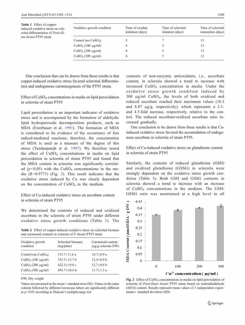
Lipid peroxidation is an important indicator of oxidativestress and is accompanied by the formation of aldehydiclipid hydroperoxide decomposition products, such asMDA (Esterbauer et al. 1991). The formation of MDAis considered to be evidence of the occurrence of freeradical-mediated reactions; therefore, the concentrationof MDA is used as a measure of the degree of thisstress (Yazdanpanah et al. 1997). We therefore testedthe effect of CuSO4 concentrations in media on lipidperoxidation in sclerotia of strain PT95 and found thatthe MDA content in sclerotia was significantly correlat-ed (p<0.05) with the CuSO4 concentrations in the me-dia (R=0.9771) (Fig. 2). This result indicates that theoxidative stress induced by Cu was clearly dependenton the concentration of CuSO4 in the medium.
Effect of Cu-induced oxidative stress on ascorbate contentin sclerotia of strain PT95
We determined the contents of reduced and oxidizedascorbate in the sclerotia of strain PT95 under differentoxidative stress growth conditions (Table 3). The
contents of non-enzymic antioxidants, i.e., ascorbatecontent, in sclerotia showed a trend to increase withincreased CuSO4 concentration in media. Under theoxidat ive s t ress growth condi t ion induced by300 μg/ml CuSO4, the levels of both oxidized andreduced ascorbate reached their maximum values (10.3and 8.87 μg/g, respectively), which represents a 3.1-and 4.5-fold increase, respectively, relative to the con-trol. The reduced ascorbate/oxidized ascorbate ratio in-creased gradually.
One conclusion to be drawn from these results is that Cu-induced oxidative stress favored the accumulation of endoge-nous ascorbate in sclerotia of strain PT95.
Effect of Cu-induced oxidative stress on glutathione contentin sclerotia of strain PT95
Similarly, the contents of reduced glutathione (GSH)and oxidized glutathione (GSSG) in sclerotia werestrongly dependent on the oxidative stress growth con-dition (Table 3). Both GSH and GSSG contents insclerotia showed a trend to increase with an increaseof CuSO4 concentrations in the medium. The GSH/GSSG ratio was maintained at a high level in all
Table 1 Effect of copper-induced oxidative stress on scle-rotial differentiation of Penicilli-um thomii PT95 strain
Oxidative growth condition Time of exudateinitiation (days)
Time of sclerotialinitiation (days)
Time of sclerotialmaturation (days)
Control (no CuSO4) 5 7 13
CuSO4 (100 μg/ml) 4 5 12
CuSO4 (200 μg/ml) 4 5 12
CuSO4 (300 μg/ml) 4 5 12
Table 2 Effect of copper-induced oxidative stress on sclerotial biomassand carotenoid contents in sclerotia of P. thomii PT95 strain
Oxidative growthcondition
Sclerotial biomass(mg/plate)
Carotenoid content(μg/g sclerotia DW)
Control (no CuSO4) 315.7±11.6 a 10.7±0.9 a
CuSO4 (100 μg/ml) 352.3±12.7 b 12.4±0.9 b
CuSO4 (200 μg/ml) 422.3±19.8 c 12.7±0.8 b
CuSO4 (300 μg/ml) 493.7±10.4 d 11.7±1.3 a
DW, Dry weight
Values are presented as themean ± standard error (SE). Values in the samecolumn followed by different lowercase letters are significantly differentat p<0.05 according to Duncan’s multiplerange test
Fig. 2 Effect of CuSO4 concentrations in media on lipid peroxidation ofsclerotia of Penicillium thomii PT95 strain based on malondialdehyde(MDA) content. Results represent mean values of 3 independent experi-ments± standard deviation (SD)
Ann Microbiol (2015) 65:1505–1514 1509

treatments and showed a 1.08-fold increase relative tothe control, possible due to nonprotein thiol (NP−SH)synthesis. Under the high oxidative stress condition(200–300 μg/ml CuSO4), the GSH/GSSG ratio graduallyincreased, reaching a maximum at 300 μg/ml (1.46-foldincrease).
Effect of Cu-induced oxidative stress on SOD activityin sclerotia of strain PT95
Data on SOD activity of sclerotia of strain PT95 is shown inFig. 3. Based on our results, increasing the CuSO4 concentra-tion in the medium from 0 to 100 μg/ml correlated with asignificant increase in SOD activity, while increasing theCuSO4 concentration in the medium from 100 to 300 μg/mldid not cause a significant increase SOD activity. This obser-vation suggests that SOD activity in sclerotia subjected to theoxidative stress condition induced by CuSO4 at a concentra-tion of 100 μg/ml had reached its maximum value of 135.93
U/g DW. The values of SOD activities under different oxida-tive stress conditions showed a moderately negative correla-tion with the contents of reduced and oxidized ascorbate,respectively (R red= −0.45, R oxi= −0.4783), with the caroten-oid contents in sclerotia (R= −0.5411), and with the contentsof GSH and GSSG, respectively (R GSH=−0.172, R GSSG=−0.661). These results suggest that under higher oxidativestress, strain PT95 may fail to significantly enhance SODactivity in sclerotia to scavenge ROS generated by the oxida-tive stress. However, the strain may further differentiate toform more sclerotia for long-term survival or produce moreendogenous antioxidants to counter ROS formation.
Effect of Cu-induced oxidative stress on CAT activityin sclerotia of strain PT95
Information on the CAT activity of sclerotia is presented inFig. 4. CAT activity significantly increased when the CuSO4
concentration in the medium increased from 0 to 100 μg/ml,but it significantly decreased with an increase of CuSO4
concentration in the medium from 100 to 300 μg/ml. Under
Table 3 Oxidized glutathione (GSSG) and reduced glutathione (GSH) content, GSH/GSSG ratio, reduced ascorbate and oxidized ascorbate content,and reduced ascorbate/oxidized ascorbate ratio in sclerotia of P. thomii PT95 strain
Oxidative growthcondition
GSH content(μmol/g DW)
GSSG content(μmol/g DW)
GSH/GSSG
Reduced ascorbatecontent (μg/g DW)
Oxidized ascorbatecontent (μg/g DW)
Reduced ascorbate/oxidized ascorbate
Control (noCuSO4)
1.51±0.02 a 1.10±0.06 a 1.36 1.97±0.31 a 3.3±0.3 a 0.59
CuSO4
(100 μg/ml)1.75±0.01 b 1.21±0.03 b 1.45 3.53±0.5 b 4.87±0.25 b 0.72
CuSO4
(200 μg/ml)1.78±0.04 bc 1.22±0.01 b 1.46 7.47±0.5 c 9.03±0.45 c 0.83
CuSO4
(300 μg/ml)1.81±0.03 c 1.24±0.02 b 1.46 8.87±0.42 d 10.3±0.26 d 0.87
Values are presented as the mean ± standard error (SE). Values in the same column followed by different lowercase letters are significantly different atp<0.05 according to Duncan’s multiplerange test
Fig. 3 Effect of Cu-induced oxidative stress on sclerotia’s superoxidedismutase (SOD) activities of P. thomii PT95 strain. Results representmean values of 3 independent experiments± SD
Fig. 4 Effect of Cu-induced oxidative stress on catalase (CAT) activity ofthe sclerotia of P. thomii PT95 strain. Results represent mean values of 3independent experiments± SD
1510 Ann Microbiol (2015) 65:1505–1514

the oxidative stress condition induced by 100 μg/ml CuSO4,CAT activity of sclerotia reached its maximum value of 20.02U/g DW/min. The values of CAT activity had a moderatelypositive correlation with those of SOD activity (R=0.8615). Incontrast, they had a moderately negative correlation with thecontents of reduced and oxidized ascorbate (R red= −0.6290, Roxi= −0.6435), respectively, with carotenoid contents in scle-rotia (R= −0.4990), and with GSH content (R GSH=−0.392),and they had a significantly negative correlation (p<0.05)with GSSG content (R GSSG=−0.950). These results suggestthat under the relatively lower oxidative stress, strain PT95may significantly enhance CAT activity in sclerotia to scav-enge ROS generated by oxidative stress.
Effect of Cu-induced oxidative stress on GR activityin sclerotia of strain PT95
As shown in Fig. 5, GR activity increased by almost 1.19-foldwith the addition of 100 μg/ml CuSO4 to the medium relativeto the control, indicating higher enzyme activity either inresponse to the superoxide anions formed due to Cu toxicityor for the increased synthesis of glutathione to trap intracellu-lar Cu. A gradual decrease in enzyme activity was observedwith an increase in CuSO4 concentration, with GR activitydecreasing by almost 1.88-fold at 300 μg/ml CuSO4 relativeto the control. This decrease in enzyme activity is indicative ofthe severity of the oxidative stress imposed by the higherCuSO4 concentration on the test strain. The values of GRactivity showed a moderate positive correlation with those ofthe SOD and CAT activities (R SOD=0.442, R CAT=0.419,respectively). In contrast, the values of GR activity had amoderately negative correlation with the contents of reducedand oxidized ascorbate (R red=−0.924, R oxi=−0.920), withcarotenoid contents in sclerotia (R= −0.701), and with GSHand GSSG contents (R GSH=−0.845, R GSSG=−0.872),respectively.
Effect of Cu-induced oxidative stress on APX activityin sclerotia of strain PT95
Peroxidases [APX and glutathione peroxidase (GPX)] aredistributed throughout the cell and catalyze the reduction ofhydrogen peroxide (H2O2) to H2O. APX uses ascorbate aselectron donor in the first step of the ascorbate–glutathionecycle and is considered to be the most important plant perox-idase involved in H2O2 detoxification (Noctor and Foyer1998). As shown in Fig. 6, under Cu-induced oxidative stress,APX activity decreased linearly with increasing CuSO4 con-centration (R= −0.988) (p<0.05). The highest concentrationof Cu (300 μg/ml) proved to be extremely toxic, resulting in adecline in APX activity. Compared with the control, APXactivity decreased by 3.3-fold. The values of APX activityhad a moderately positive correlation with the values of SOD,CAT, and GR activities (R SOD=0.475, R CAT=0.786, R GR=0.688). The values of GR activity had a moderately negativecorrelation with the contents of reduced and oxidized ascor-bate (R red=−0.924, R oxi=−0.920), significantly (p<0.01)negative correlation with carotenoid contents in sclerotia (R= −0.994), and a moderately negative correlation with GSHand GSSG contents (R GSH=−0.881, R GSSG=−0.350).
Discussion
Georgiou (1997) advanced a theory which proposed thatsclerotial differentiation in fungi was triggered by oxidativestress. Our previous experiments (Han et al. 2005; Li et al.2006) showed that the sclerotial biomass produced by strainPT95 grown under a high oxidative stress condition was 1.23-fold greater than that produced when grown under a lowoxidative stress condition. The data of this study provideadditional evidence in support of Georgiou’s theory and arealso in accordance with the general theory of microbial
Fig. 5 Effect of Cu-induced oxidative stress on sclerotia’s glutathionereductase (GR) activities of P. thomii PT95 strain. Results represent meanvalues of 3 independent experiments± SD
Fig. 6 Effect of Cu-induced oxidative stress on sclerotia’s ascorbateperoxidase (APX) activity of P. thomii PT95 strain. Results representmean values of 3 independent experiments± SD
Ann Microbiol (2015) 65:1505–1514 1511

differentiation, which also postulates that the latter is inducedby oxidative stress (Burton and Ingold 1984).
Metal toxicity is often driven by the generation of ROSeither directly via the catalytic production of superoxide (O2
-)by the Haber–Weiss and Fenton reactions or indirectly by othermechanisms (Yamamoto et al. 2002; Boscolo et al. 2003) andparticipates in the initiation and propagation of lipid peroxida-tion. These last findings provide indirect evidence that exces-sive ROS were produced after the Cu treatment, resulting inmembrane lipid deterioration measured as MDA. Our resultsindicate that excess Cu increases oxidative stress, as was evi-dent from the increased lipid peroxidation, which is in accor-dance with previous findings showing that MDA accumulatedgreatly after Cu exposure and that the cell membrane was theprimary site affected by Cu toxicity (Thounaojam et al. 2012),possibly due to the overproduction of ROS under Cu stresswhich is highly destructive to the cell membrane. This toxiceffect resulting from the cellular oxidative stress may be allayedby several antioxidant systems. The increased activity of anti-oxidative enzymes in the sclerotia indicates the formation ofROS. In other words, the sclerotia analyzed in this studycontained sufficiently high Cu concentrations to activate ROSproduction and subsequently oxidative stress.
The protection provided by the antioxidant system againstthese ROS is complex and highly organized. In this system,the SOD constitutes the primary line of defense as itdismutates superoxide radicals to H2O2 (Fatima and Ahmad2005). H2O2 degradation to water and oxygen is carried out bythe CAT localized in the peroxisomes and, as a constituent ofascorbate–glutathione cycle, by the APX, GR, ascorbate andglutathione (Gratão et al. 2005).
However, we noted that the activities of the various anti-oxidant enzymes were affected differently by high Cu con-centrations (=high oxidative stress). SOD is the first enzymeof the detoxifying process. A disproportionate amount of thesuperoxide anion (O2
-) was catalyzed by SOD, which cata-lyzes the formation of H2O2 (Asada 2006). Under the lowoxidative stress condition (100 μg/ml), SOD activity wassignificantly increased compared to control; in contrast, underthe higher oxidation stress condition (100–300 μg/ml Cu),SOD activity decreased, possibly due to its sensitivity to highH2O2 content in the cell. This decreased SOD activity may berelated to lower levels of O2·because of the Haber–Weissreaction. Roughly paralleling the changes in SOD, CAT, andGR activities increased with increasing Cu concentrationsfrom 0 to 100 μg/ml, but decreased with an increase in Cuconcentrations from to 300 μg/ml. According to Willekenset al. (1997), it is likely that an excess production of ROScaused by heavy metals can inactivate CAT, probably byinactivating the enzyme-bound heme group. CAT is onlypresent in peroxisomes, but it is indispensable for ROS detox-ification during stress when high levels of ROS are produced.APX has a higher affinity for H2O2 than CAT and can
therefore scavenge small amounts of H2O2 in more specificlocations (Asada 1992; Radic et al. 2010). In our experiments,Cu treatments induced APX activities in the sclerotia of strainPT95 strain, suggesting that APX activity decreased linearlywith increasing CuSO4 concentration. The highest concentra-tion of Cu (300 μg/ml) proved to be extremely toxic, resultingin a decline of APX activity. The hyperactivity of peroxidaseunder Cu stress indicates its role in the constant detoxificationof H2O2. Glutathione is considered to be a very importantsignal molecule which acts as a link between environmentalstress and adaptive responses, and it is regenerated from GSTby GR activity (Navari–Izzo et al. 1997).
Carotenoids can be antioxidant since they are knownto inhibit oxidative stress by acting as quenchers ofsinglet oxygen and scavengers of hydroxyl, alkoxyl,and alkoperoxyl radicals (Burton and Ingold 1984;Simic 1992; Stratton and Liebler 1997; Georgiou andPetropoulou 2001a). Ascorbate can directly scavengeROS (including hydroxyl and superoxide radicals andH2O2) either nonenzymatically or enzymatically(McKersie and Leshem 1994). In the latter case, it isused as a substrate for the H2O2-splitting enzyme APX(Nakano and Asada 1981). It can also indirectly act asan antioxidant by regenerating the membrane-bound α-tocopherol which is involved in the scavenging ofperoxyl radicals and singlet oxygen (Schraudner et al.1997). In our experiment, carotenoids, ascorbate, andglutathione were accumulated in the sclerotia of strainPT95. However, the effect of Cu-induced oxidativestress on carotenoid content in the sclerotia was differ-ent from that on ascorbate content and glutathione con-tent. We found that the carotenoid content in sclerotiahad a weak, positive correlation with the CuSO4 con-centration in the medium (R=0.480) and that the totalascorbate content in sclerotia had a significantly positivecorrelation (p<0.05) with the CuSO4 concentrations inthe medium (R=0.915). The GSH content in sclerotiahad a significantly positive correlation (p<0.05) withthe CuSO4 concentrations in the medium (RGSH=0.924). The highest carotenoid content was obtainedfrom PDA plates supplemented with 200 μg/mlCuSO4. The total ascorbate content was the sum ofthe reduced and oxidized ascorbate content, and thehighest total ascorbate content was obtained from PDA platessupplemented with 300 μg/ml CuSO4 (19.17 μg/g dry scle-rotia). The highest GSH and GSSG contents were obtainedfrom PDA plates supplemented with 300 μg/ml CuSO4
(Table 3). These results indicate that the oxidative stressinduced by a lower amount of CuSO4 (about 200 μg/ml)favored endogenous carotenogenesis by strain PT95, whereasthe oxidative stress induced by a higher amount of CuSO4
(about 300 μg/ml) favored the accumulation of ascorbate andglutathione in sclerotia.
1512 Ann Microbiol (2015) 65:1505–1514

Conclusions
In light of these results, we suggest that higher Cu levels causeoxidative stress in P. thomii PT95 cells and may cause mem-brane damage through the production of ROS. Under the Cu-induced oxidative stress growth conditions in this study, thetime of exudate initiation, sclerotial initiation, and sclerotialmaturation of strain PT95 advanced by 1−2 days. Therefore,the data shown here can be used to illustrate how P. thomiiPT95 strain responds to its stressful environment. Cu-inducedoxidative stress favored sclerotial differentiation in strainPT95 as well as the biosynthesis of endogenous antioxidants,i.e., carotenoids, ascorbate, and glutathione. Among the anti-oxidative enzymes, SOD, CAT, APX, and GR appear to playkey roles in this fungus’ antioxidative defense mechanismsunder conditions of Cu toxicity. Our results suggest that theability of P. thomii PT95 strain to cope with metal-inducedstress is related to its ability to incite an efficient defenseagainst oxidative stress. These findings may contribute to abetter understanding of the response mechanism of producingsclerotia by this Penicillium strain following exposure tometal stress and further insights into metal–fungi interactionsin natural environments.
Acknowledgments Support for this research by the Chinese NationalNatural Science Fund (grant no. 31070048) is gratefully acknowledged.
References
Asada K (1992) Ascorbate peroxidase: a hydrogen peroxide-scavengingenzyme in plants. Physiol Plant 85:235–241
Asada K (2006) Production and scavenging of reactive oxygen species inchloroplasts and their functions. Plant Physiol 141:391–396
Bai Z, Harvey LM, McNeil B (2003) Oxidative stress in submergedcultures of fungi. Crit Rev Biotechnol 23:267–302
Baldrian P (2003) Interactions of heavy metals with white–rot fungi.Enzyme Microb Technol 32:78–91
Blaudez D, Botton B, Chalot M (2000) Cadmium uptake and subcellularcompartmentation in the ectomycorrhizal fungus Paxillus involutus.Microbiology 146:1109–1117
Borkow G, Gabbay J (2005) Copper as a biocidal tool. Curr Med Chem12(18):2163–2175
Boscolo PRS, Menossi M, Jorge RA (2003) Aluminum induced oxida-tive stress in maize. Phytochemistry 62:181–189
Brady D, Glaum D, Duncan JR (1994) Copper tolerance inSaccharomyces cerevisiae. Lett Appl Microbiol 18:245–250
Burton WG, Ingold UK (1984) β-carotene: an unusual type of lipidantioxidant. Science 224(4649):569–573
Chillappagari S, Seubert A, Trip H, Kuipers OP, Marahiel MA, MiethkeM (2010) Copper stress affects iron homeostasis by destabilizingiron–sulfur cluster formation in Bacillus subtilis. J Bacteriol192(10):2512–2524
Dávila Costa JS, Albarracín VH, Abate CM (2011) Responses of envi-ronmental Amycolatopsis strains to copper stress. EcotoxicolEnviron Saf 74(7):2020–2028
Dupont CL, Grass G, Rensing C (2011) Copper toxicity and the origin ofbacterial resistance—new insights and applications. Metallomics3(11):1109–1118
Elleuch A, Chaâbene Z, Grubb Douglas C, Drira N, Mejdoub H,Khemakhem B (2013) Morphological and biochemical behaviorof fenugreek (Trigonella foenum–graecum) under copper stress.Ecotoxicol Environ Saf 98:46–53
Esterbauer H, Schaur JR, Zollner H (1991) Chemistry and biochemistryof 4-hydroxynonenal, malonadehyde and related aldehydes. FreeRadic Biol Med 11(1):81–128
Fatima RA, Ahmad M (2005) Certain antioxidant enzymes of Alliumcepa as biomarkers for the detection of toxic heavy metals in wastewater. Sci Total Environ 346:256–273
Fujs S, Gazdag Z, Poljšak B, Stibilj V, Milačič R, Pesti M (2005) Theoxidative stress response of the yeast Candida intermedia to copper,zinc, and selenium exposure. J Basic Microbiol 45:125–135
Gadd GM (1993) Interactions of fungi with toxic metals. New Phytol124:25–60
Georgiou CD (1997) Lipid peroxidation in Sclerotium rolfsii: a new lookinto the mechanism of sclerotial biogenesis in fungi. Mycol Res101(4):460–464
Georgiou CD, Petropoulou KP (2001a) Role of erythroascorbate andascorbate in sclerotial differentiation in Sclerotinia sclerotiorum.Mycol Res 105(11):1364–1370
Georgiou CD, Petropoulou KP (2001b) The role of ascorbic acid in thedifferentiation of sclerotia in Sclerotinia minor. Mycopathologia154(2):71–77
Georgiou CD, Petropoulou KP (2001c) Effect of the antioxidant ascorbicacid on sclerotial differentiation in Rhizoctonia solani. Plant Pathol50(5):594–600
Georgiou CD, Tairis N, Polycratis A (2001a) Production ofβ-carotene bySclerotinia sclerotiorum and its role in sclerotium differentiation.Mycol Res 105(9):1110–1115
Georgiou CD, Zervoudakis G, Tairis N, Kornaros M (2001b) β-Caroteneproduction and its role in sclerotial differentiation of Sclerotiumrolfsii. Fungal Genet Biol 34(1):11–20
Georgiou CD, Zervoudakis G, Petropoulou PK (2003) Ascorbic acidmight play a role in sclerotial differentiation of Sclerotium rolfsii.Mycologia 95(2):308–316
Giannopolitis CN, Ries SK (1977) Superoxide dismutases: I. occurrencein higher plants. Plant Physiol 59(2):309–314
Gokhale NH, Cowan JA (2005) Inactivation of human angioten-sin converting enzyme by copper peptide complexes con-taining ATCUN motifs. Chem Commun (Camb) 47(47):5916–5918
Gratão PL, Polle A, Lea PJ, Azevedo RA (2005)Making the life of heavymetal- stressed plants a little easier. Funct Plant Biol 32:481–494
Griffith OW (1980) Determination of glutathione and glutathione disul-fide using glutathione reductase and 2-vinylpyridine. Anal Biochem106:207–212
Guillén Y, Machuca Á (2008) The effect of copper on the growth ofwood–rotting fungi and a blue–stain fungus. World J MicrobiolBiotechnol 24(1):31–37
Han JR (1998) Sclerotia growth and carotenoid production of Penicilliumsp. PT95 during solid-state fermentation of corn meal. BiotechnolLett 20(11):1063–1065
Han JR, Wang XJ, Yuan XE (1998) Studies on the production of carot-enoids in sclerotia of PT95 strain of Penicillium. Microbiology25(6):319–321 (In Chinese)
Han JR, Zhao WJ, Gao YY, Yuan JM (2005) Effect of oxidativestress and exogenous β-carotene on sclerotial differentiationand carotenoid yield of Penicillium sp. PT95. Lett ApplMicrobiol 40(6):412–417
Hayashi Y, Mutoh N (1994a) Cadystin (phytochelatin) in fungi. In:Winkelmann G, Winge DR (eds) Metal ions in fungi. MarcelDekker, New York, pp 339–359
Ann Microbiol (2015) 65:1505–1514 1513

Hayashi Y, Mutoh N (1994b) Cadystin (phytochelatin) in fungi. In:Winkelmann G, Winge DR (eds) Metal ions in fungi. MarcelDekker, New York, pp 311–337
Hodges MD, DeLong JM, Forney CF, Prange RK (1999) Improving thethiobarbituric acid-reactive substances assay for estimating lipidperoxidation in plant tissues containing anthocyanin and other in-terfering compounds. Planta 207(4):604–611
Jamieson DJ (1998) Oxidative stress responses of the yeastSaccharomyces cerevisiae. Yeast 14:1511–1527
Joho M, Inouhe M, Tohoyama H, Murayama T (1995) Nickel resistancemechanisms in yeasts and other fungi. J Ind Microbiol 14:164–168
Krumova EZ, Pashova SB, Dolashka–Angelova PA, Stefanova T,Angelova MB (2009) Biomarkers of oxidative stress in the fungalstrain Humicola lutea under copper exposure. Process Biochem44(3):288–295
Krumova ET, Stoitsova SR, Paunova-Krasteva TS, Pashova SB,Angelova MB (2012) Copper stress and filamentous fungusHumicola lutea 103—ultrastructural changes and activities of keymetabolic enzymes. Can J Microbio 58(12):1335–1343
Li LJ, Liu XM, Guo YP, Ma EB (2005) Activity of the enzymes of theantioxidative system in cadmium–treated Oxya chinensis(Orthoptera Acridoidae). Environ Toxicol Pharmacol 20(3):412–416
Li XL, Cui XH, Han JR (2006) Sclerotial biomass and carotenoid yield ofPenicillium sp. PT95 under oxidative growth conditions and in thepresence of antioxidant ascorbic acid. J Appl Microbiol 101(3):725–731
Martino E, Franco B, Piccoli G, Stocchi V, Perotto S (2002) Influence ofzinc ions on protein secretion in a heavy metal tolerant strain of theericoid mycorrhizal fungusOidiodendron maius. Mol Cell Biochem231:179–185
McKersie BD, Leshem YY (1994) Oxidative stress. In: McKersie BD,Leshem YY (eds) Stress and stress coping in cultivated plants.Kluwer, Dordrecht, pp 15–54
Merian E (1991) Metals and their compounds in the environment. VCHVerlag, Weinheim
Mohr H, Schopfer P (1995) Plant physiology. Springer, BerlinMukherjee A, Das D, Mondal SK, Biswas R, Das TK, Boujedaini N,
Khuda–Bukhsh AR (2010) Tolerance of arsenate–induced stress inAspergillus niger, a possible candidate for bioremediation.Ecotoxicol Environ Saf 73:172–182
Nakano Y, Asada K (1981) Hydrogen peroxide is scavenged byascorbate-specific peroxidase in spinach chloroplasts. Plant CellPhysiol 22(5):867–880
Navari–Izzo F, Meneguzzo S, Loggini B, Vazzana C, Sgherri CLM(1997) The role of the glutathione system during dehydration ofBoea hygroscopica. Physiol Plant 99:23–30
Noctor G, Foyer CH (1998) Ascorbate and glutathione: keeping activeoxygen under control. Annu Rev Plant Biol 49:249–279
Peña MMO, Lee J, Thiele DJ (1999) A delicate balance: homeostaticcontrol of copper uptake and distribution. J Nutr 129(7):1251–1260
Pitt JI (2000) A laboratory guide to common Penicillium species, 3rd edn.CSIRO Division of Food Processing, North Ryde
Pócsi I, Prade RA, Penninckx J (2004) Glutathione altruistic metabolite infungi. Adv Microbial Physiol 49:1–76
Radic S, Babic M, Skobic D, Roje V, Pevalek–Kozlina B (2010)Ecotoxicological effects of aluminum and zinc on growth andantioxidants in Lemna minor L. Ecotoxicol Environ Saf 73:336–342
Ray AA (1985) SAS users guide: statistics. SAS Institute, CaryRizzo DM, Blanchette RA, Palmer MA (1992) Biosorption of metal ions
by Armillaria rhizomorphs. Can J Bot 70:1515–1520Romero–Isart N, Vašák M (2002) Advances in the structure and chemis-
try of metallothioneins. J Inorg Biochem 88:388–396Schraudner M, Langebartels J, Sandermann H (1997) Changes in the
biochemical status of plant cell induced by the environmental pol-lutant ozone. Physiol Plant 100(2):274–280
Sharma SS, Dietz KJ (2009) The relationship between metal toxicity andcellular redox imbalance. Trends Plant Sci 14(1):43–50
Simic GM (1992) Carotenoid free radicals. In: Packer L (ed) Methods inenzymology. Academic, New York, pp 444–453
Stratton PS, Liebler DC (1997) Determination of singlet oxygen-specificversus radical-mediated lipid peroxidation in photosensitized oxida-tion of lipid bilayers: Effect of β-carotene and α-tocopherol.Biochemistry 36(42):12911–12920
Sun HS, Wang HN, Wang YY (2007) Study on activities of glutathionereductase in the haemolymph of Chlamys farreri. Mar Sci Bull 26:108–112
Suresh K, Subramanyam C (1996) Isolation and characterization of acopper containing protein from blue cell walls of Neurosporacrassa. Indian J Exp Biol 34:671–677
Thounaojam TC, Panda P, Mazumdar P, Kumar D, Sharma GD, Sahoo L(2012) Excess copper induced oxidative stress and response ofantioxidants in rice. Plant Physiol Biochem 53:33–39
Uauy R, Olivares M, Gonzalez M (1998) Essentiality of copper inhumans. Am J Clin Nutr 67[Suppl]:952S–959S
Wang S, Teng S, Fan M (2010) Interaction between heavy metals andaerobic granular sludge. In: Santosh Kumar Sarkar (ed)Environmental management. Sciyo, Croatia, pp 173–188
Willekens H, Chamnongpol S, DaveyM, Schraudner M, Langebartels C,Van Montagu M, Inzé D, Van CampW (1997) Catalase is a sink forH2O2 and is indispensable for stress defence in C-3 plants. EMBO J16:4806–4816
Yamamoto Y, Kobayashi Y, Devi SR, Rikiishi S, Matsumoto H (2002)Aluminum toxicity is associated with mitochondrial dysfunctionand the production of reactive oxygen species in plant cells. PlantPhysiol 128:63–72
Yazdanpanah M, Luo XP, Lau R, Greenberg M, Fisher LJ, Lehotay DC(1997) Cytotoxic aldehydes as possible markers for childhood can-cer. Free Radic Biol Med 23(6):870–878
Zervoudakis G, Tairis N, Salahas G, Georgiou CD (2003) β-Caroteneproduction and sclerotial differentiation in Sclerotinia minor. MycolRes 107(5):624–631
1514 Ann Microbiol (2015) 65:1505–1514