THE J BIOLOGICAL C Printed in U.S.A. Oxidative ... fileOxidative Modification of Aldose Reductase...
12
Oxidative Modification of Aldose Reductase Induced by Copper Ion DEFINITION OF THE METAL-PROTEIN INTERACTION MECHANISM* Received for publication, July 11, 2002, and in revised form, August 12, 2002 Published, JBC Papers in Press, August 14, 2002, DOI 10.1074/jbc.M206945200 Ilaria Cecconi, Andrea Scaloni‡, Giulio Rastelli§, Maria Moroni, Pier Giuseppe Vilardo, Luca Costantino§, Mario Cappiello, Donita Garland¶, Deborah Carper¶, J. Mark Petrash, Antonella Del Corso, and Umberto Mura** From the Dipartimento di Fisiologia e Biochimica, Universita ` di Pisa, via S. Maria, 55, 56100 Pisa, Italy, the ‡Proteomics and Mass Spectrometry Laboratory, I.A.B.B.A.M., National Research Council, 80147 Napoli, Italy, the §Dipartimento di Scienze Farmaceutiche, Universita ` di Modena e Reggio Emilia, 41100 Modena, Italy, the ¶National Eye Institute, National Institutes of Health, Bethesda, Maryland 20892, and the Departments of Ophthalmology and Visual Sciences and of Genetics, Washington University School of Medicine, St. Louis, Missouri 63110 Aldose reductase (ALR2) is susceptible to oxidative inactivation by copper ion. The mechanism underlying the reversible modification of ALR2 was studied by mass spectrometry, circular dichroism, and molecular model- ing approaches on the enzyme purified from bovine lens and on wild type and mutant recombinant forms of the human placental and rat lens ALR2. Two equivalents of copper ion were required to inactivate ALR2: one re- mained weakly bound to the oxidized protein whereas the other was strongly retained by the inactive enzyme. Cys 303 appeared to be the essential residue for enzyme inactivation, because the human C303S mutant was the only enzyme form tested that was not inactivated by copper treatment. The final products of human and bovine ALR2 oxidation contained the intramolecular disulfide bond Cys 298 -Cys 303 . However, a Cys 80 -Cys 303 disulfide could also be formed. Evidence for an intra- molecular rearrangement of the Cys 80 -Cys 303 disulfide to the more stable product Cys 298 -Cys 303 is provided. Molecular modeling of the holoenzyme supports the ob- served copper sequestration as well as the generation of the Cys 80 -Cys 303 disulfide. However, no evidence of con- ditions favoring the formation of the Cys 298 -Cys 303 disul- fide was observed. Our proposal is that the generation of the Cys 298 -Cys 303 disulfide, either directly or by rear- rangement of the Cys 80 -Cys 303 disulfide, may be induced by the release of the cofactor from ALR2 undergoing oxidation. The occurrence of a less interactive site for the cofactor would also provide the rationale for the lack of activity of the disulfide enzyme forms. Transition metals have a relevant role among systems that can induce or modulate oxidative stress. They are both able to promote the formation of reactive oxygen species (1) and to act as cofactors in enzymatic systems devoted to counteract oxida- tive stress. The important role of the copper ion as an effective prosthetic group for special protein functions and its role as a potential toxic agent in cell function are handled by the cell through a fine control of the free copper level by highly efficient metal chelating proteins (2, 3). In this regard, it is worth noting the extensive cell damage associated with pathologies resulting from both excess and a deficit of copper, such as Wilson’s and Menkes’s disease, respectively (4, 5). When the concentration of free copper increases, either by environmental or pathological causes, cell damage likely occurs (6 –10). The effectiveness of copper ion in inducing protein as well as nucleic acid oxidation, by eliciting the generation of reactive oxygen species through a Fenton-type reaction, is well documented (11–16). Moreover, because of its ability to bind proteins and nucleic acids, copper has the potential to specifi- cally promote in situ oxidative modification reactions. Thus, it is important to define the mechanisms underlying processes induced by copper-protein interaction. The effect of the copper ion on aldose reductase (alditol: NADP oxidoreductase, EC 1.1.1.21) (ALR2), 1 isolated from bovine lens was previously described (17). It appears that the enzyme, which was previously shown to be especially suscep- tible to thiol-mediated oxidation (18 –22), is highly sensitive to Cu(II). The enzyme is readily inactivated by the metal ion through an oxygen independent modification process. The mod- ified enzyme, fully reactivated in the presence of dithiothreitol, was postulated to contain an intramolecular disulfide bond and to carry two equivalents of bound copper ion. Based on the characterization of the inactivation process and on the meas- urement of the redox state of the bound copper on the enzyme, it was concluded that the metal ion responsible for ALR2 inac- tivation was directly involved in a site specific oxidation mech- anism of the enzyme. In this paper, the rationale for the definition of the copper binding site(s) and the formation of the disulfide bond in ALR2 is put forward through mass spectrometry, circular dichroism, and molecular modeling approaches on the enzyme and its mutants from different species. EXPERIMENTAL PROCEDURES Materials NADPH, D,L-glyceraldehyde, dithiothreitol, GSH, EDTA, endopro- teinase Lys-C, iodoacetamide, DTT, and myoglobin were purchased from Sigma. Bathocuproinedisulfonic acid was from Jannsen Pharma- * This work was supported in part by grants from the Italian Board for Education, University and Research (MIUR), Pisa University, and the National Research Council. The costs of publication of this article were defrayed in part by the payment of page charges. This article must therefore be hereby marked “advertisement” in accordance with 18 U.S.C. Section 1734 solely to indicate this fact. ** To whom correspondence should be addressed: Dept. di Fisiologia e Biochimica, via S. Maria, 55, 56100 Pisa, Italy. Tel.: 39-050-500292; Fax: 39-050-502583; E-mail: [email protected]. 1 The abbreviations used are: ALR2, aldose reductase; b-ALR2, bo- vine lens ALR2; h-ALR2, human placental recombinant ALR2; h-C298S, h-C80S, h-C303S, cysteine to serine mutated h-ALR2; r-ALR2, rat lens recombinant ALR2; r-C298S, cysteine to serine mu- tated r-ALR2; CAM, carboxamidomethyl; DTT, dithiothreitol; GS- ALR2, glutathione-modified ALR2; LC-ESIMS, liquid chromatography- electrospray ionization mass spectrometry; 2-ME, 2-mercaptoethanol; MD, molecular dynamics; MM, molecular mechanics. THE JOURNAL OF BIOLOGICAL CHEMISTRY Vol. 277, No. 44, Issue of November 1, pp. 42017–42027, 2002 Printed in U.S.A. This paper is available on line at http://www.jbc.org 42017 by guest on February 20, 2019 http://www.jbc.org/ Downloaded from
Transcript of THE J BIOLOGICAL C Printed in U.S.A. Oxidative ... fileOxidative Modification of Aldose Reductase...

Oxidative Modification of Aldose Reductase Induced by Copper IonDEFINITION OF THE METAL-PROTEIN INTERACTION MECHANISM*
Received for publication, July 11, 2002, and in revised form, August 12, 2002Published, JBC Papers in Press, August 14, 2002, DOI 10.1074/jbc.M206945200
Ilaria Cecconi, Andrea Scaloni‡, Giulio Rastelli§, Maria Moroni, Pier Giuseppe Vilardo,Luca Costantino§, Mario Cappiello, Donita Garland¶, Deborah Carper¶, J. Mark Petrash�,Antonella Del Corso, and Umberto Mura**
From the Dipartimento di Fisiologia e Biochimica, Universita di Pisa, via S. Maria, 55, 56100 Pisa, Italy, the ‡Proteomicsand Mass Spectrometry Laboratory, I.A.B.B.A.M., National Research Council, 80147 Napoli, Italy, the §Dipartimento diScienze Farmaceutiche, Universita di Modena e Reggio Emilia, 41100 Modena, Italy, the ¶National Eye Institute,National Institutes of Health, Bethesda, Maryland 20892, and the �Departments of Ophthalmology and Visual Sciencesand of Genetics, Washington University School of Medicine, St. Louis, Missouri 63110
Aldose reductase (ALR2) is susceptible to oxidativeinactivation by copper ion. The mechanism underlyingthe reversible modification of ALR2 was studied by massspectrometry, circular dichroism, and molecular model-ing approaches on the enzyme purified from bovine lensand on wild type and mutant recombinant forms of thehuman placental and rat lens ALR2. Two equivalents ofcopper ion were required to inactivate ALR2: one re-mained weakly bound to the oxidized protein whereasthe other was strongly retained by the inactive enzyme.Cys303 appeared to be the essential residue for enzymeinactivation, because the human C303S mutant wasthe only enzyme form tested that was not inactivatedby copper treatment. The final products of human andbovine ALR2 oxidation contained the intramoleculardisulfide bond Cys298-Cys303. However, a Cys80-Cys303
disulfide could also be formed. Evidence for an intra-molecular rearrangement of the Cys80-Cys303 disulfideto the more stable product Cys298-Cys303 is provided.Molecular modeling of the holoenzyme supports the ob-served copper sequestration as well as the generation ofthe Cys80-Cys303 disulfide. However, no evidence of con-ditions favoring the formation of the Cys298-Cys303 disul-fide was observed. Our proposal is that the generation ofthe Cys298-Cys303 disulfide, either directly or by rear-rangement of the Cys80-Cys303 disulfide, may be inducedby the release of the cofactor from ALR2 undergoingoxidation. The occurrence of a less interactive site forthe cofactor would also provide the rationale for thelack of activity of the disulfide enzyme forms.
Transition metals have a relevant role among systems thatcan induce or modulate oxidative stress. They are both able topromote the formation of reactive oxygen species (1) and to actas cofactors in enzymatic systems devoted to counteract oxida-tive stress. The important role of the copper ion as an effectiveprosthetic group for special protein functions and its role as apotential toxic agent in cell function are handled by the cellthrough a fine control of the free copper level by highly efficient
metal chelating proteins (2, 3). In this regard, it is worth notingthe extensive cell damage associated with pathologies resultingfrom both excess and a deficit of copper, such as Wilson’s andMenkes’s disease, respectively (4, 5).
When the concentration of free copper increases, either byenvironmental or pathological causes, cell damage likely occurs(6–10). The effectiveness of copper ion in inducing protein aswell as nucleic acid oxidation, by eliciting the generation ofreactive oxygen species through a Fenton-type reaction, is welldocumented (11–16). Moreover, because of its ability to bindproteins and nucleic acids, copper has the potential to specifi-cally promote in situ oxidative modification reactions. Thus, itis important to define the mechanisms underlying processesinduced by copper-protein interaction.
The effect of the copper ion on aldose reductase (alditol:NADP� oxidoreductase, EC 1.1.1.21) (ALR2),1 isolated frombovine lens was previously described (17). It appears that theenzyme, which was previously shown to be especially suscep-tible to thiol-mediated oxidation (18–22), is highly sensitive toCu(II). The enzyme is readily inactivated by the metal ionthrough an oxygen independent modification process. The mod-ified enzyme, fully reactivated in the presence of dithiothreitol,was postulated to contain an intramolecular disulfide bond andto carry two equivalents of bound copper ion. Based on thecharacterization of the inactivation process and on the meas-urement of the redox state of the bound copper on the enzyme,it was concluded that the metal ion responsible for ALR2 inac-tivation was directly involved in a site specific oxidation mech-anism of the enzyme.
In this paper, the rationale for the definition of the copperbinding site(s) and the formation of the disulfide bond in ALR2is put forward through mass spectrometry, circular dichroism,and molecular modeling approaches on the enzyme and itsmutants from different species.
EXPERIMENTAL PROCEDURES
Materials
NADPH, D,L-glyceraldehyde, dithiothreitol, GSH, EDTA, endopro-teinase Lys-C, iodoacetamide, DTT, and myoglobin were purchasedfrom Sigma. Bathocuproinedisulfonic acid was from Jannsen Pharma-
* This work was supported in part by grants from the Italian Boardfor Education, University and Research (MIUR), Pisa University, andthe National Research Council. The costs of publication of this articlewere defrayed in part by the payment of page charges. This article musttherefore be hereby marked “advertisement” in accordance with 18U.S.C. Section 1734 solely to indicate this fact.
** To whom correspondence should be addressed: Dept. di Fisiologiae Biochimica, via S. Maria, 55, 56100 Pisa, Italy. Tel.: 39-050-500292;Fax: 39-050-502583; E-mail: [email protected].
1 The abbreviations used are: ALR2, aldose reductase; b-ALR2, bo-vine lens ALR2; h-ALR2, human placental recombinant ALR2;h-C298S, h-C80S, h-C303S, cysteine to serine mutated h-ALR2;r-ALR2, rat lens recombinant ALR2; r-C298S, cysteine to serine mu-tated r-ALR2; CAM, carboxamidomethyl; DTT, dithiothreitol; GS-ALR2, glutathione-modified ALR2; LC-ESIMS, liquid chromatography-electrospray ionization mass spectrometry; 2-ME, 2-mercaptoethanol;MD, molecular dynamics; MM, molecular mechanics.
THE JOURNAL OF BIOLOGICAL CHEMISTRY Vol. 277, No. 44, Issue of November 1, pp. 42017–42027, 2002Printed in U.S.A.
This paper is available on line at http://www.jbc.org 42017
by guest on February 20, 2019http://w
ww
.jbc.org/D
ownloaded from

ceutical. All electrophoresis reagents and isoelectric focusing standardswere from Bio-Rad. Ampholine PAG plates, pH 4.0–6.5, for isoelectricfocusing were from Amersham Biosciences. Copper(II) chloride and allinorganic chemicals were of reagent grade and were from BDH. TheALR2 inhibitor (S)(�)-6-fluoro-2,3-dihydrospiro[4H-1-benzopyran-4,4�-imidazolidine]-2�,5�-dione (Sorbinil) (23) was a gift from Dr. G. Caccia,Laboratori Baldacci S.p.A., Pisa, Italy. The complex (bathocuproinedis-ulfonic acid)2Cu(I) was a gift from Dr. R. L. Levine, Laboratory ofBiochemistry, NHLBI, National Institutes of Health, Bethesda, MD.�-Glutamyl-cysteinyl-2-[3H]glycine ([3H]GSH), 1 Ci/mol was purchasedfrom PerkinElmer Life Sciences.
Generation of Mutated Enzymes and Enzyme Purification
The purification of b-ALR2 was performed as previously described(24). The pure native enzyme (specific activity 1.12 units/mg) wasstored at 4 °C in 10 mM sodium phosphate buffer, pH 7.0 (S-buffer),supplemented with 2 mM DTT.
Expression of h-ALR2 in Escherichia coli was done as previouslydescribed (25). Recombinant aldose reductase was extracted from hostcells by osmotic shock and stored at �70 °C until used. Wild type andmutated forms of h-ALR2 were purified to electrophoretic homogeneityby the same chromatographic steps used for the bovine lens enzyme(24); the pure enzymes were stored at 4 °C in S-buffer supplementedwith 2 mM DTT. The specific activities of h-ALR2 and its C298S, C80S,and C303S mutants were 3.9, 9.8, 3.1, and 6.2 units/mg, respectively.
E. coli expressing r-ALR2 and its mutants was grown as previouslydescribed (26). The cells were washed twice in 20 mM imidazole buffer,pH 7.2, and centrifuged. The cell pellet was resuspended in 10 ml of thesame buffer, sonicated, and stored at �70 °C until used. r-ALR2 and itsmutants were purified to electrophoretic homogeneity by the samechromatographic steps used for the bovine lens enzyme (24); the pureenzymes were stored at 4 °C in S-buffer supplemented with 2 mM DTT.The specific activities of r-ALR2 and its H200Q, H110Q, H41Q, H187Q,and C298S mutants were 4.5, 5.2, 0.6, 4.0, 4.0, and 5.2 units/mg,respectively.
Measurement of Enzyme Activity
The ALR2 activity and sensitivity to inhibition by Sorbinil weremeasured as previously described by using D,L-glyceraldehyde as sub-strate (17).
Enzyme Inactivation
Before use, the enzyme forms were extensively dialyzed againstS-buffer. If not otherwise specified, copper treatment of h-ALR2 formswas performed by supplementing the enzyme after dialysis with astoichiometric amount of NADP�. When r-ALR2 was used, 30 �M DTTwas present in the dialysis buffer and 4 �M DTT was constantly presentin all further incubations.
All tested enzyme forms, from 3 to 8 �M final concentrations, wereincubated for the proper time at 25 °C in S-buffer supplemented withCuCl2 to give final ratios of [Cu(II)]/[enzyme] from 0.5 to 5, as specifi-cally indicated. At the end of the incubation, 0.5 mM EDTA was addedand the enzyme activity was measured. To detect copper bound toALR2, the samples were extensively dialyzed at 4 °C against S-buffercontaining 0.5 mM EDTA.
Measurement of Copper
The concentration of Cu(I) was determined by a complexometricmethod as previously described (17) by measuring the formation of thecomplex between the metal ion and bathocuproinedisulfonic acid.
Circular Dichroism Analysis
Circular dichroism spectra were obtained on a Jasco J40AS spec-tropolarimeter with a cylindrical 10-mm path length cuvette kept at10 °C. A spectral bandwidth of 2 nm was used.
Alkylation of Aldose Reductase Samples with Iodoacetamide
To block reduced cysteines, ALR2 samples were alkylated with 1.1 M
iodoacetamide in 0.25 M Tris-HCl, 1.25 mM EDTA, containing 6 M
guanidinium chloride, pH 7.0, at room temperature for 1 min in thedark. Proteins were freed from salt and reagent excess by passing thereaction mixture through an analytical Vydac C4 column as previouslyreported (22). Protein samples were manually collected and lyophilized.
ESIMS Analysis
Electrospray mass spectra of intact protein species were recorded byusing an API-100 single quadrupole mass spectrometer (Applied Bio-
systems) equipped with an atmospheric pressure ionization source aspreviously reported (22). Mass calibration was performed by means ofthe multiply charged ions from a separate injection of horse heartmyoglobin (molecular mass 16,951.5 Da). All masses are reported asaverage values.
Enzymatic Hydrolysis
Samples of carboxamidomethylated aldose reductase (150 �g) weredigested with endoproteinase Lys-C in 0.4% ammonium bicarbonate,pH 8.0, at 37 °C overnight, using an enzyme/substrate ratio of 1:100(w/w).
LC-ESIMS Analysis
ALR2 digests were analyzed using a LCQ Deca mass spectrometer(ThermoFinnigan) equipped with an electrospray source connected to aHP1100 chromatographic system (Agilent, Palo Alto, CA). Peptide mix-tures were separated on a narrow bore Vydac C18 column (The Separa-tion Group) using a linear gradient from 5 to 70% acetonitrile contain-ing 0.1% trifluoroacetic acid, over a period of 65 min, at a flow rate of 0.2ml/min. The column effluent was split 1:1 into the mass spectrometerconnected on-line. The remaining part was spectrophotometrically de-tected at 220 nm. In the last case, peptides were manually collected andlyophilized for further characterization. Spectra were acquired in therange m/z 250–2000. Data were elaborated using the Excalibur soft-ware provided by the manufacturer. The instrument was calibratedusing a mixture of caffeine, MRFA peptide, and Ultramark 1621.
A determination of the relative abundance of the peptides containingCys80, Cys298, and Cys303 was performed as previously reported by Vinciet al. (27). Briefly, because different peptides containing a specific Cysresidue could exist, the ion current for peptides containing a specificCys residue (in reduced or oxidized form) was obtained by summing theion current relative to all peptides containing that cysteine (in reducedor oxidized form). To obtain the relative abundance of a specific cysteinein reduced or oxidized form, this value was divided by the ion currentproduced by all peptides containing Cys80, Cys298, and Cys303. Becausedifferent peptides may ionize with different efficiencies, it was notpossible to evaluate the absolute abundance of each reduced or oxidizedcysteine. It was, nevertheless, possible to compare the trends in theoxidation of the different cysteine residues.
Protein Sequence Analysis
Automated N-terminal degradation of the purified peptides was per-formed by using Procise 491 protein sequencer (Applied Biosystems)equipped with a 140C microgradient apparatus and a 785A UV detector(Applied Biosystems) for the automated identification of phenylthiohy-dantoin-derivative.
Other Methods
Protein concentration was determined according to Bradford (28)using bovine serum albumin as the standard. Electrophoretic and iso-electrofocusing analyses were performed as previously described (17).Radioactivity measurements were done using a Beckman LS5000CEscintillation counter and Optiphase Hi Safe as scintillation fluid with acounting efficiency of 50% as determined by the tritium standardquench curve of the instrument.
Molecular Modeling
Molecular mechanics and molecular dynamics simulations were per-formed with the sander_classic module of AMBER6 (29), using theCornell et al. (30) force field. Calculations were performed on a IBM-SP3 computer. Graphical display and manipulations were performed onSilicon Graphics O2 workstations using MIDAS (31).
Force Field Parameters of Copper—Force field parameters for cop-per(II) have been developed in the present work and tested on plasto-cyanin. An ionic (nonbonded) approach (32) was adopted in modelingthe metal. The van der Waals R* of copper(II) was interpolated from alinear plot of the R* values of the metal ions contained in the AMBERforce field against their atomic radii (33); similarly, the well depth � wasinterpolated from an exponential plot of the � values of the same metalions versus their atomic radii. Values of R* � 1.26 Å and � � 0.0123kcal/mol were obtained.
The coordination geometry of copper extracted from the crystal struc-ture of plastocyanin (34) was used for charge calculations of copper; tothis aim, the C� carbons of the four amino acids coordinating copper(His37, His87, Cys84, and Met92) were truncated with methyl groups, andcysteine was modeled as anionic (35, 36). The atomic charge of Cu(II)was obtained from an electrostatic potential fit to STO-3G and 6–31G*
Copper-induced Inactivation of Aldose Reductase42018
by guest on February 20, 2019http://w
ww
.jbc.org/D
ownloaded from
ab initio wave functions, using GAUSSIAN94 (37), followed by standardRESP fit (38, 39). In the first case, a STO-3G basis set for copper and thewhole coordination sphere was used, whereas a mixed 6–31G* basis setfor the amino acids and a STO-3G basis set for copper was used in asecond case. Atomic charges of copper of �0.71 and �1.13 were ob-tained, respectively. In both cases, the formal �2 charge of copperturned out to be significantly reduced upon coordination. As plastocya-nin might not be an appropriate model for the copper coordination inALR2, which is unknown, charge calculations were repeated for asystem comprised of copper and six coordinating water molecules, usingSTO-3G. We still found that the charge of copper reduces to �1.08, afinding that confirms that the charge of the metal has to be reducedfrom �2 in liquid simulations, regardless of the nature of the residuesthat coordinate the metal.
To test the reliability of the present R*, �, and charge parameters ofcopper, MM and MD calculations were performed on the whole plasto-cyanin molecule using AMBER. Hydrogens were added to the proteinand then optimized, keeping the heavy atoms of the protein fixed attheir original positions. Counterions were placed around the chargedresidues at the surface of the protein to neutralize the system. Theparameters for Na� and Cl� were taken from the works of Åqvist (40)and Jorgensen (41), respectively. Crystallographic water molecules bur-ied inside the protein were maintained and a 15-Å spherical cap ofTIP3P (42) water molecules centered on copper was added, resulting in83 additional waters.
Three independent MM and MD simulations were performed bysetting the charge on copper at �2 (formal charge), �1.13 (mixed6–31G*/STO-3G), and �0.71 (ST0–3G). In each case, 3,000 steps ofconjugate-gradient minimization with MM were performed on the wa-ter molecules first, followed by 10,000 steps minimization of the proteinresidues at 12-Å distance from copper and all the water molecules. A10-Å cut-off for the nonbonded interactions was adopted. Moleculardynamics was performed for 100 ps at 27 °C, using SHAKE (43) toconstrain bond lengths at their equilibrium values. Coordinates werecollected every 0.1 ps for analysis, with the last 20 ps averaged foranalysis. The root mean square deviations between the averaged struc-tures and the crystal structure of plastocyanin (34) have been analyzed.Taking into account the root mean square deviation values correspond-ing to the backbone atoms of the protein, values of 0.50, 0.36, and 0.28were obtained for the simulations employing the �2, �1.13, and �0.71charge on copper, respectively. Root mean square deviations limited tothe coordination sphere of copper (i.e. copper, His37, His87, Cys84, andMet92) gave a similar trend (0.47, 0.28, and 0.20 for the three simula-tions). Therefore, MD with the formal charge of �2 on copper gave theworst results both in terms of reproducing the crystal structure ofplastocyanin and the coordination geometry of copper. In contrast, the�0.71 simulation gave the best results and this charge was usedthroughout for the aldose reductase simulations.
Noncovalent Interactions between ALR2 and Copper—As it will beshown, an ad hoc strategy was devised to simulate the modification ofALR2 as a two-step process in which copper initially forms a noncova-lent complex with ALR2 and, subsequently, induces the disulfide bridgeformation.
Two copper ions were docked into the structure of the human ALR2holoenzyme (44). Because Cys80, Cys298, and Cys303 are the three cys-teines involved in the formation of a disulfide bridge, copper ions wereinitially positioned to interact with these residues. One copper ion waspositioned close to Cys80 and one close to Cys303 to investigate theformation of the Cys80-Cys303 disulfide, and one copper ion was posi-tioned close to Cys298 and one close to Cys303 for the Cys298-Cys303
disulfide. When coordinating copper, cysteines were assigned a depro-tonated form (35, 36). The ALR2 structures have been prepared using aprocedure similar to that described for plastocyanin, with hydrogensadded and counterions placed. The parameters for the cofactor weretaken from previous work. Structures were solvated with spherical capsof more than 2000 TIP3P (42) water molecules centered on the center ofmass of ALR2.
The following protocol was adopted for minimization and dynamics.A few steps of minimization with MM were performed on the two copperions keeping the protein fixed at its original position to adjust theirinitial position with respect to the two cysteines. Prior to energy min-imization of ALR2, only the water molecules were energy minimizedand then subjected to 50 ps of MD at 27 °C to let the solvent equilibratearound the solute. Then, 5000 steps of minimization were performed onthe whole system. 300 ps of MD at 27 °C was then performed startingfrom the minimized structure, using the same conditions described forplastocyanin. The whole structure was allowed to move during MD. MDwas continued for over 800 ps in the case of the enzyme loaded with the
two copper ions close to Cys298 and Cys303.Disulfide Bond Formation—Because the sulfur atoms of Cys80 and
Cys303, after the 300 ps MD with copper, turned out to be much closerthan the corresponding atoms in the crystal structure of the holoen-zyme, the last minimized structure obtained from the noncovalentsimulation described in the force field parameter section was used asthe starting point for building a covalent disulfide bond between thesecysteines. Five thousand steps of minimization and 800 ps of MD at27 °C were performed on the enzyme carrying the Cys80-Cys303 disulfidewith coordinates collected for the subsequent analysis.
Because, after 800 ps of MD, Cys298 and Cys303 were not sufficientlyclose to form a disulfide in the noncovalent complexes, a differentstrategy was used to build a model structure carrying this disulfide.Using the homology modeling software Model er6 (45), the sequence ofALR2 was artificially aligned with itself, with the only difference beingthat while the template did not contain any disulfide (the crystalstructure of the holoenzyme), a disulfide was explicitly requested in themodeled structure. This approach produced an initial structure of ALR2carrying the disulfide; the quality of the structure was evaluated usingPROCHECK (46). Then, the two copper ions were docked close to Cys298
and Cys303, and the structure was refined with MM and MD using thesame protocol described above. 5000 steps of minimization were per-formed, and the structure was equilibrated with 900 ps of MD.
RESULTS
Copper-dependent Inactivation of Human and Rat Recombi-nant ALR2—The recombinant h-ALR2 and r-ALR2 werereadily inactivated by low concentrations of Cu(II) (Fig. 1). Therate and extent of inactivation were dependent on copper ionconcentration. Moreover, in both cases enzyme activity wasrecovered upon addition of DTT. Following incubation with
FIG. 1. Inactivation of human and rat aldose reductase in-duced by copper ion. ALR2 at the final concentration of 3.5 �M wasincubated at 25 °C both in the absence (�) and presence of the followingCuCl2 �M concentrations: 1.75 (●), 3.5 (Œ, �), 7.0 (f, �). The incuba-tions were performed either in S-buffer for h-ALR2 (panel A) or inS-buffer containing 4 �M DTT for r-ALR2 (panel B). At the timesindicated by the arrows, 3 mM DTT was added and the mixtures wereagain incubated at 25 °C (dashed lines). Closed and open symbols referto the enzyme activity measured in the absence and presence of 10 �M
Sorbinil, respectively.
Copper-induced Inactivation of Aldose Reductase 42019
by guest on February 20, 2019http://w
ww
.jbc.org/D
ownloaded from

CuCl2 and extensive dialysis at 4 °C against EDTA, r-ALR2and h-ALR2 contained 1.8 � 0.1 and 2.1 � 0.1 equivalents oftotal metal ion per enzyme mole, respectively. This is consist-ent with that reported for the b-ALR2 (17). Moreover, EDTA oro-phenanthroline (1 mM), when initially present or when addedat different times in the Cu(II)/ALR2 incubations mixtures,was able to prevent or block the enzyme modification (data notshown).
Some distinct differences were observed among the bovine,rat, and human enzymes. The rat lens enzyme, possibly be-cause of the presence of an extra cysteine residue with respectto the human and bovine enzymes (47), is not stable unless lowDTT concentrations (in the micromolar range) are present. Onthe other hand, h-ALR2, because of its reduced ability to retainthe bound cofactor with respect to b-ALR2,2 requires supple-mentation with NADP�, which is always present during en-zyme manipulation and incubation at a concentration ratio of[NADP�]/[enzyme] of 1:1. Despite species-specific differencesin the amino acid sequence among these enzymes, the highsequence homology can be ascribed as the reason for the sub-stantially identical behavior of these enzymes with respect tocopper-induced inactivation. Rat and human enzymes, mu-tated at different Cys residues (Fig. 2), as well as the ratenzyme mutated at several His residues (data not shown), arealso inactivated by copper and their activities could be rescuedby DTT treatment. The h-C298S and r-C298S enzymes were assensitive to inactivation by copper as their respective wild typeenzymes (Fig. 2). The h-C80S enzyme was slightly less suscep-tible to copper-induced inactivation than the native enzyme,but was still reactivated by DTT.
The h-C303S enzyme was insensitive to copper treatment;this enzyme retained 90% of the initial activity after 90 min ofincubation in the presence of copper ion at a ratio of [Cu(II)]/[ALR2] of 2 (Fig. 2). A significant loss of enzyme activity wasobserved only when the concentration of copper was raised to
[Cu(II)]/[ALR2] ratios higher than 3. However, under theseconditions enzyme inactivation paralleled protein aggregationand enzyme activity was not recovered with DTT treatment(data not shown). The h-C303S enzyme also differed from wildtype and other human mutated forms in the content of boundcopper after treatment with the metal ion. Only 1.0 � 0.1 eq oftotal copper ion per enzyme mole was detected on Cu(II)-treated, but still active, h-C303S. The h-C303S enzyme becamesensitive to copper only when the pyridine cofactor, normallysupplemented in the incubating mixtures of human enzymes,was omitted. However, under these conditions the activity ofthe h-C303S enzyme was recovered by incubation for 3 h at37 °C with 0.5 mM EDTA. This was the only case among all ofthe ALR2 enzyme forms tested in which the inactivation couldbe reversed without addition of DTT. Thus, it appeared thatCys303 was the most relevant residue for the copper-inducedinactivation of ALR2. The susceptibility to inactivation by cop-per was tested for a series of His mutants of r-ALR2 (i.e.H200Q, H110Q, H41Q, H187Q); in all cases at ratios of [Cu(II)]/[enzyme] of 3, the mutated enzymes were inactivated and theactivity was recovered upon treatment with DTT (data notshown).
Effect of Thiol Compounds on the Copper-modified ALR2—DTT was able to rescue aldose reductase activity from Cu(II)-inactivated r-ALR2 and h-ALR2 (Fig. 1) as previously shownfor the bovine lens enzyme (17). In fact, treatment with thisreducing agent generated enzyme forms with a specific activityand sensitivity to inhibition by Sorbinil comparable with thoseof the respective native enzymes. GSH and 2-ME allowed arecovery of the enzyme activity, which was consistent with thegeneration of enzyme forms carrying the specific thiol reagentlinked to the protein by a mixed disulfide bond. This conclusionwas drawn from the results obtained with both b-ALR2 andh-ALR2 which, after inactivation by copper ion and removal ofexcess metal ion by extensive dialysis against EDTA, weretreated at 37 °C with different reducing thiol compounds. Inparticular, for b-ALR2 (Fig. 3) 2-ME caused an increase of theactivity �2-fold that expected for the native ALR2, and theenzyme was scarcely affected by Sorbinil. These properties are2 I. Cecconi, unpublished observations.
FIG. 2. Effect of copper ion on human and rat Cys mutantrecombinant ALR2 enzymes. Human C298S (●), C80S (Œ), andC303S (f), and rat C298S (�) ALR2 mutated enzymes, were incubatedat the final concentration of 3.5 �M at 25 °C in the presence of 7 �M
CuCl2 and enzyme activity was measured. At the time indicated by thearrow, 3 mM DTT was added and the mixtures were incubated again at25 °C. Dashed and dotted lines refer to human and rat wild type,respectively, incubated as above in the presence of CuCl2 (data takenfrom Fig. 1A and B). The shaded area refers to the activity of thedifferent enzymes incubated in the absence of CuCl2.
FIG. 3. Effect of thiol reducing conditions on copper-modifiedbovine lens ALR2. Following CuCl2 treatment for 90 min at 25 °C (ata [Cu(II)]/[enzyme] ratio of 2:1) and extensive dialysis against S-buffersupplemented with 0.5 mM EDTA, b-ALR2 at the final concentration of5.3 �M was incubated at 25 °C in S-buffer in the presence of 3 mM of thefollowing thiol compounds: DTT (●, E), 2-ME (Œ, �), GSH (f, �),cysteine (�, ƒ). Closed and open symbols refer to the enzyme activitymeasured in the absence and presence of 10 �M Sorbinil, respectively.
Copper-induced Inactivation of Aldose Reductase42020
by guest on February 20, 2019http://w
ww
.jbc.org/D
ownloaded from

all comparable with those of a previously characterized 2-ME-modified ALR2 form (18, 48). GSH treatment of the copper-modified b-ALR2 induced a progressive recovery of enzymeactivity and susceptibility to Sorbinil inhibition that was com-patible with the formation of native ALR2. However, in thiscase, during the GSH-dependent reactivation process, the gen-eration of GS-ALR2, an intermediate enzyme form, was ob-served. In fact, an enzyme form that did not bind the MatrexOrange A resin, and was not sensitive to Sorbinil (49), wasdetectable (data not shown). Finally, cysteine appeared to be apoor reducing agent of the oxidized enzyme, inducing a verymodest recovery of ALR2 activity without a parallel recovery ofthe susceptibility to Sorbinil inhibition.
Concerning the human enzyme (data not shown), the activityof the Cu(II)-inactivated ALR2 was rescued by thiol compoundsin a slightly different fashion than observed for the bovineenzyme. In this case, GSH was unable to induce a full reductionof the inactivated enzyme. Instead, GS-ALR2, in which Cys298
was previously shown to be involved in the formation of themixed disulfide bond (19), is the only product of the reaction. Infact, after GSH treatment of the Cu(II)-inactivated h-ALR2,only one protein band in isoelectric focusing analysis (pI 5.9)was detectable; this enzyme form was insensitive to Sorbiniland did not bind the Matrex Orange A. When [3H]GSH wasused to recover enzyme activity from the Cu(II)-modifiedh-C80S enzyme, an incorporation of radioactivity consistentwith the insertion of 1 (0.82 � 0.02) glutathionyl residue perenzyme mole was observed. A further support of the involve-ment of Cys298 in the modification of the cysteine redox state ofthe enzyme subjected to copper treatment comes from the effectof the metal ion on the h-C298S enzyme. As shown above (Fig.2) this form was readily inactivated by Cu(II), and the enzymeactivity was fully recovered upon treatment with DTT. In thecase of h-C80S and h-C298S enzymes, GSH and 2-ME werelargely ineffective, allowing recoveries of only 30–40% of theexpected enzyme activity value (data not shown).
Structural Analysis of Bovine and Human ALR2 and TheirProducts following Cu(II) Treatment—The amino acid se-quences of b-ALR2, h-ALR2, and the h-C298S mutant wereverified by ESIMS; the measured masses were 35,961.8 � 3.4Da, 35,721.3 � 3.1 Da, and 35,704.9 � 3.9 Da, respectively, inperfect agreement with the theoretical values (35,961.2,35,722.5, and 35,706.2 Da, respectively).
b-ALR2 and h-ALR2 (3.5 �M) were incubated in S-buffer
either in the absence or presence of 7 �M CuCl2 for 180 min,quickly alkylated, and then analyzed by ESIMS as describedunder “Experimental Procedures.” In the case of untreatedb-ALR2 and h-ALR2, the spectra showed a single component at36,360.9 � 2.9 Da and 36,120.8 � 3.2 Da, respectively, corre-sponding to a protein species containing seven carboxamidom-ethyl groups (theoretical values 36,360.5 Da and 36,121.5 Da).These results were consistent with the expected fully reducedform for both ALR2 species and demonstrated that the alkyla-tion reaction went to completion. Similarly, the spectra ofCu(II)-treated b-ALR2 and h-ALR2 showed, in both cases, amain component at 36,244.9 � 2.5 and 36,005.8 � 2.7 Da,respectively, corresponding to an ALR2 form containing anintramolecular disulfide bond and five carboxamidomethylgroups (theoretical values 36,244.4 and 36,005.5 Da, respec-tively), with traces of fully reduced species.
To identify the amino acids involved in the intramoleculardisulfide bond observed following Cu(II) treatment, all enzymespecies were digested with endoproteinase Lys-C. The peptidemixtures obtained gave similar peptide maps when analyzedby LC-ESIMS. The fractions obtained were eventually charac-terized by Edman degradation for their peptide components. Inall cases, peptides were identified within the protein sequenceon the basis of their molecular mass and enzyme specificity.Furthermore, ESIMS analysis allowed the determination of theredox state of the cysteine residues present in each peptide.The peptides observed in each fraction are reported in Table I.As expected, the peptide maps obtained for the Cu(II)-treatedspecies were almost identical to those of the native ones, withthe differences being limited only to specific fractions.
In the case of untreated b-ALR2 and h-ALR2, the fractioneluting at 51.7 min contained components with molecularmasses of 6,457.9 � 0.9 and 6,418.3 � 1.3 Da that were as-signed to the bovine and human peptide-(263-315)-CAM2, re-spectively. Edman degradation analysis confirmed the natureof these peptides, demonstrating the presence of a carboxam-idomethyl group at both Cys298 and Cys303. Minor componentsoriginating from hydrolysis at Lys282 (bovine) and Lys274 andLys307 (human) were also observed. In all the other peptidesdetected, cysteines were present as carboxamidomethylatedresidues.
In the case of Cu(II)-treated b-ALR2 and h-ALR2, peptides-(263-315)-CAM2 were present only in traces (Table I). However,the fraction eluting at 50.5 min presented main signals at mass
TABLE ILC-ESIMS analysis of native and Cu(II)-treated b-ALR2, h-ALR2, and human C298S mutant digests
Protein samples were alkylated under denaturing conditions prior to enzyme endoproteinase Lys-C digestion. CAM1/2 indicated mono- anddicarboxamidomethylated species for the h-ALR2 C298S mutant and b- or h-ALR2, respectively.
Time b-ALR2 Cu(II)-treated b-ALR2 h-ALR2 Cu(II)-treated
h-ALR2 h-C298S Cu(II)-treatedh-C298S Peptide
min Mass (Da)
4.5 562.3 � 0.5 562.4 � 0.4 563.3 � 0.7 562.1 � 0.5 563.3 � 0.7 562.1 � 0.5 (90–94)-CAM10.2 1018.3 � 0.6 1018.7 � 0.4 960.1 � 0.8 959.7 � 0.9 960.4 � 0.7 960.7 � 0.3 (86–94)-CAM14.7 1025.6 � 0.9 1025.7 � 0.8 1040.1 � 0.7 1039.9 � 0.5 1040.5 � 0.4 1039.7 � 0.3 (195–202)-CAM26.9 1122.6 � 0.6 1121.7 � 0.6 1136.6 � 0.5 1137.1 � 0.4 1136.3 � 0.9 1136.5 � 0.8a (78–85)-CAM33.0 2218.8 � 0.8 2217.9 � 0.5 2218.5 � 0.5 2217.9 � 0.5 2218.1 � 0.7 2217.4 � 0.3 (177–194)-CAM37.4 3297.5 � 0.9 3297.2 � 0.8 3327.7 � 0.7 3327.5 � 0.8 3327.4 � 0.9 3327.3 � 0.7 (33–61)-CAM38.0 3035.3 � 1.0 3035.9 � 0.8 3091.3 � 1.0 3091.9 � 0.8 3091.6 � 1.1 3091.5 � 0.9 (62–85)-CAM40.2 4091.5 � 0.9 4091.8 � 0.7a (283–315)-CAM240.9 3975.6 � 1.3 (283–315)-S-S42.3 3980.7 � 0.9 3980.5 � 1.2a 3908.4 � 0.7 (275–307)-CAM1/243.0 3864.9 � 0.8 (275–307)-S-S46.4 5045.9 � 1.1 5046.3 � 1.3a 4972.7 � 0.8 4972.5 � 1.1a (275–315)-CAM1/247.3 4929.7 � 0.6 (275–315)-S-S50.5 6341.7 � 0.7 6302.0 � 1.0 (263–315)-S-S51.7 6457.9 � 0.9 6457.7 � 1.4a 6418.3 � 1.3 6418.9 � 1.1a 6345.7 � 1.4 6345.4 � 1.5a (263–315)-CAM1/253.9 5992.7 � 0.9 (78–85)�(275–315)-S-S
7365.2 � 1.5 (78–85)�(263–315)-S-Sa Traces.
Copper-induced Inactivation of Aldose Reductase 42021
by guest on February 20, 2019http://w
ww
.jbc.org/D
ownloaded from

6,341.7 � 0.7 and 6,302.0 � 1.0 Da that were associated withthe bovine and human peptide-(263-315), respectively, whereCys298 and Cys303 were involved in an intramolecular disulfidebridge. Peptide sequencing of both fractions confirmed thishypothesis as demonstrated from the presence of the corre-sponding phenylthiohydantoin-derivative at the relative cycles.Also, in this case minor components originating from hydroly-sis at Lys282 (bovine) and Lys274 and Lys307 (human) wereobserved.
The analysis of all the other fractions shown in Table Irevealed, in all the enzyme species, that Cys44, Cys80, Cys92,Cys186, and Cys199 were in a fully carboxamidomethylatedform. Therefore, these results clearly demonstrate that Cu(II)treatment induces in both bovine and human enzymes thesame molecular rearrangement resulting in the specific S–Sbridge pairing between Cys298 and Cys303.
Following the alkylation reaction, ESIMS analysis of theh-C298S enzyme showed a single component at 36,049.4 � 3.4Da corresponding to a protein species containing six carboxam-idomethyl groups (theoretical value 36,048.5 Da). Further-more, the spectrum of the Cu(II)-treated h-C298S enzymeshowed a main molecular species, whose molecular mass(35,932.9 � 3.2 Da) was consistent with an ALR2 form contain-ing an intramolecular disulfide bond and four carboxamidom-ethyl groups (theoretical value 35,932.5 Da), and traces of thefully reduced species. Peptide mapping experiments on theoxidized enzyme showed the occurrence of a clear signal at7,365.2 � 1.5 Da that was associated with peptides-(78-85) and-(263-315), containing Cys80 and Cys303 joined by a disulfidebridge (Table I). The nature of this species was confirmed byEdman degradation. A minor component originating from hy-drolysis at Lys274 was also observed. Traces of peptides-(78-85)-CAM and -(263-315)-CAM were also present. These data dem-onstrated that in the case of h-C298S mutant, ALR2 oxidationcan proceed through the alternative formation of a specific S–Sbridge between Cys80 and Cys303.
Time Course of Disulfide Generation—To definitively clarifythe mechanism of Cu(II)-induced oxidation of b-ALR2, proteinaliquots were taken at different times during copper treatmentand quickly alkylated with iodoacetamide. Samples were di-gested as previously reported and a determination of the rela-tive abundance of the peptides containing Cys80, Cys298, andCys303 was calculated from the LC-ESIMS analysis by using
the approach of Vinci et al. (27). The results reported in Fig. 4clearly show that the ALR2 inactivation parallels Cys oxida-tion. The oxidation initially proceeds with the simultaneousformation of the disulfide Cys80-Cys303 and Cys298-Cys303.However, the relative amount of the disulfide Cys80-Cys303
decreases as reaction proceeds, whereas a concomitant increasein the disulfide Cys298-Cys303 concentration can be observed.These results suggest that the disappearance of the disulfideCys80-Cys303 at long times of incubation can be tentativelyassociated with an intramolecular disulfide rearrangementthat results only in the Cys298-Cys303 species as already re-ported in Table I. As expected, a parallel decrease in the rela-tive abundance of peptides containing these cysteines in thereduced form was observed.
Molecular Modeling of the Modified ALR2 Structures—Toprovide a description of the ALR2 structural changes associ-ated with the Cu(II)-induced oxidation, thus generating theobserved intramolecular disulfides, a molecular modeling ap-proach was used. Simulations were performed to investigateboth steps of the ALR2 modification by copper (i.e. the earlyformation of a noncovalent complex between copper and ALR2and the subsequent formation of the disulfide bond).
Noncovalent Interactions between ALR2 and Copper—Aftertwo copper ions were positioned on the human holoenzyme asdescribed under “Experimental Procedures,” MM and MD inwater were performed on the noncovalent complexes. MD sim-ulations performed on the Cu(II)�Cys80-Cys303 complex resultedin a substantial conformational rearrangement of the ALR2C-terminal end, as graphically reported in Fig. 5A. Comparedwith the crystal structure, the C terminus carrying Cys303
moves significantly toward Cys80 thereby reducing the distancebetween the two S atoms from 6.9 Å to an average value of 3.7Å. It is interesting to observe that the conformation of thesegment carrying Cys80 is almost unaltered during MD, so thisconformational change may be ascribed only to the C terminus.This finding is in agreement with the relative B-factor of thetwo regions, and finds precedents in the substantial conforma-tional reorganization of segment 298–303 observed in the crys-tal structures of ALR2 complexed with the inhibitors Zopolres-tat (50) and Tolrestat (51).
In the structures of this noncovalent complex, a stable com-plex between Cys80, Cys303, and the two copper ions was de-tected (Fig. 5A, inset). Each copper ion coordinates both cys-
FIG. 4. Time course of copper-in-duced disulfide generation on bovinelens ALR2. b-ALR2 (7.5 �M) was incu-bated in the presence of 15 �M CuCl2 at25 °C. At different times the enzyme ac-tivity was measured (●). At the sametimes aliquots were withdrawn and therelative amounts of peptides carryingCys80, Cys298, and Cys303 in different re-dox states were determined by LC-ESIMSanalysis (see “Experimental Procedures”).The symbols refer to: �, all reduced Cys;f, Cys80-Cys303 disulfide; Œ, Cys298-Cys303 disulfide.
Copper-induced Inactivation of Aldose Reductase42022
by guest on February 20, 2019http://w
ww
.jbc.org/D
ownloaded from
teines at an average distance of 2.3 Å. The formation of thisnoncovalent complex allows a substantial reduction of the dis-tance between the two cysteines, with important consequenceson the subsequent formation of the disulfide bond. Cys80 andCys303 are close to each other and the nearby copper ions are ableto undergo electron transfer in the following covalent steps.
In contrast to what was observed for Cys80 and Cys303, MDsimulations aimed at modeling the structures of noncovalentcomplexes between copper and Cys298 and Cys303 did not yielda significant shortening of the distance between these twocysteines. In fact, Cys298 and Cys303, although able to coordi-nate the two copper ions, are separated by 12.3 Å in the crystalstructure, and MD reduced their distance only to 11.3 � 0.5 Å.A substantial rearrangement of the C-terminal segment wouldresult in a shortened distance between these residues. Indeed,this rearrangement was already observed in the thermal rear-rangement of Cys- and Cys-Gly mixed disulfide containingALR2 (22). Modeling a process in which Cys298 and Cys303
approach each other as a result of copper binding would pre-sumably require simulation times much longer than those usedin the present work.
Disulfide Bond Formation—Starting from the structure ofthe noncovalent complex between copper and Cys80 and Cys303
(see above), a disulfide bond was imposed and the structurecarrying the disulfide Cys80-Cys303 was equilibrated with 800ps of MD at 27 °C. The averaged structures collected during
MD are graphically reported in Fig. 5B. Compared with thenoncovalent structures described above, the C-terminal endmoves even closer to the segment carrying Cys80 in the disul-fide-containing structure. Cys298 is still far away from Cys80
and Cys303. The loops L4 and L7, adjacent to the C-terminalend, exhibited the highest conformational flexibility duringMD. Whereas one of the two copper ions that coordinate Cys80
and Cys303 in the noncovalent complex is now significantlydistant from both cysteines and free to interact with watermolecules and loop L4 (Cu2 in Fig. 5B), the other copper ion(Cu1) remains completely embedded into the protein, interact-ing with Thr113, Trp111, Leu300, and only one water molecule.
As the 300 ps MD simulation with the copper ions close toCys298 and Cys303 failed to reach a noncovalent complex inwhich these cysteines were sufficiently close to conceive theformation of a disulfide, a starting structure with the Cys298-Cys303 disulfide bond was modeled using Model er6, and thenthis structure was refined with 900 ps MD. Fig. 6 reports theaveraged structures collected during MD. As expected, forma-tion of the disulfide bond results in a significant refolding of theC-terminal segment. Three folded conformations of the C-ter-minal segment were sampled during MD, as it can be inferredfrom the clustering of conformations reported in the figure. Inall cases, these conformations were significantly different fromthose observed in the crystal structure of ALR2, in which thissegment is in an extended conformation. One of the two copperions (Cu2) is, again, significantly distant from Cys298 andCys303 and free to interact with water molecules and residuesat the enzyme surface. The other (Cu1) remains embedded intothe protein in a position similar to that already described forthe Cys80-Cys303 disulfide, even though solvent-exposed posi-tions of this copper were also sampled during MD.
ALR2 Oxidation and Active Site—Tyr48 and His110 are tworesidues playing a fundamental role in the catalytic reductionof aldehydes by ALR2 (52, 53). A close inspection of the activesite architecture reveals that Tyr48 and His110 are still properlylocated with respect to the C4-carbon of the nicotinamide of thecofactor after formation of the Cys80-Cys303 and Cys298-Cys303
disulfides. Fig. 7 reports a superimposition between these anda few nearby residues in the crystal structure of the holoen-zyme and those in the MD disulfide-containing ALR2 struc-tures. Whereas the phenol ring of Tyr48 superimposes verywell, slight differences in the position of His110 have beenobserved. However, despite these differences, the average dis-tance between the reactive C-4 of nicotinamide and the N�-2nitrogen of His110 was 5.6 � 0.4 Å in the Cys80-Cys303 disulfidestructure and 5.4 � 0.6 Å in the Cys298-Cys303 disulfide struc-ture. These compare well with the value of 5.1 Å in the ALR2crystal structure. Similarly, the distances between C-4 of nic-otinamide and the phenol oxygen of Tyr48 were 4.6 � 0.4 and4.4 � 0.4 Å in the Cys80-Cys303 and Cys298-Cys303 MD struc-tures, respectively. These also compare well with the distanceof 4.5 Å in the crystal structure. In contrast, major differenceswere observed in the conformation of the side chain of Trp111,which is more affected by the nearby disulfides Cys80-Cys303
and Cys298-Cys303 (Fig. 7, A and B, respectively). Conforma-tional changes at Trp111 clearly depend on the C terminusrearrangement caused by the formation of the disulfides. Basedon these results, the possibility that slight differences atHis110, coupled with marked differences at Trp111, might resultin an impaired kinetics cannot be ruled out. On the other hand,a small aldehyde like D-glyceraldehyde could still be manuallydocked into the structures of the modified active sites with thecarbonyl of the aldehyde hydrogen bonded to Tyr48 and His110,and without steric conflicts with the Trp111 side chain.
FIG. 5. Molecular dynamics simulations of ALR2 interactingwith copper at Cys80 and Cys303 residues. MD at 27 °C was per-formed for copper-ALR2 noncovalent complex (panel A) and for theCys80-Cys303 disulfide-containing enzyme form (panel B). Insets in bothpanels show the magnification of the structure region in which the twocopper ions were found. Yellow line refers to the structure of the ho-loenzyme from crystallographic data. Only the C� trace of ALR2 isshown.
Copper-induced Inactivation of Aldose Reductase 42023
by guest on February 20, 2019http://w
ww
.jbc.org/D
ownloaded from

Enzyme Cofactor Binding following Copper-induced Inacti-vation—The possibility of a loss of the pyridine cofactor, follow-ing treatment of b-ALR2 with copper ion, was considered byevaluating the relative amount of NADP� bound to the copper-modified enzyme by CD spectroscopy. This evaluation was pos-sible because the addition of DTT to the ALR2-cofactor complexgenerates a dichroic signal that peaks at 335 nm with anintensity proportional to the complex concentration (24). Fig. 8shows the appearance of such a dichroic signal after addition of5 mM DTT to copper-modified b-ALR2 previously dialyzedagainst S-buffer. A further increase of the signal intensity wasobserved when 10 �M NADP� was added to the enzyme sample.No further changes in the 280–370 nm spectral region wereobserved by further increases of the cofactor concentration to20 �M (data not shown). The relative intensity of the CD signalat 335 nm of the DTT-treated, copper-modified ALR2 beforeand after addition of saturating NADP� accounted for a loss of�60% of the pyridine cofactor from the Cu(II)-inactivated en-zyme. This was evaluated by taking into account the dilutionfactor of 1.12 on the CD signal after addition of NADP�. Thesame dilution factor was adopted to correct the protein contri-bution to the overall observed elongation.
DISCUSSION
The human and rat recombinant forms of aldose reductaseexhibited the same response to copper ion as the bovine lensenzyme. Both were inactivated by Cu(II) and ALR2 activitycould be recovered by treating the inactive enzyme with DTTbut not with EDTA or o-phenanthroline. Copper remainedbound on the protein after inactivation. Whereas 1 eq of copperper enzyme mol is directly detectable as Cu(I), a total of 2 eq ofcopper per enzyme mol can be detected only after a 3-h treat-
FIG. 8. Circular dichroic spectra of Cu(II)-modified ALR2.Spectra were obtained as described under “Experimental Procedures”with 10 �M Cu(II)-modified b-ALR2 in the following conditions: curve 1,S-buffer; curve 2, S-buffer supplemented with 5 mM DTT; curve 3,sample of curve 2 (1 ml) supplemented with 120 �l of 0.1 mM NADP� in5 mM DTT. A superimposable spectrum was obtained when the sampleof curve 2 was supplemented of 120 �l of 0.2 mM NADP� solution. Nodifferences were observed in the 280–370 nm region of the S-bufferspectrum (curve 4) after addition of both 5 mM DTT and 10 �M NADP�.
FIG. 6. Molecular dynamics simula-tions of ALR2 interacting with cop-per containing the Cys298-Cys303 di-sulfide. MD at 27 °C was performed forCys298-Cys303 disulfide-containing ALR2interacting with copper. Yellow line refersto the structure of the holoenzyme fromcrystallographic data. Only the C� traceof ALR2 is shown.
FIG. 7. Active site comparison be-tween Cys80-Cys303 and Cys298-Cys303
disulfide-containing ALR2. Superim-position of Tyr48 and His110 and a fewnearby residues at the active site in thecrystal structure of the holoenzyme (inyellow) and those in the MD disulfide-containing ALR2 structures. A, Cys80-Cys303; and B, Cys298-Cys303 disulfide-containing ALR2.
Copper-induced Inactivation of Aldose Reductase42024
by guest on February 20, 2019http://w
ww
.jbc.org/D
ownloaded from
ment at 37 °C of the inactive enzymes with either 3 mM DTT or0.5 mM EDTA.
Cys303 is the only residue whose presence is essential forALR2 inactivation. In fact, the h-C303S mutant reacts withcopper quite differently from all other enzyme forms tested.When this mutant was incubated with copper in the presence ofa stoichiometric amount of NADP�, the enzyme activity re-mained essentially stable until the copper concentration wasraised to values causing protein aggregation (17). Furthermore,when NADP� was not supplemented to the enzyme prepara-tion, the copper treatment caused C303S mutant inactivation,which did not require DTT to be reversed, at variance with allother enzymes. In fact, in this case treatment with EDTA wassufficient to rescue the enzyme activity.
In this study, mass spectrometric analysis of Cu(II)-inacti-vated ALR2 of bovine and human, as well as, the mappinganalysis of their peptides (Table I), revealed that the finalproduct generated by copper treatment is an enzyme formcarrying an intramolecular disulfide bond Cys298-Cys303. Theinvolvement of Cys298 in the disulfide bridge explains the spe-cial reactivity of the oxidized ALR2 form with respect to mono-thiol compounds (Fig. 3). In fact, it appears clear that both GSHand 2-ME can disrupt the disulfide bond by targeting Cys298
and generate mixed disulfide enzyme forms either as interme-diate species (i.e. reduction of oxidized b-ALR2 by GSH) and/oras end products (i.e. reduction of oxidized h-ALR2 and b-ALR2by GSH and 2-ME). In this regard, the apparent lack of effec-tiveness of Cys in rescuing the enzyme activity, both in termsof recovery of the native enzyme form and of generation of theCys-ALR2 mixed disulfide form, is in complete agreement withthe rather low, unusual stability of Cys-ALR2 that was shownto rearrange at 37 °C back to the Cys298-Cys303 disulfide-con-taining enzyme (22). The behavior of the h-C80S enzyme is inline with these results. However, the inactivation observed forthe h-C298S enzyme following Cu(II) treatment (Fig. 2) and thepresence of the intramolecular disulfide Cys80-Cys303, verifiedfor the oxidized human mutant (Table I), raise the question onthe mechanism underlying the modification processes. Indeed,whereas the kinetics of copper-induced inactivation of b-ALR2is paralleled by the accumulation of the Cys298-Cys303 disul-fide, a transient formation of the Cys80-Cys303 disulfide bondwas also observed (Fig. 4). Thus the disulfide Cys80-Cys303 canbe generated not only in the Cys298 mutants, but also in thenative enzyme. The transient appearance of the Cys80-Cys303
disulfide enzyme and the progressive accumulation of theCys298-Cys303 disulfide enzyme (Fig. 4) necessarily imply the
occurrence of an intramolecular rearrangement of the formerenzyme form to the latter. Even though the results related tothe h-C80S enzyme indicates the possible direct formation ofthe Cys298-Cys303 disulfide, the exact oxidation pathway oc-curring on the native enzyme that gives rise to the endproduct of the copper-induced ALR2 oxidation remains anopen question.
By inspection of the distances between the sulfur atoms ofthe cysteines in the crystal structure of the h-ALR2/NADP�
holoenzyme (44) it turns out that while Cys298 and Cys303 aretoo far apart (12.3 Å) to be likely candidates for formation of adisulfide bond, Cys80 and Cys303 are relatively closer (6.9 Å).However, given that these cysteines are not close to a bondingdistance, the formation of disulfide bridges must be accompa-nied by remarkable conformational changes in ALR2.
The structural changes allowing the generation of intramo-lecular disulfide bonds were investigated by a molecular mod-eling approach. Indeed, simulations were performed with theaim to investigate both steps of the ALR2 modification bycopper (i.e. the early formation of a noncovalent complex be-tween copper and ALR2 and the subsequent formation of thedisulfide bond). Following the definition of the force field pa-rameters for copper, a four-centered complex between Cys80,Cys303, and 2 copper ions could be envisaged for ALR2. Duringa molecular dynamics simulation of the copper-ALR2 noncova-lent complex, the distance between the sulfur atoms of Cys80
and Cys303 dropped significantly (Fig. 5A). In this configurationeach copper ion coordinates both cysteines. The closer distancesbetween the two cysteines and between these residues and thecopper ions were compatible with an electron transfer processthat can proceed through the formation of the disulfide Cys80-Cys303. Molecular dynamics simulations of the ALR2 structurecontaining the disulfide were then performed to allow the con-formational changes arising from the disulfide modification(Fig. 5B). One interesting observation was that, following thedisulfide formation, the two copper ions appeared to interact toa lesser extent with the cysteine residues. In fact, whereas Cu2significantly drifted apart from both residues (�10 Å) interact-ing with water molecules, Cu1, yet at non-coordinating dis-tance from both the cysteines, appeared to remain completelyembedded in the protein (Fig. 5B). This finding is in agreementwith a previous experimental observation (17) that only one ofthe two copper ions bound to ALR2 can be detected by directbathocuproinedisulfonic acid titration (Cu2 in our case). More-over, the second copper ion (Cu1), which would be trapped bythe enzyme upon formation of the disulfide, became detectable
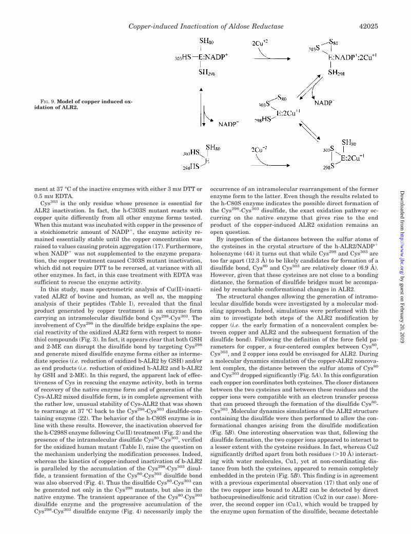
FIG. 9. Model of copper induced ox-idation of ALR2.
Copper-induced Inactivation of Aldose Reductase 42025
by guest on February 20, 2019http://w
ww
.jbc.org/D
ownloaded from
only after the reduction of the enzyme (i.e. after removal of thedisulfide bond) or after prolonged treatment of the modifiedALR2 at 37 °C with EDTA.
Cys298 remained too far apart from other cysteines to partic-ipate in any disulfide bond. Any attempt to shorten its distancefrom Cys303 through the interaction with copper ions by MMand MD approaches using the crystal structure of theNADP��ALR2 complex failed. A substantial reduction of thedistance between these residues could occur only after a refold-ing of the C-terminal segment. Conformational changes at theC terminus were previously observed in the crystal structuresof ALR2 complexed with the inhibitors Zopolrestat (50) andTolrestat (51), but in none of these structures were Cys298 andCys303 closer than 11 Å. However, different experiments involv-ing formation of mixed disulfides at 37 °C showed that thermalrearrangements of Cys- and Cys-Gly mixed disulfide contain-ing ALR2 likely occurred by closer Cys298-Cys303 interactions(22). Nevertheless, when the disulfide Cys303-Cys298 was im-posed, the two copper ions, initially positioned at a coordinatingdistance between Cys303 and Cys298, drifted apart and becamesituated in a way similar to that described in the case of theCys80-Cys303 disulfide (Fig. 6).
One possible structural change that could allow Cys303 to movecloser and interact either directly with Cys298 (as may be the caseof the C80S enzyme) or with the disulfide bond Cys80-Cys303, mayderive from the loss of the pyridine cofactor from the enzymeundergoing oxidation. A schematic representation of ALR2modification induced by the copper ion is depicted in Fig. 9.
The loss of the cofactor should induce significant changes inthe ALR2 conformation. In fact, the release of the cofactor,which is the rate-limiting step in the direction of aldehydereduction, is known to be associated with remarkable confor-mational changes of loop 7 (54–57). Indeed, loop 7, which isadjacent to the Cys298-Cys303 disulfide (Fig. 6), showed thehighest conformational variability during MD, supporting thehypothesis that formation of the disulfide might affect theNADP� binding site and NADP� release. Such an event mayalso give the rationale for the lack of activity of the disulfideenzyme forms. In fact, from a closer view of the active site ofthe disulfide-carrying enzymes (Fig. 7), besides the confor-mational change observed at Trp111, which may possibly af-fect enzyme activity, there are no apparent reasons for theoxidized holoenzyme to be inactive. Unfortunately, the highvalues of the kinetic constants of the reversible movement ofloop 7 in the mechanism of action (i.e. 0.5 s�1) (55), as well asthe lack of useful crystal structure data on the ALR2 apoen-zyme, made it very difficult to handle this problem by amolecular modeling approach. However, the involvement ofthe release of NADP� in the conformational rearrangementsleading Cys303 to reach at bonding distance Cys298 is sup-ported by the CD data. In fact, the analysis of CD spectra ofthe Cu(II)-modified b-ALR2 reveals that 60% of the NADP�,usually firmly bound to native ALR2, was lost after coppertreatment (Fig. 8).
In conclusion, the site-specific oxidative action of copperion on ALR2 ends with one of the most easy to predict mod-ifications (i.e. thiol oxidation to disulfides) that this metal ionis able to induce on target proteins. In this case, the evolutionof the oxidative insult on the protein structure leads to anenzyme form that is inactive but still convertible to the activenative enzyme and to an efficient copper sequestration.Whether such an apparent scavenging action exerted byALR2 contributes to the control of oxidative insult is still amatter of investigation.
Acknowledgments—We are indebted to Dr. Nando Benimeo(Industrie Alimentari Carni, Castelvetro, Modena) for the kind supply
of bovine lenses, and to Dr. Giovanni Sorlini and the veterinary staff ofIN.AL.CA. for their valuable cooperation in bovine lens collection. Weacknowledge Prof. Maurizio Zandomeneghi (Department of Chemistry,University of Pisa) for valuable expertise in the CD analysis. We thankCentro Interdipartimentale di Calcolo Elettronico, University of Mod-ena, for access to the computers.
REFERENCES
1. Halliwell, B., and Gutteridge, M. C. (1984) Biochem. J. 219, 1–142. Stuckel, J., Wallace, A. C., Cohen, F. E., and Prusiner, S. B. (1998) Biochem-
istry 37, 7185–71933. Linder, M. C., and Hazegh-Azam, M. (1996) Am. J. Clin. Nutr. 63, 797S–811S4. Yuan, D. S., Stearman, R., Dancis, A., Dunn, T., Beeler, T., and Klausner, R. D.
(1995) Proc. Natl. Acad. Sci. U. S. A. 92, 2632–26365. Petris, M. J., Mercer, J. F., Culvenor, J. G., Lockhart, P., Gleeson, P. A., and
Camakaris, J. (1996) EMBO J. 15, 6084–60956. Ogihara, H., Ogihara, T., Miki, M., Yasuda, H., and Mino, M. (1995) Pediatr.
Res. 37, 219–2267. Tallis, G. A., Kitchener, M. I., and Thomas, A. C. (1990) Clin. Chem. 36,
568–5708. Harman, D. (1965) J. Gerontol. 20, 151–1539. Brewer, G. J., and Yuzbasiyan-Gurkan, V. (1992) Medicine 71, 139–164
10. Lin, J. (1977) Jpn. J. Ophthalmol. 41, 130–13711. Ueda, J., Saito, N., and Ozawa, T. (1996) Arch. Biochem. Biophys. 325, 65–7612. Li, Y., Trush, M. A., and Yager, J. D. (1994) Carcinogenesis 15, 1421–142713. Kobayashi, S., Ueda, K., Morita, J., Sakai, H., and Komano, T. (1988) Biochim.
Biophys. Acta 949, 143–14714. Gutteridge, J. M., and Halliwell, B. (1982) Biochem. Pharmacol. 31, 2801–280515. Aruoma, O. I., Halliwell, B., Gajewski, E., and Dizdaroglu, M. (1991) Biochem.
J. 273, 601–60416. Garland, D. (1990) Exp. Eye Res. 50, 677–68217. Cecconi, I., Moroni, M., Vilardo, P. G., Dal Monte, M., Borella, P., Rastelli, G.,
Costantino, L., Garland, D., Carper, D., Petrash, J. M., Del Corso, A., andMura, U. (1998) Biochemistry 37, 14167–14174
18. Giannessi, M., Del Corso, A., Cappiello, M., Voltarelli, M., Marini, I.,Barsacchi, D., Garland, D., Camici, M., and Mura, U. (1993) Arch. Biochem.Biophys. 300, 423–429
19. Cappiello, M., Voltarelli, M., Cecconi, I., Vilardo, P. G., Dal Monte, M., Marini,I., Del Corso, A., Wilson, D. K., Quiocho, F. A., Petrash, J. M., and Mura, U.(1996) J. Biol. Chem. 271, 33539–33544
20. Liu, S.-Q., Bhatnagar, A., and Srivastava, S. K. (1992) Biochim. Biophys. Acta1120, 329–366
21. Cappiello, M., Voltarelli, M., Giannessi, M., Cecconi, I., Camici, G., Manao, G.,Del Corso, A., and Mura, U. (1994) Exp. Eye Res. 58, 491–501
22. Vilardo, P. G., Scaloni, A., Amodeo, P., Barsotti, C., Cecconi, I., Cappiello, M.,Lopez Mendez, B., Rullo, R., Dal Monte, M., Del Corso, A., and Mura, U.(2001) Biochemistry 40, 11985–11994
23. Inagaki, K., Miwa, I., Yashiro, T., and Okuda, J. (1982) Chem. Pharm. Bull. 30,3244–3254
24. Del Corso, A., Barsacchi, D., Giannessi, M., Tozzi, M. G., Camici, M., Houben,J. L., Zandomeneghi, M., and Mura, U. (1990) Arch. Biochem. Biophys. 283,512–518
25. Petrash, J. M., Harter, T. M., Devine, C. S., Olins, P. O., Bhatnagar, A., Liu, S.,and Srivastava, S. K. (1992) J. Biol. Chem. 267, 24833–24840
26. Old, S. E., Sato, S., Kador, P. F., and Carper, D. A. (1990) Proc. Natl. Acad. Sci.U. S. A. 87, 4942–4945
27. Vinci, F., Ruoppolo, M., Pucci, P., Freedman, R. B., and Marino, G. (2000)Protein Sci. 9, 525–535
28. Bradford, M. M. (1976) Anal. Biochem. 72, 248–25429. Case, D. A., Pearlman, D. A., Caldwell, J. W., Cheathan, T. E., III, Ross, W. S.,
Simmerling, C. L., Darden, T. A., Merz, K. M., Stanton, R. V., Cheng, A. L.,Vincent, J. J., Crowley, M., Tsui, V., Radmer, R. J., Duan, Y., Pitera, J.,Massova, I., Seibel, G. L., Singh, U. C., Weiner, P. K., and Kollman, P. A.(1999) AMBER6, University of California, San Francisco, CA
30. Cornell, W. D., Cieplak, P., Bayly, C. I., Gould, I. R., Merz, K. M., Ferguson,D. M., Spellmeyer, D. C., Fox, T., Caldwell, J. W., and Kollman, P. A. (1995)J. Am. Chem. Soc. 117, 5179–5197
31. Ferrin, T. E., Huang, C. C., Jarvis, L. E., and Langridge, L. (1988) J. Mol.Graph. 6, 13–27
32. Hay, B. P. (1993) Coord. Chem. Rev. 126, 177–23633. Weast, R. C., Astle, M. J., Beyer, W. H. (1985–1986) Handbook of Chemistry
and Physics, 66th Edition, Section F-164, CRC Press, Boca Raton, FL34. Guss, J. M., Bartunik, H. D., and Freeman, H. C. (1992) Acta Cryst. Sect. B 48,
790–81135. Ryde, U., Ollson, M. H. M., Pierloot, K., and Roos, B. O. (1996) J. Mol. Biol.
261, 586–59636. Pierloot, K., De Kerpel, J. O. A., Ryde, U., and Roos, B. O. (1997) J. Am. Chem.
Soc. 119, 218–22637. Frisch, M. J., Trucks, G. W., Schlegel, H. B., Gill, M. W., Johnson, B. G., Robb,
M. A., Cheeseman, J. R., Keith, T., Petersson, G. A., Montgomery, J. A.,Raghavachari, K., Al-Laham, M. A., Zakrzewski, V. G., and Ortiz, J. V.(1995) Gaussian94, Revision D, Gaussian, Inc., Pittsburgh, PA
38. Bayly, C. I., Cieplak, P., Cornell, W. D., and Kollman, P. A. (1993) J. Phys.Chem. 97, 10269–10277
39. Cieplak, P., Bayly, C. I., Cornell, W. D., and Kollman, P. A. (1995) J. Comput.Chem. 16, 1357–1377
40. Åqvist, J. (1990) J. Phys. Chem. 94, 8021–802441. Jorgensen, W. L., Buckner, J. K., Huston, S. E., and Rossky, P. J. (1987) J. Am.
Chem. Soc. 109, 1891–189942. Jorgensen, W. L., Chandrasekhar, J., Madura, J. D., Impey, R. W., and Klein,
M. L. (1983) J. Chem. Phys. 79, 926–935
Copper-induced Inactivation of Aldose Reductase42026
by guest on February 20, 2019http://w
ww
.jbc.org/D
ownloaded from

43. van Gusteren, W. F., and Berendsen, H. J. C. (1977) Mol. Phys. 34, 1311–132744. Wilson, D. K., Bohren, K. M., Gabbay, K. H., and Quiocho, F. H. (1992) Science
257, 81–8445. Sali, A., and Blundell, T. L. (1993) J. Mol. Biol. 234, 779–81546. Laskowski, R. A., McArthur, M. W., Moss, D. S., and Thornton, J. M. (1993)
J. Appl. Cryst. 26, 283–29147. Schade, S. Z., Sherrell, L. E., Williams, T. R., Kezdy, F. J., Heinrikson, R. L.,
Grimshaw, C. E., and Doughty, C. C. (1990) J. Biol. Chem. 265, 3628–363548. Bohren, K. M., and Gabbay, K. H. (1993) in Enzymology and Molecular Biology
of Carbonyl Metabolism (Weiner, H., ed) Vol. 4, pp. 267–277, Plenum Press,New York
49. Cappiello, M., Vilardo, P. G., Cecconi, I., Leverenz, V., Giblin, F. J., Del Corso,A., and Mura, U. (1995) Biochem. Biophys. Res. Commun. 207, 775–782
50. Wilson, D. K., Tarle, I., Petrash, J. M., and Quiocho, F. A. (1993) Proc. Natl.Acad. Sci. U. S. A. 90, 9847–9851
51. Urzhumtsev, A., Tete-Favier, F., Mitschler, A., Barbanton, J., Barth, P.,Urzhumtseva, L., Biellmann, J. F., Podjarni, A. D., and Moras, D. (1997)Structure 5, 601–612
52. Bohren, K. M., Grimshaw, C. E., Lai, C.-J., Harrison, D. H., Ringe, D., Petsko,G. A., and Gabbay, K. H. (1994) Biochemistry 33, 2021–2032
53. Tarle, I., Borhani, D. W., Wilson, D. K., Quiocho, F. A., and Petrash, J. M.(1993) J. Biol. Chem. 268, 25687–25693
54. Grimshaw, C. E., Shabbaz, M., and Putney, G. C. (1990) Biochemistry 29,9947–9955
55. Grimshaw, C. E., Bohren, K. M., Lai, C.-J., and Gabbay, K. H. (1995) Biochem-istry 34, 14356–14365
56. Grimshaw, C. E., Bohren, K. M., Lai, C.-J., and Gabbay, K. H. (1995) Biochem-istry 34, 14366–14373
57. Kubiseski, T. J., Hindman, D. J., Morjana, N. A., and Flynn, T. G. (1992)J. Biol. Chem. 267, 6510–6517
Copper-induced Inactivation of Aldose Reductase 42027
by guest on February 20, 2019http://w
ww
.jbc.org/D
ownloaded from

Antonella Del Corso and Umberto MuraLuca Costantino, Mario Cappiello, Donita Garland, Deborah Carper, J. Mark Petrash, Ilaria Cecconi, Andrea Scaloni, Giulio Rastelli, Maria Moroni, Pier Giuseppe Vilardo,
OF THE METAL-PROTEIN INTERACTION MECHANISMOxidative Modification of Aldose Reductase Induced by Copper Ion: DEFINITION
doi: 10.1074/jbc.M206945200 originally published online August 14, 20022002, 277:42017-42027.J. Biol. Chem.
10.1074/jbc.M206945200Access the most updated version of this article at doi:
Alerts:
When a correction for this article is posted•
When this article is cited•
to choose from all of JBC's e-mail alertsClick here
http://www.jbc.org/content/277/44/42017.full.html#ref-list-1
This article cites 54 references, 13 of which can be accessed free at
by guest on February 20, 2019http://w
ww
.jbc.org/D
ownloaded from