Conformations Single-Stranded Bacteriophage M13*authors.library.caltech.edu/8680/1/FORpnas70.pdf ·...
8
Proceedings of the National Academy of Sciences Vol. 67, No. 3, pp. 1534-1541, November 1970 Conformations of the Single- Stranded DNA of Bacteriophage M13* Arleen B. Forsheitt and Dan S. Ray MOLECULAR BIOLOGY INSTITUTE AND DEPARTMENT OF ZOOLOGY, UNIVERSITY OF CALIFORNIA, LOS ANGELES, CALIF. 90024 Communicated by Norman Davidson, August 28, 1970 Abstract. At least two conformations of M13 single-stranded DNA have been demonstrated by measuring differences in sedimentation coefficient and by direct visualization in the electron microscope. Which form is obtained from infected cells and/or intact phage depends on the pH, ionic strength, and tem- perature. The slower-sedimenting form can be converted to the faster-sedi- menting, single-stranded form by low ionic strength, alkali treatment, forma- mide, or formaldehyde, but not by exposure to 100'C in 1.0 M NaCl. The ability to assume either conformation appears to be a function of the nucleic acid alone. Whether or not these different conformations are of biological significance is still unknown. M13 is a small fibrous bacteriophage with a genome consisting of a circular, single-stranded DNA molecule of molecular weight 2 X 10⁶. The base composi- tion is inconsistent with Watson-Crick pairing and the optical density at 260 nm increases gradually with increasing temperature in contrast to the sharp melting of double-stranded DNA.1,2 The absorbance at 260 nm and the sedi- mentation coefficient vary greatly with ionic strength, suggesting a flexible con- formation with significant base stacking in aqueous solution.3' 4 We have isolated from M13-infected cells, and from intact phage, a circular, single-stranded DNA form which sediments in 1.0 M NaCl at a rate 1.16 times as fast as that of RF I, in contrast to the form usually observed which sediments at a rate 1.31-1.35 times that of RF I. The isolation and characterization of the slower-sedi- menting single-stranded DNA (SS*), and the interconversions between the slow- and fast-sedimenting(SS) forms, are described below. Materials and Methods. Sources of materials, media, bacterial growth, phage infection and assay, lysis of cells, centrifugation techniques, and radioactivity assays have all been described (ref. 5, and manuscript in preparation). Buffers used are: (1) Tris-EDTA, 10 mM Tris (pH 8.0)-i mM EDTA; (2) Tris- EDTA-NaCl is Tris-EDTA with the indicated molarity of NaCl. Radioactive M13 single-stranded DNA, used as a sedimentation marker, was prepared as follows: labeled phage were incubated with 100 Ag/ml pronase and 0.1% sodium dodecyl sulfate for 20 min at 60'C in Tris-EDTA or Tris-EDTA-1 M NaCl, then were deproteinized by adding one-half volume of 3 M NaCl04 and one volume of chloroform-octanol (9:1) and vortex mixing. The aqueous phase was then dialyzed against Tris-EDTA. Material was prepared for electron microscopy by the method described by Davis, Simon, and Davidson.6 The spreading solution contained 0.5 M NH4C2H302-1 mM EDTA-10 mM Tris, pH 7.5, and the hypophase contained 0.25 M NH4C2H3020.5 mM 1534
Transcript of Conformations Single-Stranded Bacteriophage M13*authors.library.caltech.edu/8680/1/FORpnas70.pdf ·...

Proceedings of the National Academy of SciencesVol. 67, No. 3, pp. 1534-1541, November 1970
Conformations of the Single-Stranded DNAof Bacteriophage M13*
Arleen B. Forsheitt and Dan S. RayMOLECULAR BIOLOGY INSTITUTE AND DEPARTMENT OF ZOOLOGY, UNIVERSITY OF CALIFORNIA,
LOS ANGELES, CALIF. 90024
Communicated by Norman Davidson, August 28, 1970
Abstract. At least two conformations of M13 single-stranded DNA have beendemonstrated by measuring differences in sedimentation coefficient and bydirect visualization in the electron microscope. Which form is obtained frominfected cells and/or intact phage depends on the pH, ionic strength, and tem-perature. The slower-sedimenting form can be converted to the faster-sedi-menting, single-stranded form by low ionic strength, alkali treatment, forma-mide, or formaldehyde, but not by exposure to 100'C in 1.0 M NaCl. Theability to assume either conformation appears to be a function of the nucleicacid alone. Whether or not these different conformations are of biologicalsignificance is still unknown.
M13 is a small fibrous bacteriophage with a genome consisting of a circular,single-stranded DNA molecule of molecular weight 2X 10⁶. The base composi-tion is inconsistent with Watson-Crick pairing and the optical density at 260nm increases gradually with increasing temperature in contrast to the sharpmelting of double-stranded DNA.1,2 The absorbance at 260 nm and the sedi-mentation coefficient vary greatly with ionic strength, suggesting a flexible con-formation with significant base stacking in aqueous solution.3' 4 We have isolatedfrom M13-infected cells, and from intact phage, a circular, single-stranded DNAform which sediments in 1.0 M NaCl at a rate 1.16 times as fast as that of RF I,in contrast to the form usually observed which sediments at a rate 1.31-1.35times that of RF I. The isolation and characterization of the slower-sedi-menting single-stranded DNA (SS*), and the interconversions between theslow- and fast-sedimenting(SS) forms, are described below.
Materials and Methods. Sources of materials, media, bacterial growth, phageinfection and assay, lysis of cells, centrifugation techniques, and radioactivity assayshave all been described (ref. 5, and manuscript in preparation).
Buffers used are: (1) Tris-EDTA, 10 mM Tris (pH 8.0)-i mM EDTA; (2) Tris-EDTA-NaCl is Tris-EDTA with the indicated molarity of NaCl. Radioactive M13single-stranded DNA, used as a sedimentation marker, was prepared as follows: labeledphage were incubated with 100 Ag/ml pronase and 0.1% sodium dodecyl sulfate for 20min at 60'C in Tris-EDTA or Tris-EDTA-1 M NaCl, then were deproteinized by addingone-half volume of 3 M NaCl04 and one volume of chloroform-octanol (9:1) and vortexmixing. The aqueous phase was then dialyzed against Tris-EDTA.
Material was prepared for electron microscopy by the method described by Davis,Simon, and Davidson.6 The spreading solution contained 0.5 M NH4C2H302-1 mMEDTA-10 mM Tris, pH 7.5, and the hypophase contained 0.25 M NH4C2H3020.5 mM
1534

VOL. 67, 1970 DNA CONFORMATIONS OF M13 PHAGE 1535
EDTA-5 mM Tris, pH 7.4. The preparations were shadowed at low angle with plati-num-palladium and examined in a Phillips model 200 electron microscope. (We areindebted to Dr. John Fessler, Mrs. June Baumer, and Dr. Frederick Eiserling for helpand advice with electron microscopy.)
Results. Isolation and characterization of SS* from infected cells: WhenM13-infected Escherichia coli are lysed with lysozyme-EDTA and sarcosyl de-tergent in Tris-EDTA-0.1 M NaCl, the intracellular, single-stranded, DNAcosediments with a viral SS marker at 1.31-1.35 times the rate of RF I in asucrose gradient that contains 1.0 M NaCl. In contrast, if infected cells arelysed with lysozyme-EDTA (in Tris-EDTA-0.1 M NaCl) without sarcosyl,the single-stranded DNA quantitatively appears as the slower-sedimentingSS* form, which sediments 1.16 times as fast as RF I (Fig. 1). SS* can be ob-
30-ss
6
020
_E C')
10 N
RFI 2
RFIE
C 00 10 20 30 40
Fraction no.
FIG. 1. Isolation of intracellular SS*. A culture of E. coli K12 5274 was grown to 1 X 10⁸cells/ml and infected with \113 at a phage to cell ratio of 40. The cells were labled from 3 to 60min after infection with 20 ,Ci/ml [3H] deoxythymidine. At the end of the labeling period, thecells were washed, lysed with lysozyme-EDTA, mixed with a [31P]SS marker, and analyzed byvelocity sedimentation through a 5-20% linear sucrose gradient in Tris-EDTA-1 M NaCl for16 hr at 24,000 rpm and 50C in a SW27 rotor. Sedimentation is from left to right. *-4,1H cpm; O-O, 32p cpm.
tained after lysozyme-EDTA and sarcosyl lysis, however, if the NaCl concen-tration is 1.0 M during lysis. SS*, under these lysis conditions, is more stableat high ionic strength.SS* purified by sucrose gradient sedimentation is stable at 370C in 1.0 M
NaCl, but not in 0.1 1\I NaCl. The SS* form is stable in 0.1 M NaCl at 370Conly in the presence of the crude lysate. The conversion of SS* to SS, in sar-cosyl-treated lysates in 0.1 MI NaCl, may possibly be due to denaturation ofproteins or disruption of the lipoprotein cell membrane, either of which may beresponsible for the stabilization of the structure under these conditions.
In general, agents or conditions which disrupt hydrogen bonds result in con-version of SS* to SS. At salt concentrations of 0.01 MI or lower, SS* is converted

1536 BIOCHEMISTRY: FORSHEIT AND RAY PROC. N. A. S.
to SS even at 50C. Alkali (0.1 N NaOH), formamide (40%o), and formaldehyde(6%) act similarly, although exposure to 100'C for 10 min in Tris-EDTA-1.0M NaCl does not convert SS* to SS. In Tris-EDTA-0.5 to 1.0 M NaCl, SS*is unaffected by pronase, sodium dodecyl sulfate, sarcosyl detergent, ether orchloroform extraction, deproteinization with perchlorate and chloroform-octanol, or deproteinization with phenol and/or ethanol precipitation.
Isolation of SS* from infectious M13 bacteriophage particles: The slower-sedimenting single-stranded DNA can be obtained from M13 by heating thephage in Tris-EDTA-1.0 M NaCl at 100'C for 10 min, or by deproteinizationof the phage with NaClO4 and chloroform-octanol (9: 1) followed by dialysis at50C against Tris-EDTA that contains from 0.5 to 1.0 M NaCl (Fig. 2). Thedeproteinizing procedure for obtaining SS* requires phage particles as the start-ing material. If SS DNA is deproteinized with NaCl04 and chloroform-octanol(9:1) and dialysed against Tris-EDTA-1.0 1\1 NaCl at 5°C or below, it remains
o- (a) -8M13 SS
-6
20 14
-2
0 0
ss4
4 4
Fraction no.
FIG...Isolation of SS from intact M13 phage particles. A sample of [3H]deoxythymi-dine-labeled M13 was deproteinized with sodium perchlorate and chloroform-octanol anddivided into two portions. One part was dialyzed against Tris-EDTA--1.0 M NaCl and theother against Tris-EDTA. Each sample was mixed with a ["2P]SS marker and analyzed byvelocity sedimentation through a linear 5-20% sucrose gradient in Tris-EDTA-1.0 M NaCl for2.75 hr at 60,000 rpm and 5°C in a SW65 rotor. Sedimentation is from right to left. (a) ['H]-M13 phage (b) [3H]SS* (deproteinized [3H]M13 dialyzed against Tris-EDTA-1.0 M NaCl),(c) ['H]SS (deproteinized [3H]M13 dialyzed against Tris-EDTA). *-e ',Hcpm; O-O.32p cpm.
VOL. 67, 1970 DNA CONFORMATIONS OF M13 PHAGE 1537
as SS. Deproteinization of the phage, followed by dialysis against Tris-EDTA,produces the fast-sedimenting SS form (Fig. 2c). Sedimentation of the phageprior to deproteinization is shown in Fig. 2a. (The small, fast-sedimentingpeak of phage material in Fig. 2a is thought to be an aggregation artifact in ourphage preparations.) The properties of SS* obtained from phage are identicalto those of the SS* isolated from infected cells.Both the slow-sedimenting SS* and the fast-sedimenting SS have identical
ultraviolet absorption spectra in the range from 220 nm to 320 nm and showAxol/A ratios, in Tris-EDTA-1.0 M NaCl, of 1.67. These data, and the re-sistance of SS* to proteolytic enzymes and to the protein denaturing conditionsdescribed above, suggested that this was not a protein-mediated conversion.Failure to label SS* with [3H]leu or [35S]sulfate supported this hypothesis. Theradioactive phage used contained label such that 0.5-1% of the phage proteinscould have been detected in association with the DNA.Heating and cooling of SS DNA: The above data suggested that SS* and
SS are two different conformations of the viral nucleic acid per se. To furthertest this hypothesis, we attempted to convert SS back to the slower sedimentingSS*. At the same time, a similar single-stranded DNA from OX174 (providedby R. Schekman) was examined. A 32P-labeled sample of DNA from M13 anda 32P-labeled sample of DNA from 4X174 were separately heated in Tris-EDTA-1.0 M NaCl to 800C, allowed to cool slowly over a period of 6 hr, and analyzed byvelocity sedimentation through a neutral sucrose gradient, in 1.0 M NaCl, inthe presence of the appropriate 3H-labeled SS-marker DNA. [3H]RF markerswere also added in the case of M13. The results are shown in Fig. 3.
For M13 DNA, the unheated 32P-labeled sample cosedimented with the markerof [3H]M13 SS DNA, at a rate 1.31 times that of RF I, whereas the heated andcooled sample sedimented at 1.22 times the rate of RF I (Fig. 3a and b). For4OX174, a similar result was obtained, although the difference between the sedi-mentation coefficients was not as great (Figs. 3c and d). If the DNA was quick-cooled on ice, similar results were observed but with more forward skewing ofthe DNA.
If SS in Tris-EDTA is dialyzed overnight against Tris-EDTA-1.0 M NaClat 23 or 370C, a result very similar to that obtained by heating and cooling isobserved. At 230C, SS is converted to a form sedimenting at 1.22 times therate of RF I, while at 370C SS appears as a form sedimenting at 1.25 times therate of RF I in 1.0 M NaCl. Under the conditions used, SS sedimented at1.32 times the rate of RF I. Dialysis of SS against Tris--EDTA-1.0 M NaCl at50C does not alter its sedimentation rate.
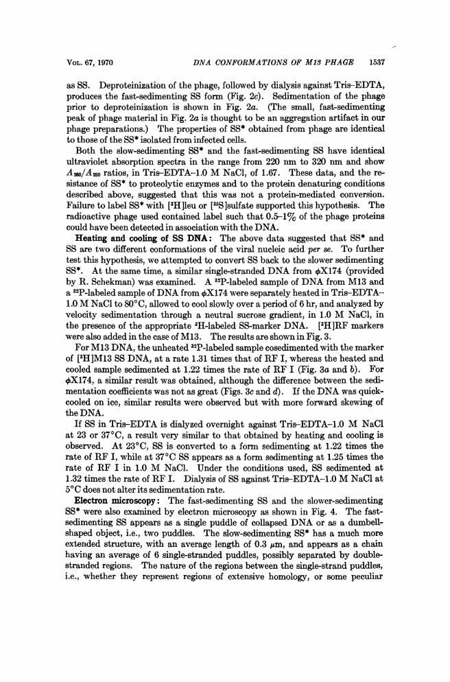
Electron microscopy: The fast-sedimenting SS and the slower-sedimentingSS* were also examined by electron microscopy as shown in Fig. 4. The fast-sedimenting SS appears as a single puddle of collapsed DNA or as a dumbell-shaped object, i.e., two puddles. The slow-sedimenting SS* has a much moreextended structure, with an average length of 0.3 ,m, and appears as a chainhaving an average of 6 single-stranded puddles, possibly separated by double-stranded regions. The nature of the regions between the single-strand puddles,i.e., whether they represent regions of extensive homology, or some peculiar

(a) 6
RFII
(b)
12 SSRFI 12
8 RFI8~~~RF
O () ~10 20 30 40 0
X 2
(C)f StS EX(1 ) SS
0
8- 4
C D~~~~~~~~~~~~~~~~~~~~~~~~~C
I010 20 30 40
Fraction noFIG. 3. Heating and cooling of SS. 3H-labeled- and 32P-labeled-M13 55 was prepared as
described. 3H-labeled- and 32P-labeled-+X174 SS was prepared by phenol treatment of thephage, alcohol precipitation of the aqueous phase, and resuspension in Tris-EDTA. The 3H-labeled 55 from the appropriate phase was used as a marker in each case. A sample of each[32P] 5 preparation was sedimented (untreated) as a control, and a second sample was heatedto 800C for 30 min in Tris-EDTA-1.0 M NaCl and allowed to cool slowly to room temperaturefor 6 hr. 3H-labeled-M13 RF markers were added to the M13 samples. Velocity sedimenta-tion was as described in Fig. 2. Sedimentation is from right to left. (a) 32P-labeled-M13 55,unheated; (b) 32P-labeled-M13 SS, heated to 800C and cooled; (c) 32P-labeled-+X174 SS,unheated; (d) 32P-labeled-+X174 SS, heated to 800C and cooled. *-- *, 'H cpm, O-O.32p cpm.
VOL. 67, 1970 DNA CONFORMATIONS OF M13 PHAGE 1539
sX{;itb~~~~~~~~~~~~I'.:'.~~~~~~~~~~~~~~~~~~~~~~~~~~.~~~ ~ ~ ~ ~ ~ ~ ~ ~ 7
:O~~X
J*.4 ,.. ^^.;.A
,taa
attodfeetmanfctos b an(d3Sttesm-anfctos
C
twsigo" n par ofa -h rtrn arun anthr isukont(hogthrad-ik stutreiat ofat. dobesrne NAmlcl sda
penda
t*A, ~ ThA,
-rn t~. 2.actA6-.A
FIG. 4. Electron micrographs of the two forms of M13 single-stranded DNA. (a) and (c), SSat two different magnifications; (6) and (d) SS* at the same magmifications.
twisting of one part of the strand around another, is unknown. (The long,thread-like structure is part of a double-stranded )\ DNA molecule used as amarker.) We have been unable to utilize the formamide technique of West-moreland, et al.7 to spread the single-stranded regions because the method de-pends on very low ionic strength, a condition under which SS* is unstable.

1540 BIOCHEMISTRY: FORSHEIT AND RAY PROC. N. A. S.
Discussion. We have demonstrated, by differences in sedimentation rateand by visualization in the electron microscope, that there are at least two stableconformations of the M13 single-stranded DNA. Either conformation can beobtained from phage or from infected cells, depending on the ionic strength,pH, and temperature. It is perhaps significant that the conversion of SS* toSS occurs at conditions close to physiological conditions (0.1-0.2 M NaCl and370C).DNA is obtained in the fast-sedimenting SS form at low ionic strength, and in
the slow-sedimenting SS* form at high ionic strength. Conversion of SS* toSS, by dialysis against buffer containing 0.01 M NaCl or less, is quantitative.Conditions for quantitative conversion of SS to SS* have not yet been found.Heating and slow-cooling of SS, or dialysis of SS against 1.0 M NaCl at 23 or370C, produces a form sedimenting 1.22 or 1.25 times as fast as RF I in 1.0M NaCl. These forms sediment at rates intermediate between those of SS (1.31-1.35 times the rate of RF I) and SS* (1.16 times the rate of RF I). It is possible,therefore, that M13 DNA, and perhaps 4X174 DNA, can exist in several meta-stable, hydrogen-bonded states with different degrees of extension and, conse-quently, different sedimentation rates. Discrete metastable states have alsobeen observed for MS2 RNA.8 The results with 4X174 suggest that such con-formations could be general for all small single-stranded DNAs.The electron micrographs suggest that the fast-sedimenting SS is a very com-
pact structure under these spreading conditions. On the other hand, the slower-sedimenting structure appears more rigid and elongated, and suggests a chain ofsingle-stranded bushes separated by double-stranded regions. The extendedconformation would have a much greater frictional coefficient than the more com-pact one and would be expected to sediment more slowly, as observed. Carefulmeasurements of length and of the distances between single-strand puddles mayprovide meaningful data on whether this slow-sedimenting conformation is aunique ordered arrangement, facilitated by specific base pairing along the lengthof the molecules, or whether it is the result of random association of strandsstabilized by a few base pairs. The techniques used here, however, do not ad-equately resolve all the single-strand bushes from the assumed double-strandedregions; finer resolution is being sought. Some of the regions between single-strand puddles appear to be thicker than the double-stranded DNA used asmarker.Two other models may be considered. One is that the slower-sedimenting
form is a result of aggregation of the fast-sedimenting SS. This model is in-consistent with the dimensions observed in the electron microscope, i.e., thesingle-strand bushes of the fast-sedimenting form are much larger than the bushesof the elongated SS*. Furthermore, the aggregation reaction would probablybe dependent upon concentration whereas formation of SS* is independent ofconcentration over several orders of magnitude. Another explanation is thatin aqueous solution, the slower-sedimenting form is simply a coiled structurewith a larger radius of gyration than the fast-sedimenting form, which mighthave certain additional, specific, internal hydrogen-bonded regions permitting itto contract. On the basis of our data, we favor the interpretation suggested by

VOL. 67, 1970 DNA CONFORMATIONS OF M13 PHAGE 1541
the electron micrographs that the SS* form has single-strand "bushes," sepa-rated by doublestranded regions, whereas the SS form is a compact structurewith many random hydrogen bonds.The suggestion that there are specific double-stranded regions in the single-
stranded molecule is attractive because such double-stranded regions mightserve as enzyme recognition sites for the initial events in replication of the phageDNA. In addition, formation of a double-stranded region might provide ameans for circularization of a newly-synthesized, linear, single strand by poly-nucleotide ligase.9 The extended form might also be the necessary DNA pre-cursor for assembling the phage, the single-stranded regions being stretched out,in the phage particle by interaction with the coat protein. The biological signif-icance of either conformation, however, is still unknown. Whatever the bio-logical roles, a knowledge of the conformations of the single-stranded DNA ofM13 is an important first step to understanding and elucidating these roles.
Abbreviations: RF, replicative form; RF I, the covalently closed, double-stranded, ringform; SS, fast-sedimenting single-stranded DNA; SS*, slow-sedimenting single-stranded DNA.
* Supported by NSF grant GB 18074, University of California Cancer Research Funds, andan NSF predoctoral traineeship to A.B.F.
t Division of Chemistry and Chemical Engineering, California Institute of Technology,Pasadena, Calif.
1 Salivar, W. C., H. Tzagoloff, and D. Pratt, Virology, 24, 359 (1964).2 Marvin, D. A., and B. Hohn, Bacteriol. Rev., 33, 172 (1969).3 Studier, F. W., J. Mol. Biol., 11, 373 (1965).4 Studier, F. W., J. Mol. Biol., 41, 199 (1969).5 Ray, D. S., and R. Schekman, Biochim. Biophys. Acta, 179, 398 (1969); Ray, D. S., and
R. Schekman, J. Mol. Biol., 43, 645 (1969).6 Davis, R. W., M. Simon, and N. Davidson, in Methods in Enzymology, eds., L. Grossman
and K. Moldave (New York and London: Academic Press, 1970) vol. 12, part C: in press.7 Westmoreland, B. C., W. Szybalski, and H. Ris, Science, 163, 1343 (1969).8 Strauss, J., and R. L. Sinsheimer, J. Mol. Biol., 34, 453 (1968).9 Schaller, H., H. Voss, and S. Gucker, J. Mol. Biol., 44, 445 (1969).