Manual and automated preparation of single-stranded DNA ...
25
Manual and automated preparation of single-stranded DNA libraries for the sequencing of DNA from ancient biological remains and other sources of highly degraded DNA Marie-Theres Gansauge ✉ , Ayinuer Aximu-Petri, Sarah Nagel and Matthias Meyer ✉ It has been shown that highly fragmented DNA is most efficiently converted into DNA libraries for sequencing if both strands of the DNA fragments are processed independently. We present an updated protocol for library preparation from single-stranded DNA, which is based on the splinted ligation of an adapter oligonucleotide to the 3′ ends of single DNA strands, the synthesis of a complementary strand using a DNA polymerase and the addition of a 5′ adapter via blunt-end ligation. The efficiency of library preparation is determined individually for each sample using a spike-in oligonucleotide. The whole workflow, including library preparation, quantification and amplification, requires two work days for up to 16 libraries. Alternatively, we provide documentation and electronic protocols enabling automated library preparation of 96 samples in parallel on a Bravo NGS Workstation (Agilent Technologies). After library preparation, molecules with uninformative short inserts (shorter than ~30-35 base pairs) can be removed by polyacrylamide gel electrophoresis if desired. This protocol is an update to Nat. Protoc. 8, 737–748 (2013): https://doi.org/10.1038/nprot.2013.038 Introduction Development of the protocol The preparation of DNA libraries is a critical step in the preparation of samples for high-throughput DNA sequencing, particularly if only small quantities of highly degraded DNA are present, such as in ancient biological material. In 2012, we introduced a library preparation method that differed fun- damentally from previously existing methods in that it relies on the heat denaturation of double- stranded DNA fragments and the subsequent conversion of single DNA strands into library mole- cules 1,2 . Single-stranded library preparation minimizes the loss of short DNA fragments and those with single-stranded breaks, thereby increasing the number of library molecules that can be retrieved from highly degraded DNA. The power of the method was first demonstrated by the generation of a high-coverage genome sequence from just a few milligrams of bone powder obtained from a finger bone of a Denisovan individual, an extinct Pleistocene hominin 1 . Since then, it has made the recovery of additional high-quality genomes from ancient DNA possible, including those of three Neander- tals 3–5 and an early modern human 6 . Single-stranded DNA library preparation was also instrumental for the retrieval of DNA sequences from the ~430,000-year-old bear and hominin remains from Sima de los Huesos in Spain 7–9 , which are by far the oldest remains outside of permafrost that have been sequenced to date. More recently, it has allowed the recovery of Neandertal and Denisovan DNA from Pleistocene cave sediments 10 . A schematic overview of the single-stranded library preparation protocol provided here is shown in Fig. 1a. Briefly, after heat denaturation of the double-stranded DNA fragments, a biotinylated adapter is ligated to the 3′ end of each single-stranded molecule. After immobilizing the ligation products on beads, the template strand is copied by extending a primer hybridized to the adapter using a DNA polymerase, which creates a double-stranded DNA fragment to which the second adapter is joined by blunt-end ligation. The library is then released from the beads at a high Department of Evolutionary Genetics, Max Planck Institute for Evolutionary Anthropology, Leipzig, Germany. ✉ e-mail: [email protected]; [email protected] NATURE PROTOCOLS | VOL 15 | AUGUST 2020 | 2279–2300 | www.nature.com/nprot 2279 PROTOCOL UPDATE https://doi.org/10.1038/s41596-020-0338-0 1234567890():,; 1234567890():,;
Transcript of Manual and automated preparation of single-stranded DNA ...

Manual and automated preparation ofsingle-stranded DNA libraries for the sequencingof DNA from ancient biological remains andother sources of highly degraded DNAMarie-Theres Gansauge ✉, Ayinuer Aximu-Petri, Sarah Nagel and Matthias Meyer ✉
It has been shown that highly fragmented DNA is most efficiently converted into DNA libraries for sequencing if bothstrands of the DNA fragments are processed independently. We present an updated protocol for library preparation fromsingle-stranded DNA, which is based on the splinted ligation of an adapter oligonucleotide to the 3′ ends of single DNAstrands, the synthesis of a complementary strand using a DNA polymerase and the addition of a 5′ adapter via blunt-endligation. The efficiency of library preparation is determined individually for each sample using a spike-in oligonucleotide.The whole workflow, including library preparation, quantification and amplification, requires two work days for up to 16libraries. Alternatively, we provide documentation and electronic protocols enabling automated library preparation of96 samples in parallel on a Bravo NGS Workstation (Agilent Technologies). After library preparation, molecules withuninformative short inserts (shorter than ~30−35 base pairs) can be removed by polyacrylamide gel electrophoresisif desired.
This protocol is an update to Nat. Protoc. 8, 737–748 (2013): https://doi.org/10.1038/nprot.2013.038
Introduction
Development of the protocolThe preparation of DNA libraries is a critical step in the preparation of samples for high-throughputDNA sequencing, particularly if only small quantities of highly degraded DNA are present, such as inancient biological material. In 2012, we introduced a library preparation method that differed fun-damentally from previously existing methods in that it relies on the heat denaturation of double-stranded DNA fragments and the subsequent conversion of single DNA strands into library mole-cules1,2. Single-stranded library preparation minimizes the loss of short DNA fragments and thosewith single-stranded breaks, thereby increasing the number of library molecules that can be retrievedfrom highly degraded DNA. The power of the method was first demonstrated by the generation of ahigh-coverage genome sequence from just a few milligrams of bone powder obtained from a fingerbone of a Denisovan individual, an extinct Pleistocene hominin1. Since then, it has made the recoveryof additional high-quality genomes from ancient DNA possible, including those of three Neander-tals3–5 and an early modern human6. Single-stranded DNA library preparation was also instrumentalfor the retrieval of DNA sequences from the ~430,000-year-old bear and hominin remains from Simade los Huesos in Spain7–9, which are by far the oldest remains outside of permafrost that have beensequenced to date. More recently, it has allowed the recovery of Neandertal and Denisovan DNAfrom Pleistocene cave sediments10.
A schematic overview of the single-stranded library preparation protocol provided here is shownin Fig. 1a. Briefly, after heat denaturation of the double-stranded DNA fragments, a biotinylatedadapter is ligated to the 3′ end of each single-stranded molecule. After immobilizing the ligationproducts on beads, the template strand is copied by extending a primer hybridized to the adapterusing a DNA polymerase, which creates a double-stranded DNA fragment to which the secondadapter is joined by blunt-end ligation. The library is then released from the beads at a high
Department of Evolutionary Genetics, Max Planck Institute for Evolutionary Anthropology, Leipzig, Germany. ✉e-mail: [email protected];[email protected]
NATURE PROTOCOLS | VOL 15 |AUGUST 2020 | 2279–2300 |www.nature.com/nprot 2279
PROTOCOL UPDATEhttps://doi.org/10.1038/s41596-020-0338-0
1234
5678
90():,;
1234567890():,;
NNNNNNNN
NNNNNNNN
NNNNNNNN
INDEX
INDEX
INDEX
INDEX
INDEX
INDEX
INDEX
INDEX
INDEX
INDEX
INDEX
INDEX
INDEX
INDEXINDEXINDEX
INDEX
INDEX
INDEX
INDEX
a
Size selection
Indexing
Single-stranded library
preparation
qPC
R(all m
olecules)qP
CR
(spike-in)
Sequencing
Optional
Single-stranded library
preparation
b
Steps 1–5
Steps 6–9
Steps 10–20
Steps 21–23
Steps 24–27
Steps 28–31
Steps 1–31
Steps 32–37
Steps 38–42
Steps 43–46
Steps 47–52
Steps 53–58, 60Step 62
Step 61
Steps 63–66
Step 68
Steps 67–69
Step 70
Sequencing
Step 70
Tube preparation
Step 59
PROTOCOL UPDATE NATURE PROTOCOLS
2280 NATURE PROTOCOLS | VOL 15 |AUGUST 2020 | 2279–2300 |www.nature.com/nprot
temperature, and its concentration is determined using quantitative PCR (qPCR). A final amplifi-cation by PCR introduces the full-length adapter sequences required for sequencing using Illuminatechnology as well as pairs of sample-specific indices.
Since the first detailed protocol for single-stranded library preparation was published in NatureProtocols in 20132, the method has been continuously refined. Some of the previously reportedimprovements are as follows. (1) Bst DNA polymerase was replaced by the Klenow fragment ofEscherichia coli DNA polymerase I in the primer extension step, which eliminated the blunt-endrepair step that was previously required before the blunt-end ligation of the second adapter11. (2) Theligation of the first adapter to the single-stranded sample molecules is now performed using splinter-mediated ligation with T4 DNA ligase at 37 °C instead of using CircLigase at 60 °C12,13. The change inligase greatly reduced reagent costs, improved the robustness of the protocol and made it compatiblewith pipetting on automated liquid handling systems10 where tubes cannot be sealed to avoid eva-poration. (3) An oligonucleotide spike-in was introduced that allows monitoring the efficiency oflibrary preparation for each sample and detecting inhibition of enzymes that might result fromimpurities in the sample DNA14.
In addition to the previously published modifications above, the protocol provided here encom-passes modified oligonucleotide sequences, which make the method robust to fluctuations in thesynthesis quality of the oligonucleotides used. This has been achieved by replacing spacer mod-ifications in the adapter and splinter oligonucleotides by 2′-O-methyl-ribonucleotides, which arecheaper and easier to synthesize and effectively prevent incorporation of fragmented oligonucleotidesinto the library. Beyond other small optimizations that improve ease of use, we also describe anoptional size selection procedure that allows for efficient removal of library molecules with extremelyshort inserts (<35 base pairs (bp)) after library amplification. Such molecules, which can result fromthe extraction of extremely short DNA fragments from highly degraded samples, are usually unin-formative and consume unnecessary sequencing capacity if they make up a substantial proportion ofthe library15. Finally and importantly, we provide electronic protocol files (https://zenodo.org/record/3631147) and a Supplementary Manual that allow automated library preparation in a 96-well formaton a Bravo NGS Workstation (Agilent Technologies). Automation of sample preparation is becomingincreasingly important for studies where the analysis of hundreds or thousands of samples isdesirable, such as the characterization of ancient environmental DNA.
Applications and comparison with other methodsSeveral studies comparing library preparation methods have confirmed that yields of library mole-cules from ancient biological material are substantially higher with the single-stranded method thanwith double-stranded library preparation12,16,17. The average improvement over double-strandedmethods was estimated to ~ten-fold, but gains can be expected to be even higher with particularlyheavily degraded material. We therefore recommend the use of single-stranded library preparationfor all cases where destructive sampling should be kept to a minimum or where DNA preservation is
Fig. 1 | Schematic overview of the workflow described in this protocol. a, Single-stranded library preparation isinitiated by heat denaturation of the sample DNA and the removal of 5′ and 3′ phosphate groups (if present).Subsequently, an adapter oligonucleotide (red) carrying a 5′ phosphate and a 3′ biotin (indicated by a circle) is ligatedto the 3′ end of each DNA fragments using T4 DNA ligase. This reaction is enabled by a splinter oligonucleotidecarrying a stretch of eight random nucleotides at its 3′ end. The ligation products as well as excess adapters areimmobilized on streptavidin-covered magnetic beads (gray circles), and splinter oligonucleotides are removed by awash step at an elevated temperature. A primer is hybridized to the adapter oligonucleotide and a copy of the templatestrand synthesized using the Klenow fragment of E. coli DNA polymerase I. The primer contains phosphorothioatebackbone modifications to prevent its exonucleolytic degradation (not depicted). Unincorporated primers are removedthrough a bead wash at an elevated temperature, preventing the formation of adapter dimers during the subsequentblunt-end ligation of a second, double-stranded adapter (blue), a reaction again catalyzed by T4 DNA ligase. One ofthe adapter strands carries a 3′ dideoxy modification to prevent self-ligation of adapters. The library strand is releasedfrom the beads by heat denaturation. b, After library preparation, the yield of library molecules as well as the numberof successfully converted control oligonucleotides, which were spiked-into the sample DNA before library preparation,is determined using two probe-based qPCR assays. Libraries are amplified and indexed via PCR with 5′ tailed primersusing optimal cycle numbers inferred from qPCR. Libraries are sequenced directly or undergo an optional size-selectionstep. For size selection, one of the library strands is biotinylated using a four-cycle PCR, the library is immobilized onstreptavidin-coated beads and the non-biotinylated strand is isolated by incubation with an alkaline solution. Sizeseparation is performed on a denaturing polyacrylamide gel. Markers loaded to the left and right of the library guidethe excision of gel slice containing library molecules of desired lengths. The gel slice is crushed by centrifugationthrough a perforated tube and the DNA is extracted from the gel, amplified by PCR and sequenced.
NATURE PROTOCOLS PROTOCOL UPDATE
NATURE PROTOCOLS | VOL 15 |AUGUST 2020 | 2279–2300 |www.nature.com/nprot 2281

expected to be very poor. This might include, for example, material older than ~50,000 years that wasnot preserved under permafrost conditions or material from warm environments. For relatively well-preserved material, simpler double-stranded methods remain a viable alternative18,19. As lab auto-mation eliminates most of the manual handling steps, there is no benefit, in our view, to performingdouble-stranded library preparation if liquid handling systems are used.
An additional benefit of single-stranded library preparation is that it retains the full-lengthsequences and original strand orientation of the DNA molecules. This becomes apparent whencomparing the effect of the presence of uracils on sequences generated with single- and double-stranded library preparation. Uracils result from cytosine deamination in ancient DNA and accu-mulate preferentially at the ends of molecules. Whereas uracils manifest as cytosine-to-thymine(C-to-T) substitutions in single-stranded libraries1, libraries prepared with double-stranded methodscarry both C-to-T and guanine-to-adenine (G-to-A) substitutions20. The latter are caused by blunt-end repair, in which a DNA polymerase with 3′−5′ exonuclease activity is used to add or removenucleotides from the 3′ ends of molecules. Although deamination-induced sequence differencescomplicate the differentiation between base damage and real sequence differences, they can also beexploited to distinguish authentic ancient DNA from recent contamination in silico during sequenceanalysis8,21 or even physically at the stage of library preparation using uracil selection methods22,23.The latter methods are also based on single-stranded library preparation and have proven useful formassive sequence generation from samples that are particularly heavily contaminated with recenthuman DNA23. However, these methods are not a suitable alternative to regular single-strandedlibrary preparation for routine applications.
The utility of single-stranded library preparation is not confined to ancient DNA research. Studiesby us and other groups have shown that single-stranded library preparation increases the number ofDNA molecules from formalin-fixed tissue that can be made accessible for sequencing by severalhundred-fold or even thousand-fold12,24. In addition, the method has been shown to improve therecovery of sequences from cell-free DNA and to more accurately retain the size distribution and basecomposition of the sample DNA12,25,26. Single-stranded library preparation is thus gaining substantialpopularity also in biomedical research. Over the years, variations of the original single-strandedlibrary preparation method, as well as new approaches to single-stranded library preparation,have been reported for various applications, such as cell-free DNA sequencing27,28, methylationmapping29, chromatin and DNA replication analyses30–32 or Rad-Seq of museum samples33. Inaddition, commercial kits for single-stranded library preparation are available from Swift Biosciences(e.g. the Accel-NGS 1S Plus DNA Library Kit) and Claret Bioscience (e.g. the SRSLY NGS LibraryPrep Kit). Whereas the Swift Biosciences kit relies on a workflow similar to the one described here(addition of a first adapter and primer extension and ligation of a second adapter), the SRSLY methodprovides a simpler workflow that is based on the simultaneous splinted ligation of two adapters toboth ends of molecules34. In contrast to the protocol presented here, both commercial systemsinclude size-selective purification steps after adapter ligation, which are likely to reduce the yield oflibrary molecules, especially those with short inserts. This might be particularly detrimental for veryheavily degraded material such as the Sima de los Huesos fossils, where almost all surviving moleculesare shorter than 45 bp8. However, no direct comparisons have been made between those kits, or anyother of the aforementioned methods, and single-stranded library preparation as described here, andnone of them has been applied to highly degraded ancient DNA in published research. As it is unclearwhether there are large differences in performance, we do not advise against using a kit, especially ifonly a small number of reactions need to be performed. In this case, the initial investment in reagentsfor the protocol described here might be higher than the price of a kit. For large numbers of samples(hundreds or thousands), our protocol allows single-stranded library preparation, indexing, libraryquantification and purification at a total cost of only ~12.50 € per sample for reagents (based onGerman list prices for reagents).
Experimental designLaboratory environmentIt is common practice that sample preparation for genetic analysis of ancient biological material isperformed in a dedicated ancient DNA clean room to minimize contamination with exogenous DNAfrom humans and other sources35. Such facilities do not usually exist in laboratories working withother types of material. As single-stranded library preparation was developed to enable sequencing oftrace amounts of highly degraded DNA, it is critical, irrespective of the material under study, to create
PROTOCOL UPDATE NATURE PROTOCOLS
2282 NATURE PROTOCOLS | VOL 15 |AUGUST 2020 | 2279–2300 |www.nature.com/nprot
a workspace for DNA extraction and library preparation that reduces the risk of DNA contaminationas much as possible. This could, for example, be a fume hood that was thoroughly cleaned with asodium hypochlorite (bleach) solution36 before starting the experiments and that is located in alaboratory where no large quantities of DNA, such as PCR products, are handled. In addition, cleanpipettes, filter tips and fresh reagents should be used and the negative controls closely monitored.
Preparing sample DNA for library preparationThe protocol described here was optimized for the conversion of up to 50 ng of short single- ordouble-stranded DNA molecules (smaller than ~250 bp) into DNA libraries. The efficiency of librarypreparation can decrease if larger quantities or longer DNA fragments are used as input for librarypreparation. However, this is hardly ever a concern when working with ancient bones, teeth orsediments. For this type of material, we recommend DNA extraction from 50 mg of sample powder,ideally using a silica-based method, such as specified in the protocol by Rohland et al.37, and thesubsequent conversion of 20% of the DNA extract into a single-stranded library (e.g. using 10 µl ofinput if the extract volume is 50 µl). We have processed thousands of samples with this strategy overthe last years and rarely observed inefficiencies in library preparation that appeared to be due to asaturation with input DNA. Quantification of the DNA extract is therefore unnecessary, in ourexperience. In fact, it is a strength of the method presented here that it allows successful librarypreparation and sequencing also in cases where DNA concentrations are so low that they cannot beaccurately determined using spectrophotometry or fluorescence measurements with intercalatingdyes, which is frequently the case with ancient biological material. Instead, the protocol presentedhere provides an indirect measure of the DNA content of the sample material through counting thenumber of library molecules that are obtained by qPCR.
ControlsTo verify successful library preparation, we recommend the inclusion of several controls with eachexperiment. These are, first, a positive control consisting of 0.1 pmol (~6 × 10¹⁰ molecules) of a 40-nucleotide oligonucleotide (CL304, see Table 1 for oligonucleotide sequences). If library preparationwas successful, this control should yield at least 6 × 10⁹ library molecules, indicating a conversion rateof molecules into the library of greater than 10%. Second, one or more library negative controlswithout sample DNA should be included, as well as an extraction negative control consisting of amock reaction that was carried through the DNA extraction process. Depending on the synthesisquality of the adapter and splinter oligonucleotides, the negative controls typically yield between107 and 5 × 10⁸ library molecules and are dominated by artifacts generated during library pre-paration, for example through the incorporation of adapter or splinter oligonucleotides into thelibrary. All negative controls should be included in sequencing to estimate the level of contaminationwith human or other undesired DNA (see ‘Anticipated results’ section below). Third, in addition todedicated positive control reactions, ~6 million copies of oligonucleotide CL304 are spiked into eachlibrary preparation reaction. The number of spike-in molecules is so small that their contribution tothe final library is negligible. However, through a qPCR assay that measures the number of controllibrary molecules generated in each reaction, the spike-in makes it possible to determine the efficiencyof library preparation separately for each sample relative to the negative controls (see Box 1). Thisallows for the detection of sporadic inefficiencies in library preparation, which can result, for example,from pipetting errors or the presence of inhibitory substances that were co-purified duringDNA extraction.
Uracil-DNA-glycosylase treatmentAlthough deamination-aware algorithms are available that allow highly accurate genotype callingfrom high-coverage sequence data38, the analysis of low-coverage sequence data from ancient DNA iscomplicated by the occurrence of deamination-induced C-to-T substitutions. In the previous librarypreparation protocol2, we suggested treatment of the sample DNA with uracil-DNA-glycosylase(UDG) before library preparation to reduce the effect of deamination on sequence analysis. Twoenzymes are available for this purpose. The first, E. coli UDG, excises uracils from the interior ofmolecules but shows reduced activity for uracils located at the 5′ terminus or within the two terminal3′ nucleotides of molecules1,39. The second, Archaeoglobus fulgidus (Afu) UDG, is more efficient inthe removal of terminal uracils2.
Even though E. coli UDG treatment is presented as an option in the current protocol, in recentyears we have omitted UDG treatment of sample DNA. This decision was based on the notion that
NATURE PROTOCOLS PROTOCOL UPDATE
NATURE PROTOCOLS | VOL 15 |AUGUST 2020 | 2279–2300 |www.nature.com/nprot 2283
uracil excision fragments the DNA further, thereby reducing library yields. In addition, it is oftenpreferable to retain the full deamination signal to maximize the power for enriching sequences fromdeaminated molecules in downstream analyses to deplete contamination. In many cases, populationgenetic analyses focus on the analysis of variants that were pre-ascertained in high-quality genomes. Ifthese variants are transversions, the effect of deamination in the analysis is usually negligible. Fur-thermore, as single-stranded library preparation fully retains the information about which strand wassequenced, the orientation of sequence reads can be taken into account to distinguish between C-to-Tsubstitutions that are possibly caused by deamination and G-to-A substitutions that are not, allowingtransition variants to be confidently identified in ancient DNA sequences. On the other hand, UDGtreatment can help to reduce mapping bias by minimizing the number of differences between thesequenced fragments and the reference genome, which might be particularly useful when workingwith organisms for which no good reference genome is available.
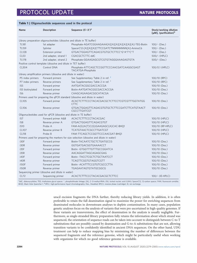
Table 1 | Oligonucleotide sequences used in the protocol
Name Description Sequence (5′–3′)a Stock/working dilution[µM], (purification)b
Library preparation oligonucleotides (dissolve and dilute in TE buffer)
TL181 1st adapter Phosphate-AGATCGGAAGAAA[A][A][A][A][A][A][A]-TEG-Biotin 100/- (Des.)
TL159 Splinter SpacerC12-[A][A][A]CTTCCGATCTNNNNNNNN[A]-AminoC6 100/- (Des.)
CL128 Extension primer GTGACTGGAGTTCAGACGTGTGCTCTTCC*G*A*T*C*T 100/- (Des.)
CL53 2nd adapter, strand 1 CGACGCTCTTC-ddC 500/- (HPLC)
TL178 2nd adapter, strand 2 Phosphate-GGAAGAGCGTCGTGTAGGGAAAGAGTGTA 500/- (Des.)
Positive control template (dissolve and dilute in TET buffer)
CL304 Control DNA Phosphate-ATTCAGCTCCGGTTCCCAACGATCAAGGCGAGTTACATGA-Phosphate
100/0.1 (HPLC)
Library amplification primers (dissolve and dilute in water)
P5 index primers Forward primers See Supplementary Table 2 in ref. 2 100/10 (RPC)
P7 index primers Reverse primers See Supplementary Table 2 in ref. 2 100/10 (RPC)
IS5 Forward primer AATGATACGGCGACCACCGA 100/10 (Des.)
IS5 biotinylated Forward primer Biotin-AATGATACGGCGACCACCGA 100/10 (Des.)
IS6 Reverse primer CAAGCAGAAGACGGCATACGA 100/10 (Des.)
Primers used for preparing the qPCR standard (dissolve and dilute in water)
CL105 Forward primer ACACTCTTTCCCTACACGACGCTCTTCCTCGTCGTTTGGTATGGCTTC
100/10 (Des.)
CL106 Reverse primer GTGACTGGAGTTCAGACGTGTGCTCTTCCGATCTTCATGTAACTCGCCTTGATCGT
100/10 (Des.)
Oligonucleotides used for qPCR (dissolve and dilute in TE buffer)
IS7 Forward primer A&B ACACTCTTTCCCTACACGAC 100/10 (HPLC)
IS8 Reverse primer A GTGACTGGAGTTCAGACGTGT 100/10 (HPLC)
IS10 Probe A FAM-A{G}A{T}C{G}GAAGAGC{A}CAC-BHQ1 100/10 (RP-HPLC)
CL107 Reverse primer B TCATGTAACTCGCCTTGATCGT 100/10 (HPLC)
CL118 Probe B FAM-TTCAGCTCCGGTTCCCAACGAT-BHQ1 100/10 (HPLC)
Primers used for preparing the markers for size selection (dissolve and dilute in water)
i30F Forward primer Biotin-TACAATCTGCTCTGATGCCG 100/10 (Des.)
i30R Reverse primer GGTGATGACGGTGAAAACCT 100/10 (Des.)
i35F Forward primer Biotin -GTGGTTTGTTTGCCGGATCA 100/10 (Des.)
i35R Reverse primer AACAGGATTAGCAGAGCGAG 100/10 (Des.)
i40F Forward primer Biotin -TACCTCGCTCTGCTAATCCT 100/10 (Des.)
i40R Reverse primer TCAGTTCGGTGTAGGTCGTT 100/10 (Des.)
i150F Forward primer Biotin -ACATTTCCGTGTCGCCCTTA 100/10 (Des.)
i150R Reverse primer TGAGAATAGTGTATGCGGCG 100/10 (Des.)
Sequencing primer (dissolve and dilute in water)
CL72 Sequencing primer ACACTCTTTCCCTACACGACGCTCTTCC 100/- (IE-HPLC)
addC, dideoxycytidine; TEG, triethylene glycol spacer; *, phosphothioate linkage; [N], 2′-O-methyl-RNA; {N}, locked nucleic acid (LNA); SpacerC12, 12-carbon spacer; FAM, fluorescein amidite;BHQ1, Black Hole Quencher-1. bHPLC, high-performance liquid chromatography; Des., Desalted; RP(C), reverse phase (cartridge); IE, ion exchange.
PROTOCOL UPDATE NATURE PROTOCOLS
2284 NATURE PROTOCOLS | VOL 15 |AUGUST 2020 | 2279–2300 |www.nature.com/nprot

Library amplificationThe protocol described here involves the amplification of libraries by PCR. This step is necessary toprevent the loss of molecules during sequencing and to ‘immortalize’ the content of a library so thatit remains available for future use—for example, to target parts of the genome by hybridizationcapture40–42. When amplifying libraries for the first time, pairs of sample-specific indices are intro-duced so that libraries from multiple samples and controls can be sequenced simultaneously43. Owingto the low concentration of template DNA in these indexing PCR reactions, a hot-start enzyme,AccuPrime Pfx DNA polymerase, is used to suppress the formation of primer dimers. The proof-reading activity of the enzyme does not interfere with the amplification of uracil-containing librarymolecules, as the template strands created during library preparation are copies of the original samplemolecules. Subsequent re-amplification of libraries, which is necessary, for example, as part of thesize-selection protocol or hybridization capture, is performed with Herculase II Fusion DNA poly-merase, a less costly enzyme with proofreading but no hot-start activity.
Although the two DNA polymerases included in this protocol have been chosen to minimizelength and base composition biases that are associated with PCR amplification of heterogeneoustemplate molecules44, it is advisable, if possible, to perform only as many PCR cycles as necessary toobtain large enough quantities of library for downstream experiments. For manual library prepara-tion, we suggest to determine the optimal PCR cycle number separately for each sample based on theamplification plots obtained from the qPCR measurement (see Box 2 for instructions). For automatedlibrary preparation, where all libraries are amplified simultaneously in plate format, it is inevitable toset a fixed cycle number for all libraries. To be compatible with high-throughput screening, wesuggest that this number is chosen so that all reactions reach PCR plateau (see Supplementary
Box 1 | Detecting sporadic inefficiencies in library preparation
The probe-based qPCR assay depicted below (Steps 32–37 of the protocol) allows for independently determiningthe number of library molecules obtained from the control oligonucleotide in each reaction, including the negativecontrols containing no sample DNA. In theory, the number of control library molecules should be uniform acrossall reactions if library preparation was correctly performed for all samples. However, impurity of the sample DNAmight occasionally lead to an inhibition of enzymatic reactions during library preparation, reducing librarypreparation efficiency. Library preparation efficiency can be estimated by comparing the number of control librarymolecules generated in the sample libraries to those in the negative controls, as indicated below.
Efficiency =sample/average
of controls
IS7
CL107
CL118
qPCR measurement for control library molecules
For samplelibaries
For negativecontrols
Box 2 | Determining the optimal cycle number for library amplification using qPCR amplificationplots
1 Analyze the qPCR amplification plot independently for each sample to determine the number of cycles untilwhich amplification remains in the exponential phase.
2 Determine the relative quantity of sample DNA that will be used for indexing PCR, taking differences inreaction volume, input volume and the library dilution into account.
3 Assuming full PCR efficiency (i.e. a doubling of molecules in each cycle), determine the number of cycles thathave to be subtracted to compensate for the above differences in the amplification reaction.
Flu
ores
cenc
e
# cycles0 10 20
Reactionvolume
(µl)
Input ofDNA(µl)
Dilution
qPCR 25 1 1:50
IndexingPCR
100 20 -
e.g. Sample 2 (16 cycles in qPCR)
4× the reaction volume 2n = 4, n = 2 (2 cycles more)20× more sample DNA 2n = 20, n = 4.3 (~4 cycles less)50× less diluted 2n = 50, n = 5.6 (~6 cycles less)
Optimal cycle number of library
amplification:
16 – 8 = 8
1. 2.
4 20 50
3.
PCR plateau
Sample 2Sample 1
NATURE PROTOCOLS PROTOCOL UPDATE
NATURE PROTOCOLS | VOL 15 |AUGUST 2020 | 2279–2300 |www.nature.com/nprot 2285
Manual). This strategy accepts a small increase in PCR bias but enables pooling of all libraries inequal volumes before sequencing, as all libraries reach similar DNA concentrations in PCR plateau.
If libraries are amplified into PCR plateau, heteroduplexes are formed45—that is, molecules withsingle- and double-stranded regions that result from re-hybridization of the two library strands viatheir adapter sequences. These structures do not interfere with hybridization capture or sequencing,but they prevent the use of capillary gel electrophoresis for quantifying libraries before sequencingand might also distort concentration measurements based on fluorescent double-stranded DNAbinding dyes or spectrophotometry. If necessary, heteroduplexes can be removed by subjecting 500 ngof a purified library, or a pool of purified libraries, to a PCR with primers IS5 and IS6 as described inSteps 66 and 68 but using only a single PCR cycle (see Extended Data Fig. 1 for an overview of libraryamplification schemes used in this protocol).
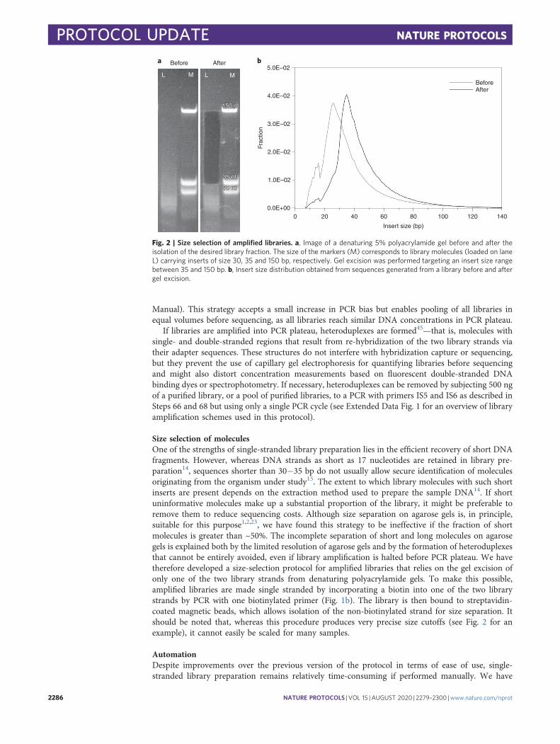
Size selection of moleculesOne of the strengths of single-stranded library preparation lies in the efficient recovery of short DNAfragments. However, whereas DNA strands as short as 17 nucleotides are retained in library pre-paration14, sequences shorter than 30−35 bp do not usually allow secure identification of moleculesoriginating from the organism under study15. The extent to which library molecules with such shortinserts are present depends on the extraction method used to prepare the sample DNA14. If shortuninformative molecules make up a substantial proportion of the library, it might be preferable toremove them to reduce sequencing costs. Although size separation on agarose gels is, in principle,suitable for this purpose1,2,23, we have found this strategy to be ineffective if the fraction of shortmolecules is greater than ~50%. The incomplete separation of short and long molecules on agarosegels is explained both by the limited resolution of agarose gels and by the formation of heteroduplexesthat cannot be entirely avoided, even if library amplification is halted before PCR plateau. We havetherefore developed a size-selection protocol for amplified libraries that relies on the gel excision ofonly one of the two library strands from denaturing polyacrylamide gels. To make this possible,amplified libraries are made single stranded by incorporating a biotin into one of the two librarystrands by PCR with one biotinylated primer (Fig. 1b). The library is then bound to streptavidin-coated magnetic beads, which allows isolation of the non-biotinylated strand for size separation. Itshould be noted that, whereas this procedure produces very precise size cutoffs (see Fig. 2 for anexample), it cannot easily be scaled for many samples.
AutomationDespite improvements over the previous version of the protocol in terms of ease of use, single-stranded library preparation remains relatively time-consuming if performed manually. We have
150 nt
35 nt
30 nt
Before After
L M L M
150 nt
35 nt
30 nt
a b5.0E–02
4.0E–02
3.0E–02
2.0E–02
1.0E–02
0.0E+000 20 40 60 80
Insert size (bp)
100 120 140
BeforeAfter
Frac
tion
Fig. 2 | Size selection of amplified libraries. a, Image of a denaturing 5% polyacrylamide gel before and after theisolation of the desired library fraction. The size of the markers (M) corresponds to library molecules (loaded on laneL) carrying inserts of size 30, 35 and 150 bp, respectively. Gel excision was performed targeting an insert size rangebetween 35 and 150 bp. b, Insert size distribution obtained from sequences generated from a library before and aftergel excision.
PROTOCOL UPDATE NATURE PROTOCOLS
2286 NATURE PROTOCOLS | VOL 15 |AUGUST 2020 | 2279–2300 |www.nature.com/nprot

therefore developed a version of the protocol that is compatible with liquid handling on the BravoNGS Workstation (Agilent Technologies). The electronic protocol files and instructions on how touse them are provided in a Supplementary Manual and on the Zenodo website (https://zenodo.org/record/3631147). However, we caution that, apart from the investment required for acquisition of thisliquid handling system, successful implementation and subsequent maintenance of the automatedprotocol requires substantial expertise in protocol development and troubleshooting on the system.The reason for this is that some parameters, such as pipetting distances and the force of the grippers,have to be adjusted slightly differently on each machine to ensure consistent and reliable performance(see the Supplementary Manual for advice). In addition, open liquid handling on 96-well platesgreatly increases the risk of cross-contamination among samples. We strongly advise to test for thispossibility after the implementation of the protocol and whenever further adjustments are made. Thiscan be achieved, for example, by generating libraries from the positive control oligonucleotide CL304as well as water controls that are alternatingly distributed across the entire plate in a chess-board-likesetup. A possible carryover of the oligonucleotide to neighboring wells can then be detected using theqPCR assay B described in Steps 32−37.
Limitations of the protocol and expertise requiredAlthough the implementation of automated single-stranded library preparation is challenging andshould be attempted only as part of a sustained effort to develop a large-scale sample preparationpipeline, a laboratory technician with experience in performing qPCR and library preparation shouldnot face problems implementing the manual version of single-stranded library preparation describedhere. One general constraint of the protocol is that the adapter sequences were specifically designed forIllumina sequencers and that libraries cannot easily be sequenced using other technologies. In addi-tion, one of the adapter sequences differs from standard Illumina adapters by a 5-bp deletion, whichrequires the use of a custom sequencing primer for the first insert read (see Extended Data Fig. 1 foradapter sequences and binding sites of primers used for library amplification and sequencing). Wehave not encountered difficulties with replacing sequencing primers on our in-house HiSeq and MiSeqmachines as well as when outsourcing projects to large sequencing centers. Nonetheless, it is advisableto clarify upfront with the sequencing unit whether the necessary modifications can be made.
Materials
Reagents● Ancient or damaged DNA or oligonucleotide sample to be sequenced (see ‘Reagent setup’section below)
● Oligonucleotide solutions for library preparation and amplification (see Table 1 and ‘Reagent setup’section below)
● Homemade buffers (see ‘Reagent setup’ section below)● Water, HPLC-grade (Merck, cat. no. 270733)● 5 M NaCl solution (Merck, cat. no. S5150-1L)● 1 M Tris-HCl solution, pH 8.0 (AppliChem, cat. no. A4577,0500)● 0.5 M EDTA solution, pH 8.0 (AppliChem, cat. no. A4892,1000)● Tween-20 (Merck, cat. no. P5927-100ML); also prepare a 2% (vol/vol) solution in water● 20× SSC buffer (Thermo Fisher Scientific, cat. no. AM9763)● 20% (wt/vol) SDS solution (Thermo Fisher Scientific, cat. no. AM9820) ! CAUTION Can cause skinand eye irritation. Wear gloves and eye protection.
● 3 M Sodium acetate, pH 5.2 (Merck, cat. no. S7899-500ML) ! CAUTION Can cause skin and eyeirritation. Wear gloves and eye protection.
● T4 RNA ligase reaction buffer (NEB, cat. no. B0216L), including 50% (wt/vol) PEG 8000● ATP solution (100 mM; Thermo Fisher Scientific, cat. no. R0441)● Klenow fragment, including 10× reaction buffer (Thermo Fisher Scientific, cat. no. EP0052)● T4 polynucleotide kinase 10 U µl−1 (Thermo Fisher Scientific, cat. no. EK0031)● FastAP thermosensitive alkaline phosphatase (Thermo Fisher Scientific, cat. no. EF0651)● T4 DNA ligase, high concentrated 30 U µl−1 (Thermo Fisher Scientific, cat. no. EL0013)● T4 DNA ligase, 5 U µl−1 (Thermo Fisher Scientific, cat. no. EL0012), including 10× reaction buffer and50% (wt/vol) PEG-4000
● Herculase II fusion DNA polymerase, including 5× Herculase II reaction buffer (Agilent, cat. no.600679)
NATURE PROTOCOLS PROTOCOL UPDATE
NATURE PROTOCOLS | VOL 15 |AUGUST 2020 | 2279–2300 |www.nature.com/nprot 2287

● Optional: USER enzyme (NEB, cat. no. M5505L)● Dynabeads MyOne Streptavidin C1 (Life Technologies, cat. no. 65001)● 25 mM each dNTP (Thermo Fisher Scientific, cat. no. R1121)● Maxima Probe qPCR Master Mix (2×; Thermo Fisher Scientific, cat. no. K0261)● AccuPrime Pfx DNA polymerase (Thermo Fisher Scientific, cat. no. 12344-024), including 10×AccuPrime reaction mix
● MinElute PCR Purification Kit (Qiagen, cat. no. 28006)● QIAquick Nucleotide Removal Kit (Qiagen, cat. no. 28306)● pUC 19 vector (NEB, cat. no. N3041S)● SYBR Gold Nucleic Acid Gel Stain (Thermo Fisher Scientific, cat. no. S11494)● 20/100 Ladder (IDT, cat. no. 51-05-15-02)● 5× Novex TBE Running Buffer (Thermo Fisher Scientific, cat. no. LC6675)● 2× TBE-Urea Sample Buffer (Bio-Rad, cat. no. 1610768)● Agilent DNA 1000 Kit (Agilent, cat. no. 5067-1504)
Equipment● Latex gloves (Roth, cat. no. L949.1)● Nitrile gloves (Ansell Health Care, cat. no. 112-2688)● 2-ml Safe-lock tubes (Eppendorf, cat. no. 0030120094)● 1.5-ml Safe-lock LoBind tubes (Eppendorf, cat. no. 0030120086)● 0.5-ml Safe-lock LoBind tubes (Eppendorf, cat. no. 0030121023)● 0.2-ml PCR eight-tube strips (Eppendorf, cat. no. 0030124359)● 96-well semi-skirted PCR plate (Thermo Fisher Scientific, cat. no. AB2400)● MicroAmp optical eight-cap strips (Thermo Fisher Scientific, cat. no. 4323032)● 50-ml tubes (Merck, cat. no. CLS430921)● Scalpel (Braun, cat. no. 10567364)● Sterican needle, 0.9 × 40 mm (Braun, cat. no. 2050798)● Racks for 0.2-ml, 0.5-ml and 1.5-ml tubes (many suppliers)● Magnetic-ring stand (96-well; Thermo Fisher Scientific, cat. no. AM10050)● Magnetic rack for 1.5-ml tubes (MagJET Separation Rack; Thermo Fisher Scientific, cat. no. MR02)● Rotator for 1.5-ml tubes (VWR, cat. no. 13916-822)● 5% Criterion TBE-urea polyacrylamide gel (Bio-Rad, cat. no. 3450086)● Criterion cell and PowerPac basic power supply (Bio-Rad, cat. no. 165-6019)● Rocking shaker (Grant-Bio PMR-30, Kisker, cat. no. 144201)● Dark reader transilluminator (Clare Chemical Research, cat. no DR196)● Microcentrifuge with adapters for 0.2-ml, 0.5-ml, 1.5-ml and 2-ml tubes (NeoLab, cat. no. D-8550)● Vortex mixer (Scientific Industries, cat. no. SI-0256) and, optionally, Centrifuge/Vortex MultispinMSC-6000 (BioSan, cat. no. BS-010211-AAL)
● Benchtop centrifuge for 1.5-ml and 2-ml tubes (Eppendorf, cat. no. 5420000318)● Plate centrifuge for 96-well PCR plates (Eppendorf, cat. no. 5948000913)● Thermal cyclers with lid heating—one for 0.2-ml PCR tubes and one for 0.5-ml tubes (DNA EngineThermal CyclerPTC-200 Thermo Cycler, MJ Research, cat. no. 8252-30-0001)
● Benchtop thermo mixer (ThermoMixer C; Eppendorf, cat. no. 5382000015)● 96-well qPCR system (CFX96 Touch Real-Time PCR Detection System; Bio-Rad, cat. no. 1855195)● 2100 Bioanalyzer (Agilent, cat. no. G2939BA)● Spectrophotosmeter (NanoDrop 2000/2000c, cat. no. ND-2000)● UV cross-linker (Bio-Link BLX 254, Vilber, cat. no. 611110811)● Illumina sequencing instrument (MiSeq, HiSeq, NovaSeq platforms) and related sequencing chemistry
Reagent setup
c CRITICAL All buffers and reagents below will suffice for at least 48 reactions. c CRITICAL Steps 1and 2 of the protocol are sensitive to contamination with exogenous DNA. We recommendUV-decontamination of the TET buffer and water used in these steps as well as when preparing thecontrol oligonucleotide dilutions. For this purpose, prepare at least three 1-ml aliquots of TET bufferand one of water in 1.5-ml tubes and UV-C (254-nm wavelength) irradiate them with a dose of 7 J/cm2
PROTOCOL UPDATE NATURE PROTOCOLS
2288 NATURE PROTOCOLS | VOL 15 |AUGUST 2020 | 2279–2300 |www.nature.com/nprot

in a cross-linker. Note that decontamination in 50-ml Falcon tubes with closed caps is ineffective owingto the thickness of the plastic walls.Bind and wash (B&W) buffer I (50 ml): Combine 47.125 ml of water, 1 ml of 5 M NaCl, 500 μl of 1 MTris-HCl (pH 8.0), 100 μl of 0.5 M EDTA (pH 8.0), 25 μl of Tween 20 and 1.25 ml of 20% (wt/vol)SDS. Store at room temperature (20−25 °C). Shelf life is 2 months.Wash buffer II (50 ml): Combine 48.375 ml of water, 1 ml of 5 M NaCl, 500 μl of 1 M Tris-HCl (pH 8.0),100 μl of 0.5 M EDTA (pH 8.0) and 25 μl of Tween 20. Store at room temperature for up to 1 year.Stringency wash buffer (50 ml): Combine 49.5 ml of water, 250 μl of 20% (wt/vol) SDS and 250 μl of20× SSC buffer. Store at room temperature. Shelf life is 2 months.TE buffer (50 ml): Combine 49.4 ml of water, 500 μl of 1 M Tris-HCl (pH 8.0) and 100 μl of 0.5 MEDTA (pH 8.0). Store at room temperature for up to 1 year.TET buffer (50 ml): Combine 49.375 ml of water, 500 μl of 1 M Tris-HCl (pH 8.0), 100 μl of 0.5 MEDTA (pH 8.0) and 25 μl of Tween 20. Store at room temperature for up to 1 year.Gel elution buffer (50 ml): Combine 44.375 ml of water, 5 ml 5 M NaCl, 500 μl of 1 M Tris-HCl (pH 8.0),100 μl of 0.5 M EDTA (pH 8.0) and 25 μl of Tween 20. Store at room temperature for up to 1 year.Melt buffer (1 ml): Combine 850 μl water, 125 μl 1M NaOH and 25 µl 2% (vol/vol) Tween 20. Alwaysprepare freshly.
Decontamination and hybridization of adapter/splinter mix for single-stranded ligation (60 µl)In the first tube of a 0.2-ml PCR eight-tube strip, combine 18 µl of water, 3 µl of 10× T4 RNA ligasereaction buffer, 6 µl of 100 µM adapter oligonucleotide TL181, 1.5 µl of 10 U µl−1 Klenow fragmentand 1.5 µl of 10 U µl−1 T4 polynucleotide kinase. In another tube of the strip, combine 12 µl of water,3 µl of 10× T4 RNA ligase reaction buffer, 12 µl of 100 µM splinter oligonucleotide TL159, 1.5 µl of10 U µl−1 Klenow fragment and 1.5 µl of 10 U µl−1 T4 polynucleotide kinase. Vortex the tubes andbriefly spin down in a microcentrifuge. Incubate the tube for 20 min at 37 °C in a thermal cycler,followed by 1 min at 95 °C to inactivate the enzyme. The oligonucleotides are now purified fromundesired synthesis artifacts and contaminant DNA. Combine the contents of both tubes in a tube ofa fresh eight-tube strip and incubate the mix at 95 °C for 10 s, followed by a ramp to 10 °C at 0.5 °Cs−1 in a thermal cycler. Transfer 60 µl of adapter/splinter mix with a final concentration of 10/20 µMinto a 1.5-ml tube. Store at −20 °C and thaw at room temperature before use.
Hybridization of double-stranded adapter for second ligation (100 µl)In one tube of an 0.2-ml PCR eight-tube strip, combine 9.5 μl of TE buffer, 0.5 μl of 5 M NaCl, 20 μlof 500 μM oligonucleotide CL53 and 20 μl of 500 μM oligonucleotide TL178. Vortex the tubes andbriefly spin down in a microcentrifuge. Hybridize the adapter oligonucleotides by incubating thereaction mix in a thermal cycler at 95 °C for 10 s, followed by a ramp to 14 °C at 0.1 °C s−1. Add 50 µlof TE for a final double-stranded adapter concentration of 100 µM. Store at −20 °C for up to 1 yearand thaw at room temperature when used.
Preparation of positive control oligonucleotide aliquotsDilute 100 µM stock solution of oligonucleotide CL304 to 0.1 µM by combining 999 µl of TET(UV-decontaminated) and 1 µl of stock solution. Mix well and prepare 10-µl aliquots in eight-tubestrips. Store at −20 °C for up to 1 year. Before starting a library preparation experiment, thaw onetube at room temperature, vortex the tube, spin the liquid down and prepare a 0.5 nM dilution bycombining 5 µl of the diluted oligonucleotide with 995 µl of TET. Vortex the tube, spin the liquiddown and prepare a final 10 pM oligonucleotide dilution by combining 2 µl of the previous dilutionwith 98 µl of TET. Vortex the tube and spin the liquid down.
Preparation of qPCR standardPerform a 50-µl PCR assay with AccuPrime Pfx DNA polymerase using 10 ng of pUC19 plasmidDNA as the template and primers CL105 and CL106 (Table 1). Follow the manufacturer’s instruc-tions and choose an annealing temperature of 60 °C for 30 cycles. Purify the PCR product using theMinElute PCR Purification Kit. Elute the DNA in 20 µl of TET and determine its concentration usinga spectrophotometer. The expected concentration range is 50–100 ng µl−1. Use 10 ng of the purifiedPCR product as template for a second amplification reaction with an arbitrary combination of P5 andP7 indexing primers. Purify the PCR product using the MinElute PCR Purification Kit and measurethe DNA concentration on a DNA 1000 chip using Bioanalyzer 2100. Prepare a ten-fold dilutionseries in TET ranging from 109 to 102 copies per µl. Store at −20 °C for up to 1 year.
NATURE PROTOCOLS PROTOCOL UPDATE
NATURE PROTOCOLS | VOL 15 |AUGUST 2020 | 2279–2300 |www.nature.com/nprot 2289

Preparation of single-stranded markers for gel excisionFor each desired size marker, perform a 100-µl PCR assay with AccuPrime Pfx DNA polymeraseusing the respective primer pair provided in Table 1 (one biotinylated and one non-biotinylatedprimer) and 10 ng of pUC19 plasmid as the template. Follow the manufacturer’s instructions andchoose an annealing temperature of 60 °C for 30 cycles. Purify the PCR product with the MinElutePCR Purification Kit. Elute the DNA in 20 µl of TET and determine its concentration using aspectrophotometer. The expected concentration range is 100−200 ng µl−1. Transfer 2 µg of DNA to afresh tube and fill up to 20 µl with water. Resuspend the stock solution of Dynabeads MyOneStreptavidin C1 by vortexing. Transfer 100 μl of bead suspension into a 1.5-ml tube, pellet the beadsusing a magnetic rack and discard the supernatant. Perform two bead washes by resuspending thebeads in 500 µl of B&W buffer I, pelleting the beads in a magnetic rack and discarding the super-natant. Resuspend the beads in 250 μl of B&W buffer I. Add the prepared PCR product (20 µl) to thebead suspension and repeatedly invert the tubes upside down on a rotator for 15 min at roomtemperature. Pellet the beads in a magnetic rack, discard the supernatant and wash the beads with500 μl of wash buffer II as described above. Pellet the beads again, discard the supernatant andresuspend the beads in 50 μl of melt buffer. Let the tubes stand at room temperature for 5 min, pelletthe beads and collect the supernatants. Add 10 μl of sodium acetate (3 M, pH 5.2) and purify theDNA using the Nucleotide Removal Kit following the manufacturer’s instructions but using MinElutecolumns (from the MinElute PCR Purification Kit) and eluting in 20 μl of TET. Measure the DNAconcentration using a spectrophotometer. The expected concentration range is 20–40 ng µl−1. Dilutethe marker with TET to 20 ng µl−1. Store at −20 °C for up to 1 year.
Procedure
Single-stranded DNA library preparationUracil removal, dephosphorylation and heat denaturation ● Timing 30 min1 Prepare the DNA samples in 0.5-ml tubes by pipetting the desired volume of DNA extract and
filling with TET to 30 µl. We recommend preparation of no more than 16 libraries in parallel,including at least one positive control (CL304) and one library preparation negative control withwater instead of sample DNA.
Optional: If removal of internal uracils is desired, fill tubes to only 29 µl and add 1 µl of USERenzyme mix (1 U µl−1). Mix gently by flicking the tubes with a finger, spin tubes briefly in amicrocentrifuge and incubate them in a thermal cycler for 1 h at 37 °C. Then continue with thenext step.
2 In a 1.5-ml tube, prepare a master mix containing the reagents below. Multiply the volumes by thenumber of reactions that are to be performed plus one in excess to ensure that the master mixsuffices for all samples. Mix the reagents by vortexing before adding the enzyme and mix gently byflicking the tube with a finger thereafter.
c CRITICAL STEP Always prepare a fresh 10 pM dilution of CL304 (see ʻReagent setupʼ).
Reagent Volume (μl) perreaction
Final concentration in reactionafter Step 3/Step7
Water 3.6 —
T4 RNA ligation buffer (10×) 8 1.75×/1×
Tween 20 (2% (vol/vol)) 2 0.09%/0.05%
Spike-in positive control CL304 (10 pM) 1 0.22 pM/0.125 pM
Fast AP (1 U µl−1) 1 0.02 U µl−1/inactive
3 Add 15.6 µl of the reaction master mix to the prepared DNA to obtain a total volume of 45.6 µl.4 Mix the tube contents by firmly flicking the side of each tube. Spin the tubes briefly in a
microcentrifuge and transfer to a PCR cycler.5 Incubate reactions at 37 °C for 10 min and at 95 °C for 2 min. Hold reactions at 4 °C afterwards.
Continue with the next step during the incubation phase.
Splinted ligation of first adapter ● Timing 1.5 h6 In a 2-ml tube, prepare a master mix containing the reagents below. Multiply the volumes by the
number of reactions that are to be performed plus one in excess. Pre-mix the reagents for 5 min by
PROTOCOL UPDATE NATURE PROTOCOLS
2290 NATURE PROTOCOLS | VOL 15 |AUGUST 2020 | 2279–2300 |www.nature.com/nprot

repeatedly inverting the tube upside down on a rotator before adding the enzyme. Repeatedly invertthe tubes upside down on a rotator at least 10 min thereafter to ensure that the reagents areproperly mixed.
c CRITICAL STEP PEG-8000 is highly viscous; pipette slowly. Vortexing instead of rotation is notrecommended, as it does not achieve homogenous mixing of the reagents.
Reagent Volume (μl) per reaction Final concentration in reaction
PEG-8000 (50% (wt/vol)) 32 20%
ATP (100 mM) 0.4 0.5 mM
TL181/TL159 (10/20 µM) 1 0.125/0.25 µMT4 DNA ligase high conc. (30 U µl−1) 1 0.375 U µl−1
Total 34.4 —
7 Add 34.4 µl of the reaction master mix to each sample from Step 5 to obtain a final reaction volumeof 80 µl.
8 Mix the tube contents repeatedly by firmly flicking the side of each tube. Spin tubes briefly in amicrocentrifuge and transfer to a PCR cycler.
c CRITICAL STEP Proper mixing is critical. Visually inspect each tube and ensure that there are nostreaks before moving on to the next step.
9 Incubate at 37 °C for 1 h and at 95 °C for 2 min. Hold at 10 °C afterwards.
j PAUSE POINT Samples can be stored at −20 °C for several days.
Immobilization of ligation products on beads ● Timing 45 min10 Resuspend the stock solution of Dynabeads MyOne Streptavidin C1 by vortexing. For each
reaction, transfer 20 μl of bead suspension into a 2-ml tube (e.g. 320 µl for 16 reactions). Pellet thebeads using a magnetic rack and discard the supernatant.
11 Add 500 µl of B&W buffer I and resuspend the beads by vortexing. Spin the tubes briefly in amicrocentrifuge, place on a magnetic rack and discard the supernatant. Repeat this step for a totalof two washes.
12 Resuspend the beads in a volume of B&W buffer I corresponding to 100 µl per reaction plus100 µl in excess (e.g. 1.7 ml for 16 reactions). Split the suspension into 100-µl aliquots in1.50-ml tubes
13 Add 100 µl of B&W buffer I to the ligation reactions from Step 9, mix by pipetting up and downand transfer the diluted ligation reaction to the bead suspension prepared in Step 12. The finalvolume of the bead suspension is 280 µl.
14 Repeatedly invert the tubes upside down on a rotator for 20 min at room temperature.
First bead wash ● Timing 20 min15 Spin the beads briefly in a microcentrifuge. Pellet the beads in a magnetic rack and discard the
supernatant.16 Add 200 µl of B&W buffer I and resuspend the beads by vortexing. Briefly spin the tubes in a
microcentrifuge, pellet the beads in a magnetic rack and discard the supernatant.17 Add 100 µl of Stringency wash buffer and resuspend the beads by vortexing. Incubate the tubes for
3 min at 45 °C in a thermo mixer without shaking. Pellet the beads in a magnetic rack and discardthe supernatant.
c CRITICAL STEP In this step, vortex slowly to avoid spilling liquid on the tube walls and cap.If spilling occurs, spin the tubes in a microcentrifuge and resuspend the beads more gently. Werecommend using a spin-mix-spin device (see Equipment) for spinning and vortexing to performboth actions in the most time-effective manner.
18 Add 200 µl of wash buffer II and resuspend the beads by vortexing. Briefly spin the tubes in amicrocentrifuge and leave them in a rack on the bench.
Second-strand synthesis ● Timing 45 min19 In a 2-ml tube, prepare a master mix containing the reagents below. Multiply the volumes by the
number of reactions that are to be performed plus one in excess. Mix the reagents by vortexingbefore adding the enzyme and mix gently by flicking the tube with a finger thereafter.
NATURE PROTOCOLS PROTOCOL UPDATE
NATURE PROTOCOLS | VOL 15 |AUGUST 2020 | 2279–2300 |www.nature.com/nprot 2291
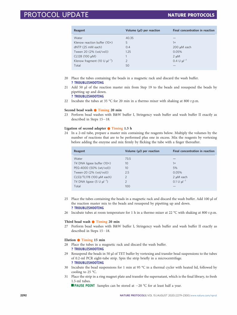
Reagent Volume (μl) per reaction Final concentration in reaction
Water 40.35 —
Klenow reaction buffer (10×) 5 1×
dNTP (25 mM each) 0.4 200 µM each
Tween 20 (2% (vol/vol)) 1.25 0.05%
CL128 (100 μM) 1 2 µMKlenow fragment (10 U µl−1) 2 0.4 U µl−1
Total 50 —
20 Place the tubes containing the beads in a magnetic rack and discard the wash buffer.? TROUBLESHOOTING
21 Add 50 µl of the reaction master mix from Step 19 to the beads and resuspend the beads bypipetting up and down.? TROUBLESHOOTING
22 Incubate the tubes at 35 °C for 20 min in a thermo mixer with shaking at 800 r.p.m.
Second bead wash ● Timing 20 min23 Perform bead washes with B&W buffer I, Stringency wash buffer and wash buffer II exactly as
described in Steps 15−18.
Ligation of second adapter ● Timing 1.5 h24 In a 2-ml tube, prepare a master mix containing the reagents below. Multiply the volumes by the
number of reactions that are to be performed plus one in excess. Mix the reagents by vortexingbefore adding the enzyme and mix firmly by flicking the tube with a finger thereafter.
Reagent Volume (μl) per reaction Final concentration in reaction
Water 73.5 —
T4 DNA ligase buffer (10×) 10 1×
PEG-4000 (50% (wt/vol)) 10 5%
Tween-20 (2% (vol/vol)) 2.5 0.05%
CL53/TL178 (100 µM each) 2 2 µM each
T4 DNA ligase (5 U µl−1) 2 0.1 U µl−1
Total 100 —
25 Place the tubes containing the beads in a magnetic rack and discard the wash buffer. Add 100 µl ofthe reaction master mix to the beads and resuspend by pipetting up and down.? TROUBLESHOOTING
26 Incubate tubes at room temperature for 1 h in a thermo mixer at 22 °C with shaking at 800 r.p.m.
Third bead wash ● Timing 20 min27 Perform bead washes with B&W buffer I, Stringency wash buffer and wash buffer II exactly as
described in Steps 15−18.
Elution ● Timing 15 min28 Place the tubes in a magnetic rack and discard the wash buffer.
? TROUBLESHOOTING29 Resuspend the beads in 50 µl of TET buffer by vortexing and transfer bead suspensions to the tubes
of 0.2-ml PCR eight-tube strip. Spin the strip briefly in a microcentrifuge.? TROUBLESHOOTING
30 Incubate the bead suspensions for 1 min at 95 °C in a thermal cycler with heated lid, followed bycooling to 25 °C.
31 Place the strip in a ring magnet plate and transfer the supernatant, which is the final library, to fresh1.5-ml tubes.
j PAUSE POINT Samples can be stored at −20 °C for at least half a year.
PROTOCOL UPDATE NATURE PROTOCOLS
2292 NATURE PROTOCOLS | VOL 15 |AUGUST 2020 | 2279–2300 |www.nature.com/nprot
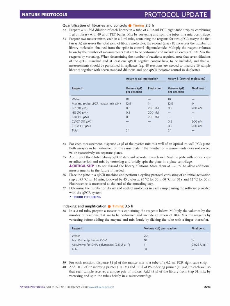
Quantification of libraries and controls ● Timing 2.5 h32 Prepare a 50-fold dilution of each library in a tube of a 0.2-ml PCR eight-tube strip by combining
1 µl of library with 49 µl of TET buffer. Mix by vortexing and spin the tubes in a microcentrifuge.33 Prepare two master mixes, each in a 2-ml tube, containing the reagents for two qPCR assays: the first
(assay A) measures the total yield of library molecules; the second (assay B) measures the number oflibrary molecules obtained from the spike-in control oligonucleotide. Multiply the reagent volumesbelow by the number of measurements that are to be performed and include an excess of 10%. Mix thereagents by vortexing. When determining the number of reactions required, note that seven dilutionsof the qPCR standard and at least one qPCR negative control have to be included, and that allmeasurements should be performed in replicates (e.g. 48 reactions are needed to measure 16 samplelibraries together with seven standard dilutions and one qPCR negative control in duplicate).
Assay A (all molecules) Assay B (control molecules)
Reagent Volume (μl)per reaction
Final conc. Volume (μl)per reaction
Final conc.
Water 10 — 10 —
Maxima probe qPCR master mix (2×) 12.5 1× 12.5 1×
IS7 (10 µM) 0.5 200 nM 0.5 200 nM
IS8 (10 µM) 0.5 200 nM — —
IS10 (10 µM) 0.5 200 nM — —
CL107 (10 µM) — — 0.5 200 nM
CL118 (10 µM) — — 0.5 200 nM
Total 24 24 —
34 For each measurement, dispense 24 µl of the master mix to a well of an optical 96-well PCR plate.Both assays can be performed on the same plate if the number of measurements does not exceed96 or successively on separate plates.
35 Add 1 µl of the diluted library, qPCR standard or water to each well. Seal the plate with optical capsor adhesive foil and mix by vortexing and briefly spin the plate in a plate centrifuge.
c CRITICAL STEP Do not discard the library dilutions. Store them at −20 °C to allow additionalmeasurements in the future if needed.
36 Place the plate in a qPCR machine and perform a cycling protocol consisting of an initial activationstep at 95 °C for 10 min, followed by 45 cycles at 95 °C for 30 s, 60 °C for 30 s and 72 °C for 30 s.Fluorescence is measured at the end of the annealing step.
37 Determine the number of library and control molecules in each sample using the software providedwith the qPCR system.? TROUBLESHOOTING
Indexing and amplification ● Timing 3.5 h38 In a 2-ml tube, prepare a master mix containing the reagents below. Multiply the volumes by the
number of reactions that are to be performed and include an excess of 10%. Mix the reagents byvortexing before adding the enzyme and mix firmly by flicking the tube with a finger thereafter.
Reagent Volume (μl) per reaction Final conc.
Water 20 —
AccuPrime Pfx buffer (10×) 10 1×
AccuPrime Pfx DNA polymerase (2.5 U µl−1) 1 0.025 U µl−1
Total 31 —
39 For each reaction, dispense 31 µl of the master mix to a tube of a 0.2-ml PCR eight-tube strip.40 Add 10 µl of P7 indexing primer (10 µM) and 10 µl of P5 indexing primer (10 µM) to each well so
that each sample receives a unique pair of indices. Add 49 µl of the library from Step 31, mix byvortexing and spin the tubes briefly in a microcentrifuge.
NATURE PROTOCOLS PROTOCOL UPDATE
NATURE PROTOCOLS | VOL 15 |AUGUST 2020 | 2279–2300 |www.nature.com/nprot 2293
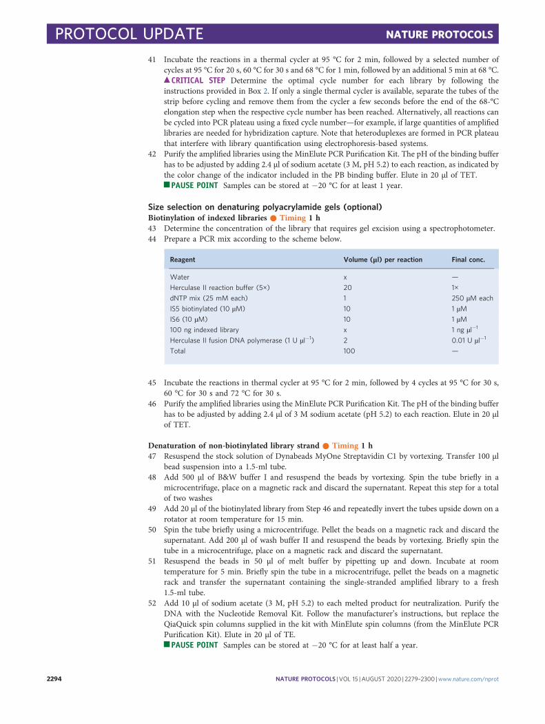
41 Incubate the reactions in a thermal cycler at 95 °C for 2 min, followed by a selected number ofcycles at 95 °C for 20 s, 60 °C for 30 s and 68 °C for 1 min, followed by an additional 5 min at 68 °C.
c CRITICAL STEP Determine the optimal cycle number for each library by following theinstructions provided in Box 2. If only a single thermal cycler is available, separate the tubes of thestrip before cycling and remove them from the cycler a few seconds before the end of the 68-°Celongation step when the respective cycle number has been reached. Alternatively, all reactions canbe cycled into PCR plateau using a fixed cycle number—for example, if large quantities of amplifiedlibraries are needed for hybridization capture. Note that heteroduplexes are formed in PCR plateauthat interfere with library quantification using electrophoresis-based systems.
42 Purify the amplified libraries using the MinElute PCR Purification Kit. The pH of the binding bufferhas to be adjusted by adding 2.4 µl of sodium acetate (3 M, pH 5.2) to each reaction, as indicated bythe color change of the indicator included in the PB binding buffer. Elute in 20 µl of TET.
j PAUSE POINT Samples can be stored at −20 °C for at least 1 year.
Size selection on denaturing polyacrylamide gels (optional)Biotinylation of indexed libraries ● Timing 1 h43 Determine the concentration of the library that requires gel excision using a spectrophotometer.44 Prepare a PCR mix according to the scheme below.
Reagent Volume (μl) per reaction Final conc.
Water x —
Herculase II reaction buffer (5×) 20 1×
dNTP mix (25 mM each) 1 250 µM each
IS5 biotinylated (10 µM) 10 1 µMIS6 (10 µM) 10 1 µM100 ng indexed library x 1 ng µl−1
Herculase II fusion DNA polymerase (1 U µl−1) 2 0.01 U µl−1
Total 100 —
45 Incubate the reactions in thermal cycler at 95 °C for 2 min, followed by 4 cycles at 95 °C for 30 s,60 °C for 30 s and 72 °C for 30 s.
46 Purify the amplified libraries using the MinElute PCR Purification Kit. The pH of the binding bufferhas to be adjusted by adding 2.4 µl of 3 M sodium acetate (pH 5.2) to each reaction. Elute in 20 µlof TET.
Denaturation of non-biotinylated library strand ● Timing 1 h47 Resuspend the stock solution of Dynabeads MyOne Streptavidin C1 by vortexing. Transfer 100 μl
bead suspension into a 1.5-ml tube.48 Add 500 µl of B&W buffer I and resuspend the beads by vortexing. Spin the tube briefly in a
microcentrifuge, place on a magnetic rack and discard the supernatant. Repeat this step for a totalof two washes
49 Add 20 µl of the biotinylated library from Step 46 and repeatedly invert the tubes upside down on arotator at room temperature for 15 min.
50 Spin the tube briefly using a microcentrifuge. Pellet the beads on a magnetic rack and discard thesupernatant. Add 200 µl of wash buffer II and resuspend the beads by vortexing. Briefly spin thetube in a microcentrifuge, place on a magnetic rack and discard the supernatant.
51 Resuspend the beads in 50 μl of melt buffer by pipetting up and down. Incubate at roomtemperature for 5 min. Briefly spin the tube in a microcentrifuge, pellet the beads on a magneticrack and transfer the supernatant containing the single-stranded amplified library to a fresh1.5-ml tube.
52 Add 10 μl of sodium acetate (3 M, pH 5.2) to each melted product for neutralization. Purify theDNA with the Nucleotide Removal Kit. Follow the manufacturer’s instructions, but replace theQiaQuick spin columns supplied in the kit with MinElute spin columns (from the MinElute PCRPurification Kit). Elute in 20 μl of TE.
j PAUSE POINT Samples can be stored at −20 °C for at least half a year.
PROTOCOL UPDATE NATURE PROTOCOLS
2294 NATURE PROTOCOLS | VOL 15 |AUGUST 2020 | 2279–2300 |www.nature.com/nprot

Size selection on a denaturing polyacrylamide gel ● Timing 2 d53 Take a precast 5% polyacrylamide urea gel from the fridge and let it equilibrate to room temperature.54 Prepare the sample for loading on a denaturing gel by combining the reagents below in a tube of a
0.2-ml PCR eight-tube strip.
Reagent Volume (μl) per reaction Final conc.
Water 7 —
Single-stranded amplified library 3 —
TBE-Urea sample buffer (2×) 10 1×
Total 20 —
55 Prepare a marker DNA mix by combining the reagents below in another tube of the eight-tube strip.
Reagent Volume (μl) per reaction Final conc.
Water (to 40 µl) 18.8 —
30-nucleotide marker (20 ng µl−1) 0.4 0.4 ng µl−1
35-nucleotide marker (20 ng µl−1) 0.4 0.4 ng µl−1
150-nucleotide marker (20 ng µl−1) 0.4 0.4 ng µl−1
TBE-Urea sample buffer (2×) 20 1×
Total 40 —
c CRITICAL STEP The markers suggested here are optimal for isolating library molecules withinsert sizes between 35 and 150 bp. The 30-bp marker is included to allow a visual control of sizeseparation. Adjust the composition of markers as needed if other insert sizes should be selected.
56 Incubate the DNA and marker mixes at 95 °C for 2 min in a thermal cycler.57 Mount the gel in the electrophoresis chamber and add TBE buffer to the top and bottom reservoirs.
Keep the packaging of the gel for later use. Remove the plastic comb and flush the gel pockets withTBE buffer by pipetting up and down. Load 20 µl of sample DNA into one of the pockets and 20 µlof marker DNA into each of the neighboring pockets (see Fig. 2a).
c CRITICAL STEP Load the sample and markers to the center of the gel, as the DNA does notalways run straight at the sides.
58 Apply a current of 200 V for 55 min (~12 V cm−1).59 In the meantime, heat up a needle or syringe with a diameter of approximately 0.9 mm using a
Bunsen burner or lighter and pierce a hole into the bottom of a 0.5-ml tube (see Fig. 1b, Tubepreparation, Step 59)
60 Use the packaging of the gel as reservoir for staining by adding 100 ml of TBE buffer and 10 µl ofSYBR Gold dye (10,000×). Gently shake the reservoir to mix dye and buffer on a rocking shaker.Break the gel cartridge open and remove the front cover containing the top reservoir. Submerge thegel in the reservoir while still attached to the plastic back of the cartridge. Stain for 5 min by gentlyshaking the reservoir on a rocking shaker.
61 Place the gel on the Dark Reader Transilluminator and make two horizontal cuts through the sampleand marker lanes, cutting along the marker bands. Then make two vertical cuts flanking the samplelane. Halve the gel slice lengthways and transfer both pieces into the 0.5-ml tube prepared in Step 59.Possible carryover of marker DNA is not critical, as the marker cannot be amplified and sequenced.? TROUBLESHOOTING
62 Place the pierced 0.5-ml tube containing the gel slice inside a 2-ml tube (see Fig. 1b for anillustration) and spin for 1 min at 10,000g in a benchtop centrifuge to break the gel into small piecesand collect them in the 2-ml tube.
63 Discard the empty 0.5-ml tube. Add 300 μl of gel elution buffer to the mashed gel and incubateovernight at room temperature in a thermo mixer with constant shaking at 800 r.p.m.
j PAUSE POINT Overnight incubation64 Spin the tube for 1 min at 10,000g in a benchtop centrifuge to pellet the gel. Transfer the supernatant
to a 15-ml Falcon tube and add 2.4 ml of PN buffer included with the Nucleotide Removal Kit. Add10 μl of 3 M sodium acetate (pH 5.2). Vortex the mixture and centrifuge the tube for 1 min at 1,500gat room temperature. Residual gel will pellet at the bottom of the tube. Carefully transfer 700 µl ofsupernatant to a MinElute column and centrifuge for 30 s at 10,000g. Discard the flow-through and
NATURE PROTOCOLS PROTOCOL UPDATE
NATURE PROTOCOLS | VOL 15 |AUGUST 2020 | 2279–2300 |www.nature.com/nprot 2295

repeat the transfer of supernatant to the column and centrifugation three times. Continue DNApurification from the PE buffer wash steps as detailed in the manual of the Nucleotide Removal Kit.Elute in 20 µl of TE buffer.
c CRITICAL STEP Avoid carryover of gel to the silica spin column. Leave some buffer in the Falcontube rather than disturbing the pellet.
65 Determine the concentration of library molecules by qPCR as described in Steps 32−37.66 To amplify the gel-excised library, prepare a PCR mix in one tube of a 0.2-ml PCR eight-tube strip
by combining the reagents below.
Reagent Volume (μl) per reaction Final conc.
Water 59 —
Herculase II reaction buffer (5×) 20 1×
dNTP (25 mM each) 1 250 µMIS5 (10 µM) 4 0.4 µMIS6 (10 µM) 4 0.4 µMGel-excised library 10 —
Herculase II fusion DNA Polymerase (1 U µl−1) 2 0.01 U µl−1
Total 100 —
67 Incubate the reactions in a thermal cycler at 95 °C for 2 min, followed by a selected number ofcycles at 95 °C for 10 s, 60 °C for 30 s and 72 °C for 30 s.
c CRITICAL STEP Determine the optimal cycle number that avoids PCR plateau by following theinstructions provided in Box 2.
68 Purify the amplified libraries using the MinElute PCR Purification Kit. The pH of the binding buffer hasto be adjusted by adding 2.4 µl of 3 M sodium acetate (pH 5.2) to each reaction. Elute in 20 µl of TET.
Library quantification and sequencing ● Timing 1−3 d69 Determine the concentration of each library using a DNA 1000 chip on the Bioanalyzer 2100 or
similar capillary electrophoresis systems. Alternatively, pool the libraries if multiplex sequencing isdesired and determine the concentration of the pool.
c CRITICAL STEP If indexing PCR has reached plateau, it is necessary to remove heteroduplexes beforeperforming capillary gel electrophoresis (see ‘Experimental design’ section in the Introduction for details).? TROUBLESHOOTING
70 For sequencing, follow the instructions provided by Illumina for multiplexed sequencing, butreplace the first read sequencing primer by CL72. A fresh dilution of this primer should be preparedbefore each sequencing run by combining 10 µl of 100 µM CL72 with 1.99 ml of hybridizationbuffer provided in the kit containing the sequencing reagents. We recommend paired-endsequencing (e.g. 2 × 75 bp) for ancient DNA. Approximately 1 million sequence reads per samplelibrary are usually sufficient to determine the proportion of mapped sequences, their lengthdistribution and DNA damage profiles, and to estimate the content of informative molecules in thelibrary (see ‘Anticipated results’ section). Fewer sequences (~200,000) are needed from the negativecontrols that were included during DNA extraction and/or library preparation.
Troubleshooting
Troubleshooting advice can be found in Table 2.
Table 2 | Troubleshooting table
Step Problem Possible reason Solution
21, 25, 29 Beads appear clumpy Beads dried out Usually not critical. Continue library preparation
20, 25, 28 Foam remains on the beads afterremoval of the wash buffer andbefore adding reaction mix orelution buffer
Detergent in the wash buffer causesfoaming if beads are resuspended tooharshly
Does not affect library yield; try to resuspendbeads more gently (centrifuge/vortex multispindevice recommended)
Table continued
PROTOCOL UPDATE NATURE PROTOCOLS
2296 NATURE PROTOCOLS | VOL 15 |AUGUST 2020 | 2279–2300 |www.nature.com/nprot
Timing
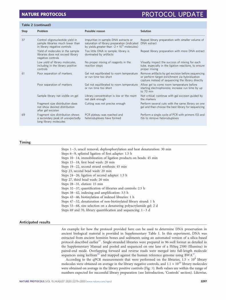
Steps 1−5, uracil removal, dephosphorylation and heat denaturation: 30 minSteps 6−9, splinted ligation of first adapter: 1.5 hSteps 10−14, immobilization of ligation products on beads: 45 minSteps 15−18, first bead wash: 20 minSteps 19−22, second strand synthesis: 45 minStep 23, second bead wash: 20 minSteps 24−26, ligation of second adapter: 1.5 hStep 27, third bead wash: 20 minSteps 28−31, elution: 15 minSteps 32−37, quantification of libraries and controls: 2.5 hSteps 38−42, indexing and amplification: 3.5 hSteps 43−46, biotinylation of indexed libraries: 1 hSteps 47−52, denaturation of non-biotinylated library strand: 1 hSteps 53−68, size selection on a denaturing polyacrylamide gel: 2 dSteps 69 and 70, library quantification and sequencing: 1−3 d
Anticipated results
An example for how the protocol provided here can be used to determine DNA preservation inancient biological material is provided in Supplementary Table 1. In this experiment, DNA wasextracted from ancient hominin bones and sediments using an automated version of a silica-basedprotocol described earlier37. Single-stranded libraries were prepared in 96-well format as detailed inthe Supplementary Manual and pooled and sequenced on one lane of a HiSeq 2500 (Illumina) inpaired-end mode. Overlapping forward and reverse reads were merged into full-length moleculesequences using leeHom46 and mapped against the human reference genome using BWA47.
According to the qPCR measurements that were performed on the libraries, 1.5 × 10⁸ librarymolecules were obtained on average in the library negative controls, and 1.1 × 10¹⁰ library moleculeswere obtained on average in the library positive controls (Fig. 3). Both values are within the range ofnumbers expected for successful library preparation (see Introduction, ‘Controls’ section). Likewise,
Table 2 (continued)
Step Problem Possible reason Solution
37 Control oligonucleotide yield insample libraries much lower thanin library negative controls
Impurities in sample DNA extracts orsaturation of library preparation (indicatedby yields greater than ~2 × 10¹¹ molecules)
Repeat library preparation with smaller volume ofDNA extract
Yield of molecules in the samplelibraries does not exceed librarynegative controls
Too little DNA in sample; library isdominated by artifacts
Repeat library preparation with more DNA extract
Low yield of library molecules,including in the library positivecontrols
No proper mixing of reagents in thereaction steps
Visually inspect the success of mixing for eachtube, especially in the ligation reactions, to ensureproper mixing
61 Poor separation of markers Gel not equilibrated to room temperatureor run time too short
Remove artifacts by gel excision before sequencingor perform target enrichment via hybridizationcapture instead of sequencing the library directly
Poor separation of markers Gel not equilibrated to room temperatureor run time too short
Allow gel to come room temperature beforestarting electrophoresis; increase run time by upto 70 min
Sample library not visible on gel Library concentration is low or the roomnot dark enough
Not critical; continue with gel excision guided bythe markers
Fragment size distribution doesnot show desired distributionafter gel excision
Cutting was not precise enough Perform several cuts with the same library on onegel and then choose the best library for sequencing
69 Fragment size distribution showsa secondary peak of unexpectedlylong library molecules
PCR plateau was reached andheteroduplexes have formed
Perform a single cycle of PCR with primers IS5 andIS6 to remove heteroduplexes
NATURE PROTOCOLS PROTOCOL UPDATE
NATURE PROTOCOLS | VOL 15 |AUGUST 2020 | 2279–2300 |www.nature.com/nprot 2297
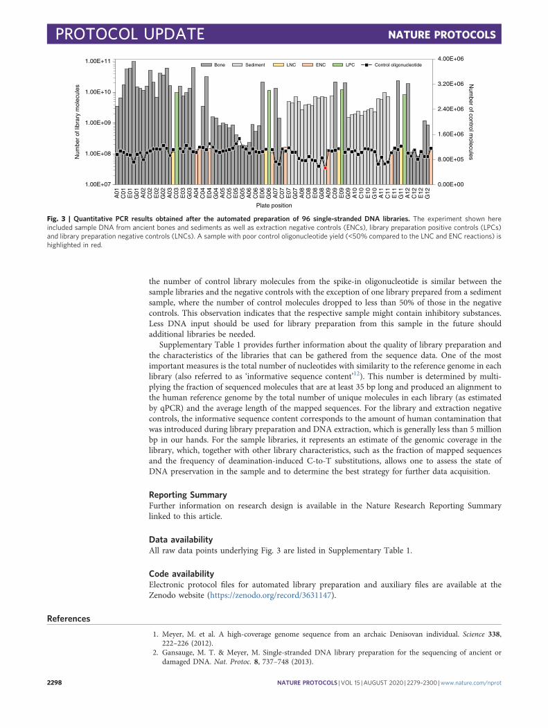
the number of control library molecules from the spike-in oligonucleotide is similar between thesample libraries and the negative controls with the exception of one library prepared from a sedimentsample, where the number of control molecules dropped to less than 50% of those in the negativecontrols. This observation indicates that the respective sample might contain inhibitory substances.Less DNA input should be used for library preparation from this sample in the future shouldadditional libraries be needed.
Supplementary Table 1 provides further information about the quality of library preparation andthe characteristics of the libraries that can be gathered from the sequence data. One of the mostimportant measures is the total number of nucleotides with similarity to the reference genome in eachlibrary (also referred to as ‘informative sequence content’12). This number is determined by multi-plying the fraction of sequenced molecules that are at least 35 bp long and produced an alignment tothe human reference genome by the total number of unique molecules in each library (as estimatedby qPCR) and the average length of the mapped sequences. For the library and extraction negativecontrols, the informative sequence content corresponds to the amount of human contamination thatwas introduced during library preparation and DNA extraction, which is generally less than 5 millionbp in our hands. For the sample libraries, it represents an estimate of the genomic coverage in thelibrary, which, together with other library characteristics, such as the fraction of mapped sequencesand the frequency of deamination-induced C-to-T substitutions, allows one to assess the state ofDNA preservation in the sample and to determine the best strategy for further data acquisition.
Reporting SummaryFurther information on research design is available in the Nature Research Reporting Summarylinked to this article.
Data availabilityAll raw data points underlying Fig. 3 are listed in Supplementary Table 1.
Code availabilityElectronic protocol files for automated library preparation and auxiliary files are available at theZenodo website (https://zenodo.org/record/3631147).
References
1. Meyer, M. et al. A high-coverage genome sequence from an archaic Denisovan individual. Science 338,222–226 (2012).
2. Gansauge, M. T. & Meyer, M. Single-stranded DNA library preparation for the sequencing of ancient ordamaged DNA. Nat. Protoc. 8, 737–748 (2013).
0.00E+00
8.00E+05
1.60E+06
2.40E+06
3.20E+06
4.00E+06
1.00E+07
1.00E+08
1.00E+09
1.00E+10
1.00E+11
A01
C01
E01
G01
A02
C02
E02
G02
A03
C03
E03
G03
A04
C04
E04
G04
A05
C05
E05
G05
A06
C06
E06
G06
A07
C07
E07
G07
A08
C08
E08
G08
A09
C09
E09
G09
A10
C10
E10
G10
A11
C11
E11
G11
A12
C12
E12
G12
Num
ber of control moleculesN
umbe
r of
libr
ary
mol
ecul
es
Plate position
Bone Sediment LNC ENC LPC Control oligonucleotide
Fig. 3 | Quantitative PCR results obtained after the automated preparation of 96 single-stranded DNA libraries. The experiment shown hereincluded sample DNA from ancient bones and sediments as well as extraction negative controls (ENCs), library preparation positive controls (LPCs)and library preparation negative controls (LNCs). A sample with poor control oligonucleotide yield (<50% compared to the LNC and ENC reactions) ishighlighted in red.
PROTOCOL UPDATE NATURE PROTOCOLS
2298 NATURE PROTOCOLS | VOL 15 |AUGUST 2020 | 2279–2300 |www.nature.com/nprot

3. Prüfer, K. et al. The complete genome sequence of a Neanderthal from the Altai Mountains. Nature 505,43–49 (2014).
4. Prüfer, K. et al. A high-coverage Neandertal genome from Vindija Cave in Croatia. Science 358, 655–658(2017).
5. Mafessoni, F. et al. A high-coverage Neandertal genome from Chagyrskaya Cave. Preprint at https://www.biorxiv.org/content/10.1101/2020.03.12.988956v1 (2020).
6. Fu, Q. et al. Genome sequence of a 45,000-year-old modern human from western Siberia. Nature 514,445–449 (2014).
7. Dabney, J. et al. Complete mitochondrial genome sequence of a Middle Pleistocene cave bear reconstructedfrom ultrashort DNA fragments. Proc. Natl Acad. Sci. USA 110, 15758–15763 (2013).
8. Meyer, M. et al. A mitochondrial genome sequence of a hominin from Sima de los Huesos. Nature 505,403–406 (2014).
9. Meyer, M. et al. Nuclear DNA sequences from the Middle Pleistocene Sima de los Huesos hominins. Nature531, 504–507 (2016).
10. Slon, V. et al. Neandertal and Denisovan DNA from Pleistocene sediments. Science 356, 605–608 (2017).11. Korlević, P. et al. Reducing microbial and human contamination in DNA extractions from ancient bones and
teeth. Biotechniques 59, 87–93 (2015).12. Gansauge, M. T. et al. Single-stranded DNA library preparation from highly degraded DNA using T4 DNA
ligase. Nucleic Acids Res. 45, e79 (2017).13. Gansauge, M. T. & Meyer, M. A method for single-stranded ancient DNA library preparation. Methods Mol.
Biol. 1963, 75–83 (2019).14. Glocke, I. & Meyer, M. Extending the spectrum of DNA sequences retrieved from ancient bones and teeth.
Genome Res. 27, 1230–1237 (2017).15. de Filippo, C., Meyer, M. & Prüfer, K. Quantifying and reducing spurious alignments for the analysis of ultra-
short ancient DNA sequences. BMC Biol. 16, 121 (2018).16. Bennett, E. A. et al. Library construction for ancient genomics: single strand or double strand? Biotechniques
56, 289–290 (2014).17. Wales, N. et al. New insights on single-stranded versus double-stranded DNA library preparation for ancient
DNA. Biotechniques 59, 368–371 (2015).18. Meyer, M. & Kircher, M. Illumina sequencing library preparation for highly multiplexed target capture and
sequencing. Cold Spring Harb. Protoc. 2010, pdb prot5448 (2010).19. Caroe, C. et al. Single-tube library preparation for degraded DNA. Methods Ecol. Evol. 9, 410–419 (2018).20. Briggs, A. W. et al. Patterns of damage in genomic DNA sequences from a Neandertal. Proc. Natl Acad. Sci.
USA 104, 14616–14621 (2007).21. Skoglund, P. et al. Separating endogenous ancient DNA from modern day contamination in a Siberian
Neandertal. Proc. Natl Acad. Sci. USA 111, 2229–2234 (2014).22. Gansauge, M. T. & Meyer, M. Selective enrichment of damaged DNA molecules for ancient genome
sequencing. Genome Res. 24, 1543–1549 (2014).23. Bokelmann, L. et al. A genetic analysis of the Gibraltar Neanderthals. Proc. Natl Acad. Sci. USA 116,
15610–15615 (2019).24. Stiller, M. et al. Single-strand DNA library preparation improves sequencing of formalin-fixed and paraffin-
embedded (FFPE) cancer DNA. Oncotarget 7, 59115–59128 (2016).25. Snyder, M. W., Kircher, M., Hill, A. J., Daza, R. M. & Shendure, J. Cell-free DNA comprises an in vivo
nucleosome footprint that informs its tissues-of-origin. Cell 164, 57–68 (2016).26. Burnham, P. et al. Single-stranded DNA library preparation uncovers the origin and diversity of ultrashort
cell-free DNA in plasma. Sci. Rep. 6, 27859 (2016).27. Turchinovich, A. et al. Capture and Amplification by Tailing and Switching (CATS). An ultrasensitive
ligation-independent method for generation of DNA libraries for deep sequencing from picogram amountsof DNA and RNA. RNA Biol. 11, 817–828 (2014).
28. Karlsson, K. et al. Amplification-free sequencing of cell-free DNA for prenatal non-invasive diagnosis ofchromosomal aberrations. Genomics 105, 150–158 (2015).
29. Raine, A., Manlig, E., Wahlberg, P., Syvanen, A. C. & Nordlund, J. SPlinted Ligation Adapter Tagging(SPLAT), a novel library preparation method for whole genome bisulphite sequencing. Nucleic Acids Res. 45,e36 (2017).
30. Wu, J., Dai, W., Wu, L. & Wang, J. SALP, a new single-stranded DNA library preparation method especiallyuseful for the high-throughput characterization of chromatin openness states. BMC Genomics 19, 143(2018).
31. Ding, J., Taylor, M. S., Jackson, A. P. & Reijns, M. A. Genome-wide mapping of embedded ribonucleotidesand other noncanonical nucleotides using emRiboSeq and EndoSeq. Nat. Protoc. 10, 1433–1444 (2015).
32. Petryk, N. et al. Replication landscape of the human genome. Nat. Commun. 7, 10208 (2016).33. Tin, M. M., Economo, E. P. & Mikheyev, A. S. Sequencing degraded DNA from non-destructively sampled
museum specimens for RAD-tagging and low-coverage shotgun phylogenetics. PLoS ONE 9, e96793 (2014).34. Troll, C. J. et al. A ligation-based single-stranded library preparation method to analyze cell-free DNA and
synthetic oligos. BMC Genomics 20, 1023 (2019).35. Knapp, M., Clarke, A. C., Horsburgh, K. A. & Matisoo-Smith, E. A. Setting the stage - building and working
in an ancient DNA laboratory. Ann. Anat. 194, 3–6 (2012).
NATURE PROTOCOLS PROTOCOL UPDATE
NATURE PROTOCOLS | VOL 15 |AUGUST 2020 | 2279–2300 |www.nature.com/nprot 2299
36. Kampmann, M. L., Borsting, C. & Morling, N. Decrease DNA contamination in the laboratories. Forensic Sci.Int. Genet. 6, E577–E578 (2017).
37. Rohland, N., Glocke, I., Aximu-Petri, A. & Meyer, M. Extraction of highly degraded DNA from ancientbones, teeth and sediments for high-throughput sequencing. Nat. Protoc. 13, 2447–2461 (2018).
38. Prüfer, K. snpAD: an ancient DNA genotype caller. Bioinformatics 34, 4165–4171 (2018).39. Varshney, U. & van de Sande, J. H. Specificities and kinetics of uracil excision from uracil-containing DNA
oligomers by Escherichia coli uracil DNA glycosylase. Biochemistry 30, 4055–4061 (1991).40. Burbano, H. A. et al. Targeted investigation of the Neandertal genome by array-based sequence capture.
Science 328, 723–725 (2010).41. Fu, Q. et al. DNA analysis of an early modern human from Tianyuan Cave, China. Proc. Natl Acad. Sci. USA
110, 2223–2227 (2013).42. Haak, W. et al. Massive migration from the steppe was a source for Indo-European languages in Europe.
Nature 522, 207–211 (2015).43. Kircher, M., Sawyer, S. & Meyer, M. Double indexing overcomes inaccuracies in multiplex sequencing on the
Illumina platform. Nucleic Acids Res 40, e3 (2012).44. Dabney, J. & Meyer, M. Length and GC-biases during sequencing library amplification: a comparison of
various polymerase-buffer systems with ancient and modern DNA sequencing libraries. Biotechniques 52,87–94 (2012).
45. Thompson, J. R., Marcelino, L. A. & Polz, M. F. Heteroduplexes in mixed-template amplifications: formation,consequence and elimination by ‘reconditioning PCR’. Nucleic Acids Res. 30, 2083–2088 (2002).
46. Renaud, G., Stenzel, U. & Kelso, J. leeHom: adaptor trimming and merging for Illumina sequencing reads.Nucleic Acids Res. 42, e141 (2014).
47. Li, H. & Durbin, R. Fast and accurate short read alignment with Burrows–Wheeler transform. Bioinformatics25, 1754–1760 (2009).
AcknowledgementsWe thank L. Bokelmann, E. Essel, L. Lippik, J. Richter, B. Schellbach and A. Weihmann for help in the lab, D. Massilani, S. Pääbo,B. Vernot and E. Zavala for helpful discussions, I. Bünger for help with installing software, J. Kelso and J. Visagie for help with dataprocessing and L. Jauregui for comments on the manuscript. This work was funded by the Max Planck Society.
Author contributionsM.-T.G. and M.M. developed the manual protocol. A.A.-P. and M.M. developed the automated protocol version, which was tested andoptimized by S.N. M.M. and M.-T.G. wrote the paper with help from A.A-P. and S.N.
Competing interestsThe authors declare no competing interests.
Additional informationExtended data is available for this paper at https://doi.org/10.1038/s41596-020-0338-0.
Supplementary information is available for this paper at https://doi.org/10.1038/s41596-020-0338-0.
Correspondence and requests for materials should be addressed to M-T.G. or M.M.
Peer review information Nature Protocols thanks Eva-Maria Geigl and the other, anonymous, reviewer(s) for their contribution to thepeer review of this work.
Reprints and permissions information is available at www.nature.com/reprints.
Received: 19 November 2019; Accepted: 17 April 2020;Published online: 1 July 2020
Related linksKey references using this protocolSlon, V. et al. Science 356, 605−608, (2017): https://doi.org/10.1126/science.aam9695Hajdinjak, M. et al. Nature 555, 652−656 (2018): https://doi.org/10.1038/nature26151Slon, V. et al. Nature 561, 113–116 (2018): https://doi.org/10.1038/s41586-018-0455-x
Protocol update to:Gansauge, M. & Meyer, M. Nat. Protoc. 8, 737–748 (2013): https://doi.org/10.1038/nprot.2013.038
PROTOCOL UPDATE NATURE PROTOCOLS
2300 NATURE PROTOCOLS | VOL 15 |AUGUST 2020 | 2279–2300 |www.nature.com/nprot

Extended Data Fig. 1 | Overview of the adapter sequences and library amplification primers used in this protocol. All sequences are shown in 5′–3′orientation.
NATURE PROTOCOLS PROTOCOL UPDATE
NATURE PROTOCOLS |www.nature.com/nprot

1
nature research | reporting summ
aryO
ctober 2018
Corresponding author(s): Matthias Meyer
Last updated by author(s): Feb 3, 2020
Reporting SummaryNature Research wishes to improve the reproducibility of the work that we publish. This form provides structure for consistency and transparency in reporting. For further information on Nature Research policies, see Authors & Referees and the Editorial Policy Checklist.
StatisticsFor all statistical analyses, confirm that the following items are present in the figure legend, table legend, main text, or Methods section.
n/a Confirmed
The exact sample size (n) for each experimental group/condition, given as a discrete number and unit of measurement
A statement on whether measurements were taken from distinct samples or whether the same sample was measured repeatedly
The statistical test(s) used AND whether they are one- or two-sided Only common tests should be described solely by name; describe more complex techniques in the Methods section.
A description of all covariates tested
A description of any assumptions or corrections, such as tests of normality and adjustment for multiple comparisons
A full description of the statistical parameters including central tendency (e.g. means) or other basic estimates (e.g. regression coefficient) AND variation (e.g. standard deviation) or associated estimates of uncertainty (e.g. confidence intervals)
For null hypothesis testing, the test statistic (e.g. F, t, r) with confidence intervals, effect sizes, degrees of freedom and P value noted Give P values as exact values whenever suitable.
For Bayesian analysis, information on the choice of priors and Markov chain Monte Carlo settings
For hierarchical and complex designs, identification of the appropriate level for tests and full reporting of outcomes
Estimates of effect sizes (e.g. Cohen's d, Pearson's r), indicating how they were calculated
Our web collection on statistics for biologists contains articles on many of the points above.
Software and codePolicy information about availability of computer code
Data collection Data processing was performed using the software supplied with the qPCR and DNA sequencing instruments specified in the manuscript. Summary statistics were generated using bwa, samtools and other software specified in the manuscript. Electronic protocol files for automated library preparation are published as part of this study.
Data analysis No special code or software was required for data analysis.
For manuscripts utilizing custom algorithms or software that are central to the research but not yet described in published literature, software must be made available to editors/reviewers. We strongly encourage code deposition in a community repository (e.g. GitHub). See the Nature Research guidelines for submitting code & software for further information.
DataPolicy information about availability of data
All manuscripts must include a data availability statement. This statement should provide the following information, where applicable: - Accession codes, unique identifiers, or web links for publicly available datasets - A list of figures that have associated raw data - A description of any restrictions on data availability
All molecule and sequence counts presented in Figure 3 are available in Supplementary Table 1.
2
nature research | reporting summ
aryO
ctober 2018
Field-specific reportingPlease select the one below that is the best fit for your research. If you are not sure, read the appropriate sections before making your selection.
Life sciences Behavioural & social sciences Ecological, evolutionary & environmental sciences
For a reference copy of the document with all sections, see nature.com/documents/nr-reporting-summary-flat.pdf
Life sciences study designAll studies must disclose on these points even when the disclosure is negative.
Sample size The paper details a method. The performance of the method was demonstrated by providing results from an experiment with 96 samples, which is the number of samples that fit on a microtiter plate.
Data exclusions No data were excluded, results from all samples are shown.
Replication Positive and negative controls were performed in several replicates to demonstrate the reproducibility of the method.
Randomization Not relevant, no comparisons were made between groups of samples.
Blinding Not relevant, all qPCR and sequence data were obtained using the instruments' software regardless of sample type or origin.
Reporting for specific materials, systems and methodsWe require information from authors about some types of materials, experimental systems and methods used in many studies. Here, indicate whether each material, system or method listed is relevant to your study. If you are not sure if a list item applies to your research, read the appropriate section before selecting a response.
Materials & experimental systemsn/a Involved in the study
Antibodies
Eukaryotic cell lines
Palaeontology
Animals and other organisms
Human research participants
Clinical data
Methodsn/a Involved in the study
ChIP-seq
Flow cytometry
MRI-based neuroimaging
PalaeontologySpecimen provenance DNA extracts from ancient bone and sediment samples were used solely to exemplify the technical feasibility of data generation
using the method presented. Only technical parameters are shown, with no reference to the age or provenance of the material, which are irrelevant in this context and are not reported in the manuscript.
Specimen deposition Material, or leftovers thereof, are stored at the Max Planck Institute for Evolutionary Anthropology in Leipzig, Germany.
Dating methods No dates were obtained for this study.
Tick this box to confirm that the raw and calibrated dates are available in the paper or in Supplementary Information.