Community Health Impact prenatal diagnosis by … the prevalence of congenital anomalies at birth,...
7
20ournal of Epidemtiology and Community Health 1994;48:290-296 Impact of prenatal diagnosis by ultrasound on the prevalence of congenital anomalies at birth in southern France C Julian-Reynier, N Philip, C Scheiner, Y Aurran, F Chabal, A Maron, A Gombert, S Ayme Abstract Study objective - The aims were (1) to assess whether termination of pregnancy after prenatal screening by ultrasound affected the prevalence of congenital anomalies at birth, and (2) to examine the trend of this pattern over time. Design - This study deals with congenital anomalies, possibly detectable prenatally or at birth, which were classified as iso- lated and multiple anomalies; chromo- somal anomalies were not included. The prevalence rates of congenital anomalies at birth were determined from case registration data in the Marseille district, France, from the registry of congenital malformations (Eurocat no 22), which covers 23 500 births a year. The x2 test for homogeneity in proportions was used to test whether the differences in the total prevalence rates were significant over time. Setting - The population was defined as all children born to parents living in the Marseille district between January 1 1984 and December 31 1990. Patients - Among the 164 509 pregnancy outcomes monitored during the study, 1795 children with a single congenital an- omaly and 288 with multiple congenital anomalies detectable at birth were assessed. Measurements and main results - The percentage of pregnancy terminations was higher in the case of multiple anom- alies (16%) than with single ones (7-5%). Leaving aside the lethal birth defects, this percentage became 7-9% in the case of multiple anomalies and 4-3% with isolated ones. A significant increase (p < 0-001) occurred over the seven year study period in the total percentage of terminations because of isolated anomal- ies but not in that involving multiple ones. The increase observed in the for- mer case was found to be mainly attrib- utable to an increase in the number of terminations of pregnancy undertaken because of anomalies which were either lethal or associated with very low survi- val rates (p < 0-001). Conclusions - Termination of pregnancy after prenatal ultrasound examination was found to have a definite impact on the prevalence at birth of lethal and con- genital anomalies with a low survival rate, and this impact tended to increase over time. No such impact was observed in the case of congenital anomalies asso- ciated with high survival rates. (J Epidemiol Community Health 1994;48:290-296) Congenital anomalies are one of the main causes of perinatal mortality and childhood morbidity and constitute a heavy economic burden for most industrialised countries. In mid 1989, they accounted for 30% of the deaths in the European populations analysed by Kalter,' corresponding to a perinatal death rate of around 0-85%. The former proportion has increased over the last few decades because of a reduction in perinatal mortality as a whole and not any change in the frequency of these anomalies, which has remained at a level of about 2% (Eurocat Report 4). In parallel, the emergence of prenatal diagnosis techniques and the legal availability of termination of pregnancy for medical or social reasons (since 1975 in France) have increased people's expec- tation that their children will be born without any malformations. During 1990, 23% of all the conceptions recorded in the region studied were terminated for social reasons by the 10th week of gestation (special analysis: Regional Directorate of Social and Health Programs). In France, prenatal diagnosis of chromo- somal anomalies by karyotyping has been available since the early 70s. In 1979, a strictly regulated structured network was set up to organise fetal karyotyping for pregnant women "at high risk" because of their age or their antecedents. In the meantime, prenatal ultra- sound examination was not subject to any particular ruling and gradually became the principal means of screening for congenital anomalies in the general population of preg- nant women. In the region of Provence,2 95% of all pregnant women had at least one ultra- sound examination duling their pregnancy in 1987, and there was an average of 3 5 ultra- sound examinations per pregnancy. In France, because there is no upper gestational age by which a pregnancy must be terminated when the fetus is severely malformed, late ultra- sound examinations are still thought to be an informative means of screening congenital an- omalies. This study aimed to assess the impact of termination of pregnancy after chromosomal anomalies, over a seven year period. We also proposed to examine a question which, to our knowledge, has not so far been investigated- INSERM U242 Centre de Genetique Medicale, H6pital d'Enfants de la Timone, 13385 Marseille cedex 5, France C Julian-Reynier N Philip Y Aurran F Chabal A Maron A Gombert S Ayme Laboratoire d'Anatomo- Pathologie, H6pital d'Adultes de la Timone, Marseille, France C Scheiner Correspondence to Dr C Julian-Reynier. Accepted for publication February 1994 290 on 18 May 2019 by guest. Protected by copyright. http://jech.bmj.com/ J Epidemiol Community Health: first published as 10.1136/jech.48.3.290 on 1 June 1994. Downloaded from
-
Upload
truongkiet -
Category
Documents
-
view
213 -
download
0
Transcript of Community Health Impact prenatal diagnosis by … the prevalence of congenital anomalies at birth,...
20ournal of Epidemtiology and Community Health 1994;48:290-296
Impact of prenatal diagnosis by ultrasound on
the prevalence of congenital anomalies at birth insouthern France
C Julian-Reynier, N Philip, C Scheiner, Y Aurran, F Chabal, A Maron, A Gombert,S Ayme
AbstractStudy objective - The aims were (1) toassess whether termination of pregnancyafter prenatal screening by ultrasoundaffected the prevalence of congenitalanomalies at birth, and (2) to examinethe trend of this pattern over time.Design - This study deals with congenitalanomalies, possibly detectable prenatallyor at birth, which were classified as iso-lated and multiple anomalies; chromo-somal anomalies were not included. Theprevalence rates of congenital anomaliesat birth were determined from caseregistration data in the Marseille district,France, from the registry of congenitalmalformations (Eurocat no 22), whichcovers 23 500 births a year. The x2 test forhomogeneity in proportions was used totest whether the differences in the totalprevalence rates were significant overtime.Setting - The population was defined asall children born to parents living in theMarseille district between January 1 1984and December 31 1990.Patients - Among the 164 509 pregnancyoutcomes monitored during the study,1795 children with a single congenital an-omaly and 288 with multiple congenitalanomalies detectable at birth wereassessed.Measurements and main results - Thepercentage of pregnancy terminationswas higher in the case of multiple anom-alies (16%) than with single ones (7-5%).Leaving aside the lethal birth defects,this percentage became 7-9% in the caseof multiple anomalies and 4-3% withisolated ones. A significant increase(p < 0-001) occurred over the seven yearstudy period in the total percentage ofterminations because of isolated anomal-ies but not in that involving multipleones. The increase observed in the for-mer case was found to be mainly attrib-utable to an increase in the number ofterminations of pregnancy undertakenbecause of anomalies which were eitherlethal or associated with very low survi-val rates (p < 0-001).Conclusions - Termination of pregnancyafter prenatal ultrasound examinationwas found to have a definite impact onthe prevalence at birth of lethal and con-genital anomalies with a low survivalrate, and this impact tended to increase
over time. No such impact was observedin the case of congenital anomalies asso-ciated with high survival rates.
(J Epidemiol Community Health 1994;48:290-296)
Congenital anomalies are one of the maincauses of perinatal mortality and childhoodmorbidity and constitute a heavy economicburden for most industrialised countries. Inmid 1989, they accounted for 30% of thedeaths in the European populations analysedby Kalter,' corresponding to a perinatal deathrate of around 0-85%. The former proportionhas increased over the last few decades becauseof a reduction in perinatal mortality as a wholeand not any change in the frequency of theseanomalies, which has remained at a level ofabout 2% (Eurocat Report 4). In parallel, theemergence of prenatal diagnosis techniquesand the legal availability of termination ofpregnancy for medical or social reasons (since1975 in France) have increased people's expec-tation that their children will be born withoutany malformations. During 1990, 23% of allthe conceptions recorded in the region studiedwere terminated for social reasons by the 10thweek of gestation (special analysis: RegionalDirectorate of Social and Health Programs).
In France, prenatal diagnosis of chromo-somal anomalies by karyotyping has beenavailable since the early 70s. In 1979, a strictlyregulated structured network was set up toorganise fetal karyotyping for pregnant women"at high risk" because of their age or theirantecedents. In the meantime, prenatal ultra-sound examination was not subject to anyparticular ruling and gradually became theprincipal means of screening for congenitalanomalies in the general population of preg-nant women. In the region of Provence,2 95%of all pregnant women had at least one ultra-sound examination duling their pregnancy in1987, and there was an average of 3 5 ultra-sound examinations per pregnancy. In France,because there is no upper gestational age bywhich a pregnancy must be terminated whenthe fetus is severely malformed, late ultra-sound examinations are still thought to be aninformative means of screening congenital an-omalies.This study aimed to assess the impact of
termination of pregnancy after chromosomalanomalies, over a seven year period. We alsoproposed to examine a question which, to ourknowledge, has not so far been investigated-
INSERM U242Centre de GenetiqueMedicale, H6pitald'Enfants de laTimone, 13385Marseille cedex 5,FranceC Julian-ReynierN PhilipY AurranF ChabalA MaronA GombertS Ayme
Laboratoired'Anatomo-Pathologie, H6pitald'Adultes de laTimone, Marseille,FranceC Scheiner
Correspondence toDr C Julian-Reynier.Accepted for publicationFebruary 1994
290
on 18 May 2019 by guest. P
rotected by copyright.http://jech.bm
j.com/
J Epidem
iol Com
munity H
ealth: first published as 10.1136/jech.48.3.290 on 1 June 1994. Dow
nloaded from

Prenatal US and prevalence of congenital anomalies
whether the anomalies leading to terminationof pregnancy were in fact mostly lethal. Forthis purpose, we used the geographicallydefined Birth Defects Registry of the Marseilledistrict, France, which belongs to theEuropean network of birth defects registries(Eurocat).
Method
STUDY POPULATIONThe study population consisted of a total of164 509 livebirths, stillbirths, and inducedabortions surveyed by the Marseille BirthDefects Registry (Eurocat 22) from January 11984 to December 31 1990. The registrationsystem covered all pregnancies with theseoutcomes involving mothers resident in theMarseille area, and the ascertainment of con-genital anomalies was based on the use ofmultiple sources of information includingspontaneous notifications and systematic checkof the hospital records of genetic and paedia-tric clinics. This systematic check was alwayscarried out by the same registered nurse (FC)employed by the research team, who wastrained to gather information about some spe-cific malformations only; not every sponta-neous notification was included in the recordbut only the relevant ones. Since 1984, theMarseille district registry has been includingall the malformed livebirths, all the termina-tions of pregnancy after prenatal detection of afetal anomaly, and all malformed fetal deathsoccurring after 20 weeks of gestation. Theanalysis was performed on the following threegroups: terminations of pregnancy, sponta-neous deaths from the 20th week of gestation
Table I Classification of subgroups for isolated anomalies. InternationalClassification of Diseases (9th Revision) and British Paediatric Association codes(ICD9/BPA)
Group Subgroup ICD9/BPA codesincluded
Cardiac anomalies: Left heart hypoplasia 7453, 7467Heart septal defects 7454, 7459Other major cardiac anomalies 7451, 7452, 7460-66
Nervous system anomalies: Microcephaly 7421Cyclopia/arhinencephaly 7422Anencephaly 7400Encephalocele 7420Meningocele-myelomeningocele 7412Spina bifida 7419Hydrocephaly 7423Hydranencephaly/porencephaly 7424.4
Digestive system anomalies: Oesophageal atresia 7503Duodenum/intestinal atresia 7511Anus imperforate/fistula 7512
Internal urogenital anomalies: Bilateral kidney agenesia 7530Unilateral kidney agenesia 7430.1Dysplastic kidney (bilateral) 7531.6Dysplasic kidney (unilateral) 7531.6Urinary bladder hypertrophy 7536Urinary bladder extrophy 7535
Limb anomalies: Limb reduction deformities 7552-56Syndactyly 7551Polydactyly 7550
Abdominal wall defects: Exomphalos 7567.01Gastroschisis 7567.11
Diaphragmatic hernia/agenesia 7566Other anomalies: Cleft lip/palate 7490-93
Anotiaimicrotia 7442Microphtalmy 7430-31Lung-tracheal anomalies 7483-85Biliary tract atresia (intrahepatic) 7526.52Biliary tract atresia (extrahepatic) 7516.51Sacrococcygeal teratoma 2392.99
up to the 7th day of life, and the survivors atone week. In the case of terminations of preg-nancy and deaths, a special procedure34 wasused systematically to specify the aetiologyinvolved. This procedure included gross andmicroscopic examination, karyotyping on skinfibroblasts, and skeletal x ray and photographs.
CONGENITAL ANOMALIES INCLUDED IN THESTUDYThe anomalies included are listed in table 1.This list consists of anomalies which can bediagnosed prenatally and are defined by theEurocat network5 as being reliably ascertainableat birth, except that chromosomal anomalieswere excluded even if expressed by malforma-tions. This was because the main way in whichchromosomal anomalies are detected prenatallyis through systematic fetal karyotyping in "highrisk" groups. Anomalies defined as not beingreliably ascertainable at birth are listed in theAppendix. All the cases included were reviewedby clinical geneticists qualified in syndromo-logy (NP, SA) to determine the lethality of themalformation pattern and the genetic or non-genetic origin of the defect. Cases in which twoor more major anomalies not classified as syn-dromes or associations occurred together wereclassified as "multiple malformations". Themedical records of the 111 multiple malforma-tions of unknown origin and the 22 amnioticband sequences were reviewed case by case todetermine whether these infants might havesurvived until 1 year of age. Survival expec-tancy was classified into three categories: lethal,for prenatally or neonatally lethal anomalies;the low survival category in which 50% of theinfants at least can be expected to die withinthe first year of life; and the high survivalcategory with a higher chance of survival up tothe age of 1 year. Medical records were alsochecked for renal, bladder, lung-tracheal an-omalies and teratoma, because the ICD9/BPAcodes did not give these details very fully. Thereference population was the total populationof live and stillborn infants in the same area(data from the National Institutes of Statisticsand Economic Studies) plus the induced abor-tions carried out because of congenital anomal-ies.
STATISTICAL ANALYSISTotal prevalence rates with their 95% confi-dence intervals (Poisson estimation) were cal-culated by dividing the number of malformedliveborns, stillborns, and induced abortions bythe total number of liveborns, stillborns, andinduced abortions (with a congenital anomaly)in the population. The prevalence rates ofmultiple anomalies with a Mendelian originwere also calculated. The secular trend wastested with the X2 test for homogeneity inproportions.6 The two components of this testare a x2 test to determine the significance of theincrease (slope) and a x2 test to check the linearslope hypothesis (HO: the trend is linear). Thex2 values were taken to be significant when theprobability of a type I error was less than 5%.
291
on 18 May 2019 by guest. P
rotected by copyright.http://jech.bm
j.com/
J Epidem
iol Com
munity H
ealth: first published as 10.1136/jech.48.3.290 on 1 June 1994. Dow
nloaded from
2Julian-Reynier, Philip, Scheiner, Aurran, Chabal, Maron, Gombert, Ayme
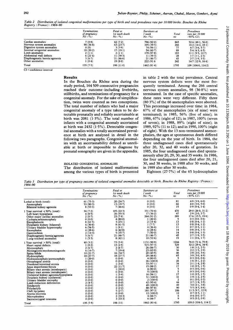
Table 2 Distribution of isolated congenital malformations per type of birth and total prevalence rate per 10 000 births. Bouches du Rh6neRegistry (France): 1984-90
Terminations Fetal or Survivors at Prevalenceof pregnancy Ist week death I week Total rate per 10000No (%) No (%) No (%) no (95% CI)
Cardiac anomalies 7 (0-8) 62 (7-2) 796 (92-0) 865 52-6 (49-1, 56-0)Nervous system anomalies 98 (36-8) 63 (23-7) 105 (39-5) 266 16-2 (14-2, 18-1)Digestive system anomalies 0 (0) 3 (3-9) 74 (96-1) 77 4 7 (3-6, 5-7)Intemal urogenital anomalies 18 (20-2) 17 (19 1) 54 (60-7) 89 5-4 (4-3, 6-5)Limb anomalies 2 (1-1) 2 (1-1) 179 (97-8) 183 11-1 (9-5, 12-7)Abdominal wall defects 6 (21-4) 11 (39-3) 11 (39-3) 28 1-7 (1-1, 2 3)Diaphragmatic hernia/agenesia 3 (6-7) 21 (46-6) 21 (46-7) 45 2 7 (1-9, 3-5)Other anomalies 1 (0-4) 19 (8-0) 222 (91-6 242 14-7 (12-9, 16-6)Total 135 (7-5) 198 (11-0) 1462 (81-4) 1795 109 (104-0, 114-2)CI = confidence interval
ResultsIn the Bouches du Rhone area during thestudy period, 164 509 consecutive pregnanciesreached their outcome -including livebirths,stillbirths, and terminations of pregnancy for a
congenital anomaly. For the sake of simplifica-tion, twins were counted as two conceptions.The total number of infants who had a majorcongenital anomaly of a type taken to be de-tectable prenatally and reliably ascertainable atbirth was 2081 (13%). The total number ofinfants with a congenital anomaly ascertainedat birth was 2432 (1-5%). Detectable congen-ital anomalies with a totally ascertained preval-ence at birth are analysed in detail in thefollowing two paragraphs. Congenital anomal-ies with an ascertainability defined as unreli-able at birth or impossible to diagnose byprenatal sonography are given in the appendix.
ISOLATED CONGENITAL ANOMALIESThe distribution of isolated malformationsamong the various types of birth is presented
in table 2 with the total prevalence. Centralnervous system defects were the most fre-quently terminated. Among the 266 centralnervous system anomalies, 98 (36 8%) were
terminated. In the case of specific anomalies,these rates were very different. Fifty three(80 3%) of the 66 anencephalies were aborted.This percentage increased over time: in 1984,67% of the anencephalies (six of nine) were
terminated; in 1985, 56% (five of nine); in1986, 67% (eight of 12); in 1987, 100% (sevenof seven); in 1988, 89% (eight of nine); in1989, 92% (11 to 12); and in 1990, 100% (eightof eight). With the 13 non-terminated anence-
phalies, the ages at spontaneous death differeddepending on the year of birth. In 1984, thethree undiagnosed cases died spontaneouslyafter 20, 32, and 40 weeks of gestation. In1985, the four undiagnosed cases died sponta-neously after 20,29, 30, and 35 weeks. In 1986,the four undiagnosed cases died after 20, 21,30, and 38 weeks, in 1988 after 30 weeks, andin 1989 also after 30 weeks.
Eighteen (27-7%) of the 65 hydrocephalies
Table 3 Distribution per type of pregnancy outcome of isolated congenital anomalies detectable at birth. Bouches du Rh6ne Registry (France):1984-90
Terminations Fetal or Survivors at Prevalenceof pregnancy Ist week death 1 week Total rate per 10 000No (%) No (%) No (%) no (95% CI)
Lethal at birth (total): 61 (75-3) 20 (24-7) 0 (0-0) 81 4-9 (3-9, 6-0)Anencephaly 53 (80-3) 13 (19-7) 0 (0-0) 66 4-0 (3-0, 5-0)Bilateral kidney agenesia 8 (53-3) 7 (46-6) 0 (0-0) 15 0 9 (0-4, 1-4)
1 Year survival <50% (total): 34 (7-2) 105 (22-3) 331 (70-4) 470 29-0 (26-0, 31-2)Left heart hypoplasia 4 (8-5) 26 (55-3) 17 (36-2) 47 2-9 (2-0, 3-7)Other major cardiac anomalies 2 (0-7) 23 (7-9) 264 (91-2) 289 17 6 (15-5, 19-6)Cyclopia/arhinencephaly 0 (0-0) 4 (80 0) 1 (20-0) 5 0-3 (0-0. 0-6)Encephalocele 8 (61-5) 3 (23 1) 2 (15-4) 13 0-8 (0-4, 1 2)Dysplastic kidney (bilateral) 4 (30-8) 8 (61-5) 1 (7-7) 13 0-8 (0-4, 1-2)Urinary bladder hypertrophy 6 (54-5) 1 (9-1) 4 (36-4) 11 0-7 (0-3, 1-1)Exomphalos 4 (28-6) 6 (42-8) 4 (28 6) 14 0-8 (0-4, 1-3)Gastroschisis 2 (143) 5 (35-7) 7 (50-0) 14 0-8 (04, 1-3)Diaphragmatic hernia/agenesia 3 (6-7) 21 (46 7) 21 (46-7) 45 2-7 (1-9, 3-5)Lung-tracheal anomalies 1 (5-3) 8 (42-1) 10 (52-6) 19 1-1 (0-6, 1-7)
1 Year survival >50% (total): 40 (3-2) 73 (5-9) 1131 (90-9) 1244 76-0 (71-4, 79-8)Heart septal defects 1 (0-2) 13 (2-5) 515 (97-3) 529 32-2 (29-4, 34-9)Microcephaly 2 (6-7) 2 (6-7) 26 (86-7) 30 1-8 (1-2, 2-5)Meningocele/myelomeningocele 6 (16-7) 7 (19-4) 23 (63-9) 36 2-2 (1-5, 2-9)Spina bifida 10 (21-7) 16 (34-7) 20 (43-5) 46 2-8 (2-0, 3-6)Hydrocephaly 18 (27-7) 18 (27-7) 29 (44-6) 65 3-9 (3-0, 4-9)Hydranencephaly/porencephaly 1 (20-0) 0 (0-0) 4 (80-0) 5 0-3 (0-0, 0-6)Oesophageal atresia 0 (0-0) 0 (0-0) 26 (100-0) 26 1-6 (0-1, 2-2)Duodenal/intestinal atresia 0 (0-0) 1 (3-0) 33 (97-1) 34 2-1 (1-4, 2-8)Anus imperforate/fistula 0 (0-0) 22 (11-7) 15 (88-2) 17 1-0 (0-5, 1-5)Biliary tract atresia (intrahepatic) 0 (0-0) 1 (20-0) 4 (80-0) 5 0-3 (0-0, 0-6)Biliary tract atresia (extrahepatic) 0 (0-0) 0 (0-0) 5 (100-0) 5 0-3 (0-0, 0-6)Unilateral kidney agenesia/hypoplasia 0 (0-0) 1 (5-9) 16 (94-1) 17 1-0 (0-5, 1-5)Dysplasic kidney (unilateral) 0 (0-0) 0 (0-0) 31 (100-0) 31 1-9 (1-2, 2-5)Urinary bladder extrophy 0 (0-0) 0 (0-0) 2 (100-0) 2 0-1 (0-0, 0-3)Limb reduction deformities 2 (4-5) 0 (0-0) 42 (95-4) 44 2-7 (1-9, 3-5)Syndactyly 0 (0-0) 0 (0-0) 49 (100-0) 49 3-0 (2-1, 3-8)Polydactyly 0 (0-0) 2 (2-2) 88 (97-8) 90 5-5 (4-3, 6-6)Cleft lip/palate 0 (0-0) 5 (2-7) 181 (97-3) 186 11-3 (9-7, 12-9)Anotia-microtia 0 (0-0) 0 (0-0) 12 (100-0) 12 0-7 (0-3, 1-1)Microphtalmy 0 (0-0) 2 (33-3) 4 (66-7) 6 0-4 (0-0. 0-7)Sacrococcygeal teratoma 0 (0-0) 3 (33-3) 6 (66-7 9 0-5 (0-2, 0-9)
Total 135 (7-5) 198 (11) 1462 (81-4) 1795 109.0 (104-0, 114-2)
292
on 18 May 2019 by guest. P
rotected by copyright.http://jech.bm
j.com/
J Epidem
iol Com
munity H
ealth: first published as 10.1136/jech.48.3.290 on 1 June 1994. Dow
nloaded from
Prenatal US and prevalence of congenital anomalies
were terminated and 27 (20%) of the 135 otherneural tube defects (meningocele, encephalo-cele, spina bifida).None of the 77 isolated digestive system
anomalies were terminated. Eighteen (20 2%)of the 89 internal urogenital anomalies wereterminated including eight (53%) of the 15with bilateral kidney agenesia and six (55%) ofthe 11 with urethral obstruction sequences(urinary or bladder hypertrophy). The appen-dix gives isolated anomalies which were notreliably established at birth. When these caseswere added, the total prevalence at birth ofisolated congenital anomalies amounted to 124out of 10 000 births.The proportion of terminations of pregnan-
cies per type of anomaly was analysed in eachof the three classes of life expectancy (table 3).Among the lethal anomalies, 75% were termi-nated but 7% of those with an estimated survi-val lower than 50% at one year and 3 2% ofthose with a high survival at one year wereterminated.
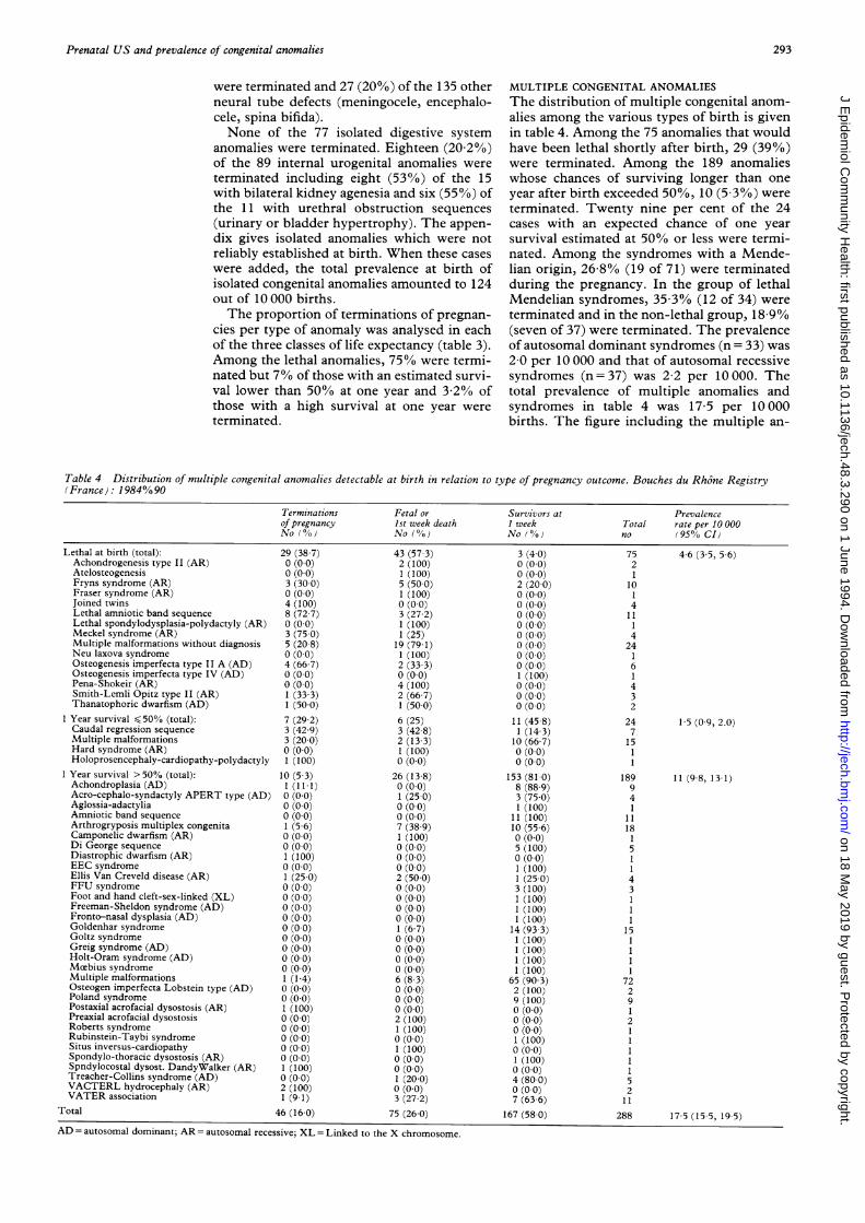
MULTIPLE CONGENITAL ANOMALIESThe distribution of multiple congenital anom-alies among the various types of birth is givenin table 4. Among the 75 anomalies that wouldhave been lethal shortly after birth, 29 (39%)were terminated. Among the 189 anomalieswhose chances of surviving longer than oneyear after birth exceeded 50%, 10 (5 3%) wereterminated. Twenty nine per cent of the 24cases with an expected chance of one yearsurvival estimated at 50% or less were termi-nated. Among the syndromes with a Mende-lian origin, 26 8% (19 of 71) were terminatedduring the pregnancy. In the group of lethalMendelian syndromes, 35 3% (12 of 34) wereterminated and in the non-lethal group, 18 9%(seven of 37) were terminated. The prevalenceof autosomal dominant syndromes (n = 33) was2 0 per 10 000 and that of autosomal recessivesyndromes (n=37) was 22 per 10000. Thetotal prevalence of multiple anomalies andsyndromes in table 4 was 17 5 per 10 000births. The figure including the multiple an-
Table 4 Distribution of multiple congenital anomalies detectable at birth in relation to type of pregnancy outcome. Bouches du Rh6ne Registry(France): 1984%90
Terminations Fetal or Survivors at Prevalenceof pregnancy Ist week death 1 week Total rate per 10000No (%) No (%) No (%) no (95% CI)
Lethal at birth (total):Achondrogenesis type II (AR)AtelosteogenesisFryns syndrome (AR)Fraser syndrome (AR)Joined twinsLethal amniotic band sequenceLethal spondylodysplasia-polydactyly (AR)Meckel syndrome (AR)Multiple malformations without diagnosisNeu laxova syndromeOsteogenesis imperfecta type II A (AD)Osteogenesis imperfecta type IV (AD)Pena-Shokeir (AR)Smith-Lemli Opitz type II (AR)Thanatophoric dwarfism (AD)
1 Year survival < 50% (total):Caudal regression sequenceMultiple malformationsHard syndrome (AR)Holoprosencephaly-cardiopathy-polydactyly
1 Year survival > 50% (total):Achondroplasia (AD)Acro-cephalo-syndactyly APERT type (AD)Aglossia-adactyliaAmniotic band sequenceArthrogryposis multiplex congenitaCamponelic dwarfism (AR)Di George sequenceDiastrophic dwarfism (AR)EEC syndromeEllis Van Creveld disease (AR)FFU syndromeFoot and hand cleft-sex-linked (XL)Freeman-Sheldon syndrome (AD)Fronto-nasal dysplasia (AD)Goldenhar syndromeGoltz syndromeGreig syndrome (AD)Holt-Oram syndrome (AD)Mctbius syndromeMultiple malformationsOsteogen imperfecta Lobstein type (AD)Poland syndromePostaxial acrofacial dysostosis (AR)Preaxial acrofacial dysostosisRoberts syndromeRubinstein-Taybi syndromeSitus inversus-cardiopathySpondylo-thoracic dysostosis (AR)Spndylocostal dysost. DandyWalker (AR)Treacher-Collins syndrome (AD)VACTERL hydrocephaly (AR)VATER association
Total
29 (38-7) 43 (57-3)n /,^AX I, I -U (0-0)0 (0-0)3 (30-0)0 (0-0)4 (100)8 (72-7)0 (0-0)3 (75-0)5 (20-8)0 (0-0)4 (66-7)0 (0-0)0 (0-0)1 (33-3)1 (50-0)7 (29-2)3 (42-9)3 (20-0)0 (0-0)1 (100)
10 (5-3)1 (11-1)0 (0-0)0 (0-0)0 (0-0)1 (5-6)0 (0-0)0 (0-0)1 (100)0 (0-0)1 (25-0)0 (0-0)0 (0-0)0 (0-0)0 (0-0)0 (0-0)0 (0-0)0 (0-0)0 (0-0)0 (0-0)1 (1-4)0 (0-0)0 (0-0)1 (100)0 (0-0)0 (0-0)0 (0-0)0 (0-0)0 (0-0)1 (100)0 (0-0)2 (100)1 (9- 1)
46 (16-0)
2 (100)1 (100)5 (50-0)1 (100)0 (0-0)3 (27-2)1 (100)1 (25)
19 (79-1)1 (100)2 (33-3)0 (0-0)4 (100)2 (66-7)1 (50-0)6 (25)3 (42-8)2 (13-3)1 (100)0 (0-0)
26 (13-8)0 (0-0)1 (25-0)0 (0-0)0 (0-0)7 (38-9)1 (100)0 (0-0)0 (0-0)0 (0-0)2 (50-0)0 (0-0)0 (0-0)0 (0-0)0 (0-0)1 (6-7)0 (0-0)0 (0-0)0 (0-0)0 (0-0)6 (8-3)0 (0-0)0 (0-0)0 (0-0)2 (100)1 (100)0 (0-0)1 (100)0 (0-0)0 (0-0)1 (20-0)0 (0-0)3 (27-2)
75 (26-0)
AD = autosomal dominant; AR = autosomal recessive; XL = Linked to the X chromosome.
3 (4-0)0 (0-0)0 (0-0)2 (20-0)0 (0-0)0 (0-0)0 (0-0)0 (0-0)0 (0-0)0 (0-0)0 (0-0)0 (0-0)1 (100)0 (0-0)0 (0-0)0 (0-0)
11 (45-8)1 (14-3)
10 (66-7)0 (0-0)0 (0-0)
153 (81-0)8 (88-9)3 (75-0)1 (100)
11 (100)10 (55-6)0 (0-0)5 (100)0 (0-0)1 (100)1 (25-0)3 (100)1 (100)1 (100)1 (100)
14 (93-3)1 (100)1 (100)1 (100)1 (100)
65 (90-3)2 (100)9 (100)0 (0-0)0 (0-0)0 (0-0)1 (100)0 (0-0)1 (100)0 (0-0)4 (80-0)0 (0-0)7 (63-6)
167 (58-0)
7521
1014
111424161432
247
1511
189941
11118
2
511
43
1
1115
172
291
211
28
4-6 (3-5, 5-6)
1-5 (0-9, 2.0)
11 (9-8, 13-1)
17-5 (155, 19-5)
293
on 18 May 2019 by guest. P
rotected by copyright.http://jech.bm
j.com/
J Epidem
iol Com
munity H
ealth: first published as 10.1136/jech.48.3.290 on 1 June 1994. Dow
nloaded from

2julian-Reynier, Philip, Scheiner, Aurran, Chabal, Maron, Gombert, Ayme
Table 5 Congenital anomalies: changes in pregnancy outcome over the years 1984-90(Bouches du Rh6ne Birth Defects Registry)
Terminations Fetal or Survivors atYear of of pregnancy perinatal deaths I week Totalbirth No ( 0/,, ) No (0%) No (%) No (100%)
1984 17 (6 10) 49 (15-1) 212(763) 2781985 13 (4 40) 45 (15 2) 238 (80 4) 2961986 26 (9 20) 42 (14 9) 214 (75-9) 2821987 33 (10 3) 37 (11 6) 249 (78 1) 3191988 25 (7 00) 43 (12-0) 289 (81 0) 3571989 32 (10-9) 39 (13 3) 222 (75 8) 2931990 35 (13 6) 18 (7 00) 205 (79 5) 258'I'otal 181 (8 7) 273 (13-1) 1629 (78 2) 2083
Table 6 Terminations of pregnancy for congenital anomalies: changes in expectedsurvival over the years 1984-90 (Bouches du Rh6ne Birth Defects Registry)
1 year survival
Lethal at birth s 50%/o > 50% TotalYear No (%} No % No % no
1984 11 (647) 3 (176) 3 (176) 171985 7 (538) 2 (154) 4 (308) 131986 13 (500) 2 (77) 11(423) 261987 14 (42 4) 8 (24 4) 11 (33 3) 331988 13 (52 0) 7 (28f0) 5 (20 0) 251989 17 (53 1) 7 (21 9) 8 (25-0) 321990 15 (42-9) 11 (31 4) 9 (28-1) 35
'I'otal 90 (49 7) 40 (22 1) 51 (28 2) 181
omalies which were not reliably ascertained atbirth (Appendix) were 24 1 per 10 000.
SECULAR TREND OF ABORTIONS INDUCED AFTER
PRENATAL DIAGNOSIS
The changes in the pregnancy outcomesinvolving isolated and multiple anomalies over
the seven year period are given in table 5. Theoverall proportion of terminations for isolatedplus multiple anomalies increased significantlybetween 1984 and 1990 (p<0001) but thetrend was non-linear (p < 0-00 1). This increasewas significant in the case of the isolatedmalformations only (p < 0001). The overallpercentage of terminations due to lethal andlow life expectancy anomalies increased signi-ficantly (p<0 001) over the seven year periodbut the increase was not significant in the case
of the high survival category (p >0 10). Table6 gives the distribution of expected survivalpercentages in the case of the aborted fetuses.The average gestational age at termination was
22 4 weeks (21-9-22-9), and this figure did not
vary significantly over the seven years.
DiscussionThis study did not aim to evaluate the effec-tiveness of prenatal ultrasound but rather itsimpact on the prevalence of malformations.Several studies have been published on
prevalence,57910 but the results are difficult to
compare with ours because the malformedindividual and not the malformation was thestatistical unit and because chromosomalaetiologies were included in some studies butnot in ours. We have dealt elsewhere with thelatter anomalies." We also excluded any anom-lies whicn were not reliably ascertainable at
birth. This led to eliminating from the analysis20 terminations of pregnancy which feature inthe appendix, involving anomalies with whichthe number of livebirths is liable to beunderestimated. This decision led to under-estimating the impact of prenatal diagnosis butreinforced the homogeneity of the compari-sons. The Marseille registry belongs to theEurocat network and the prevalence rates werecomparable with those based on other Frenchregistries during the period under analysis.'When prevalence rates are regularly publishedby registries, it is always in terms of groupsof malformations and not of the aetiologyinvolved. The only studies besides this onegiving prevalence rates specific to the aetio-logical type of the anomalies have been thosepublished by Martinez-Frias.9"' This author9obtained a 12 per 100 000 prevalence rate inthe case of automosomal dominant syndromesamong livebirths and one of 10-3 per 100 000in that of autosomal recessive disorders."'Without including the cases noted in theappendix, we obtained the following results forautosomal dominant and autosomal recessivedisorders (from table 4): 20 1 and 22 5 per100 000 livebirths, respectively. These com-parisons provided grounds for assuming thatour ascertainment was more complete thanthat of Martinez-Frias. The skeletal dysplasiaprevalence rates were 0-06% total births forachondroplasia, 0 06% for osteogenesis imper-fecta, and 0-02% for thanatophoric dwarfismand achondrogenesis. These rates are compar-able with those published,78 which indicatesthat our ascertainment was accurate.
In the Bouches du Rh6ne region, the num-ber of terminations of pregnancy after prenatalultrasound examination in the case of the con-genital anomalies included in this study wastwice as high in 1990 (14 9 per 10 000 births) asin 1984 (7 4 per 10 000 births). Terminationsof pregnancy after prenatal diagnosis reducedthe isolated anomalies by only 3 2%, takinginto account the anomalies with an expectedprobability of survival higher than 50% at theage of 1 year. In the case of multiple anomaliesor syndromes giving an equivalent chance ofsurvival, this proportion was 5 3%. On thewhole, leaving aside the malformations givinga poor chance of survival, only 3 5% of all theanomalies included in this study were termi-nated because of prenatal ultrasound examina-tion. Spontaneous deaths accounted for 11%of all the single anomalies and for 26% of themultiple ones; on the overall data, naturalselection from the 20th week of gestational agedealt with 13% of the congenital anomalies.The overall figures show that abortion
increased proportionally over the seven yearperiod, but this trend was significant only inthe case of single anomalies and not with themultiple ones. The abortion rate associatedwith lethal and low life expectancy anomaliesalso increased significantly but that due toanomalies giving high prospects of survival didnot. The impact of prenatal diagnosis dependson the effectiveness of the diagnostic tool used.Prenatal ultrasound examination is not a sensi-tive technique for screening malformations
294
on 18 May 2019 by guest. P
rotected by copyright.http://jech.bm
j.com/
J Epidem
iol Com
munity H
ealth: first published as 10.1136/jech.48.3.290 on 1 June 1994. Dow
nloaded from

Prenatal US and prevalence of congenital anomalies
among the general population of pregnantwomen: the results depend on the gestationalage, the operator, the nature of the malforma-tion, and the equipment used. Levi et al'2 in a
prospective study from 1984 to 1989 observedthat 40% of all congenital anomalies were
detected by sonography and that excellent re-
sults were obtained as regards the specificity(99 9%), positive predictive value (95 1%),and negative predictive value (98-6%). Othergeographically based studies'3 carried out in a
routine context have yielded comparable re-
sults but have a rather low sensitivity. Hospitalstudies,'4 5 even in a routine context, haveshown a higher sensitivity. The good predic-tive score shows the accuracy of sonography as
a screening tool in a routine pregnancy followup. With the development of this technique,its accuracy may predictably be improved.The decisions resulting from prenatal dia-gnosis of malformations involve ascertaining a
prognosis, which is very often difficult to as-sess: fear of legal suit may be an incentive forphysicians to terminate the pregnancy. InFrance, termination of pregnancy is an avail-able option with no upper gestational age limitwhen no treatment exists and when the prog-
nosis is thought to be very severe. Our resultsshow that termination of pregnancy was car-ried out on severe clinical cases not alwaysassociated with mental retardation (limbreduction defects for example). Central ner-vous system anomalies were the most frequentcategory of isolated malformations to be termi-nated. For the period 1984-86, the EurocatWorking Group'6 noted that 80% of all anen-
cephalies were terminated in six out of 11centres and 40% of all cases of spina bifidarecorded in four registries. Anencephaly is agood index to prenatal medical monitoring in a
population since it should always be detectablewith the ultrasound technique. Over the seven
year period studied here, the percentages ofterminations were 80% for anencephaly and22% for spina bifida, but over the last threeyears (1987-90) 95% of all fetal anencephalieswere aborted.
In conclusion, among all the cases of con-
genital anomaly defined as being readily ascer-
tainable at birth and possibly by ultrasoundexamination, the proportion in which preg-nancy was terminated after prenatal ultra-sound examination rose from 6 1% in 1984 to13 6% in 1990. About half of the terminations
AppendixIsolated atnd multiple congenital anomlalies not definitely, ascertained at birth. Distributiotn per type of pregnancyoutcome; Bouches du Rhone registry (France) 1984-90
Terminationis of Fetal or Survizvors at Total Prevalencepregnancy Ist week death I week no rate per 10 000lN'o ( % ) Nlo (}) No ( %)o ( 95°0o CI)
Isolated malformations (total): 15 (6-3) 18 (7-5) 207 (86-3) 240 15-0 (12-7, 16-4)Cervical hvgroma 9 (47-4) 10 (52-6) 0 (0-0) 19 1-1 (0-6, 1 7)Corpus callosum agenesis 1 (20-0) 2 (40-0) 2 (40-0) 5 0-3 (0-0, 0-6)Craniostenosis 1 (2-5) 0 (0-0) 39 (97-5) 40 2-4 (1-7, 3-2)Ebstein anomaly 0 (0-0) 0 (0-0) 1 (100.0) 1 0-1 (-0-1, 0-2)Hydronephrosis 1 (0-6) 2 (1-2) 162 (98-2) 165 10 (8-5, 11-6)Polvcystic kidney disease (infantile) 3 (37-5) 3 (37-5) 2 (25-0) 8 0-5 (0-1, 0-8)Polycystic kidney disease (adult AD) 0 (0-0) 1 (100 0 (0-0) 1 0-1 (-0-1, 0-2)Polycystic kidney 0 (0-0) 0 (0-0) 1 (33-3) 1 0-1 (-0-1, 0-2)
Multiple anomalies (total): 5 (4-6) 17 (15-6) 87 (79-8) 109 6-6 (5-4, 7-9)Multiple malformation without diagnosis 1 (14-3) 5 (71-5) 1 (14 3) 7 0-4 (0-1, 0-7)Acrocephalosyndactyly Saethre-Chotzen 0 (0-0) 0 (0 0) 1 (100-0) 1 0-1 (-0-1, 0 2)Albers-Schonberg syndrome (AR) 0 (0-0) 0 (0-0) 2 (100-0) 2 0-1 (-0-0, 0-3)Anus-hand-foot-ear syndrome 0 (0-0) 0 (0-0) 3 (100.0) 3 0-2 (-0-0. 0-4)Arthrogryposis - deafness 0 (0-0) 0 (0-0) 1 (100 0) 1 0-1 (-0-1, 0-2)Beals-Hecht syndrome 0 (0-0) 0 (0-0) 1 (100-0) 1 0-1 (-0-01, 0-2)Beckwith-Wiedemann syndrome 0 (0-0) 1 (100-0) 0 (0-0) 1 0-1 (-0-1, 0-2)Blepharophimosis-Ptosis-Epicanthus 0 (0 0) 0 (0-0) 3 (100.0) 3 0-2 (-0-0, 0-4)Bor syndrome 0 (0-0) 0 (0-0) 3 (100-0) 3 0-2 (-0-0. 0-4)Caffev disease 0 (0 0) 0 (0-0) 1 (100-0) 1 0-1 (-0-1, 0-2)CHARGE association 0 (0-0) 0 (0-0) 1 (100-0) 1 0-1 (-0-1, 0-2)Chondrodysplasia punctata 0 (0-0) 0 (0-0) 2 (100-0) 2 0-1 (-0-0, 0-3)Cleidocranial dysplasia 0 (0-0) 0 (0 0) 1 (100-0) 1 0-1 (-0-1, 0-2)Cornelia de Lange syndrome 1 (33-3) 0 (0-0) 2 (66-7) 3 0-2 (-0-0, 0-4)Costello syndrome 0 (0-0) 0 (0-0) 1 (100-0) 1 0-1 (-0-1, 0-2)Fetal alcohol syndrome 0 (0-0) 0 (0-0) 18 (100-0) 18 1-1 (0-6, 1-6)FG syndrome (RLX) 0 (0-0) 1 (16-7) 5 (83-3) 6 0-4 (0-0. 0-6)G syndrome (AD) 0 (0-0) 1 (50-0) 1 (50-0) 2 0-1 (-0-0, 0-3)Hallerman-Streiff-Franqois syndrome (AR) 0 (0-0) 1 (100-0) 0 (0-0) 1 0-1 (-0-1, 0-2)Ivemark syndrome 2 (33-3) 3 (50-0) 1 (16-7) 6 0-4 (0-0, 0-6)Klippel-Feil syndrome (AD) 0 (0-0) 0 (0-0) 1(100-0) 1 0-1 (-0-1, 0-2)Leber type I disease 0 (0-0) 0 (0-0) 1(100-0) 1 0-1 (-0-1, 0-2)Marshall-Smith syndrome (AD) 0 (0-0) 1 (50-0) 1(50-0) 2 0-1 (-0-0, 0-3)Noonan syndrome 0 (0-0) 0 (0-0) 3 (100-0) 3 0-2 (-0-0, 0-4)Noonan-like syndrome 0 (0-0) 0 (0-0) 1 (100-0) 1 0-1 (-0-1, 0-2)Pollit syndrome 0 (0-0) 0 (0-0) 1(100-0) 1 0-1 (-0-1, 0-2)Prader-Willi syndrome 0 (0-0) 0 (0-0) 4 (100-0) 4 0-2 (0-0, 0-5)Recklinghausen disease 0 (0-0) 0 (0-0) 7 (100-0) 7 0-4 (0-1, 0-7)Silver-Russell syndrome 0 (0-0) 0 (0-0) 2 (100-0) 2 0-1 (-0-0, 0-3)Smith-Lemli-Opitz syndrome type 1 (AR) 0 (0-0) 1 (50-0) 1 (50-0) 2 0-1 (-0-0, 0-3)Steinert disease (AD) 0 (0-0) 2 (66-6) 1(33-3) 3 0-2 (-0-0, 0-4)Sturge-Weber syndrome 0 (0-0) 0 (0-0) 1(100-0) 1 0-1 (-0-1, 0-2)TIricho-rhino-phalangeal syndromc 0 (0-0) 0 (0-0) 4 (100-0) 4 0-2 (0-0, 0-5)Tuberous sclerosis (AD) 0 (0-0) 1 (14-3) 6 (85-7) 7 0-4 (0-1, 0-7)Waardenburg syndrome, type I (AD) 1 (33-3) 0 (0-0) 2 (66-7) 3 0-18 (-0-0, 0-4)Williams syndrome 0 (0-0) 0 (0-0) 3 (100-0) 3 0-2 (-0-0, 0-4)
Total 20 (5-7) 35 (10-0) 294 (84-2) 349 21 (19, 23-4)
AR = autosomal recessive; AD = autosomal dominant; RLX = recessive linked to the chromosome X.
295
on 18 May 2019 by guest. P
rotected by copyright.http://jech.bm
j.com/
J Epidem
iol Com
munity H
ealth: first published as 10.1136/jech.48.3.290 on 1 June 1994. Dow
nloaded from

Julian-Reynier, Philip, Scheiner, Aurran, Chabal, Maron, Gombert, Ayme
involved malformations which were lethaleither before or soon after birth. The rest were
almost equally divided between conditionswith one year survival rates of 50% or less andof more than 50%.
This study was financially supported by the Ministry of Healthand The National Institute of Health and Medical Researchand by the Association Recherche et Parage.The authors would like to thank Mrs Juliette Gimbert for her
secretarial assistance in the Bouches du Rh6ne Defects Registryand, Mrs M-G Mattei and Mrs Joelle Le Houcq for theircytogenetic expertise.
1 Kalter H. Five decades international trends in the relationof perinatal mortality and congenital malformations: still-birth and neonatal death compared. Int Epidemiol199 1;20:173-179.
2 Macquart-Moulin G, Baret C, Julian C, Fancello G, Vin-cent A, Ayme S. Surveillance antenatale et risques deprematurite et d'hypotrophie feetale. Gynecol Obstet BiolReprod (Paris) 1992;21:9-18.
3 Ayme S, Julian C, Maurin N, Gambarelli D, Sudan N,Giraud F. Le registre des mortnes des Bouches du Rh6ne:bilan de trois ans de fonctionnement. journal de GinitiqueHumaine 1985;3-4:265-274.
4 Julian-Reynier C, Battista RN, Ayme S. Feasibility andperformance of post-mortem examination to determinethe aetiology of congenital anomalies in a population of1019 foetal and perinatal deaths. European journal ofPublic Health 1993;3:153-158.
5 Eurocat Working Group. Eurocat Report 4. Surveillance ofcongenital anomalies, years 1980-1988. Brussels: Depart-ment of Epidemiology, Catholic University of Louvain,1991.
6 Armitage P. Statistical methods in medical research. Oxford:Blackwell Scientific Publications, 1971.
7 Orioli IM. Castilla EE, Barbosa-Neto JG. The birth preval-ence rates for the skeletal dysplasia. Med Genet1986;23:328-32.
8 Andersen PE. Prevalence of lethal osteochondrodysplasiasin Denmark. Am Med Genet 1989;32:484-9.
9 Martinez-Frias ML, Bermejo E, Cereijo A, Sanchez M,Lopez M, Gonzalo C. Epidemiological aspects of mende-lian syndromes in a Spanish population sample: I. Auto-somal dominant malformation syndromes. Am MedGenet 1991;38:622-5.
10 Martinez-Frias ML, Bermejo E, Cereijo A, Sanchez M,Lopez M, Gonzalo C. Epidemiological aspects of mende-lian syndromes in a Spanish population sample: II. Auto-somal recessive malformation. syndromes. Am MedGenet 1991;38:626-9.
11 Ayme S, Julian C, Stoll C, Dott B, Dufeuil C, Goujard J.Impact du diagnostic antenatal sur la prevalence desanomalies chromosomiques a la naissance. In: Rechercheet politiques de santi: I'apport des registres de morbiditi.Paris: INSERM, 1992, 219-25.
12 Levi S, Hyjazi Y, Schaaps JP, Deffoort P, Coulon R,Buekens P. Sensitivity and specificity of routine antenatalscreening for congenital anomalies by ultrasound theBelgian multicentric study. Ultrasound Obstet Gynecol1991;1:102-10.
13 Stoll C, Alembik Y, Dott B, Roth M-P, Finck S. Evaluationof prenatal diagnosis by a registry of congenital anomalies.Prenatal Diagnosis 1992;12:263-70.
14 Luck C. Value of routine ultrasound scanning at 19 weeks: a
four year study of 8849 deliveries. BMJ 1992;304:1474-8.15 Shirley IM, Bottomley F, Robinson VP. Routine radio-
grapher screening for fetal abnormalities by ultrasound inan unselected low risk population. Br Radiol1992;65:564-9.
16 Eurocat Working Group. Prevalence of neural tube defectsin 20 regions of Europe and the impact of prenataldiagnosis, 1980-1986. Epidemiol Community Health1991;45:52-8.
296
on 18 May 2019 by guest. P
rotected by copyright.http://jech.bm
j.com/
J Epidem
iol Com
munity H
ealth: first published as 10.1136/jech.48.3.290 on 1 June 1994. Dow
nloaded from