Clase 4 ua nstemi
31
Síndrome Coronario Agudo UA – NSTEMI Unstable Angina Non ST Elevation Myocardial Infarction
-
Upload
daniel-alejandro -
Category
Education
-
view
150 -
download
3
Transcript of Clase 4 ua nstemi
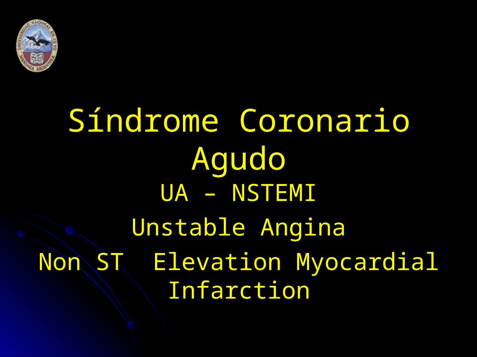
Síndrome Coronario Agudo
UA – NSTEMI
Unstable Angina
Non ST Elevation Myocardial Infarction
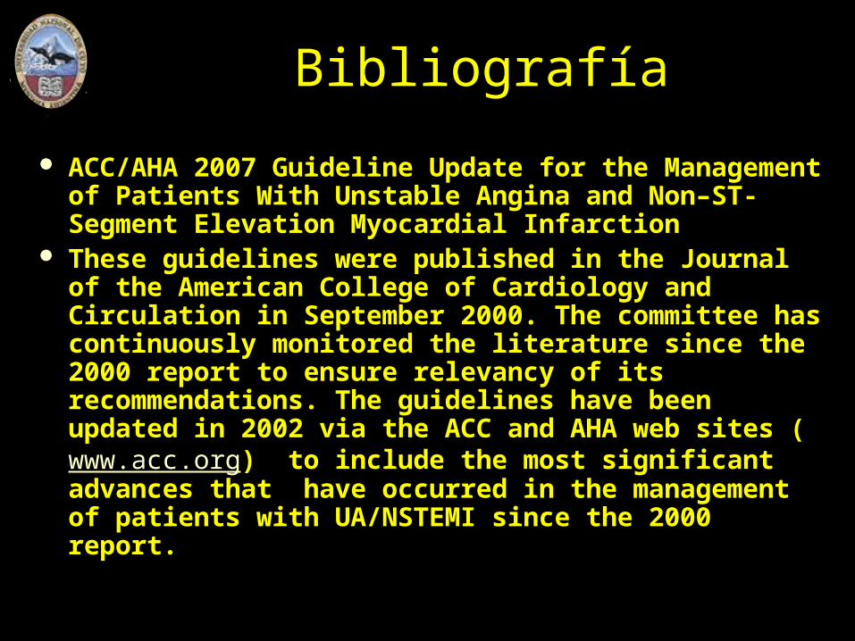
Bibliografía
ACC/AHA 2007 Guideline Update for the Management of Patients With Unstable Angina and Non–ST-Segment Elevation Myocardial Infarction
These guidelines were published in the Journal of the American College of Cardiology and Circulation in September 2000. The committee has continuously monitored the literature since the 2000 report to ensure relevancy of its recommendations. The guidelines have been updated in 2002 via the ACC and AHA web sites (www.acc.org) to include the most significant advances that have occurred in the management of patients with UA/NSTEMI since the 2000 report.
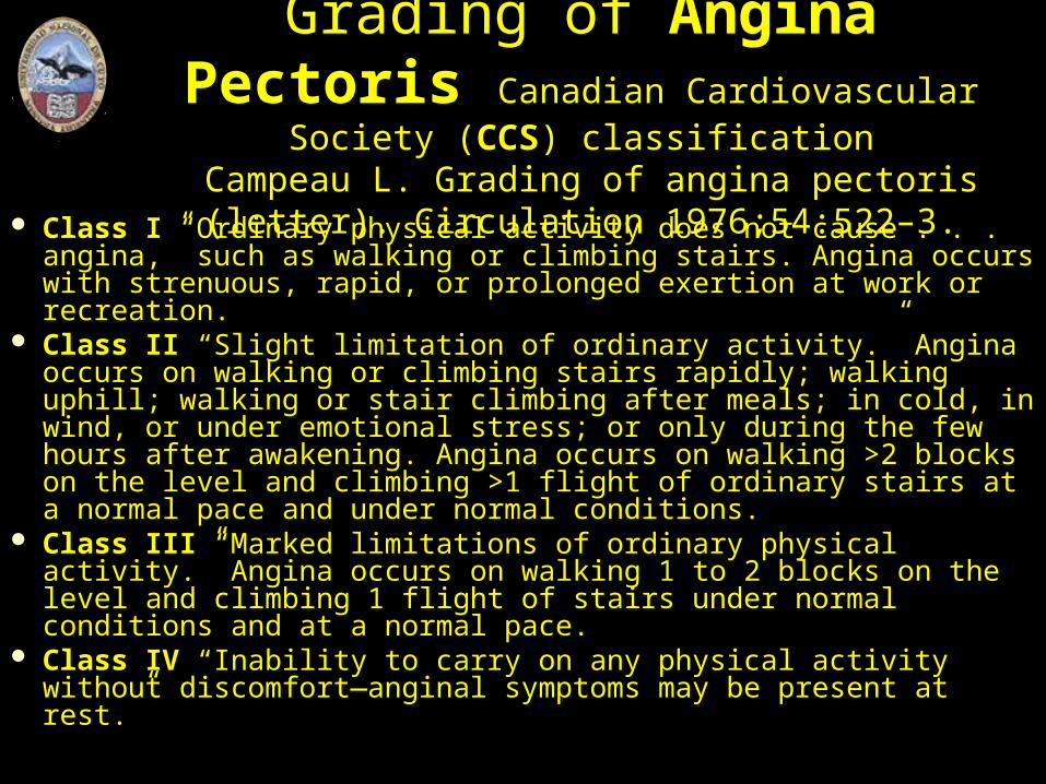
Grading of Angina Pectoris Canadian Cardiovascular Society (CCS) classification
Campeau L. Grading of angina pectoris (letter). Circulation 1976;54:522–3.
Class I “Ordinary physical activity does not cause . . . angina,” such as walking or climbing stairs. Angina occurs with strenuous, rapid, or prolonged exertion at work or recreation.
Class II “Slight limitation of ordinary activity.” Angina occurs on walking or climbing stairs rapidly; walking uphill; walking or stair climbing after meals; in cold, in wind, or under emotional stress; or only during the few hours after awakening. Angina occurs on walking >2 blocks on the level and climbing >1 flight of ordinary stairs at a normal pace and under normal conditions.
Class III “Marked limitations of ordinary physical activity.” Angina occurs on walking 1 to 2 blocks on the level and climbing 1 flight of stairs under normal conditions and at a normal pace.
Class IV “Inability to carry on any physical activity without discomfort—anginal symptoms may be present at rest.”
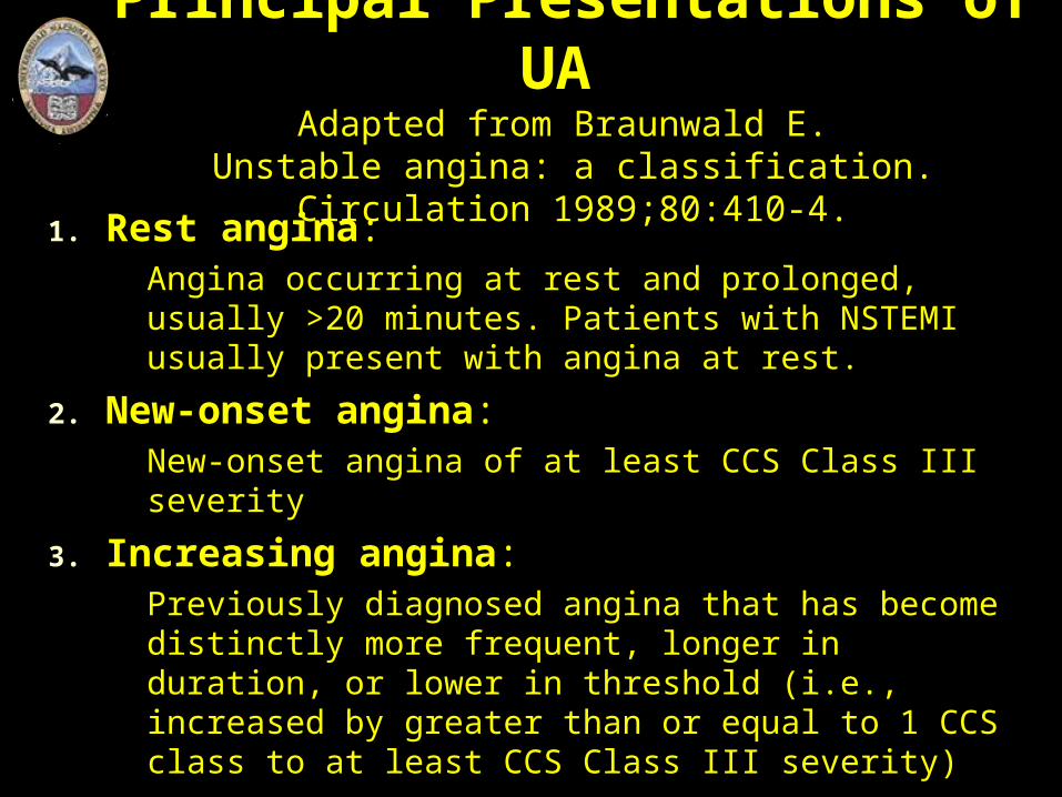
Principal Presentations of UA Adapted from Braunwald E.
Unstable angina: a classification. Circulation 1989;80:410-4.
1. Rest angina: Angina occurring at rest and prolonged, usually >20 minutes. Patients with NSTEMI usually present with angina at rest.
2. New-onset angina: New-onset angina of at least CCS Class III severity
3. Increasing angina: Previously diagnosed angina that has become distinctly more frequent, longer in duration, or lower in threshold (i.e., increased by greater than or equal to 1 CCS class to at least CCS Class III severity)
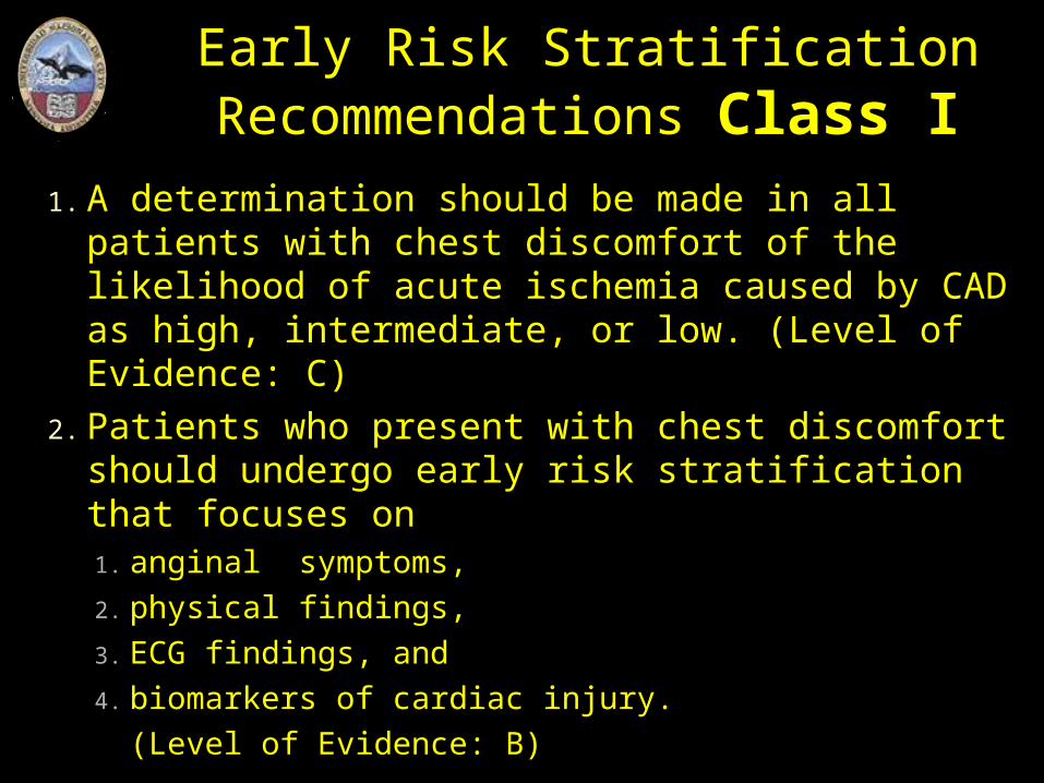
Early Risk Stratification Recommendations Class I
1. A determination should be made in all patients with chest discomfort of the likelihood of acute ischemia caused by CAD as high, intermediate, or low. (Level of Evidence: C)
2. Patients who present with chest discomfort should undergo early risk stratification that focuses on 1. anginal symptoms,
2. physical findings,
3. ECG findings, and
4. biomarkers of cardiac injury.
(Level of Evidence: B)
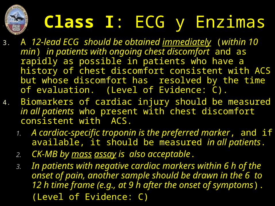
Class I: ECG y Enzimas3. A 12-lead ECG should be obtained immediately (within 10
min) in patients with ongoing chest discomfort and as rapidly as possible in patients who have a history of chest discomfort consistent with ACS but whose discomfort has resolved by the time of evaluation. (Level of Evidence: C).
4. Biomarkers of cardiac injury should be measured in all patients who present with chest discomfort consistent with ACS.
1. A cardiac-specific troponin is the preferred marker, and if available, it should be measured in all patients.
2. CK-MB by mass assay is also acceptable. 3. In patients with negative cardiac markers within 6 h of the
onset of pain, another sample should be drawn in the 6 to 12 h time frame (e.g., at 9 h after the onset of symptoms). (Level of Evidence: C)
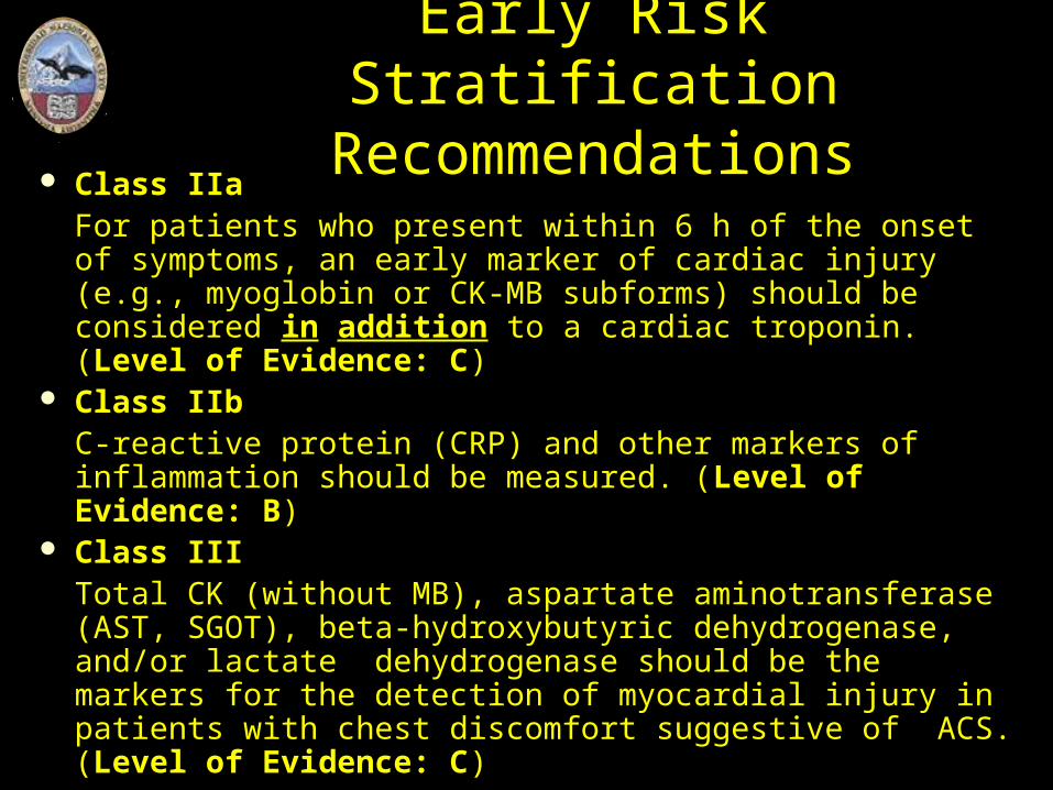
Class IIa For patients who present within 6 h of the onset of symptoms, an early marker of cardiac injury (e.g., myoglobin or CK-MB subforms) should be considered in addition to a cardiac troponin. (Level of Evidence: C)
Class IIb C-reactive protein (CRP) and other markers of inflammation should be measured. (Level of Evidence: B)
Class III Total CK (without MB), aspartate aminotransferase (AST, SGOT), beta-hydroxybutyric dehydrogenase, and/or lactate dehydrogenase should be the markers for the detection of myocardial injury in patients with chest discomfort suggestive of ACS. (Level of Evidence: C)
Early Risk Stratification Recommendations
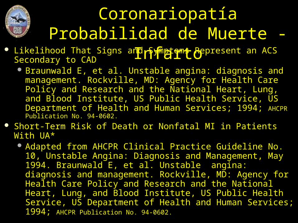
Probabilidad de CoronariopatíaProbabilidad de Muerte - Infarto
Likelihood That Signs and Symptoms Represent an ACS Secondary to CAD Braunwald E, et al. Unstable angina: diagnosis and
management. Rockville, MD: Agency for Health Care Policy and Research and the National Heart, Lung, and Blood Institute, US Public Health Service, US Department of Health and Human Services; 1994; AHCPR Publication No. 94-0602.
Short-Term Risk of Death or Nonfatal MI in Patients With UA* Adapted from AHCPR Clinical Practice Guideline No. 10,
Unstable Angina: Diagnosis and Management, May 1994. Braunwald E, et al. Unstable angina: diagnosis and management. Rockville, MD: Agency for Health Care Policy and Research and the National Heart, Lung, and Blood Institute, US Public Health Service, US Department of Health and Human Services; 1994; AHCPR Publication No. 94-0602.
* Guía … no “mandato”
Short-Term Risk of Death or Nonfatal MI in Patients With UA* *Estimation of the short-term risks of death and
nonfatal cardiac ischemic events in UA is a complex multivariable problem that cannot be fully specified in a table such as this; therefore, this table is meant to offer general guidance and illustration rather than rigid algorithms.

Enfermedad Coronaria Aterosclerótica“High Likelihood”
Feature Any of the following features:
History Chest or left arm pain or discomfort as chief symptom reproducing prior documented angina. Known history of CAD, including MI.
Examination Transient MR, hypotension, diaphoresis, pulmonary edema, or rales
ECG New, or presumably new, transient ST-segment deviation (> 0.05 mV) or T-wave inversion (>/= 0.2 mV) with symptoms
Cardiac markers Elevated cardiac TnI, TnT, or CK-MB

Enfermedad Coronaria Aterosclerótica“Intermediate Likelihood”
Feature Ausencia de hallazgos de “high likelihood”Any of the following features:
History Chest or left arm discomfort as chief symptom. Age > 70 years. Diabetes mellitus. Male sex.
Examination Extracardiac vascular disease
ECG Fixed Q waves. Abnormal ST segments or T waves not documented to be new.
Cardiac markers Normal
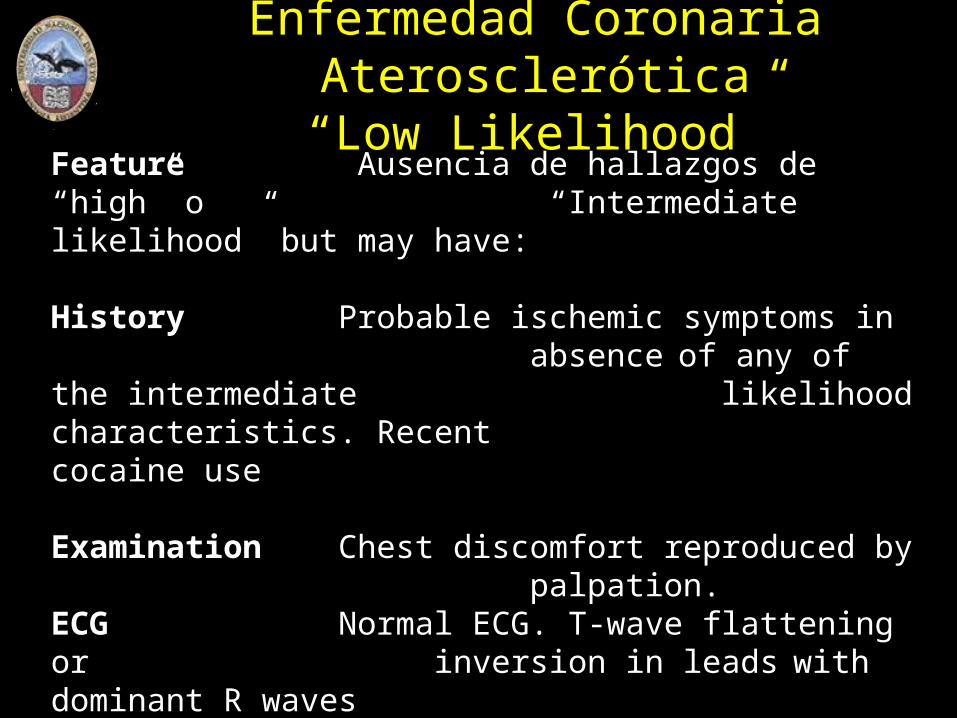
Enfermedad Coronaria Aterosclerótica“Low Likelihood”
Feature Ausencia de hallazgos de “high” o “Intermediate likelihood” but may have:
History Probable ischemic symptoms in absence of any of the intermediate likelihood characteristics. Recent cocaine use
Examination Chest discomfort reproduced by palpation.
ECG Normal ECG. T-wave flattening or inversion in leads with dominant R waves
Cardiac markers Normal
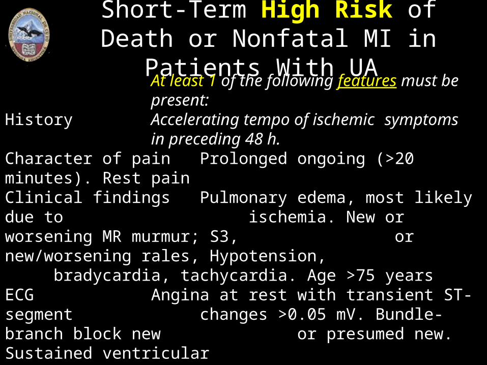
Short-Term High Risk of Death or Nonfatal MI in Patients With UA
At least 1 of the following features must be present:
History Accelerating tempo of ischemic symptoms in preceding 48 h.
Character of pain Prolonged ongoing (>20 minutes). Rest pain
Clinical findings Pulmonary edema, most likely due to ischemia. New or worsening MR murmur; S3, or new/worsening rales, Hypotension, bradycardia, tachycardia. Age >75 years
ECG Angina at rest with transient ST-segment changes >0.05 mV. Bundle-branch block new or presumed new. Sustained ventricular tachycardia
Cardiac markers Elevated (TnT or TnI >0.1 ng/mL)
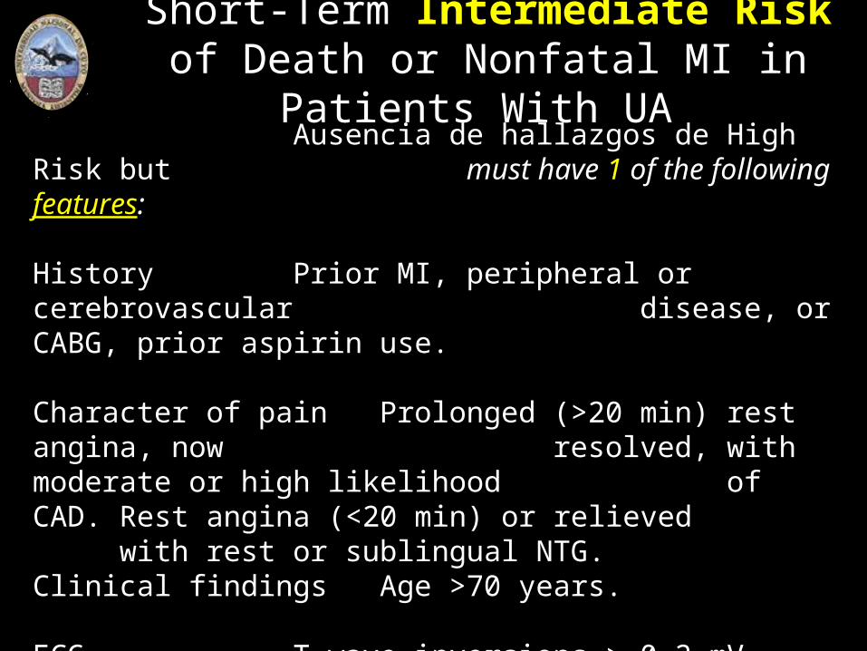
Short-Term Intermediate Risk of Death or Nonfatal MI in Patients With UA
Ausencia de hallazgos de High Risk but must have 1 of the following features:
History Prior MI, peripheral or cerebrovascular disease, or CABG, prior aspirin use.
Character of pain Prolonged (>20 min) rest angina, now resolved, with moderate or high likelihood of CAD. Rest angina (<20 min) or relieved with rest or sublingual NTG.
Clinical findings Age >70 years. ECG T-wave inversions > 0.2 mV. Pathological
Q waves Cardiac markers TnT > 0.01 ng/ml but < 0.1 ng/mL.
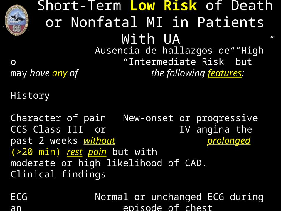
Short-Term Low Risk of Death or Nonfatal MI in Patients With UA
Ausencia de hallazgos de “High” o “Intermediate Risk” but may have any of the following features:
History
Character of pain New-onset or progressive CCS Class III or IV angina the past 2 weeks without prolonged (>20 min) rest pain but with moderate or high likelihood of CAD.
Clinical findings
ECG Normal or unchanged ECG during an episode of chest discomfort
Cardiac markers TnT < 0,01 ng/mL (< 0,03 ng% – VN -)
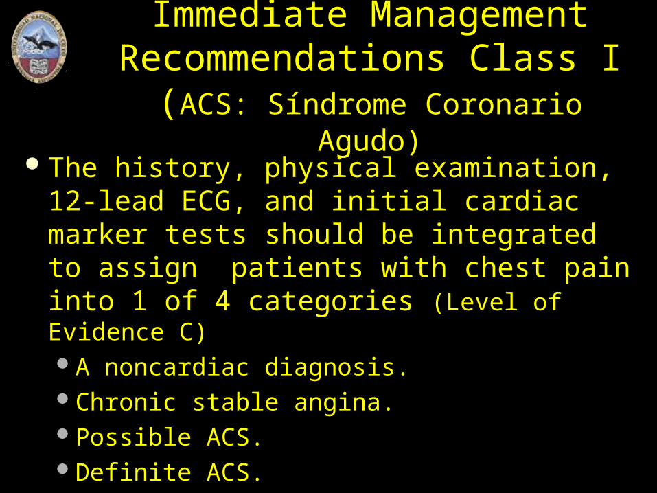
Immediate ManagementRecommendations Class I
(ACS: Síndrome Coronario Agudo)The history, physical examination, 12-lead
ECG, and initial cardiac marker tests should be integrated to assign patients with chest pain into 1 of 4 categories (Level of Evidence C)A noncardiac diagnosis. Chronic stable angina. Possible ACS. Definite ACS.
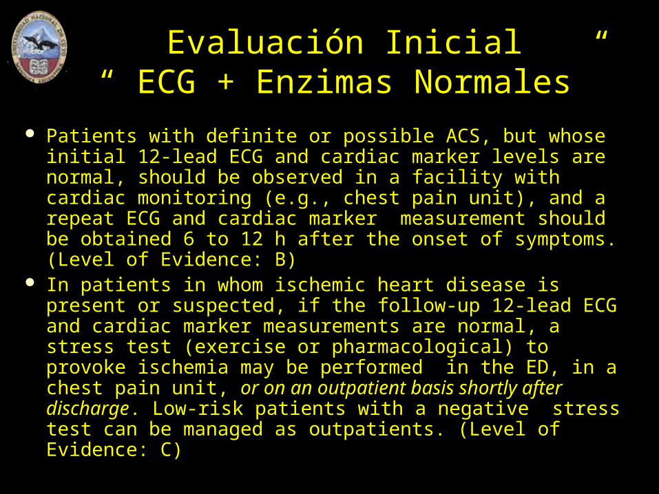
Evaluación Inicial “ ECG + Enzimas Normales”
Patients with definite or possible ACS, but whose initial 12-lead ECG and cardiac marker levels are normal, should be observed in a facility with cardiac monitoring (e.g., chest pain unit), and a repeat ECG and cardiac marker measurement should be obtained 6 to 12 h after the onset of symptoms. (Level of Evidence: B)
In patients in whom ischemic heart disease is present or suspected, if the follow-up 12-lead ECG and cardiac marker measurements are normal, a stress test (exercise or pharmacological) to provoke ischemia may be performed in the ED, in a chest pain unit, or on an outpatient basis shortly after discharge. Low-risk patients with a negative stress test can be managed as outpatients. (Level of Evidence: C)

Evaluación Inicial¡ACS con Supra ST!
Patients with definite ACS and ST-segment elevation should be evaluated for immediate reperfusion therapy. (Level of Evidence: A)
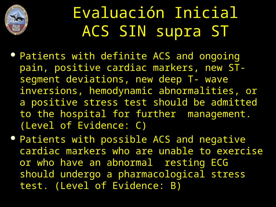
Evaluación InicialACS SIN supra ST
Patients with definite ACS and ongoing pain, positive cardiac markers, new ST-segment deviations, new deep T- wave inversions, hemodynamic abnormalities, or a positive stress test should be admitted to the hospital for further management. (Level of Evidence: C)
Patients with possible ACS and negative cardiac markers who are unable to exercise or who have an abnormal resting ECG should undergo a pharmacological stress test. (Level of Evidence: B)
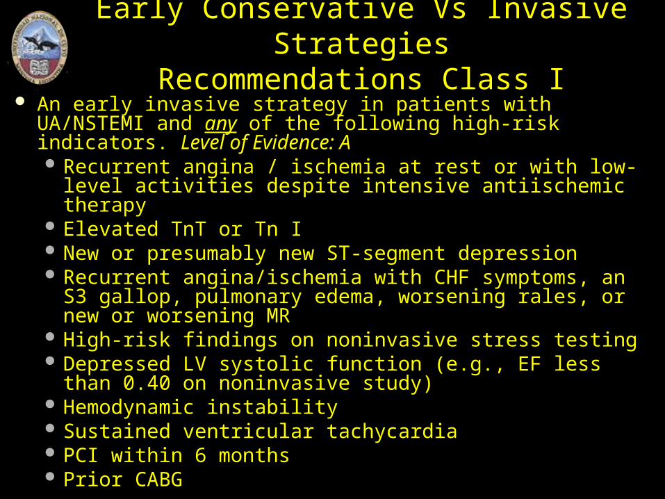
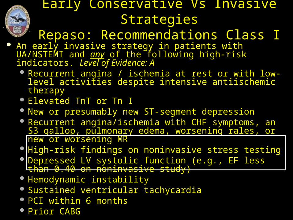
Early Conservative Vs Invasive StrategiesRecommendations Class I
An early invasive strategy in patients with UA/NSTEMI and any of the following high-risk indicators. Level of Evidence: A Recurrent angina / ischemia at rest or with low-level
activities despite intensive antiischemic therapy Elevated TnT or Tn I New or presumably new ST-segment depression Recurrent angina/ischemia with CHF symptoms, an S3
gallop, pulmonary edema, worsening rales, or new or worsening MR
High-risk findings on noninvasive stress testing Depressed LV systolic function (e.g., EF less than 0.40 on
noninvasive study) Hemodynamic instability Sustained ventricular tachycardia PCI within 6 months Prior CABG
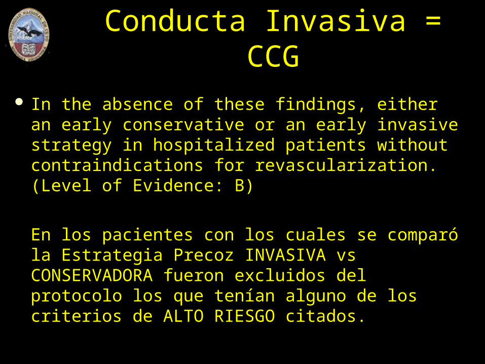
Conducta Invasiva = CCG
In the absence of these findings, either an early conservative or an early invasive strategy in hospitalized patients without contraindications for revascularization. (Level of Evidence: B)
En los pacientes con los cuales se comparó la Estrategia Precoz INVASIVA vs CONSERVADORA fueron excluidos del protocolo los que tenían alguno de los criterios de ALTO RIESGO citados.
Early Conservative Vs Invasive StrategiesRepaso: Recommendations Class I
An early invasive strategy in patients with UA/NSTEMI and any of the following high-risk indicators. Level of Evidence: A Recurrent angina / ischemia at rest or with low-level
activities despite intensive antiischemic therapy Elevated TnT or Tn I New or presumably new ST-segment depression Recurrent angina/ischemia with CHF symptoms, an S3
gallop, pulmonary edema, worsening rales, or new or worsening MR
High-risk findings on noninvasive stress testing Depressed LV systolic function (e.g., EF less than 0.40 on
noninvasive study) Hemodynamic instability Sustained ventricular tachycardia PCI within 6 months Prior CABG
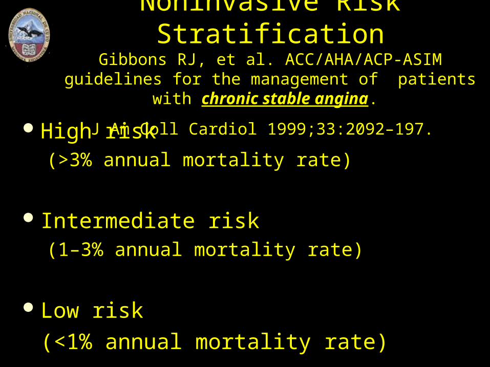
Noninvasive Risk StratificationGibbons RJ, et al. ACC/AHA/ACP-ASIM guidelines for
the management of patients with chronic stable angina.
J Am Coll Cardiol 1999;33:2092–197. High risk
(>3% annual mortality rate)
Intermediate risk (1–3% annual mortality rate)
Low risk
(<1% annual mortality rate)
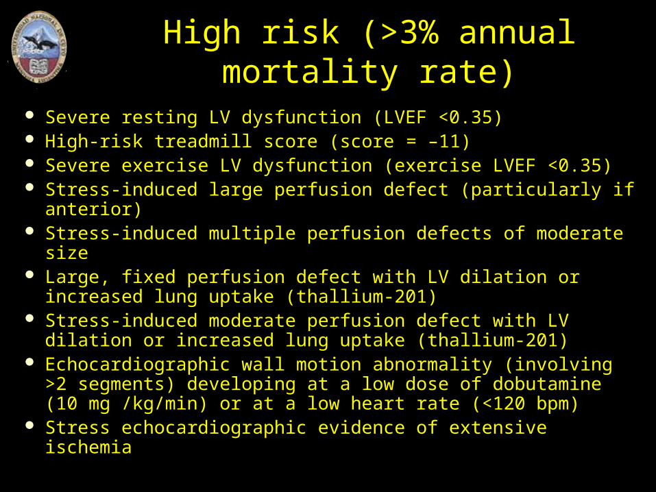
High risk (>3% annual mortality rate)
Severe resting LV dysfunction (LVEF <0.35) High-risk treadmill score (score = –11) Severe exercise LV dysfunction (exercise LVEF <0.35) Stress-induced large perfusion defect (particularly if anterior) Stress-induced multiple perfusion defects of moderate size Large, fixed perfusion defect with LV dilation or increased
lung uptake (thallium-201) Stress-induced moderate perfusion defect with LV dilation or
increased lung uptake (thallium-201) Echocardiographic wall motion abnormality (involving >2
segments) developing at a low dose of dobutamine (10 mg /kg/min) or at a low heart rate (<120 bpm)
Stress echocardiographic evidence of extensive ischemia
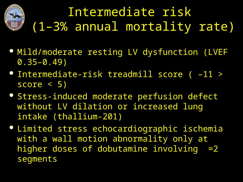
Intermediate risk (1–3% annual mortality rate)
Mild/moderate resting LV dysfunction (LVEF 0.35–0.49)
Intermediate-risk treadmill score ( –11 > score < 5) Stress-induced moderate perfusion defect without
LV dilation or increased lung intake (thallium-201) Limited stress echocardiographic ischemia with a
wall motion abnormality only at higher doses of dobutamine involving =2 segments
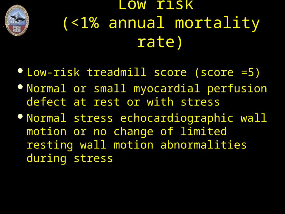
Low risk (<1% annual mortality rate)
Low-risk treadmill score (score =5)Normal or small myocardial perfusion
defect at rest or with stressNormal stress echocardiographic wall
motion or no change of limited resting wall motion abnormalities during stress

CCG y ACS sin Supra STConducta Médica
Tratamiento FarmacológicoAnticoagulantes – AntiagregantesBeta bloqueantes“Estatinas”NitritosOtros
IECAAnticálcicos
¿Conveniencia de Tratamiento Invasivo?Cirugía de Revascularización (CABG)Angioplastía (PCI)
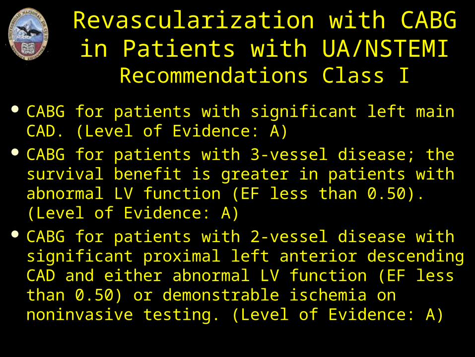
Revascularization with CABG in Patients with UA/NSTEMI
Recommendations Class I
CABG for patients with significant left main CAD. (Level of Evidence: A)
CABG for patients with 3-vessel disease; the survival benefit is greater in patients with abnormal LV function (EF less than 0.50). (Level of Evidence: A)
CABG for patients with 2-vessel disease with significant proximal left anterior descending CAD and either abnormal LV function (EF less than 0.50) or demonstrable ischemia on noninvasive testing. (Level of Evidence: A)
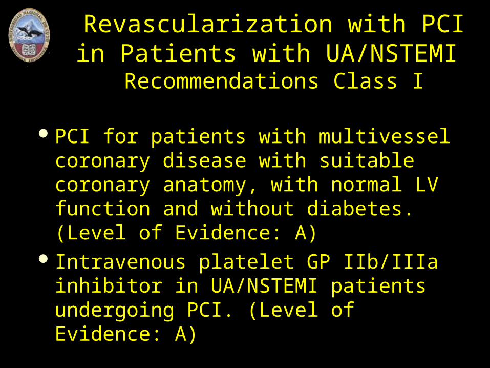
Revascularization with PCI in Patients with UA/NSTEMI
Recommendations Class I
PCI for patients with multivessel coronary disease with suitable coronary anatomy, with normal LV function and without diabetes. (Level of Evidence: A)
Intravenous platelet GP IIb/IIIa inhibitor in UA/NSTEMI patients undergoing PCI. (Level of Evidence: A)
Revascularization with PCI or CABG in Patients with UA/NSTEMI
Recommendations Class I
PCI or CABG for patients with 1- or 2-vessel CAD without significant proximal left anterior descending CAD but with a large area of viable myocardium and high-risk criteria on noninvasive testing. (Level of Evidence: B)
¡A trabajar!
Curso de CardiologíaDepartamento de Medicina Interna
Facultad de Ciencia Médicas – UN de Cuyo
Prof. Dr. Raúl Edgar Ortego