Case Report Primary T-Cell Non-Hodgkin Lymphoma of the...
5
Case Report Primary T-Cell Non-Hodgkin Lymphoma of the Vagina J. L. Herraiz, 1 A. Llueca, 1 Y. Maazouzi, 1 D. Piquer, 1 A. Palmeiro, 2 and E. Calpe 1 1 Department of Obstetrics and Gynecology, General University Hospital of Castellon, Avenida Benicassim S/N, Castell´ on de la Plana, 12004 Castell´ o, Spain 2 Department of Pathology, General University Hospital of Castellon, Avenida Benicassim S/N, Castell´ on de la Plana, 12004 Castell´ o, Spain Correspondence should be addressed to J. L. Herraiz; [email protected] Received 1 March 2015; Accepted 19 May 2015 Academic Editor: Konstantinos Dafopoulos Copyright © 2015 J. L. Herraiz et al. is is an open access article distributed under the Creative Commons Attribution License, which permits unrestricted use, distribution, and reproduction in any medium, provided the original work is properly cited. e primary vaginal T-cell non-Hodgkin lymphoma is a rare form of lymphoma. Most of the previously published cases were about B-cell non-Hodgkin lymphomas. We present the case of a vaginal mass in an 82-year-old patient presenting vaginal bleeding. e results of the immunohistological studies of the mass revealed the presence of a cytotoxic T-cell non-Hodgkin lymphoma, which is the least common subtype. 1. Introduction e primary affectation of the female genital tract in non- Hodgkin lymphomas is rare and represents 1.5–2% of all extraganglionar primary lymphomas. e most commonly affected areas are in the ovaries (50%) and cervix, while the uterine and vaginal affectation remains extremely rare (6%) [1]. e average age of onset is in the fiſth decade of life [2, 3] and the most common histology is usually that of diffuse large B-cell lymphoma (DLBCL). e patient’s motive for consultation is usually due to abnormal vaginal bleeding [4]. Despite the lack of standard treatments for the non- Hodgkin vaginal lymphoma, various treatment algorithms are used which include chemotherapy and radiotherapy, eliciting good results in initial stages of the disease [5]. We hereby present an unusual case of a vaginal T-cell lymphoma. Most of the reported cases were of B-cell non- Hodgkin lymphomas [1, 6]. 2. Case Presentation We present the case of an 82-year-old patient with no personal history of interest that came for a consultation in our gynecology department prompted by an episode of vaginal bleeding lasting various days. Her obstetrical/gynecological background includes 2 vaginal births and menopause at the age of 50. During the exploration, we found a mass, hard and friable in consistency, occupying the entire vaginal cavity. We biopsied the tumor, and the histologic results revealed the presence of medium-sized cells with granular nuclei and vascular destruction accompanied by necrosis. e immunohistochemistry showed CD3 (+), CD56 (-), CD8 (-), EBV (+), CD4(+), and granzyme B (+). e studies’ conclusions were that of a NK/cytotoxic T-cell extranodal lymphoma (Figures 1 and 2). A thoracoabdominal pelvic CT scan was performed, confirming the solid mass described in our exploration: a 7.5 × 7.5 × 9 cm mass with a rounded morphology, occupying the vesical-uterine space. ere were no pathological findings in the uterus and ovaries. ere was no sign of retroperitoneal or pelvic lymphadenopathies, nor were there lesions at the extrapelvic level (Figure 3). e Magnetic Resonance Imag- ing (MRI) corroborated the findings at the vesical-uterine level, which were previously mentioned in the CT scan results (Figure 4). e PET scan showed the previously discovered mass in the vesical-uterine space, presenting a high metabolic activity (Figure 5). e rest of the studies showed no evidence of increased tumor extension. e complete blood count (CBC), LDH, B2 microglobulin, and albumin levels were within the range of normality. e patient rejected any type of treatment. She received palliative care at home and died two months aſter having been diagnosed. Hindawi Publishing Corporation Case Reports in Obstetrics and Gynecology Volume 2015, Article ID 893083, 4 pages http://dx.doi.org/10.1155/2015/893083
Transcript of Case Report Primary T-Cell Non-Hodgkin Lymphoma of the...
Case ReportPrimary T-Cell Non-Hodgkin Lymphoma of the Vagina
J. L. Herraiz,1 A. Llueca,1 Y. Maazouzi,1 D. Piquer,1 A. Palmeiro,2 and E. Calpe1
1Department of Obstetrics and Gynecology, General University Hospital of Castellon, Avenida Benicassim S/N,Castellon de la Plana, 12004 Castello, Spain2Department of Pathology, General University Hospital of Castellon, Avenida Benicassim S/N, Castellon de la Plana,12004 Castello, Spain
Correspondence should be addressed to J. L. Herraiz; [email protected]
Received 1 March 2015; Accepted 19 May 2015
Academic Editor: Konstantinos Dafopoulos
Copyright © 2015 J. L. Herraiz et al. This is an open access article distributed under the Creative Commons Attribution License,which permits unrestricted use, distribution, and reproduction in any medium, provided the original work is properly cited.
The primary vaginal T-cell non-Hodgkin lymphoma is a rare form of lymphoma.Most of the previously published cases were aboutB-cell non-Hodgkin lymphomas. We present the case of a vaginal mass in an 82-year-old patient presenting vaginal bleeding. Theresults of the immunohistological studies of the mass revealed the presence of a cytotoxic T-cell non-Hodgkin lymphoma, whichis the least common subtype.
1. Introduction
The primary affectation of the female genital tract in non-Hodgkin lymphomas is rare and represents 1.5–2% of allextraganglionar primary lymphomas. The most commonlyaffected areas are in the ovaries (50%) and cervix, while theuterine and vaginal affectation remains extremely rare (6%)[1].
The average age of onset is in the fifth decade of life [2, 3]and the most common histology is usually that of diffuselarge B-cell lymphoma (DLBCL). The patient’s motive forconsultation is usually due to abnormal vaginal bleeding [4].
Despite the lack of standard treatments for the non-Hodgkin vaginal lymphoma, various treatment algorithmsare used which include chemotherapy and radiotherapy,eliciting good results in initial stages of the disease [5].
We hereby present an unusual case of a vaginal T-celllymphoma. Most of the reported cases were of B-cell non-Hodgkin lymphomas [1, 6].
2. Case Presentation
We present the case of an 82-year-old patient with nopersonal history of interest that came for a consultation in ourgynecology department prompted by an episode of vaginalbleeding lasting various days. Her obstetrical/gynecologicalbackground includes 2 vaginal births and menopause at
the age of 50. During the exploration, we found a mass,hard and friable in consistency, occupying the entire vaginalcavity. We biopsied the tumor, and the histologic resultsrevealed the presence of medium-sized cells with granularnuclei and vascular destruction accompanied by necrosis.The immunohistochemistry showedCD3 (+), CD56 (−), CD8(−), EBV (+), CD4(+), and granzyme B (+). The studies’conclusions were that of a NK/cytotoxic T-cell extranodallymphoma (Figures 1 and 2).
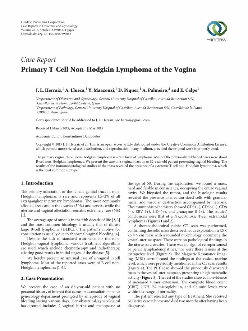
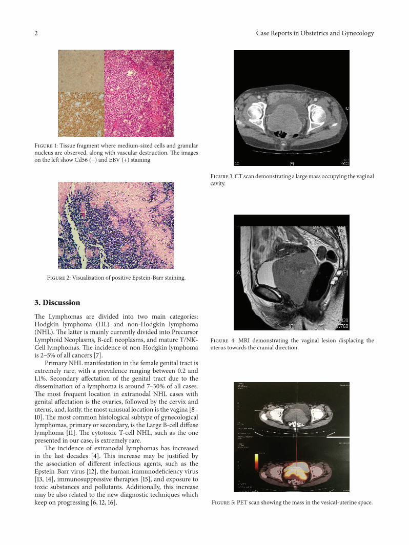
A thoracoabdominal pelvic CT scan was performed,confirming the solidmass described in our exploration: a 7.5×7.5 × 9 cm mass with a rounded morphology, occupying thevesical-uterine space. There were no pathological findings inthe uterus and ovaries. There was no sign of retroperitonealor pelvic lymphadenopathies, nor were there lesions at theextrapelvic level (Figure 3). The Magnetic Resonance Imag-ing (MRI) corroborated the findings at the vesical-uterinelevel, whichwere previouslymentioned in the CT scan results(Figure 4). The PET scan showed the previously discoveredmass in the vesical-uterine space, presenting a highmetabolicactivity (Figure 5).The rest of the studies showed no evidenceof increased tumor extension. The complete blood count(CBC), LDH, B2 microglobulin, and albumin levels werewithin the range of normality.
The patient rejected any type of treatment. She receivedpalliative care at home and died twomonths after having beendiagnosed.
Hindawi Publishing CorporationCase Reports in Obstetrics and GynecologyVolume 2015, Article ID 893083, 4 pageshttp://dx.doi.org/10.1155/2015/893083
2 Case Reports in Obstetrics and Gynecology
Figure 1: Tissue fragment where medium-sized cells and granularnucleus are observed, along with vascular destruction. The imageson the left show Cd56 (−) and EBV (+) staining.
Figure 2: Visualization of positive Epstein-Barr staining.
3. Discussion
The Lymphomas are divided into two main categories:Hodgkin lymphoma (HL) and non-Hodgkin lymphoma(NHL). The latter is mainly currently divided into PrecursorLymphoid Neoplasms, B-cell neoplasms, and mature T/NK-Cell lymphomas. The incidence of non-Hodgkin lymphomais 2–5% of all cancers [7].
Primary NHL manifestation in the female genital tract isextremely rare, with a prevalence ranging between 0.2 and1.1%. Secondary affectation of the genital tract due to thedissemination of a lymphoma is around 7–30% of all cases.The most frequent location in extranodal NHL cases withgenital affectation is the ovaries, followed by the cervix anduterus, and, lastly, themost unusual location is the vagina [8–10]. The most common histological subtype of gynecologicallymphomas, primary or secondary, is the Large B-cell diffuselymphoma [11]. The cytotoxic T-cell NHL, such as the onepresented in our case, is extremely rare.
The incidence of extranodal lymphomas has increasedin the last decades [4]. This increase may be justified bythe association of different infectious agents, such as theEpstein-Barr virus [12], the human immunodeficiency virus[13, 14], immunosuppressive therapies [15], and exposure totoxic substances and pollutants. Additionally, this increasemay be also related to the new diagnostic techniques whichkeep on progressing [6, 12, 16].
Figure 3: CT scan demonstrating a largemass occupying the vaginalcavity.
Figure 4: MRI demonstrating the vaginal lesion displacing theuterus towards the cranial direction.
Figure 5: PET scan showing the mass in the vesical-uterine space.
Case Reports in Obstetrics and Gynecology 3
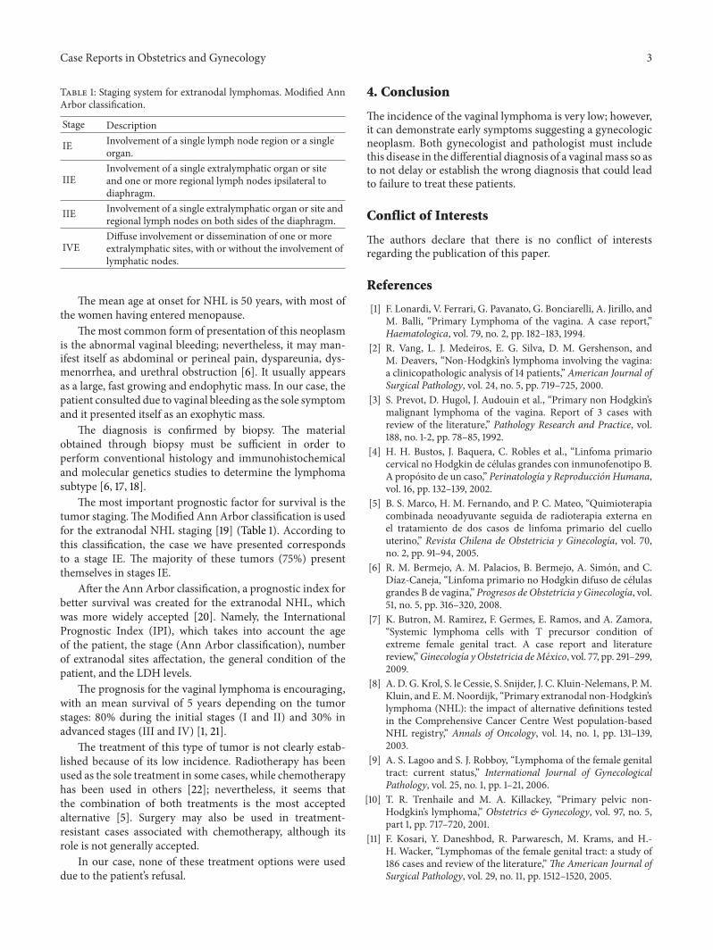
Table 1: Staging system for extranodal lymphomas. Modified AnnArbor classification.
Stage Description
IE Involvement of a single lymph node region or a singleorgan.
IIEInvolvement of a single extralymphatic organ or siteand one or more regional lymph nodes ipsilateral todiaphragm.
IIE Involvement of a single extralymphatic organ or site andregional lymph nodes on both sides of the diaphragm.
IVEDiffuse involvement or dissemination of one or moreextralymphatic sites, with or without the involvement oflymphatic nodes.
The mean age at onset for NHL is 50 years, with most ofthe women having entered menopause.
Themost common form of presentation of this neoplasmis the abnormal vaginal bleeding; nevertheless, it may man-ifest itself as abdominal or perineal pain, dyspareunia, dys-menorrhea, and urethral obstruction [6]. It usually appearsas a large, fast growing and endophytic mass. In our case, thepatient consulted due to vaginal bleeding as the sole symptomand it presented itself as an exophytic mass.
The diagnosis is confirmed by biopsy. The materialobtained through biopsy must be sufficient in order toperform conventional histology and immunohistochemicaland molecular genetics studies to determine the lymphomasubtype [6, 17, 18].
The most important prognostic factor for survival is thetumor staging.TheModified Ann Arbor classification is usedfor the extranodal NHL staging [19] (Table 1). According tothis classification, the case we have presented correspondsto a stage IE. The majority of these tumors (75%) presentthemselves in stages IE.
After the Ann Arbor classification, a prognostic index forbetter survival was created for the extranodal NHL, whichwas more widely accepted [20]. Namely, the InternationalPrognostic Index (IPI), which takes into account the ageof the patient, the stage (Ann Arbor classification), numberof extranodal sites affectation, the general condition of thepatient, and the LDH levels.
The prognosis for the vaginal lymphoma is encouraging,with an mean survival of 5 years depending on the tumorstages: 80% during the initial stages (I and II) and 30% inadvanced stages (III and IV) [1, 21].
The treatment of this type of tumor is not clearly estab-lished because of its low incidence. Radiotherapy has beenused as the sole treatment in some cases, while chemotherapyhas been used in others [22]; nevertheless, it seems thatthe combination of both treatments is the most acceptedalternative [5]. Surgery may also be used in treatment-resistant cases associated with chemotherapy, although itsrole is not generally accepted.
In our case, none of these treatment options were useddue to the patient’s refusal.
4. Conclusion
The incidence of the vaginal lymphoma is very low; however,it can demonstrate early symptoms suggesting a gynecologicneoplasm. Both gynecologist and pathologist must includethis disease in the differential diagnosis of a vaginalmass so asto not delay or establish the wrong diagnosis that could leadto failure to treat these patients.
Conflict of Interests
The authors declare that there is no conflict of interestsregarding the publication of this paper.
References
[1] F. Lonardi, V. Ferrari, G. Pavanato, G. Bonciarelli, A. Jirillo, andM. Balli, “Primary Lymphoma of the vagina. A case report,”Haematologica, vol. 79, no. 2, pp. 182–183, 1994.
[2] R. Vang, L. J. Medeiros, E. G. Silva, D. M. Gershenson, andM. Deavers, “Non-Hodgkin’s lymphoma involving the vagina:a clinicopathologic analysis of 14 patients,” American Journal ofSurgical Pathology, vol. 24, no. 5, pp. 719–725, 2000.
[3] S. Prevot, D. Hugol, J. Audouin et al., “Primary non Hodgkin’smalignant lymphoma of the vagina. Report of 3 cases withreview of the literature,” Pathology Research and Practice, vol.188, no. 1-2, pp. 78–85, 1992.
[4] H. H. Bustos, J. Baquera, C. Robles et al., “Linfoma primariocervical no Hodgkin de celulas grandes con inmunofenotipo B.A proposito de un caso,” Perinatologıa y Reproduccion Humana,vol. 16, pp. 132–139, 2002.
[5] B. S. Marco, H. M. Fernando, and P. C. Mateo, “Quimioterapiacombinada neoadyuvante seguida de radioterapia externa enel tratamiento de dos casos de linfoma primario del cuellouterino,” Revista Chilena de Obstetricia y Ginecologıa, vol. 70,no. 2, pp. 91–94, 2005.
[6] R. M. Bermejo, A. M. Palacios, B. Bermejo, A. Simon, and C.Dıaz-Caneja, “Linfoma primario no Hodgkin difuso de celulasgrandes B de vagina,” Progresos de Obstetricia y Ginecologıa, vol.51, no. 5, pp. 316–320, 2008.
[7] K. Butron, M. Ramirez, F. Germes, E. Ramos, and A. Zamora,“Systemic lymphoma cells with T precursor condition ofextreme female genital tract. A case report and literaturereview,”Ginecologıa y Obstetricia deMexico, vol. 77, pp. 291–299,2009.
[8] A. D. G. Krol, S. le Cessie, S. Snijder, J. C. Kluin-Nelemans, P.M.Kluin, and E.M. Noordijk, “Primary extranodal non-Hodgkin’slymphoma (NHL): the impact of alternative definitions testedin the Comprehensive Cancer Centre West population-basedNHL registry,” Annals of Oncology, vol. 14, no. 1, pp. 131–139,2003.
[9] A. S. Lagoo and S. J. Robboy, “Lymphoma of the female genitaltract: current status,” International Journal of GynecologicalPathology, vol. 25, no. 1, pp. 1–21, 2006.
[10] T. R. Trenhaile and M. A. Killackey, “Primary pelvic non-Hodgkin’s lymphoma,” Obstetrics & Gynecology, vol. 97, no. 5,part 1, pp. 717–720, 2001.
[11] F. Kosari, Y. Daneshbod, R. Parwaresch, M. Krams, and H.-H. Wacker, “Lymphomas of the female genital tract: a study of186 cases and review of the literature,”The American Journal ofSurgical Pathology, vol. 29, no. 11, pp. 1512–1520, 2005.

4 Case Reports in Obstetrics and Gynecology
[12] S. Domingo, A. Perales, V. Torres, M. J. Alcaraz, and A. Pellicer,“Epstein-Barr virus positivity in primary vaginal lymphoma,”Gynecologic Oncology, vol. 95, no. 3, pp. 719–721, 2004.
[13] D. C. Pham, T. H. Guthrie, and B. Ndubisi, “HIV-associatedprimary cervical non-Hodgkin’s lymphoma and two othercases of primary pelvic non-Hodgkin’s lymphoma,”GynecologicOncology, vol. 90, no. 1, pp. 204–206, 2003.
[14] A. Klepfish, A. Schattner, L. Shvidel et al., “Succesful treatmentof aggressive HIV-associated non-hodgkin’s lymphoma withcombination chemotherapy, biotherapy with rituximab andHAART: presentation of a therapeutic option,” Leukemia andLymphoma, vol. 44, no. 2, pp. 349–351, 2003.
[15] K. Yoshinaga, J.-I. Akahira, H. Niikura et al., “A case of primarymucosa-associated lymphoid tissue lymphoma of the vagina,”Human Pathology, vol. 35, no. 9, pp. 1164–1166, 2004.
[16] D. N. Lanjewar and D. D. Dongaonkar, “HIV-associatedprimary non-Hodgkin’s lymphoma of ovary: a case report,”Gynecologic Oncology, vol. 102, no. 3, pp. 590–592, 2006.
[17] O. Akbayir, K. Gungorduk, A. Gulkilik, E. Yavuz, A. I. Tekirdag,and E. Odabas, “Successful treatment of primary vaginal diffuselarge B-cell lymphoma using chemotherapy,” Taiwanese Journalof Obstetrics and Gynecology, vol. 47, no. 3, pp. 334–337, 2008.
[18] N. van Renterghem, P. de Paepe, R. van den Broecke, C.Bourgain, and R. Serreyn, “Primary lymphoma of the cervixuteri: a diagnostic challenge. Report of two cases and reviewof the literature,” European Journal of Gynaecological Oncology,vol. 26, no. 1, pp. 36–38, 2005.
[19] R. Vang, L. J. Medeiros, G. N. Fuller, A. H. Sarris, and M.Deavers, “Non-Hodgkin’s lymphoma involving the gynecologictract: a review of 88 cases,”Advances in Anatomic Pathology, vol.8, no. 4, pp. 200–217, 2001.
[20] A. S.Huerta, L. P. Abad, J.M. Saera, and J. Gomez-Codina, “Fac-tores pronostico de los linfomas oseos primarios. Importanciadel Indice Pronostico Internacional,” Oncologıa, vol. 27, no. 9,pp. 55–57, 2004.
[21] E. M. Guldrıs N, M. P. Vazquez C, E. Carballo N, M. PortoQ, L. Heliodoro Alba O, and B. Iglesias R, “Linfoma noHodgkin primario de vagina,” Revista Chilena de Obstetricia yGinecologıa, vol. 78, no. 1, pp. 68–71, 2013.
[22] M. Signorelli, A. Maneo, S. Cammarota et al., “Conservativemanagement in primary genital lymphomas: the role of chemo-therapy,”GynecologicOncology, vol. 104, no. 2, pp. 416–421, 2007.

Submit your manuscripts athttp://www.hindawi.com
Stem CellsInternational
Hindawi Publishing Corporationhttp://www.hindawi.com Volume 2014
Hindawi Publishing Corporationhttp://www.hindawi.com Volume 2014
MEDIATORSINFLAMMATION
of
Hindawi Publishing Corporationhttp://www.hindawi.com Volume 2014
Behavioural Neurology
EndocrinologyInternational Journal of
Hindawi Publishing Corporationhttp://www.hindawi.com Volume 2014
Hindawi Publishing Corporationhttp://www.hindawi.com Volume 2014
Disease Markers
Hindawi Publishing Corporationhttp://www.hindawi.com Volume 2014
BioMed Research International
OncologyJournal of
Hindawi Publishing Corporationhttp://www.hindawi.com Volume 2014
Hindawi Publishing Corporationhttp://www.hindawi.com Volume 2014
Oxidative Medicine and Cellular Longevity
Hindawi Publishing Corporationhttp://www.hindawi.com Volume 2014
PPAR Research
The Scientific World JournalHindawi Publishing Corporation http://www.hindawi.com Volume 2014
Immunology ResearchHindawi Publishing Corporationhttp://www.hindawi.com Volume 2014
Journal of
ObesityJournal of
Hindawi Publishing Corporationhttp://www.hindawi.com Volume 2014
Hindawi Publishing Corporationhttp://www.hindawi.com Volume 2014
Computational and Mathematical Methods in Medicine
OphthalmologyJournal of
Hindawi Publishing Corporationhttp://www.hindawi.com Volume 2014
Diabetes ResearchJournal of
Hindawi Publishing Corporationhttp://www.hindawi.com Volume 2014
Hindawi Publishing Corporationhttp://www.hindawi.com Volume 2014
Research and TreatmentAIDS
Hindawi Publishing Corporationhttp://www.hindawi.com Volume 2014
Gastroenterology Research and Practice
Hindawi Publishing Corporationhttp://www.hindawi.com Volume 2014
Parkinson’s Disease
Evidence-Based Complementary and Alternative Medicine
Volume 2014Hindawi Publishing Corporationhttp://www.hindawi.com