Case Report - Hindawi Publishing Corporation as interradicular, periapical, pseudomultilocular,...
5
Hindawi Publishing Corporation Case Reports in Dentistry Volume 2012, Article ID 482758, 4 pages doi:10.1155/2012/482758 Case Report Type B Idiopathic Bone Defect of Mandible: An Etiopathogenic Dilemma Aakarsh V. Jhamb, 1 Parul A. Jhamb, 2 Aparna Dave, 3 and Vishwa Prakash Shetty 3 1 Department of Oral & Maxillofacial Surgery, ESIC Dental College, Rohini, New Delhi 110 089, India 2 Department of Oral & Maxillofacial Pathology, Santosh Dental College & Hospital, Ghaziabad 201 009, India 3 Department of Oral & Maxillofacial Pathology, SGT Dental College & Hospital, Gurgaon 122 505, India Correspondence should be addressed to Aakarsh V. Jhamb, [email protected] Received 1 March 2012; Accepted 4 April 2012 Academic Editors: P. G. Arduino and N. Brezniak Copyright © 2012 Aakarsh V. Jhamb et al. This is an open access article distributed under the Creative Commons Attribution License, which permits unrestricted use, distribution, and reproduction in any medium, provided the original work is properly cited. Etiopathogenesis of the pathologic lesions forms the basis for formulation of appropriate intervention and further prevention. There is still a vast unknown field that has to be explored to know the causative reason behind certain benign & malignant lesions. Idiopathic bone defects are nonodontogenic pseudocystic cavities that are seen in the long bones & jaw bones. Radiographic interpretation is at times inadequate in diagnosis of odontogenic & nonodontogenic radiolucent lesions involving jaw bones. Histopathology has different criteria to segregate this lesion. In this paper, we discuss a case of type B histopathological variant of idiopathic bone defect that may suggest an alternative pathogenesis from type A variant. 1. Introduction Radiolucent lesions in the jaw can occur in varied forms and may represent innocent anatomic variations, benign or malignant processes. Appropriate diagnosis relies on a detailed history, astute clinical sense, and cognition of the fact that pathology can present in unexpected forms. Cystic lesions are the most frequently encountered pathologic radiolucencies in the jaw [1]. This spectrum consists of pathological entities such as ameloblastoma, odontogenic keratocyst, and odontogenic myxoma. Most of these lesions cause symptomatic, expansile swelling that is quite obvious clinically; yet certain asymptomatic radiolucent pathologic lesions occur in the jaws that pose a significant problem in the diagnosis due to lack of definite objective or subjective clinical symptoms [2]. Idiopathic bone defect is one such intriguing lesion that occurs in the jaws relatively rarely. We elucidate a case of idiopathic bone defect of mandible with its postoperative course and discuss the pertinent review of literature. 2. Report of a Case A 35-years-old female visited our clinic in August 2009 with a complaint of swelling on right side of face, which had slowly increased in size over last one year before the patient approached for treatment. There was no history of trauma or any previous infection in the affected area. The past medical and surgical history was noncontributory. On physical examination the facial symmetry was maintained. Intraorally, slight expansion of the buccal cortex was seen in mandibular first molar region on right side (Figure 1). On palpation, the swelling was nontender, bony hard and local- ized over the alveolus of the mandibular first molar region on the buccal side. The teeth in the involved area were normal. There was no paresthesia or cervical lymphadenopathy. Radiographic examination revealed a well-localized unilocular radiolucency in the mandibular first molar area on the right side involving the second premolar and second molar roots. The alveolar bone around the first molar roots was resorbed more than two-thirds of the root length, and
Transcript of Case Report - Hindawi Publishing Corporation as interradicular, periapical, pseudomultilocular,...

Hindawi Publishing CorporationCase Reports in DentistryVolume 2012, Article ID 482758, 4 pagesdoi:10.1155/2012/482758
Case Report
Type B Idiopathic Bone Defect of Mandible:An Etiopathogenic Dilemma
Aakarsh V. Jhamb,1 Parul A. Jhamb,2 Aparna Dave,3 and Vishwa Prakash Shetty3
1 Department of Oral & Maxillofacial Surgery, ESIC Dental College, Rohini, New Delhi 110 089, India2 Department of Oral & Maxillofacial Pathology, Santosh Dental College & Hospital, Ghaziabad 201 009, India3 Department of Oral & Maxillofacial Pathology, SGT Dental College & Hospital, Gurgaon 122 505, India
Correspondence should be addressed to Aakarsh V. Jhamb, [email protected]
Received 1 March 2012; Accepted 4 April 2012
Academic Editors: P. G. Arduino and N. Brezniak
Copyright © 2012 Aakarsh V. Jhamb et al. This is an open access article distributed under the Creative Commons AttributionLicense, which permits unrestricted use, distribution, and reproduction in any medium, provided the original work is properlycited.
Etiopathogenesis of the pathologic lesions forms the basis for formulation of appropriate intervention and further prevention.There is still a vast unknown field that has to be explored to know the causative reason behind certain benign & malignant lesions.Idiopathic bone defects are nonodontogenic pseudocystic cavities that are seen in the long bones & jaw bones. Radiographicinterpretation is at times inadequate in diagnosis of odontogenic & nonodontogenic radiolucent lesions involving jaw bones.Histopathology has different criteria to segregate this lesion. In this paper, we discuss a case of type B histopathological variant ofidiopathic bone defect that may suggest an alternative pathogenesis from type A variant.
1. Introduction
Radiolucent lesions in the jaw can occur in varied formsand may represent innocent anatomic variations, benignor malignant processes. Appropriate diagnosis relies on adetailed history, astute clinical sense, and cognition of thefact that pathology can present in unexpected forms. Cysticlesions are the most frequently encountered pathologicradiolucencies in the jaw [1]. This spectrum consists ofpathological entities such as ameloblastoma, odontogenickeratocyst, and odontogenic myxoma. Most of these lesionscause symptomatic, expansile swelling that is quite obviousclinically; yet certain asymptomatic radiolucent pathologiclesions occur in the jaws that pose a significant problem inthe diagnosis due to lack of definite objective or subjectiveclinical symptoms [2]. Idiopathic bone defect is one suchintriguing lesion that occurs in the jaws relatively rarely. Weelucidate a case of idiopathic bone defect of mandible withits postoperative course and discuss the pertinent review ofliterature.
2. Report of a Case
A 35-years-old female visited our clinic in August 2009with a complaint of swelling on right side of face, whichhad slowly increased in size over last one year before thepatient approached for treatment. There was no history oftrauma or any previous infection in the affected area. Thepast medical and surgical history was noncontributory. Onphysical examination the facial symmetry was maintained.Intraorally, slight expansion of the buccal cortex was seen inmandibular first molar region on right side (Figure 1). Onpalpation, the swelling was nontender, bony hard and local-ized over the alveolus of the mandibular first molar region onthe buccal side. The teeth in the involved area were normal.There was no paresthesia or cervical lymphadenopathy.
Radiographic examination revealed a well-localizedunilocular radiolucency in the mandibular first molar areaon the right side involving the second premolar and secondmolar roots. The alveolar bone around the first molar rootswas resorbed more than two-thirds of the root length, and

2 Case Reports in Dentistry
Figure 1: Intraoral view shows slight expansion of the buccal cortexin the region of mandibular first molar on right side.
Figure 2: Orthopantomograph reveals a well-localized unilocularradiolucency in the mandibular first molar area on the right sideinvolving the second premolar and second molar roots. Resorptionof alveolar bone and loss of lamina dura around the first molar rootsis also present.
there was loss of lamina dura around the associated vitalteeth (Figure 2). Chest radiographs and blood chemistrywere normal. Aspiration with a wide bore needle wasattempted but yielded no results as it could not penetrate theradiolucent lesion.
A differential diagnosis of ameloblastoma, central giantcell granuloma, odontogenic keratocyst, and traumatic bonecyst was made. It was decided to take an incisional biopsyafter extraction of the involved first molar under localanesthesia. Upon raising the mucoperiosteal flap, the under-lying buccal cortex showed expansion and a bluish tinge(Figure 3). A small drill hole was made in the bone to allowinsertion of a needle for aspiration. The bony window wasthen enlarged to take incisional biopsy. The bony cavitywas found to be almost empty with scanty serosanguinousfluid. A provisional diagnosis of solitary bone cyst wasmade at the time of surgical exploration. The involved teethwere extracted and sufficient bleeding was induced in thecavity after curettage of the cavity walls. The wound wasclosed primarily with 3-0 silk sutures. Histopathologicalexamination of the curetted tissue exhibited osseous cysticwall and overlying fibrovascular tissue with no epitheliallining (Figure 4). A thick fibrovascular wall with underlyingdysplastic bone could be appreciated which was suggestiveof type B simple bone cyst [3]. Postoperatively, the woundhealed uneventfully (Figure 5). The patient was followed upfor next one year, as type B lesion has chances of recurrence
Figure 3: Intraoperative view shows expansion of the underlyingbuccal cortex with a bluish tinged surface is seen.
Figure 4: H&E stained section of the curetted tissue exhibitsosseous cystic wall and overlying fibrovascular tissue with noepithelial lining. (10x).
when compared with type A. After 1 year the radiographsshowed complete bony healing (Figure 6).
3. Discussion
Idiopathic bone defect has been referred by various names:hemorrhagic cyst, traumatic bone cyst, simple bone cyst,solitary bone cyst, unicameral bone cyst, progressive bonecavity, extravasation cyst [4, 5]. However, WHO has recom-mended the use of term solitary bone cyst since 1992 and inits more recent meeting in 2005 this entity has been classifiedunder bone-related lesions [6]. It was also classified as a pseu-docyst because it shows no epithelial lining. According to arecently proposed concept SBC is considered to be a synovialcyst arising from a juxtaepiphyseal error with intraosseousincorporation of synovial tissue [7]. As per the previousreports, development of this lesion has been attributed to theintrusion of salivary gland tissue, mechanical pressure of thesurrounding tissue, and the pressure of the facial artery as themost probable factors [8]. It has been classified by differentauthors as either (i) stafne type (ii) cystic type, or (i) type A,(ii) type B on radiographic and histopathological assessment[3]. Matsumura et al. stated that type B lesion results fromcystic degeneration of certain type of fibroosseous lesionsand type A might be caused by intramedullary hemorrhageand its sequelae. Most of the type B cysts are known to
Case Reports in Dentistry 3
Figure 5: Clinical photograph of postoperative healed lesion.
Figure 6: Orthopantamograph taken after 1 year shows completebony healing.
displace mandibular canal and cause buccolingual expansionin comparison with the contralateral arch as seen in this case[3].
This lesion presents mostly as an osteolytic lesion andforms a cavity with either a geodic or polymorphousshape [9]. It may be empty or filled with blood, serum,or serosanguinous fluid. No cholesterol crystals have beenreported and with time the amount of fluid diminishes[2]. This lesion is mainly diagnosed in young patients,mostly during the second decade of life, though in thiscase the lesion was diagnosed in late third decade. The sexdistribution is reported to be quite even, although there isa male predominance for the extra facial variants [9, 10].SBC evolution is asymptomatic in the beginning and isoften discovered serendipitously during examination of apanoramic radiograph. Pain is the presenting symptom in10 to 30% of patients and swelling has been reported inup to a fourth of the patients. Other symptoms includetooth sensitivity, paresthesia, fistula, and even pathologicfracture of the jaw bone [11]. As solitary bone cyst usuallydoes not show any particular pathognomonic feature andthe presentation is mostly a vague asymptomatic radiolucentshadow, so the definitive diagnosis can be made only uponsurgical exploration and histopathological evaluation.
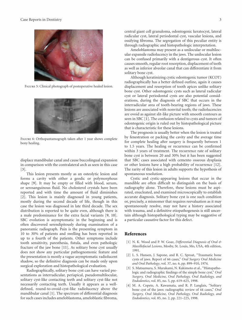
Radiographically, solitary bone cyst can have varied pre-sentations as interradicular, periapical, pseudomultilocular,solitary cyst-like contacting teeth and solitary cyst-like notnecessarily contacting teeth. Usually it appears as a well-defined, round-to-ovoid-cyst-like radiolucency above themandibular canal [1]. The spectrum of differential diagnosisfor such cases includes ameloblastoma, ameloblastic fibroma,
central giant cell granuloma, odontogenic keratocyst, lateralradicular cyst, lateral periodontal cyst, vascular lesions, andossifying fibroma. The segregation of this peculiar entity isthrough radiographic and histopathologic interpretation.
Ameloblastoma may present as a unilocular or multiloc-ular expansile radiolucency in the jaws. The unilocular lesioncan be confused primarily with a dentigerous cyst. It oftencauses smooth, regular root resorption, displacement of teethas well as inferior alveolar canal that can differentiate it fromsolitary bone cyst.
Although keratinizing cystic odontogenic tumor (KCOT)radiographically has a better defined outline, again it causesdisplacement and resorption of tooth apices unlike solitarybone cyst. Other odontogenic cysts such as lateral radicularcyst or lateral periodontal cysts are also potential consid-erations, during the diagnosis of SBC that occurs in theinterradicular area of tooth-bearing regions of jaws. Theselesions are associated with nonvital tooth; the radiolucenciesare ovoid as against slit-like picture with smooth contours asseen in SBC [1]. The confusion related to cysts and tumors ofodontogenic origin is ruled out by histopathological picturethat is characteristic for these lesions.
The prognosis is usually better when the lesion is treatedby fenestration or packing the cavity and the average timefor complete healing after surgery is frequently between 1to 1.5 years. The healing or recurrence can be confirmedwithin 3 years of treatment. The recurrence rate of solitarybone cyst is between 20 and 30% but it has been suggestedthat SBC cases associated with cemento osseous dysplasiaor other lesions have a high probability of recurrence [12].The rarity of this lesion in adults supports the hypothesis ofspontaneous resolution.
Cystic and cystic-appearing lesions that occur in themandible are often difficult to distinguish on the basis ofradiography alone. Therefore, these lesions must be aspi-rated, enucleated, and examined microscopically to establishaccurate diagnosis. Solitary bone cyst is one such conditionor, precisely, a misnomer that requires reevaluation as it mayspontaneously resolve, may not have a history associatedwith trauma, and a definitive etiopathogenesis is still uncer-tain although histopathological typing may be suggestive ofa particular causative factor for this defect.
References
[1] N. K. Wood and P. W. Goaz, Differential Diagnosis of Oral &Maxillofacial Lesions, Mosby, St. Louis, Mo, USA, 4th edition,1991.
[2] L. S. Hansen, J. Sapone, and R. C. Sproat, “Traumatic bonecysts of jaws. Report of 66 cases,” Oral Surgery Oral Medicineand Oral Pathology, vol. 37, no. 6, pp. 899–910, 1974.
[3] S. Matsumura, S. Murakami, N. Kakimoto et al., “Histopatho-logic and radiographic findings of the simple bone cyst,” OralSurgery, Oral Medicine, Oral Pathology, Oral Radiology, andEndodontics, vol. 85, no. 5, pp. 619–625, 1998.
[4] M. A. Copete, A. Kawamata, and R. P. Langlais, “Solitarybone cyst of the jaws: radiographic review of 44 cases,” OralSurgery, Oral Medicine, Oral Pathology, Oral Radiology, andEndodontics, vol. 85, no. 2, pp. 221–225, 1998.
4 Case Reports in Dentistry
[5] Y. Yanagi, J. I. Asaumi, T. Unetsubo et al., “Usefulness ofMRI and dynamic contrast-enhanced MRI for differentialdiagnosis of simple bone cysts from true cysts in the jaw,”Oral Surgery, Oral Medicine, Oral Pathology, Oral Radiologyand Endodontology, vol. 110, no. 3, pp. 364–369, 2010.
[6] I. R. H. Kramer, J. J. Pindborg, and M. Shear, HistologicalTyping of Odontogenic Tumors, Springer, New York, NY, USA,2nd edition, 1992.
[7] A. Kuhmichel and G. F. Bouloux, “Multifocal traumatic bonecysts: case report and current thoughts on etiology,” Journalof Oral and Maxillofacial Surgery, vol. 68, no. 1, pp. 208–212,2010.
[8] M. Shimizu, N. Osa, K. Okamura, and K. Yoshiura, “CTanalysis of the Stafne’s bone defects of the mandible,” Den-tomaxillofacial Radiology, vol. 35, no. 2, pp. 95–102, 2006.
[9] J. C. Harnet, T. Lombardi, P. Klewansky, J. Rieger, M. H.Tempe, and J. M. Clavert, “Solitary bone cyst of the Jaws: areview of the etiopathogenic hypotheses,” Journal of Oral andMaxillofacial Surgery, vol. 66, no. 11, pp. 2345–2348, 2008.
[10] K. Forssell, H. Forssell, R. P. Happonen, and M. Neva, “Simplebone cyst. Review of the literature and analysis of 23 cases,”International Journal of Oral and Maxillofacial Surgery, vol. 17,no. 1, pp. 21–24, 1988.
[11] K. R. Magliocca, S. P. Edwards, and J. I. Helman, “Traumaticbone cyst of the condylar region: report of 2 cases,” Journal ofOral and Maxillofacial Surgery, vol. 65, no. 6, pp. 1247–1250,2007.
[12] Y. Suei, A. Taguchi, and K. Tanimoto, “Simple bone cyst of thejaws: evaluation of treatment outcome by review of 132 cases,”Journal of Oral and Maxillofacial Surgery, vol. 65, no. 5, pp.918–923, 2007.

Submit your manuscripts athttp://www.hindawi.com
Hindawi Publishing Corporationhttp://www.hindawi.com Volume 2014
Oral OncologyJournal of
DentistryInternational Journal of
Hindawi Publishing Corporationhttp://www.hindawi.com Volume 2014
Hindawi Publishing Corporationhttp://www.hindawi.com Volume 2014
International Journal of
Biomaterials
Hindawi Publishing Corporationhttp://www.hindawi.com Volume 2014
BioMed Research International
Hindawi Publishing Corporationhttp://www.hindawi.com Volume 2014
Case Reports in Dentistry
Hindawi Publishing Corporationhttp://www.hindawi.com Volume 2014
Oral ImplantsJournal of
Hindawi Publishing Corporationhttp://www.hindawi.com Volume 2014
Anesthesiology Research and Practice
Hindawi Publishing Corporationhttp://www.hindawi.com Volume 2014
Radiology Research and Practice
Environmental and Public Health
Journal of
Hindawi Publishing Corporationhttp://www.hindawi.com Volume 2014
The Scientific World JournalHindawi Publishing Corporation http://www.hindawi.com Volume 2014
Hindawi Publishing Corporationhttp://www.hindawi.com Volume 2014
Dental SurgeryJournal of
Drug DeliveryJournal of
Hindawi Publishing Corporationhttp://www.hindawi.com Volume 2014
Hindawi Publishing Corporationhttp://www.hindawi.com Volume 2014
Oral DiseasesJournal of
Hindawi Publishing Corporationhttp://www.hindawi.com Volume 2014
Computational and Mathematical Methods in Medicine
ScientificaHindawi Publishing Corporationhttp://www.hindawi.com Volume 2014
PainResearch and TreatmentHindawi Publishing Corporationhttp://www.hindawi.com Volume 2014
Preventive MedicineAdvances in
Hindawi Publishing Corporationhttp://www.hindawi.com Volume 2014
EndocrinologyInternational Journal of
Hindawi Publishing Corporationhttp://www.hindawi.com Volume 2014
Hindawi Publishing Corporationhttp://www.hindawi.com Volume 2014
OrthopedicsAdvances in