BY - jbc.org · dilute sodium hydroxide. The capital letter, P, following imme- diately after the...
23
ARE THE PHOSPHATASES OF BONE, KIDNEY, IN- TESTINE, AND SERUM IDENTICAL? THE USE OF BILE ACIDS IN THEIR DIFFERENTIATION BY OSCAR BODANSKY (From the Children’s Medical Service and the Department oj Pathology, Bellevue Hospital, and the Department of Pediatrics, New York University College of Medicine, New York) (Received for publication, December 7, 1936) The demonstration of increased concentrations of phosphatase’ in the serum in physiological processes and pathological conditions has been pursued intensively during the past few years. Illustra- tive of many such studies are those of Kay (I), A. Bodansky and Jaffe (2), Woodard, Twombly, and Coley (3) on bone diseases, of Roberts (4) and Rothman, Meranze, and Meranze (5) on various types of jaundice, and of Gutman, Tyson, and Gutman (6) on hyperparathyroidism, Paget’s disease, multiple myeloma, and neoplastic diseases of bone. Franseen and McLean (7) have compared the phosphatase activity of plasma and bone tissue in various tumors of the bone. Investigation has also been made of the level of serum phosphatase during digestion (S), in experi- mental bile duct ligation (9, lo), and after removal of various organs (11). It has been generally assumedin such work and proof has been offered by Kay (12, 13) that the phosphatases of the different tissues and of serum are probably identical, and attempts at elucidating the nature and source of serum phosphatase in health and disease have been made by several investigators. Thus Kay (1) has offered the explanations that the high plasma phosphatase in bone diseases comesfrom the bones either as an overproduction in an attempted compensation for the lesion or because of squeez- 1 The term “phosphatase” is used in this paper to denote the enzyme acting optimally at a pH of about 9.0 on the monophosphoric esters and on hexosediphosphoric acid. 341 by guest on February 17, 2020 http://www.jbc.org/ Downloaded from
Transcript of BY - jbc.org · dilute sodium hydroxide. The capital letter, P, following imme- diately after the...

ARE THE PHOSPHATASES OF BONE, KIDNEY, IN- TESTINE, AND SERUM IDENTICAL?
THE USE OF BILE ACIDS IN THEIR DIFFERENTIATION
BY OSCAR BODANSKY
(From the Children’s Medical Service and the Department oj Pathology, Bellevue Hospital, and the Department of Pediatrics, New York
University College of Medicine, New York)
(Received for publication, December 7, 1936)
The demonstration of increased concentrations of phosphatase’ in the serum in physiological processes and pathological conditions has been pursued intensively during the past few years. Illustra- tive of many such studies are those of Kay (I), A. Bodansky and Jaffe (2), Woodard, Twombly, and Coley (3) on bone diseases, of Roberts (4) and Rothman, Meranze, and Meranze (5) on various types of jaundice, and of Gutman, Tyson, and Gutman (6) on hyperparathyroidism, Paget’s disease, multiple myeloma, and neoplastic diseases of bone. Franseen and McLean (7) have compared the phosphatase activity of plasma and bone tissue in various tumors of the bone. Investigation has also been made of the level of serum phosphatase during digestion (S), in experi- mental bile duct ligation (9, lo), and after removal of various organs (11).
It has been generally assumed in such work and proof has been offered by Kay (12, 13) that the phosphatases of the different tissues and of serum are probably identical, and attempts at elucidating the nature and source of serum phosphatase in health and disease have been made by several investigators. Thus Kay (1) has offered the explanations that the high plasma phosphatase in bone diseases comes from the bones either as an overproduction in an attempted compensation for the lesion or because of squeez-
1 The term “phosphatase” is used in this paper to denote the enzyme acting optimally at a pH of about 9.0 on the monophosphoric esters and on hexosediphosphoric acid.
341
by guest on February 17, 2020http://w
ww
.jbc.org/D
ownloaded from

342 Differentiation of Phosphatases
ing out from the cells of the injured bone under mechanical stress. A. Bodansky and Jaffe (2) and A. Bodansky (8) consider the increase in bone disease of certain types an expression of the “specific reactivity of bone” or its capacity for cellular activities; non- osseous origins of serum phosphatase in the normal are predicated because of the finding of hyperphosphatasemias after carbo- hydrate ingestion in young dogs and abundant feeding in new born puppies. A more direct experimental approach has been recently submitted by Armstrong and Banting (11) who showed that in the dog extirpation of the intestines, kidneys, spleen, pancreas, liver, testes, or epididymes does not lower the serum phosphatase and may, indeed, raise it. Bone is therefore considered as the sole source of serum phosphatase.
Preliminary to the investigation of such source and of the mechanism of the various hyperphosphatasemias is the necessity for determining whether the phosphatases of the tissues and of the serum are indeed identical or, if not, the manner in which they differ and the means by which they may be distinguished.
The present paper describes attempts to answer some portion of these questions by studying the pattern of hydrolysis by the different tissue phosphatases of several phosphoric esters, and the hydrolysis of sodium @glycerophosphate in the presence of differ- ent types of retardants, principally and usefully, the bile acids. Since the bone, kidney, and intestine have been considered to contain the highest concentrations of phosphatase (12),2 the present investigation has begun with the study of the phosphatases of these tissues from several species, including man. It is shown that the bile acids retard the hydrolysis of sodium P-glycerophos- phate by bone and kidney phosphatases but do not affect that by the intestinal phosphatases. The applicability of these findings to the study of the source and nature of serum phosphatase, in health and in disease, is then considered.
EXPERIMENTAL
Preparations of Phosphatase-In the designation of the prepara- tions used in the course of the present work, the first capital letter denotes the species from which the tissue was obtained; a second
2 Recently Folley and Kay (14) have found the mammary gland of guinea pig to contain comparable concentrations of phosphatase.
by guest on February 17, 2020http://w
ww
.jbc.org/D
ownloaded from
0. Bodansky
small letter is added, if necessary: C represents cattle, Ca cat, R rat, M man, Rb rabbit. The second capital letter represents the tissue: B represents bone, K kidney, I intestine; and the third capit,al letter merely represents stock batches of phosphatase made up at different times. The small letter, d, immediately following the hyphen designates dialysis, usually overnight against a 20-fold voIume of distihed water, changed once, as has previously been described (15). A small letter, p, following d, designates that dialysis was continued until a precipitate appeared, and that a solution was made from this precipitate with the aid of a little dilute sodium hydroxide. The capital letter, P, following imme- diately after the hyphen designates that the preparation was made as a powder by the method of Albers and Albers (16), to be described, and d following P, in this case, indicates dialysis of the solution made from this powder. Numbers following d represent different batches of the stock phosphatase extracts which were taken for dialysis.
Most of the preparations were obtained, as previously described (15, 17), by extraction and 2 to 3 day autolysis of the tissue at room temperature; 20 cc. of distilled water and 1 cc. of toluene were used per gm. of tissue. Human tissues were obtained at the autopsy table. The kidney phosphatases, Preparations CKA-Pd, CKB-Pd, and MKA-Pd, were prepared by the method of Albers and Albers (16). This consists in a 4 to 5 day autolysis of the tissue in a mixture of toluene, ethyl acetate, and 50 per cent ethyl alcohol. After completion of autolysis, ethyl alcohol is added to a concentration of 65 per cent; the precipitate contains the phos- phatase, is centrifuged, and may be dried with alcohol and ether. The dried preparation maintains its activity well. In the present studies, the precipitate was dissolved with the aid of a little dilute sodium hydroxide. The activity of such dialyzed preparations remained constant for weeks. The serum phosphatases were obtained from rabbit, cat, and man. The serum from two subjects with Paget’s disease (osteitis deformans) were used for the illustra- tion of the reaction of a pathological serum. The serum was not subjected to any purification or dialysis, but was usually diluted with water or saline and used in a concentration of 12.5 per cent by volume of the hydrolysis mixture. There was no attempt to use the enzyme preparations at a given time after extraction; it
by guest on February 17, 2020http://w
ww
.jbc.org/D
ownloaded from
344 Differentiation of Phosphatases
was considered desirable that whatever distinguishing reactions they might show should be independent of this time factor.
Substrates and Retardants-All substrates were used in a concen- tration of 0.0127 M, calculated on the basis of the phosphorus that could be liberated as inorganic phosphate. The sodium /3-glycero- phosphate was the Eastman Kodak product. Sodium hexosedi- phosphate was prepared from the calcium salt, candiolin (Winthrop Chemical Company), by treatment with a submaximal amount of sodium oxalate to avoid subsequently the possible retardant effect of the oxalate ion; the resulting solution contained, per cc. of hydrolysis mixture, 0.025 mg. of calcium and 0.045 mg. of phos- phorus as inorganic phosphate. The propylphosphoric acid was prepared as the barium salt and the benxylphosphoric acid as the potassium salt, according to Asakawa (18). The barium salt of propylphosphoric acid was dissolved in minimal hydrochloric acid, and the barium precipitated with sodium sulfate.
Conditions of experimentation necessarily limited the use of certain retardants. Thus mercuric chloride was employed in concentrations below 0.00125 M to avoid the formation of mercuric hydroxide at the pH of the hydrolysis mixture. The use of the cinchona alkaloids was limited either because of their low solu- bility at the alkaline pH of the hydrolysis mixture or because, in the subsequent Fiske-Subbarow method (19) for the determina- tion of phosphorus, they were precipitable as phosphomolybdic salts (20) and thus capable of distorting the determination of phosphorus.
Sodium taurocholate and sodium glycocholate were Pfanstiehl products; sodium desoxycholate, dehydrocholic acid, and glyco- cholic acid were Riedel-de Haen products.3 The acids were made up as the sodium salts by solution in an equivalent amount of dilute sodium hydroxide; the sodium dehydrocholate was made up just before use. In the optimal pH range (9.0 to 9.2) for the hydrolysis, the bile acids were present as the salts; reference in the text is made interchangeably to acid or salt.
The trichloroacetic acid in which the hydrolysis samples were introduced in order to stop the reaction and precipitate the protein
3 I am obliged to the Winthrop Chemical Company for the candiolin used in these experiments, to Riedel-de Haen, Incorporated, for the dehy- drocholic acid, and to Professor W. T. Dawson for the cinchona alkaloids.
by guest on February 17, 2020http://w
ww
.jbc.org/D
ownloaded from
0. Bodansky 345
was found to precipitate all the bile acids used with the exception of cholic acid. It was noted that sodium cbolate, not precipitated by the trichloroacetic acid, was precipitated in the course of the subsequent Fiske-Subbarow determination, together with molyb- date and phosphate, thus giving low and incorrect values for the latter. For this reason, the use of cholic acid was avoided in the present studies.
The possibility of precipitation of bile acids by magnesium ion was noted by Schmidt and Merrill (21) and originally by Teng- strijm (22). Of the bile acids used in the present study, only desoxycholic was precipitated by the optimal concentration of magnesium ion (0.009 M); for this reason the sodium desoxycho- late was used in a somewhat lower concentration (0.00125 M)
in the hydrolysis mixture, than the other bile acids (0.00625 M).
The bile acids, as well as other retardants, were tested under conditions corresponding to those of the experiments to insure lack of effect on the determination, by the Fiske-Subbarow method, of phosphorus.
Determination of Activity of Phosphatases-In previous papers (15, 17) it was shown that or-amino acids in very low concentra- tions increased the activity of phosphatase by preventing its in- activation during the course of the reaction. Optimal prevention of inactivation occurred at a concentration of 0.00625 M for glycine. Magnesium ion was found to increase the velocity with which the reaction started; in agreement with previous workers (12), the optimal effect occurred at concentrations of 0.001 to 0.01 M. In the presence of optimal concentrations of both magnesium and glycine, a direct proportionality was established between reaction velocity and concentration of phosphatase and this was taken to indicate that, under these conditions, the concentration of active enzyme was the same as that of the apparent concentration. In the present study, therefore, the activity has been determined in the presence of 0.00625 M glycine and a concentration of mag- nesium in the optimal range, usually 0.009 M. It was determined, on bone and intestinal phosphatase preparations, that these con- centrations were optimal for the hydrolysis in the presence of bile acids.
The desirability of comparing enzyme activity at optimal pH in studies of the effect of variation of substrate or enzyme concen-
by guest on February 17, 2020http://w
ww
.jbc.org/D
ownloaded from
346 Differentiation of Phosphatases
tration, or of the presence of retardants, is generally recognized. The precise range of pH necessary for the optimal action of phos- phatase on the substrates considered in the present work is subject to some disagreement owing to the fact that different investigators have used enzyme extracts prepared in different ways, different buffers, and different methods of measuring the.extent of enzyme action. However, in general, such work, especially that done more recently (12, 18), indicates that the pH range for optimal action is narrow (9.0 to 9.2) and the slopes of the pH-activity curve on either side of the optimum steep.
For these reasons it was necessary, as has previously been stated (15, 17), to insure in each case that the determination of the phosphatase activity was indeed conducted at the optimal pH. For each determination a series of hydrolyses, constituting in effect a very closely spaced pH-activity curve in and about the optimal range, was run. The hydrolysis showing optimal action was chosen to represent the activity. Numerous pH determina- tions, mostly calorimetric, were made and agreed with the observa- tions of previous workers that optimal activity was in the region pH 9.0 to 9.2. The presence of bile salts or other retardants, in the concentrations employed, did not alter this optimal range. This method, though somewhat laborious, has been used previ- ously in successfully eliciting precise relationships between the activity and concentration of phosphatase (15, 17).
The technique of conducting the determination of activity was as follows: To each of four test-tubes, 1 cc. of 0.1016 M sodium P-glycerophosphate (or an appropriate volume of the solution of another substrate), 0.4 cc. of 10 per cent sodium diethylbarbiturate, 0.1 cc. of 0.5 M glycine, and 0.05 cc. of 1.45 M magnesium chloride were added. The amount of sodium hydroxide was graded in the four tubes, usually from 0.0 to 0.3 cc. of 0.2 N sodium hydroxide. Distilled water was then added so that when the phosphatase extract was introduced the total volume amounted to 8 cc. In those cases in which retardants were used, appropriate volumes of solutions of these were added. The concentration of phosphatase was expressed as volumes per cent of extract present in the hydrol- ysis mixture.
The solution to be hydrolyzed and the enzyme were brought to temperature in a thermostat maintained at 25” =t 0.05’ (or any other temperature as given in the text), The phosphatase extract,
by guest on February 17, 2020http://w
ww
.jbc.org/D
ownloaded from
0. Bodansky 347
usually 1 cc., was added at a given time to the tube containing the hydrolysis mixture. The tube was immediately removed from the thermostat for a few seconds, and the contents were thoroughly mixed. Cooling was negligible.
At given time intervals samples of 1 cc. were withdrawn and added to a measured volume of trichloroacetic acid, filtered when there was a precipitate of protein or bile acid, and an aliquot taken for the determination of inorganic phosphate. The amount liberated per cc. of hydrolysis mixture was calculated. The amount of inorganic phosphate present at 0 time was calculated from the determinations of the content of inorganic phosphate in the substrate, enzyme extract, etc., if these contained any. The time course of the reaction was plotted. The reciprocal of the time in minutes necessary for the liberation of 0.05 mg. of phos-
TABLE I
Early Course of Hydrolysis Showing Inorganic Phosphate Liberated per Cc. of Hydrolysis Mixture with Optimal Activity at pH 9.1
mzn. ml. mg. 0 0.0007
33 0.0370 0.0363 46.5 0.0497 0.0490 59 0.0612 0.0605
Concentration of P as inor- gmic phosphate
P liberated as inorganic phosphate
phorus as inorganic phosphate per cc. of hydrolysis mixture was designated as Q and represented the activity of the enzyme.
A typical determination is illustrated by the following protocol. Four test-tubes containing solutions, made up as just described, were brought to 25”. At a given time, 1 cc. of bone phosphatase, Preparation CaBA-d3, similarly brought to 25”, was added to each tube, and the contents immediately mixed. Samples were taken at 33, 46.5, and 59 minutes. Since there was no protein precipi- tate when the samples were introduced into the trichloroacetic acid, it was not necessary to filter the latter. The inorganic phos- phate was determined by the Fiske-Subbarow method (19) in a final volume of 10 cc., and calculated per cc. of hydrolysis mixture. The readings for different points in the hydrolysis showing optimal activity, at pH 9.1, are given in Table I. The phosphorus liber- ated was plotted against the time; the abscissa corresponding to
by guest on February 17, 2020http://w
ww
.jbc.org/D
ownloaded from

Differentiation of Phosphatases
the ordinate 0.0500 mg. was 47.6 minutes. Q, the reciprocal, was 0.0211.
TABLE II
Rate of Hydrolysis of Phosphoric Monoesters and of Hexosediphosphoric Acid by Tissue Phosphatases
Temperature, 25”; concentration of phosphatase extract, 12.5 per cent by volume of the hydrolysis mixture, except for Preparation RbIA-d, 50 per cent; concentration of substrate, 0.0127 Y, on the basis of phosphorus liberated as inorganic phosphate. Activity is expressed as the reciprocal of time in minutes necessary for liberation of 0.05 mg. of phosphorus as inorganic phosphate per cc. of hydrolysis mixture, and also relative to the rate of action when sodium fl-glycerophosphate is used as substrate. All determinations are in the optimal pH range, 9.0 to 9.2; at optimal concentrations of glycine, 0.00625 M, and of magnesium, 0.009 M.
I Rate of action on
Sodium Preparation $$$$; Pot~?~$~Fyl-
Sodium hexosedi- Sodium propyl- phosphate phosphate
QO.03 Q0.M Relative go.05 Relative QO.06 Relative activity activity activity
Bone phosphatases
per cent per cent per cent CBH-dp 0.0083 .0.0071 85 0.0043 52 0.0007 8 CBJ-d 0.0092 0.0067 73 0.0051 55 0.0007 7 CaBA-d 0.0203 0.0060 30 0.0140 70
Kidney phosphatases
CKA-Pd RKJ-d RKH-d
Intestinal phosphatases
RIH-d3 RbIA-d CaIA-d
* This value was calculated from the activities for 12.5 per cent and 75 per cent of this preparation; proportionality between concentration of enzyme and activity for this preparation was previously reported (15).
Results
Action of Phosphatases on Phosphoric Esters--In Table II are shown the activities, Qo.05, of the tissue phosphatases on the vari-
by guest on February 17, 2020http://w
ww
.jbc.org/D
ownloaded from

0. Bodansky 349
ous substrates. When the reaction velocity, Qo.05, for the action on sodium /3-glycerophosphate was taken as 100, and the reaction velocit,ies for the hydrolysis of other substrates were compared to it, it was seen that the phosphatases of a given tissue from different species did not exhibit a constant pattern of relative actions. To illustrate, a cattle bone phosphatase, Preparation CBH-dp, showed a relative activity of 85 for the benzylphosphate and 52 for the hexosediphosphate; a cat bone phosphatase, Prepa- ration CaBA-d, showed for these two substrates relative reaction velocities of 30 and 70, respectively. The other tissue phospha- tases showed similar variations, and these variations were such as to make it impossible to differentiate an intestinal from a kidney or a bone phosphatase.
E$ect of Cinchona Alkaloids, and of Mercuric Chloride on Activity of Phosphatases-Rona and his coworkers (23, 24) found that quinine and some related alkaloids decreased the activity of the invertases and of the lipases, the latter to varying degrees, depend- ing on their source. As was mentioned earlier in this paper, the concentration in which the cinchona alkaloids could be tested for their effect on the phosphatases was limited by their solubility at the optimal pH (9.0 to 9.2) of the reaction and by the possibility of formation of a phosphomolybdate precipitate in the subsequent Fiske-Subbarow determination for phosphorus.
Rona and Reinicke (24) found that 7 X 10e6 M quinine retarded the activity of human serum lipase 50 per cent. A few preliminary experiments showed that neither quinine nor cinchonine in a concentration of 12.5 X 1O-6 nil nor quitenidine in the higher concentration of 19 X 1O-5 M had any definite effect on the hydrol- ysis of sodium fl-glycerophosphate by bone, kidney, or intestinal phosphatases.
Mercuric chloride has been found to decrease the activity of ptyalin, pepsin, invertase, and catalase (25-27). In the present study it was found that 0.00125 M mercuric chloride, within the range of the concentrations reported retardant for the other enzymes, decreased the activity of the phosphatases. These decreases were, however, not marked and did not serve to differen- tiate the phosphatases from different tissues. Thus, Qo.Db for bone phosphatase, Preparation CBJ-d, decreased from 0.0092 to 0.0075 or 19 per cent, that for kidney phosphatase, Preparation
by guest on February 17, 2020http://w
ww
.jbc.org/D
ownloaded from
350 Differentiation of Phosphatases
CKB-Pd, from 0.071 to 0.065 or 9 per cent, and that for intestinal phosphatase, Preparation CaIB-d, from 0.119 to 0.0107 or 10 per cent.
Effect of Bile Acids on Activity of Bone, Kidney, and, Intestinal Phosphatases-The salts of the bile acids have been shown to increase the activity of lipases (28, 29) and of trypsin in the acid range (30), and to decrease the activity of trypsin in the alkaline range (30). Sodium cholate has been reported to decrease the activity of kidney, bone, and liver phosphatases (31,32) of dialyzed kidney and liver lecithinases (33), and to increase the activity of these undialyzed tissue lecithinases. As was mentioned earlier in this paper, it was not found possible to study the effect of sodium cholate on the activity of the phosphatases, since this bile acid did not precipitate in the trichloroacetic acid used to stop the hydrolysis and, as appeared from experiments with known amounts of phosphorus, precipitated together with phosphate and molyb- date in the subsequent Fiske-Subbarow procedure.4
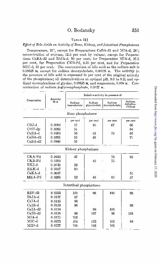
Table III shows the effect of the sodium salts of taurocholic, glycocholic, desoxycholic, and dehydrocholic acids on the rate of hydrolysis of sodium fl-glycerophosphate by bone, kidney, and intestinal phosphatases. The reaction velocities in the presence of the bile acids are expressed as percentages of the original re- action velocity. Thus for the bone phosphatase, Preparation CaBA-d3, the value for Q0.05 was 0.0211, 0.0198 in duplicate determinations; in the presence of 0.00625 M sodium taurocholate, the values for Qo.05 were 0.0114, 0.0105, 0.0104, and the average of these, 53 per cent of the reaction velocity in the absence of bile acid. Qo.os for the intestinal phosphatase, Preparation CaIB-d2, was 0.0126. In the presence of 0.00625 M sodium taurocholate, values for Q0.05 were 0.0127, 0.0121, and the average of these, 98 per cent of the original reaction velocity. In general, the error in the determination of Q, under the conditions of the present experi- ments, can be placed at less than 5 per cent.
The results show a clear cut differentiation between the effect of the bile acids on the activity of bone and kidney phosphatases,
4 Takata (31) determined inorganic phosphate by precipitation from the trichloroacetic acid filtrate with magnesia mixture, conversion of the magnesium ammonium phosphate into ammonium phosphomolybdate, and titration.
by guest on February 17, 2020http://w
ww
.jbc.org/D
ownloaded from
0. Bodansky 351
TABLE III
Effect of Bile Acids on Activity of Bone, Kidney, and Intestinal Phosphatases
Temperature, 25”, except for Preparations CaBA-d2 and MIA-d, 28”; concentration of enzyme, 12.5 per cent by volume, except for Prepara- tions CaBA-d2 and RbIA-d, 50 per cent; for Preparation MIA-d, 37.5 per cent, for Preparation CKB-Pd, 6.25 per cent, and for Preparation MIC-d, 25 per cent. The concentration of bile acid as the sodium salt is 0.00625 M, except for sodium desoxycholate, 0.00125 M. The activity in the presence of bile acid is expressed in per cent of the original activity of the phosphatase; all determinations at optimal pH, 9.0 to 9.2; and op- timal concentrations of glycine, 0.00625 M, and magnesium, 0.009 M. Con-
centration of sodium P-glycerophosphate, 0.0127 M.
Preparation
Relative activity in presence of ACtiVf”Y
CBJ-d 0.0092 CBH-dp 0.0083 CaBA-d 0.0203 CaBA-d3 0.0205 CaBA-d2 0.0940
Bone phosphatases
per cent
47 51 56 53 55
per cent p+zT cent
40 67
43 43
70
per cent
66 64 63 71
CKA-Pd 0.0463 CKB-Pd 0.0355 RKJ-d 0.0120 RKH-d 0.0017 CaKA-d 0.0047 MKA-Pd 0.0295
RIH-d3 RbIA-d CaIA-d CaIB-d CaIA-d2 CaIB-d2 MIA-d MIC-d MID-d
Kidney phosphatases
47
58 50
52
43
43
Intestinal phosphatases
0.0333 100 0.0127 97 0.0145 98 0.0119 96 0.0114 0.0126 98 0.0175 103 0.0073 104 0.0227 100
96 100 98
I 98 99 100
107 96 105
102 102 84 103 105
73 75
65
63
51 57
by guest on February 17, 2020http://w
ww
.jbc.org/D
ownloaded from

352 Differentiation of Phosphatases
on the one hand, and on that of the intestinal phosphatases, on the other. The rate of hydrolysis of sodium ,%glycerophosphate by the intestinal phosphatases is not affected by the presence of taurocholic, glycocholic, desoxycholic, nor, except for some slight retardation of the action of Preparation MC-d, by dehydrocholic acid. In contrast, the retardation of the activity of the kidney and bone phosphatases by the same concentrations of bile acids is
TABLE IV
Effect of Inactivated Phosphatase Tissue Extracts on Retardation of Phosphatase Activity by Taurocholic Acid
Temperature, 25”; concentration of substrate, sodium P-glycerophos- phate, 0.0127 M; concentration of taurocholic acid as sodium taurocholate, 0.00625 M; optimal pH, 9.0 to 9.2; and optimal concentrations of glycine, 0.00625 M, and magnesium, 0.009 M.
Phosphatase preparation Inactivated phosphatase preparation
Intestinal preparations, active
6.25% cat intestinal, 6.25% cat bone, CaIB-d CaBA-d
12.5yo human intestinal, 12.5% cat bone, MID-d CaBA-d3
11.1% human intestinal, 11.1% human kidney, MIB-d MKA-Pd
0.0059 0.0059
0.0194 0.0207
0.0047 0.0051
Bone preparations, active
12.5% cat bone, CaBA- 12.5% human intes- d3 tinal, MID-d
12.5oj, cat bone, CaBA- 12.5yo cat intestinal, d3 CaIB-d2
0.0198
0.0198
0.0110
0.0093
marked: a decrease to about 50 per cent in the presence of tauro- cholic acid, to about 45 per cent for glycocholic acid, and to about 65 per cent for desoxycholic and dehydrocholic acids. The existence of retardation of phosphatase activity by bile acids is thus characteristic of the tissue source of the phosphatase and, to the extent here investigated, independent of the species.
The results of Table IV and, as will be pointed out later, also those of Tables VI and VII, indicate that the effect of the bile
by guest on February 17, 2020http://w
ww
.jbc.org/D
ownloaded from
0. Bodansky 353
acids is probably not dependent on any substance in the tissue extracts other than the active enzymic component. Thus the addition of 12.5 per cent of the heat-inactivated cat intestinal phosphatase, Preparation CaIB-d2, did not alter, beyond experi- mental variation, the extent of retardation of the activity of the cat bone phosphatase, Preparation CaBA-d3, by sodium tauro- cholate. On the other hand, the addition of heat-inactivated human kidney phosphatase, Preparation MKA-Pd, to human intestinal phosphatase, Preparation MIB-d, did not lead to a retardation of the latter by sodium taurocholate. As the other results bring out more fully, retardation by taurocholic acid occurs if the active component is bone phosphatase; it fails to occur if the active component is intestinal phosphatase, the presence of other tissue extracts notwithstanding.
The possibility existed that the intestinal preparations might contain sufficient concentrations of bile acids which were already exerting optimal retardant effect and that further addition was without influence. Though this did not seem likely since the tissues were usually washed with saline before extraction and were subsequently dialyzed, several preparations of intestinal phos- phatase, Preparations RbIA-d, CaIB-d, and MIB-d, were tested for bile acids by the Gregory-Pascoe reaction (34). The former two preparations were negative; Preparation MIB-d, a tissue which had not been washed, yielded a faint positive reaction such as that given by only a very small fraction of the bile acid added to the hydrolysis mixture.
There is some indication in the results of Table III that the extent of retardation is dependent on the substituent groups in the cholanic acid molecule. Thus, 0.00625 M dehydrocholic acid (3,7,12-triketocholanic acid) decreased the activity of bone or kidney phosphatase to an average of about 65 per cent of the original value, while 0.00625 M taurocholic acid, the taurine-con- jugated product of 3,7,12-trihydroxycholanic acid, decreased the activity of these phosphatases to about 50 per cent, and the same concentration of glycocholic acid to about 45 per cent.
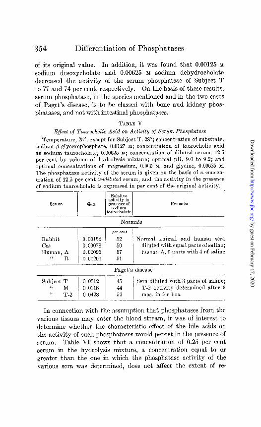
Source of Serum Phosphatase-Table V shows that the activity of serum phosphatase of rabbit, cat, and man, the last both in the normal and in two cases of Paget’s disease, is decreased in the the presence of 0.00625 M sodium taurocholate to about 50 per cent
by guest on February 17, 2020http://w
ww
.jbc.org/D
ownloaded from
354 Differentiation of Phosphatases
of its original value. In addition, it was found that 0.00125 M
sodium desoxycholate and 0.00625 M sodium dehydrocholate decreased the activity of the serum phosphatase of Subject T to 77 and 74 per cent, respectively. On the basis of these results, serum phosphatase, in the species mentioned and in the two cases of Paget’s disease, is to be classed with bone and kidney phos- phatases, and not with intestinal phosphatases.
TABLE V
E$ect of Taurocholic Acid on Activity of Serum Phosphatase
Temperature, 25”, except for Subject T, 28”; concentration of substrate, sodium /3-glycerophosphate, 0.0127 M; concentration of taurocholic acid as sodium taurocholate, 0.00625 M; concentration of diluted serum, 12.5 per cent by volume of hydrolysis mixture; optimal pH, 9.0 to 9.2; and optimal concentrations of magnesium, 0.009 M, and glycine, 0.00625 M.
The phosphatase activity of the serum is given on the basis of a concen- tration of 12.5 per cent undiluted serum, and the activity in the presence of sodium taurocholate is expressed in per cent of the original activity.
Serum 1 Qo.or / j%$$j R6tD&&S
Normals
Rabbit Cat Human, A
‘I B
per cent
0.00154 52 Normal animal and human sera 0.00078 50 diluted with equal parts of saline; 0.00095 57 human A, 6 parts with 4 of saline 0.00200 51
Paget’s disease
Subject T Sera diluted with 3 parts of saline;
:I ?-a ) !!!i%lI 1 %% 1 mos. inice box T-2 actrvlty determined after 3
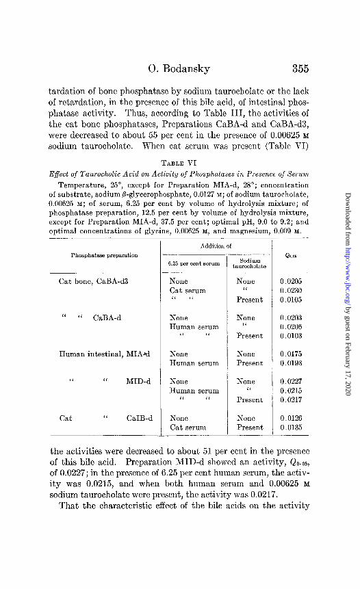
In connection with the assumption that phosphatases from the various tissues may enter the blood stream, it was of interest to determine whether the characteristic effect of the bile acids on the activity of such phosphatases would persist in the presence of serum. Table VI shows that a concentration of 6.25 per cent serum in the hydrolysis mixture, a concentration equal to or greater than the one in which the phosphatase activity of the various sera was determined, does not affect the extent of re-
by guest on February 17, 2020http://w
ww
.jbc.org/D
ownloaded from
0. Bodansky 355
tardation of bone phosphatase by sodium taurocholate or the lack of retardation, in the presence of this bile acid, of intestinal phos- phatase activity. Thus, according to Table III, the activities of the cat bone phosphatases, Preparations CaBA-d and CaBA-d3, were decreased to about 55 per cent in the presence of 0.00625 M
sodium taurocholate. When cat serum was present (Table VI)
TABLE VI
E$ect of Taurocholic Acid on Activity of Phosphatases in Presenee of Serum
Temperature, 25”, except for Preparation MIA-d, 28”; concentration of substrate, sodium ,&glycerophosphate, 0.0127 M; of sodium taurocholate, 0.00625 M; of serum, 6.25 per cent by volume of hydrolysis mixture; of phosphatase preparation, 12.5 per cent by volume of hydrolysis mixture, except for Preparation MIA-d, 37.5 per cent; optimal pH, 9.0 to 9.2; and optimal concentrations of glycine, 0.00625 M, and magnesium, 0.009 111.
Phosphatase preparation
Cat bone, CaBA-d3
L‘ “ CaBA-d
Human intestinal, MIAad
1‘ I‘ MID-d
Cat “ CaIB-d
Addition of
6.25 per cent serum Sodium taurooholate
None Cat serum L‘ ‘L
None C‘
Present
0.0205 0.0230 0.0105
None Human serum
“ “
None Human serum
None ‘I
Present
0.0203 0.0206 0.0103
None 0.0175 Present 0.0193
None Human serum
“ “
None “
Present
0.0227 0.0215 0.0217
None None 0.0126 Cat serum Present 0.0135
- QO.06
the activities were decreased to about 51 per cent in the presence of this bile acid. Preparation MID-d showed an activity, Qo.05, of 0.0227; in the presence of 6.25 per cent human serum, the activ- ity was 0.0215, and when both human serum and 0.00625 M
sodium taurocholate were present, the activity was 0.0217. That the characteristic effect of the bile acids on the activity
by guest on February 17, 2020http://w
ww
.jbc.org/D
ownloaded from
Differentiation of Phosphatases
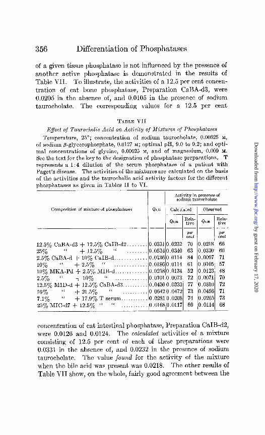
of a given tissue phosphatase is not influenced by the presence of another active phosphatase is demonstrated in the results of Table VII. To illustrate, the activities of a 12.5 per cent concen- tration of cat bone phosphatase, Preparation CaBA-d3, were 0.0205 in the absence of, and 0.0105 in the presence of sodium taurocholate. The corresponding values for a 12.5 per cent
TABLE VII
Effect of Taurocholic Acid on Activity of Mixtures of Phosphatases
Temperature, 25’; concentration of sodium taurocholate, 0.00625 M,
of sodium @-glycerophosphate, 0.0127 M; optimal pH, 9.0 to 9.2; and opti- mal concentrations of glycine, 0.00625 M, and of magnesium, 0.009 M. See the text for the key to the designation of phosphatase preparations. T represents a 1:4 dilut,ion of the serum phosphatase of a patient with Paget’s disease. The activities of the mixtures are calculated on the basis of the activities and the taurocholic acid activity factors for the different phosphatases as given in Tables II to VI.
Activity in presence of sodium taurocholate
Composition of mixture of phosphatases QO.05 Calculated Observed
QQ.06 ;;$- Qom ;;&- ---e-
Pm Pe+ cent cent
12.5% CaBA-d3 + 12.5% CaIB-d2.. . . 0.03310.0232 70 0.0218 66 25% SC + 12.5% “ 0.05340.0340 63 0.0320 60 2.5yo CaBA-d + 10% CaIB-d.. . 0.0136 0.0114 84 0.0097 71 10% “ + 2.5% “ _. . 0.01860.0114 61 0.0105 57 10% MKA-Pd + 2.5’% MIB-d.. . 0.0258 0.0134 52 0.0123 48 2.5% ‘I + 10% (‘ 0.01010.0073 72 0.0071 70 12.5% MID-d + 12.5’% CaBA-d3.. 0.0430 0.0333 77 0.0310 72 16~~ ‘( + 21.5% ‘I ,....,_... 0.06420.0472 73 0.0456 71 7.1% “ + 17.9% T serum. . 0.02810.0208 74 0.0205 73 25% MIC-d7 + 12.5’% “ “ . . . . . . . . . . 0.01680.0117 69 0.0114 68
concentration of cat intestinal phosphatase, Preparation CaIB-d2, were 0.0126 and 0.0124. The calculated activities of a mixture consisting of 12.5 per cent of each of these preparations were 0.0331 in the absence of, and 0.0232 in the presence of sodium taurocholate. The value found for the activity of the mixture when the bile acid was present was 0.0218. The other results of Table VII show, on the whole, fairly good agreement between the
by guest on February 17, 2020http://w
ww
.jbc.org/D
ownloaded from
0. Bodansky 357
values actually observed for the activities of mixtures of phos- phatases in the presence of sodium taurocholate and those calcu- lated on the basis that this bile acid exerts its characteristic effect on each of the phosphatase components of the mixture.
The above results emphasize the probability that the effect of the bile acids on the activity of phosphatase preparations (re- tardation of bone and kidney phosphatase activity, and absence of retardation of intestinal phosphatases) is an effect on the active enzymic component of the preparations. These results also indicate the possibility, as will be pointed out more fully later, of judging the source of the phosphatase in serum in various conditions by determining the extent of retardation of the activity of serum phosphatase in the presence of bile acids.
DISCUSSION
Kay (12) has considered the phosphatases of the various tissues and of plasma to be quite probably identical. Evidence for the identity of the enzymes of bone, intestine, kidney, and plasma was based on (a) the almost complete removal of phosphatase from the different tissues by the same method of extraction, (b) the same optimal pH for the action of preparations from the three tissues and plasma, (c) hydrolysis of pyrophosphate at a pH optimum (7.2 to 7.8) which was the same for the three tissues and plasma, (d) stimulation of activity by magnesium ion and maximal stimula- lation at the same concentration of magnesium by all four phosphatases, (e) the same ratio of rates of hydrolysis of a series of esters by each of the four phosphatases, (f) practically the same dissociation constant, for the four phosphatases, for the enzyme substrate compound, (g) synthesis of phosphoric esters by all four phosphatases.
Many of the above points have been verified incidentally in the course of work of other investigators. The data submit,ted in the present study, however, reveal, with respect to the retardation by the bile acids, a distinct difference between kidney, bone, and serum phosphatase, on the one hand, and intestinal phosphatase, on the other.
The question of the identity of the tissue phosphatases or, in general, of similarly acting enzymes may be considered from several view-points. In a strict sense and according to definition,
by guest on February 17, 2020http://w
ww
.jbc.org/D
ownloaded from
358 Differentiation of Phosphatases
two or more given enzyme preparations may be designated as identical only if it can be shown exhaustively that they act the same way quantitatively in every instance; that is, if they show the same pattern of rates of hydrolysis on a series of substrates, if their activities are influenced to the same extent by change in substrate concentration, retardants, preventors of inactivation, by the presence of accompanying substances, or by any other alteration in the reaction environment.
The work here presented as well as that of previous investigators on other enzymes shows that it is very difficult to ascribe “iden- tity” in this strict sense to similar preparations. Perhaps the clearest illustration of this difficulty is the finding of Falk (35) that a protein added to a given preparation of pancreatic lipase produces a different pattern of rates of hydrolysis of a series of esters. Falk, in this connection, emphasizes the necessity for consid- ering the enzyme system as a whole. Falk and his collabo- rators (36) showed in earlier work that the pattern of rates of hy- drolysis of a series of esters varies with the age of the animal, the species source of the tissue, and the age of the lipase extract.
From a second view-point, it may be assumed that in enzyme preparations showing identity of action in most respects, such differences as do exist are due to the presence of accompanying substances which vary with the particular extract. Used in this connection, the term “identity” may be conceived of as applying to the possession of a common chemical unit. In our present lack of knowledge concerning the relation between chemical structure of enzymes and their activities, the available experimental ap- proach to a decision regarding the possession of a common chemical unit is to attempt the finding of an activity characteristic which persists in spite of apparent variation in accompanying substances as these appear in different tissues or in preparations of varying degrees of purity.
The term “identity of enzymes” may be thought of in still another sense. Thus there exists the possibility that the chemical structure of an enzyme may undergo partial modification, so that its actions in certain respects remain the same but differ in others. Nelson and Papadakis (37) have shown that a highly purified yeast invertase preparation, free from melibiase, is changed by partial heat inactivation into a preparation which is different from the
by guest on February 17, 2020http://w
ww
.jbc.org/D
ownloaded from
0. Bodansky 359
original in its relative actions on the two substrates, rafhnose and sucrose; the form of the time course of the reaction and the degree of retardation by a-methylglucoside are, however, the same for the two preparations. Enzyme preparations of different tissues may, therefore, differ chemically in some respect and yet act identically on the substrates which are involved in physiological processes going on in these tissues.
In the present paper the question of the identity of the phospha- tases is considered from the first two points of view. Variations in the pattern of hydrolysis of several phosphoric esters by a given tissue phosphatase occurred with variation in the species source and were such as to make impossible the differentiation of one tissue phosphatase from another, even within a given species. The existence of such variations was not surprising, in view of the work of Falk and his collaborators on tissue lipases. In contrast, however, it proved possible to show that the activities of bone and kidney phosphatases were considerably retarded, and those of intestinal phosphatases unaffected by bile acids, independently of the age of the extract or of the animal, of the species source of the enzyme, the presence of another extract, whether enzymically active or not, and of the presence of serum. These results, then, are probably indicative of some chemical difference between the phosphatases of bone and kidney, on the one hand, and that of intestine, on the other.
Whether such a difference is physiologically significant cannot be stated at present. If it is assumed, as seems likely, that the phosphatase in serum has an extracirculatory origin and that a given tissue phosphatase does not change its capacity for retarda- tion (or lack of retardation) by bile acids when it passes into the blood stream (and in vitro it has been shown that the retardant effect is not influenced by serum), then the determination of the degree of retardation of the activity of serum phosphatase pro- vides a possible criterion for judging its source. To illustrate, a decrease to about 50 per cent in the activity of the serum phos- phatase in the normal or in Paget’s disease (Table V) indicates either (a) that the phosphatase does not come from the intestinal mucosa, but from bone, kidney, or some other tissue the phospha- tase of which is retarded to that degree by taurocholic acid or (b) that the phosphatase in the serum comes from several tissues, that
by guest on February 17, 2020http://w
ww
.jbc.org/D
ownloaded from
Differentiation of Phosphatases
some of these tissue phosphatases are retarded to more than 50 per cent, others less, but that the resultant decrease in their activity by sodium taurocholate is, fortuitously, the same as that of bone or kidney phosphatase.
To define more precisely the source of serum phosphatase in the normal and in conditions which exhibit hyperphosphatasemia, it is necessary to extend the studies here reported to other tissues which may be conceived of as sources of phosphatase in the serum. It may also be possible to emphasize the difference between different phosphatases by using other concentrations of bile acids. More- over, there is indication, from the values presented in Table III, that further study with other substituted products of cholanic acid may prove of value in determining the extent to which the similarity of bone, kidney, serum, and other tissue phosphatases persists, or else serve in their differentiation.
SUMMARY
1. The pattern of the rates of hydrolysis of sodium /3-glycero- phosphate, sodium hexosediphosphate, potassium benzylphos- phate, and sodium propylphosphate has not been found to be constant for a phosphatase from the same tissue of different species, or to be differentiable from the pattern for a different tissue of the same individual or species.
2. The cinchona alkaloids, quinine, cinchonine, and quitenidine do not, in low concentrations, decrease the rate of hydrolysis of sodium /3-glycerophosphate by tissue phosphatases. 0.00125 M
mercuric chloride decreased, in several instances, the activities of tissue phosphatases, but these effects could not serve as a basis for differentiating them.
3. Taurocholic, glycocholic, desoxycholic, and dehydrocholic acids decrease considerably the action of bone and kidney phospha- tases but do not affect that of intestinal phosphatases (with the exception of slight retardation of the action of a human intestinal preparation by dehydrocholic acid). This effect is dependent on the tissue, is independent of the age of the extract, the mode of preparation, and the animal species. The effect on a given tissue phosphatase is, as shown by studies with taurocholic acid, inde- pendent of the presence of other tissue extracts, whether enzymic- ally active or not, and of the presence of serum. This effect thus serves as a means of differentiation.
by guest on February 17, 2020http://w
ww
.jbc.org/D
ownloaded from
0. Bodansky 361
4. The question of the identity of similarly acting enzymes, in general, and of that of bone, kidney, intestinal, and serum phos- phatases, in particular, is discussed.
5. The probable source of the phosphatase in normal serum and, as an illustration of a pathological condition, in that of Paget’s disease is considered. To define more precisely the source of serum phosphatase in various conditions, it is proposed that stu- dies, similar to those here reported, be extended to other tissues and for other substituents of the cholanic acid molecule.
BIBLIOGRAPHY
1. Kay, H. D., J. Biol. Chem., 89, 249 (1930). 2. Bodansky, A., and Jaffe, H. L., Arch. Int. Med., 64, 88 (1934). 3. Woodard, H. Q., Twombly, G. H., and Coley, B. L., J. Clin. Inv.,
15, 193 (1936). 4. Roberts, W. M., Brit. Med. J., 1, 734 (1933). 5. Rothman, M. M., Meranze, D. R., and Meranze, T., Am. J. Med. SC.,
192, 526 (1936). 6. Gutman, A. B., Tyson, T. L., and Gutman, E. B., Arch. Int. Med.,
67, 379 (1936). 7. Franseen, C. C., and McLean, R., Am. J. Cancer, 24, 299 (1935). 8. Bodansky, A., J. Biol. Chem., 104, 473, 717 (1934). 9. Armstrong, A. R., King, E. J., and Harris, R. I., Canad. Med. Assn. J.,
31, 14 (1934). 10. Bodansky, A., and Jaffe, H. L., Proc. Sot. Ezp. Biol. and Med., 31,
1179 (1934). 11. Armstrong, A. R., and Banting, F. G., Canad. Med. Assn. J., 33, 243
(1935). 12. Kay, H. D., Physiol. Rev., 12, 384 (1932). 13. Kay, H. D., Biochem. J., 22,855 (1928). 14. Folley, S. J., and Kay, H. D., Biochem. J., 29, 1837 (1935). 15. Bodansky, O., J. Biol. Chem., 116, 101 (1936). 16. Albers, H., and Albers, E., 2. physiol. Chem., 232, 189 (1935). 17. Bodansky, O., J. BioZ. Chem., 114, 273 (1936). 18. Asakawa, K., J. Biochem., Japan, 11, 143 (1930). 19. Fiske, C. H., and Subbarow, Y., J. BioZ. Chem., 66, 375 (1925). 20. Henry, T. A., The plant alkaloids, London, 9 (1924). 21. Schmidt, C. L. A., and Merrill, J. A., J. Biol. Chem., 68,601 (1923-24). 22. TengstrBm, S., 2. physiol. Chem., 41, 210 (1904). 23. Rona, P., and Bloch, E., Biochem. Z., 118, 185 (1921). Rona, P.,
and Pavlovid, R., Biochem. Z., 130, 225 (1922). Rona, P., and Haas, H. E., Biochem. Z., 141, 222 (1923).
24. Rona, P., and Reinicke, D., Biochem. Z., 118, 213 (1921). 25. Senter, G., Z. physik. Chem., 61, 673 (1905). 26. Hata, S., Biochem. Z., 17, 156 (1909). 27. von Euler, H., and Svanberg, O., Fermentforschung, 3, 330 (1920).
by guest on February 17, 2020http://w
ww
.jbc.org/D
ownloaded from
Differentiation of Phosphatases
28. Willstittter, R., Waldschmidt-Leitz, E., and Memmen, F., 2. physiol. Chem., 126, 93 (1923).
29. WillstLtter, R., and Memmen, F., 2. physiol. Chem., 138, 216 (1924). 30. Vonk, H. J., Roelofsen, P. A., and Romijn, C., Z. physiol. Chem., 218,
333 (1933). 31. Takata, H., J. Biochem., Japan, 14, 61 (1931). 32. Takata, H., J. Biochem., Japan, 16, 83 (1932). 33. Takata, H., J. Biochem., Japan, 18, 63 (1933). 34. Gregory, R., and Pascoe, T. A., J. Biol. Chem., 83, 35 (1929). 35. Falk, K. G., J. Biol. Chem., 96, 53 (1932). 36. Falk, K. G., Noyes, H. M., and Sugiura, K., J. BioZ. Chem., 69, 183,
213 (1924). Noyes, H. M., and Falk, K. G., J. BioZ. Chem., ‘72, 449 (1927). Falk, K. G., and Noyes, H. M., J. BioZ. Chem., 72,489 (1927).
37. Nelson, J. M., and Papadakis, P., J. BioZ. Chem., 80, 163 (1928).
by guest on February 17, 2020http://w
ww
.jbc.org/D
ownloaded from
Oscar BodanskyIN THEIR DIFFERENTIATION
IDENTICAL?: THE USE OF BILE ACIDSKIDNEY, INTESTINE, AND SERUM
ARE THE PHOSPHATASES OF BONE,
1937, 118:341-362.J. Biol. Chem.
http://www.jbc.org/content/118/2/341.citation
Access the most updated version of this article at
Alerts:
When a correction for this article is posted•
When this article is cited•
alerts to choose from all of JBC's e-mailClick here
tml#ref-list-1
http://www.jbc.org/content/118/2/341.citation.full.haccessed free atThis article cites 0 references, 0 of which can be by guest on February 17, 2020
http://ww
w.jbc.org/
Dow
nloaded from


![JULY 4gemoranita - BMJ · JULY 22, 1922] MEMORANDA. [MD JOU3AA 127 eclamptib seizure duriing late second stage. Anaesthetic imme- diately commencedandcontinued for twoandahal-f hours,](https://static.fdocuments.us/doc/165x107/5e574b135bf3ad0bae4fb658/july-4gemoranita-bmj-july-22-1922-memoranda-md-jou3aa-127-eclamptib-seizure.jpg)