Brilliant Cresyl Blue test. - UAB Barcelona · Chapter 2: Bibliographical ... (Vishwanath. 2003)....
106
Oocyte quality Assessment of prepubertal sheep oocyte competence for in vitro embryo production by the Brilliant Cresyl Blue test. Maria Gracia Catalá Facultat de Veterinària Departament de Ciència Animal i dels Aliments Doctoral thesis Universitat Autònoma de Barcelona 2012
Transcript of Brilliant Cresyl Blue test. - UAB Barcelona · Chapter 2: Bibliographical ... (Vishwanath. 2003)....

Oocyte quality
Assessment of prepubertal sheep oocyte competence
for in vitro embryo production by the
Brilliant Cresyl Blue test.
Maria Gracia Catalá
Facultat de Veterinària
Departament de Ciència Animal i dels Aliments
Doctoral thesis
Universitat Autònoma de Barcelona
2012


La Dra Maria Teresa Paramio Nieto, Catedrática del Departamento de Ciencia Animal y de los
Alimentos de la Facultad de Veterinaria de la Universidad Autónoma de Barcelona
CERTIFICA:
Que la tesis titulada “Assessment of prepubertal sheep oocyte competence for in vitro
embryo production by the Brilliant Cresyl Blue test.” presentada por María Gracia Catalá
para optar al grado de Doctor, se realizó bajo mi dirección y con un financiamiento del
Ministerio de Ciencia e Innovación (AGL 2007 60227/GAN y AGL2011-23784) y una beca
otorgada por la Universidad Autónoma de Barcelona (UAB-2007FI00193).
I para que conste a los efectos oportunos, firmo la presente en Bellaterra (Cerdanyola del
Vallès), el 12 de marzo de 2012
Dra. Maria-Teresa Paramio Nieto


A mis padres,
A mi hermano,
A mi hermana del alma,
A Amine,


Chapter 1: General Background………………………………………………………… 10
Chapter 2: Bibliographical Revision……………………………………………………. 16
2.1. Current situation of the in vitro embryo production (IVEP) in sheep …………… 18
2.1.1. In vitro Maturation (IVM)……………………………………………………... 18
2.1.2. IVF and sperm capacitation……………………………………………………. 18
2.1.3. Intracytoplasmic Sperm Injection……………………………………………… 19
2.1.4. Parthenogenetic activation……………………………………………………... 19
2.1.5. Embryo culture and blastocyst production….…………………………………. 20
2.2. Study of the oocyte………………………………………………………………... 20
2.2.1. Meiosis: nuclear and cytoplasmic maturation…………………………………. 20
2.2.3. Maturation promoting factor…………………………………………………… 21
2.2.4. Mitochondria and ATP………………………………………………………… 22
2.2.5. Gene expression………………………………………………………………... 22
2.2.6. Non invasive oocyte quality assessment: Brilliant Cresyl Blue Strain………. 23
2.3. Parameters affecting oocyte quality……………………………………………….. 25
2.3.1. Age of donor…………………………………………………………………… 25
2.3.2. Follicular size…………………………………………………………………... 26
2.3.3. Oocyte size……………………………………………………………………... 26
2.4. Improving oocyte quality using in vitro media………………………………….... 27
2.4.1. Insulin Transferrin Selenium (ITS)……………………………………………. 28
2.4.2. Ascorbic acid …………………………………………………………………. 29
Chapter 3. Objectives……………………………………………………………………. 32
Chapter 4. Brilliant Cresyl Blue stain selects largest oocytes with highest
mitochondrial activity, maturation-promoting factor activity and embryo
developmental competence in prepubertal sheep………………………………………….....
36
Chapter 5. Effect of insulin transferrin and selenium and Ascorbic Acid in maturation
media on embryo development, MPF activity and ATP content of prepubertal sheep
oocytes selected by brilliant cresyl blue test…………………………………………………..
50
Chapter 6. Effect of oocyte quality on blastocyst development after in vitro fertilization
(IVF) and intracytoplasmic sperm injection (ICSI) in a sheep model……………………..
72
Chapter 7. General Discussion………………………………………………………….. 80
Chapter 8. Conclusions…………………………………………………………………. 86
Chapter 11. Bibliography ………………………………………………………………. 90
Thanks to, ……………………………………………………………………………… 104


10
Chapter 1:
General background

11

General background
12
Assisted reproductive technologies (ART) have been one of the major tools towards increasing
productivity in livestock industry. In this field, artificial insemination (AI) is the earliest and
most powerful among the reproductive technologies because it is easy to perform, cost-
effective, and highly successful (Vishwanath. 2003). For over the years AI has been used to
obtain offspring from genetically superior and from sub-fertile animals. Moreover, by the
1960´s, significant improvements in cryopreservation and storage of semen made AI even more
accessible to livestock producers (Vishwanath. 2003). In the modern dairy industry, new ART
techniques were developed as cryopreservation not only of semen but also of gametes and
embryos, induction of multiple ovulations, embryo transfer, in vitro fertilization (IVF), sex
determination of sperm or embryos, nuclear transfer, cloning, etc. In the small ruminants as
sheep and goat, these techniques had a lower development.
Despite the improvement in ART protocols, the pregnancy rates are still relatively low and early
embryonic mortality has been reported after embryo transfer. It has been shown that 40% of
total embryonic losses occur between day 8 and day 17 of pregnancy (Thatcher et al. 1994) and
one of the reasons could be due to the oocyte quality (Snijders et al. 2000). The oocyte quality is
used as synonymous of oocyte competence, defined as the ability of an oocyte to resume
meiosis, cleave following fertilization, develop to the blastocyst stage, induce a pregnancy and
bring the offspring to term in good health (Sirard et al. 2006). The oocyte competence is
acquired gradually during the course of folliculogenesis as the oocyte grows and its somatic
cells cohort differentiates (Eppig et al. 1994). Many factors have been shown to affect the
oocyte competence as: follicle size (Lonergan et al. 1994; Romaguera et al. 2011), phase of
follicular wave (Machatkovaa et al. 2004), hormonal stimulation (Sirard et al. 2006), maturation
environment (Warzych et al. 2007), season (Sartori et al. 2002), nutrition (Fouladi-Nashta et al.
2007) and donor’s age (Rizos et al. 2005).
The assessment of oocyte quality is one of the major objectives in ART, especially in human
where the use of the best quality oocytes to be inseminated may improve significantly the
embryo production. Consequently, multiple methods of oocyte selection have been proposed.
One of the most popular is to select the oocyte by morphology which is relatively quick and
simple; however this simple technique leads to identify more frequently the negative than the
positive aspects, and overall it is not fully satisfactory (Balaban and Urman. 2006). Other
methods have been proposed as the use of polarizing microscopy analysis (Heindryckx et al.
2011), gene expression of the granulosa cells or in the oocyte itself (Patrizio et al. 2007), polar
body biopsy to screen oocytes with chromosomal defects deriving from errors in the meiotic
division (Dawson et al. 2006). Most of these techniques are quite complicated, require

General background
13
expensive laboratory equipment and time-consuming procedures, and some of them are invasive
and consequently are not currently applicable in the clinical practice.
In research, the law is extremely restrictive in the use of human embryos; consequently using
animals has become through time a valuable tool. The bovine is the mostly used ruminant in
ART research because of its economic importance and its wide geographical distribution,
although using small ruminants as oocyte donor could be an interesting alternative. Using the
sheep (Ovis aries) can bring great benefits to reproduction research because of their younger age
to reach puberty, short gestational periods, the possibility to have more than one offspring in a
single gestation and the maintenance cost are lower than in bovine. Moreover, sheep have been
domesticated for over 10,000 years and are also widespread across the world, being adapted to
many different climatic conditions. During the past 60 years, sheep have been the subject of
considerable research starting in 1949 with an experimental super ovulation protocol (Ortavant
et al. 1949) until nowadays in which sheep receptors, genes (Leoni et al. 2007; Kyasari et al.
2012) and proteins (Grazul-Bilska et al. 2011) are being studied. In addition, using prepubertal
animals as oocyte donor has some additional benefits comparing to the use of adult donors, as
for example; reducing the generational interval (Duby et al. 1996), their ovaries produce a major
number of oocytes than adult (Koeman et al. 2003) and this oocytes could serve as a model of
low quality oocyte in research as they are characterized of having an abnormal cytoplasmic
maturation and lower ability to achieve the blastocyst stage (Revel et al. 1995; O'Brien et al.
1996; Armstrong. 2001). One of the difficulties of using ovaries coming from prepubertal
animals, is to release the complex oocyte cumulus (COCs) by traditional follicular aspiration
because of having a high percentage of antral follicles with a smaller diameter than 3 mm
(Martino et al. 1994). Consequently, the release of these oocytes is made by slicing the ovary
surface obtaining oocytes with heterogeneous diameter, different COC morphology and stage of
atresia.
The Brilliant Cresyl Blue (BCB) test has been successfully used as a non invasive methodology
to select oocytes with a higher diameter and more competent to develop up to the blastocyst
stage in cow (Pujol et al. 2004; Alm et al. 2005; Bhojwani et al. 2007; Torner et al. 2008;
Opiela et al. 2010), pig (Ericsson et al. 1993; Roca et al. 1998; El Shourbagy et al. 2006;
Egerszegi et al. 2010), goats (Rodriguez-Gonzalez et al. 2002; Rodriguez-Gonzalez et al. 2003;
Kątska-Książkiewicz et al. 2007), mouse (Wu et al. 2007) and buffalo (Manjunatha et al. 2007).
To our knowledge there are no previous reports using this stain to select oocytes by their
competence in the sheep.
The aim of this study is to test the ability of the BCB staining to select the more competent
sheep oocytes for in vitro embryo production. Also, in this work we pretend to improve the

General background
14
knowledge about oocyte competence related to their cytoplasmic and molecular performances,
their responses to different techniques of fertilization and the in vitro culture media needed to
improve the blastocyst production.

15

16
Chapter 2.
Bibliographical Revision

17
Bibliographical Revision
18
2.1. Current situation of the in vitro embryo production (IVEP) in sheep
2.1.1. In vitro maturation (IVM)
A correct maturation of the cumulus-oocyte complex (COC) is one of the most important
factors which determine the entry of the oocyte into metaphase II (MII), subsequent successful
fertilization, as well as the ability of an embryo to undergo an appropriate growth and
development. Performing the COC maturation under in vitro conditions provides an excellent
opportunity for having cheap and abundant oocytes for carrying out basic research and for the
application of emerging biotechnologies like cloning and transgenesis.
Several aspects of the IVM of sheep oocytes have been studied (Wani et al. 2000; Rao et al.
2002). Sheep COC’s are most commonly matured in Tissue Culture Medium (TCM199)
containing Earle’s salts. The supplementation of the IVM medium with epidermal growth factor
(EGF) (Guler et al. 2000), mare serum (Motlagh et al. 2008), fetal calf serum (FCS)
(Ghasemzadeh Nava and Tajik. 2000), estrous sheep serum (ESS) (Ghasemzadeh Nava and
Tajik. 2000), insulin –like growth factor (IGF-I) (Guler et al. 2000) and cysteamine (de Matos et
al. 2002) among others stimulate sheep oocyte nuclear and cytoplasmic maturation. After the
improvements in the IVM media, currently the most commonly media used to in vitro mature
sheep oocytes is the TCM199 supplemented 2 mM glutamine, 100 µM cysteamine, 0.3 mM
sodium pyruvate, 10% fetal bovine serum (FBS), 5 µg/mL FSH, 5 µg/mL LH and 1 µg/mL
estradiol (Loi et al. 2008). Moreover supplementing the IVM media with ESS instead of FBS
has also shown good results (Bebbere et al. 2010).
2.1.2. IVF and sperm capacitation.
The first reports of IVF in sheep used heparin to capacitate the ram spermatozoa reaching an
80% of fertilization but only 15% were viable embryos (Slavik et al. 1992). However, the use of
ESS instead of heparin rapidly gained importance by showing a fertilization rate of 85%
(Huneau et al. 1994) and 56% reached the blastocyst stage (Walker et al. 1994). The ESS works
through the binding-protein albumin that may facilitate the sperm capacitation by contributing
to the depletion of the sperm cholesterol membrane (Huneau et al. 1994). Ccurrently most of the
authors use the ESS to capacitate fresh and frozen semen using a concentration of 2 to 20% of
ESS (Walker et al. 1994). Bebbere et al ( 2010) using frozen ram semen capacitated with 2%
ESS reached 54% of blastocyst using adult sheep oocytes and Shirazi ( 2009) with fresh semen
and 20% ESS reached a 34% of blastocyst.
Bibliographical Revision
19
2.1.3. Intracytoplasmic Sperm Injection
Intracytoplasmic sperm injection (ICSI) consists in fertilizing a MII oocyte by the
injection of a single spermatozoon into the cytoplasm with both the acrosome and sperm
membrane intact. This technique was reported with success for the first time in hamsters
30 years ago (Uehara and Yanagimachi. 1976). Since then, the ICSI has become the
most commonly used procedure to overcome male infertility problems in human
reproduction. In animals, the ICSI has been in general used for research purposes, in
which studies with sex-sorted semen (Wilson et al. 2006), sperm mediated gene transfer
(Lavitrano et al. 2006; Pereyra-Bonnet et al. 2011) and cryopreserved oocytes (Matson
et al. 1997; Pope et al. 2012) are the most commonly reported.
The first lamb born after oocyte maturation, sperm sexing by flow cytometry and ICSI
was published by Catt et al. ( 1996). For the ICSI procedure they have directly injected
the spermatozoa into the cytoplasm without any chemical activation obtaining a low
efficiency of the technique that was traduced in 251 oocytes injected and transferred but
only one lamb arrived to term. Later, this same group reported that the manipulation by
itself was not enough to cause proper oocyte activation and that the addition of calcium
in the culture media increased the efficiency of the technique (Gomez et al. 1998). More
recently, Shirazi et al. ( 2009) tried to determine the need of an activation protocol after
sheep sperm injection, concluding that the chemical activation of oocytes must be
considered as an essential part of ICSI in this specie.
2.1.4. Parthenogenetic activation
After the entry of the sperm, mammalian oocytes exhibit an increase of the intracellular
calcium induced by the same sperm. These transient calcium peaks are propagated
throughout the fertilized oocyte in the form of a wave and initiate both the cortical
granule exocytosis and escape from the MII arrest to become a zygote [revised by (Loi
et al. 1998; Nakada and Mizuno. 1998)].
Oocyte activation protocols have been developed to induce artificially the intracellular
calcium levels in the oocyte cytoplasm. This is achieved by exposing the oocyte to a
calcium ionomicyn or ionophore and subsequently culturing it with a persistent kinase
inhibitor such as 6-DMAP (6-dimethyl amino purine). The treatment with ionomycin

Bibliographical Revision
20
alone caused the resumption of meiosis but no pronuclear formation and the 6-DMAP
alone did not cause any resumption of meiosis or pronuclear formation. So, it is
important the combination of the two compounds to reach the pronuclear stage (Susko-
Parrish et al. 1994).
In sheep, Alexander et al. ( 2006) using the combination of these two compounds
produced 21 % of blastocysts; he also showed that using cycloheximide instead of the
6-DMAP it is also possible to produce blastocyst but in a lower percentage (15%). Loi
et al. ( 1998), using the combination of ionomicyn and 6-DMAP to activate sheep
nuclear transfer oocytes, reached an efficiency of 83% of blastocyst compared to 25%
with no activation protocol.
2.1.5. Embryo culture and blastocyst production.
The most common media used during in vitro culture (IVC) of embryos is the Synthetical
Oviductal Fluid (SOF: (Tervit et al. 1972). From the beginning, this media showed good results
in culturing embryos under in vitro conditions obtaining 25 lambs born after 6 days of IVC
(Tervit and Rowson. 1974). In addition, supplementing the SOF media with serum (20% vs.
40%) (Thompson et al. 1998) and BSA (18% vs. 28%) (Carolan et al. 1995) increased
blastocyst percentage significantly. Furthermore, the addition of amino acids (aa) to this media
appears to be beneficial in sheep producing 58% of blastocyst versus 22% when the aa were not
added to the media (Walker et al. 1996).
2.2. Study of the oocyte
2.2.1. Meiosis: nuclear and cytoplasmic maturation.
In mammals, oocytes are arrested for several weeks, months or years in prophase of the first
meiotic division. During this long period, oocytes accumulate molecules of mRNA, proteins,
lipids and sugars as well as they gradually increase in size. The accumulation of all necessary
sources of energy and information during oocyte growth is essential for the final step of
oogenesis: the oocyte maturation.
Maturation consists of two interlinked and mutually dependent processes: cytoplasmic and
nuclear maturation. The cytoplasmic maturation of the oocyte includes cytoplasmic changes as
organelle redistribution, micro and macro molecular changes that occur during oocyte
maturation. These modifications contribute to the oocyte’s ability to undergo: nuclear
Bibliographical Revision
21
maturation, successful fertilization, cleavage and the development at least until the activation of
the embryonic genome.
Nuclear maturation includes chromatin changes during the oocyte maturation starting from
germinal vesicle (GV) breakdown (GVBD) through Meiosis I and Meiosis II when the oocyte is
finally arrested in the MII stage. At this moment the oocyte is physiologically prepared to
complete the second meiotic division upon fertilization. Under in vivo conditions, only fully
grown oocytes can resume meiosis which implies that cytoplasmic changes that occur before
maturation are essential for the acquisition of the developmental competence [Revised by
Marteil et al ( 2009)]. However, when oocytes are removed manually before ovulation from an
antral follicle, the separation triggers a pseudo-maturation event leading in general to the
completion of the first meiotic division and the arrest at the MII stage. This process has been
called spontaneous maturation and is believed to be induced by the removal of the oocyte
maturation inhibitor (OMI) present in the follicle where cAMP is involved [Revised by Sirard (
2011)]. A comparison between oocytes that were removed from the follicular environment and
in vitro matured compared to in vivo matured oocytes, showed the same rates of nuclear
maturation, fertilization and cleavage, but the percentage of blastocyst was significantly lower
on in vitro matured group [30% vs. 60%, revised by (Sirard and Blondin. 1996)] indicating that
the cytoplasmic competence must be different between the in vitro and the in vivo matured
oocytes.
2.2.3. Maturation Promoting Factor.
Meiosis is regulated by the maturation-promoting factor (MPF). This universal cell cycle
regulator is a heterodimer protein composed of two subunits, the catalytic subunit p34cdc2
(serine-threonine kinase activity) and the regulatory subunit cyclin B1. The association of these
two subunits is a requirement for the activation of the protein kinase activity; also the
phosphorilation of p34cdc2 on threonine 161 by the protein kinase CAK (Cdc2 activation
kinase) and dephosphorylation on threonine 14 and tyrosine 15 by Cdc25 phosphate is
necessary.
The MPF activity appears just before GVBD increasing until metaphase I (MI); its activity
decreases in the anaphase-telophase while its maximum level is reached at the MII stage (Figure
1). Incompetent goat oocytes have a limited amount of Cyclin B1 (Hue et al. 1997) and p34cdc2
(Anguita et al. 2007). In calf and lamb oocytes the MPF activity is significantly lower than in
cow and ewe oocytes (Ledda et al. 2001; Salamone et al. 2001) whereas Han D et al. ( 2010),
showed that the MPF activity of prepubertal mice oocytes was significantly higher than adult
mice oocytes, suggesting a difference in the mechanisms according to species. In prepubertal

Bibliographical Revision
22
goats, Anguita et al. ( 2007) showed higher MPF activity and competence in oocytes with a
diameter larger than 135 µm compared to the smaller diameters.
Figure 1: Schematic representation of MPF activity during oocyte maturation.
2.2.4. Mitochondria and ATP
Mitochondria are maternally inherited organelles that use oxidative phosphorylation to supply
energy as adenosine triphosphate (ATP) to the cell (Stojkovic et al. 2001). This source of ATP,
has a central role in the establishment of the developmental competence (Van Blerkom. 2004;
Van Blerkom et al. 2008). Even though mitochondria are the most abundant organelles in the
oocyte, little is known about their different functions.
The mitochondria distribution and activity change during oocyte maturation and fertilization
with the aim of bringing mitochondria to the region of the cell where a higher level of ATP
(Van Blerkom and Runner. 1984) or calcium (Sousa et al. 1997) are required. Energy in the
form of ATP is crucial; spindle formation and chromosome behavior depend on the expression
and activity of motor proteins, which use ATP as their energy source. It has been proposed that
mitochondria reorganization and ATP levels are influenced by the oocyte quality (Stojkovic
et al. 2001), compactness of the cumulus (Torner et al. 2007) and cumulus apoptosis (Torner et
al. 2004), GnRH (Dell'Aquila et al. 2009) and the microtubule cytoplasmic network (Brevini et
al. 2005) affecting the early stages of the embryo (Tarazona et al. 2006). Therefore, several
authors concluded that better quality oocytes contained significantly higher ATP levels and
produced significantly higher blastocyst rates after fertilization (Van Blerkom et al. 1995;
Stojkovic et al. 2001; Van Blerkom. 2004).
2.2.5. Gene expression
In the last few years, the study of mammalian genes has been the focus of several studies in the
belief that a good expression pattern could derive in a successful oogenesis, folliculogenesis,
fertilization and early embryonic development. In the course of acquiring the oocyte
competencies and a good embryo development a correct mRNA transcription is a crucial
process occurring in the cytoplasm (Crozet et al. 1981; Brevini-Gandolfi and Gandolfi. 2001;
MPF MPF MPF MPF
GV-Intact GVBD Metaphase I MetaphaseII

Bibliographical Revision
23
Patel et al. 2007).The mRNA content in oocytes is affected by animal nutrition (Pisani et al.
2008), donor age (Hamatani et al. 2004), follicle diameter (Caixeta et al. 2009), IVM culture
media (Saadeldin et al. 2011; Salhab et al. 2011), in vivo and in vitro conditions (Wells and
Patrizio. 2008), apoptosis (Li et al. 2009) and the cumulus cells (Adriaenssens et al. 2010)
among others.
In this thesis we are going to study the expression of four genes in relation with the oocyte
quality; two genes involved in metabolism: ATP1A1 (ATPase NaC/KC transporting a 1) and
COX1 (cytochrome c oxidase subunit 1), and two genes involved in the constitutive function of
the cell: CPEB (cytoplasmic polyadenylation-element-binding protein) and S100A10 (calcium-
binding protein).
ATP1A1 gene is translated in an enzyme responsible for the transport of Na+ out of and K+ into
the cell and that is an important key regulator of bovine blastocyst formation and is necessary
for the in vitro production of healthy bovine embryos (Watson et al. 1999). CPEB plays an
important role in the regulation of the mRNA translation targets required for oocyte maturation
(Cai et al. 2010). COX1 is a gene that produces a mitochondrial energy-transfer enzyme of the
respiratory chain. Opiela et al. ( 2010) found a high expression of COX1 in oocytes of better
quality versus lower quality oocytes. According to Tingaud-Sequeira et al. ( 2009), S100A10
plays an antiapoptotic role and that a high expression levels of S100A10 in the follicles may
have a dual function protecting follicular cells from apoptosis during atresia and acting as
chemoattractant for leukocytes and macrophages. After a microarrays of bovine oocytes, Torner
et al. ( 2008) showed a higher expression of S100A10 in lower quality than in high quality
oocytes.
2.2.6. Noninvasive oocyte quality assessment: Brilliant Cresyl Blue Stain.
With the aim of selecting the most competent oocytes, relevant results in goat (Rodriguez-
Gonzalez et al. 2002), bovine (Pujol et al. 2004; Alm et al. 2005; Bhojwani et al. 2007; Torner
et al. 2008; Opiela et al. 2010), pig (El Shourbagy et al. 2006), mouse (Wu et al. 2007) and
buffalo (Manjunatha et al. 2007) were published when the Brilliant Cresyl Blue (BCB) stain
was used to select oocytes prior to the IVM. This is a non invasive methodology that allows
selecting oocytes with larger diameters among a heterogeneous pool. Brilliant Cresyl Blue is a
compound (Figure 2; C17H20N3OCl · 1/2ZnCl2) with a molecular weight of 385.96 g/mol
which is used to determine the intracellular activity of glucose-6-phosphate dehydrogenase
(G6PDH). The G6PDH is a regulatory enzyme synthesized within the oocyte during oogenesis,
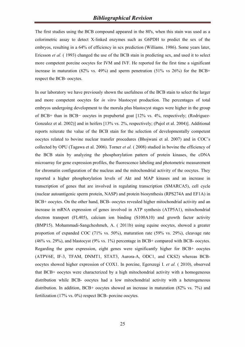
and is a component of the pentose phosphate pathway (Figure 3) that controls the flow of
carbon through this pathway and produces reducing equivalents in the form of NADPH to meet
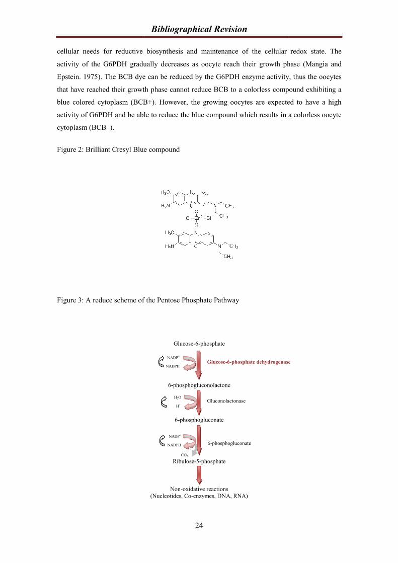
cellular needs for reductive biosynthesis and maintenance
activity of the G6PDH gradually decreases as oocyte reach their growth phase
Epstein. 1975). The BCB dye can be reduced by the G6PDH enzyme activity, thus
that have reached their growth phase cannot reduce BCB to a colorless compound exhibiting a
blue colored cytoplasm (BCB+). However, the growing oocytes are expected to have a high
activity of G6PDH and be able to reduce the blue compound which results in a colorless oocyte
cytoplasm (BCB–).
Figure 2: Brilliant Cresyl Blue compound
Figure 3: A reduce scheme of the
Bibliographical Revision
24
CO2
cellular needs for reductive biosynthesis and maintenance of the cellular redox
activity of the G6PDH gradually decreases as oocyte reach their growth phase
. The BCB dye can be reduced by the G6PDH enzyme activity, thus
that have reached their growth phase cannot reduce BCB to a colorless compound exhibiting a
BCB+). However, the growing oocytes are expected to have a high
activity of G6PDH and be able to reduce the blue compound which results in a colorless oocyte
: Brilliant Cresyl Blue compound
: A reduce scheme of the Pentose Phosphate Pathway
Glucose-6-phosphate
6-phosphogluconolactone
6-phosphogluconate
Ribulose-5-phosphate
Non-oxidative reactions
(Nucleotides, Co-enzymes, DNA, RNA)
H2O
H+
NADP+
NADPH
NADP+
NADPH Glucose-6-phosphate dehydrogenase
Gluconolactonase
6-phosphogluconate
of the cellular redox state. The
activity of the G6PDH gradually decreases as oocyte reach their growth phase (Mangia and
. The BCB dye can be reduced by the G6PDH enzyme activity, thus the oocytes
that have reached their growth phase cannot reduce BCB to a colorless compound exhibiting a
BCB+). However, the growing oocytes are expected to have a high
activity of G6PDH and be able to reduce the blue compound which results in a colorless oocyte
phosphate dehydrogenase
Bibliographical Revision
25
The first studies using the BCB compound appeared in the 80's, when this stain was used as a
colorimetric assay to detect X-linked enzymes such as G6PDH to predict the sex of the
embryos, resulting in a 64% of efficiency in sex prediction (Williams. 1986). Some years later,
Ericsson et al. ( 1993) changed the use of the BCB stain in predicting sex, and used it to select
more competent porcine oocytes for IVM and IVF. He reported for the first time a significant
increase in maturation (82% vs. 49%) and sperm penetration (51% vs 26%) for the BCB+
respect the BCB- oocytes.
In our laboratory we have previously shown the usefulness of the BCB stain to select the larger
and more competent oocytes for in vitro blastocyst production. The percentages of total
embryos undergoing development to the morula plus blastocyst stages were higher in the group
of BCB+ than in BCB− oocytes in prepubertal goat [12% vs. 4%, respectively; (Rodriguez-
Gonzalez et al. 2002)] and in heifers [13% vs. 2%, respectively; (Pujol et al. 2004)]. Additional
reports reiterate the value of the BCB stain for the selection of developmentally competent
oocytes related to bovine nuclear transfer procedures (Bhojwani et al. 2007) and in COC’s
collected by OPU (Tagawa et al. 2006). Torner et al. ( 2008) studied in bovine the efficiency of
the BCB stain by analyzing the phosphorylation pattern of protein kinases, the cDNA
microarray for gene expression profiles, the fluorescence labeling and photometric measurement
for chromatin configuration of the nucleus and the mitochondrial activity of the oocytes. They
reported a higher phosphorylation levels of Akt and MAP kinases and an increase in
transcription of genes that are involved in regulating transcription (SMARCA5), cell cycle
(nuclear autoantigenic sperm protein, NASP) and protein biosynthesis (RPS274A and EF1A) in
BCB+ oocytes. On the other hand, BCB- oocytes revealed higher mitochondrial activity and an
increase in mRNA expression of genes involved in ATP synthesis (ATP5A1), mitochondrial
electron transport (FL405), calcium ion binding (S100A10) and growth factor activity
(BMP15). Mohammadi-Sangcheshmeh, A. ( 2011b) using equine oocytes, showed a greater
proportion of expanded COC (71% vs. 50%), maturation rate (59% vs. 29%), cleavage rate
(46% vs. 29%), and blastocyst (9% vs. 1%) percentage in BCB+ compared with BCB- oocytes.
Regarding the gene expression, eight genes were significantly higher for BCB+ oocytes
(ATPV6E, IF-3, TFAM, DNMT1, STAT3, Aurora-A, ODC1, and CKS2) whereas BCB-
oocytes showed higher expression of COX1. In porcine, Egerszegi I. et al. ( 2010), observed
that BCB+ oocytes were characterized by a high mitochondrial activity with a homogeneous
distribution while BCB- oocytes had a low mitochondrial activity with a heterogeneous
distribution. In addition, BCB+ oocytes showed an increase in maturation (82% vs. 7%) and
fertilization (17% vs. 0%) respect BCB- porcine oocytes.
Bibliographical Revision
26
2.3. Parameters affecting oocyte quality
2.3.1. Age of the donor.
The age of the female donor is an important issue in ART evidencing a reduced developmental
competence when prepubertal donors are used. Armstrong et al.( 1997) suggested that the
optimal age to collect oocytes from prepubertal lambs is between 4 and 6 weeks of age as this is
the time of most follicular responsiveness. In a sheep study, in which oocytes were in vivo
fertilized and flushed from oviducts of prepubertal or adult ewes and transferred to adult
recipients, showed that only 33% of the zygotes transferred from prepubertal donors resulted in
birth compared to 73% from adult oocyte donors (Quirke and Hanrahan. 1977). Ledda et al.(
2001) reported that although prepubertal sheep oocytes reach the MII stage at the same
percentages as adult animals, they show a lower level of MPF compared to adult ones. In
addition, lambs produce 16% of cleaved zygotes that reaches the blastocyst stage, significantly
lower than the 34% produced by adult sheep donors (O'Brien et al. 1997).
Comparable results were found in other species. In cow, Revel et al. ( 1995) reported similar
rates of IVM, IVF and cleavage for calf and cow oocytes, but after 7 days of IVC, the blastocyst
percentage was significantly lower for calf than for cow oocytes (10% vs. 20%, respectively). In
pig, Grupen et al. ( 2003) showed that the rates of cleavage (92% vs. 73%) and blastocyst
formation (57%vs. 38%) were higher for adult oocytes than for prepubertal oocytes and that the
blastocysts derived from adult oocytes had more trophectoderm cells (43 vs. 30) and total cells
(51 vs. 36) than those derived from prepubertal oocytes.
This drastic reduction in blastocyst development of prepubertal donors is generally attributed to
an incomplete cytoplasmic maturation, traduced in the failure of the sperm to penetrate and
decondensate, inability to form normal male pronuclei, failure to block polyspermy, early
cleavage failure and failure to reach or survive the transition from maternal to embryonic
genome expression among others [reviewed by (Armstrong. 2001)].
2.3.2. Follicular size.
Several authors concluded that there is a correlation between the follicle diameter and the
oocyte size and its competence (Martino et al. 1994; Fair et al. 1995; Ledda et al. 1999). In
prepubertal goat and ovine the most competent oocytes are the ones coming from follicles
bigger than 3mm (Martino et al. 1994; Cognie et al. 1998). In adult goats, Crozet et al. ( 2000)
obtained a higher percentage of blastocysts (6% ,12% and 26%) using oocytes from small (2-3
mm), medium (3.1-5 mm), large (> 5 mm) follicles, respectively. Comparing adult and

Bibliographical Revision
27
prepubertal pig oocytes, Bagg et al. ( 2007) showed that rates of blastocyst development after
parthenogenetic activation of adult oocytes from three different follicles sizes (3mm, 4mm, and
5-8 mm) were similar (approximately 55%), whereas rates from prepubertal oocytes increased
with the increasing follicle size (17%, 36% and 55%, respectively). They concluded that the low
developmental competence in prepubertal porcine oocytes is associated with a greater
proportion of 3 mm follicles and not to an effect of the female age. In our laboratory we have
previously described in prepubertal goat that most of the follicles present in the ovaries were
between 2.5 and 3 mm and only 1.1% of follicles per ovary were larger than 3 mm (Martino et
al. 1994). More recently, we have reported a higher oocyte size (128 µm vs. 125 µm), higher
percentages of TUNEL positive (43% vs. 24%), higher cleavage (48% vs. 23%) and higher
blastocyst rates (20% vs. 4%) in oocytes deriving from follicles with diameter >3 mm than from
oocytes deriving from follicles with diameter <3mm. Blastocyst mean cell number did not show
significant differences between the different follicular groups (123 vs. 104 blastomeres)
(Romaguera et al. 2010). As well, significant differences were found when comparing
blastocyst rates of oocytes recovered from follicles with diameter <3 mm of prepubertal goats to
those from adult goats (5% vs. 21%, respectively). However, when prepubertal goat follicles of
>3mm were used, no differences were found comparing to adult oocytes (18%) (Romaguera et
al. 2011). In addition, Kauffold et al. ( 2005), showed an increase in blastocyst production in
oocytes coming from calf follicles with diameter > 8mm (47%) than from follicles of < 8 mm
(<15%). In addition, they found no differences in blastocyst production when comparing
oocytes from calf (47%) and cow (59%) from a follicle diameter bigger than 8mm.
2.3.3. Oocyte size.
It has been shown that oocyte size is closely related to the developmental competency. Gandolfi
et al. ( 1998) showed differences in oocyte size between cow (123 µm) and calf (118 µm)
oocytes. This difference in oocyte diameter was reflected in a significant decrease in protein
synthesis after 9 h of IVM in calf oocytes, while in cow adult it was detected after 24 h. In
prepubertal goat, oocytes from different diameter (< 110, 110-125, 125-135 and > 135 µm)
showed a positive correlation to the percentage of oocytes reaching MII stage after IVM (0%,
21%, 58% and 78%, respectively) and the percentage of blastocysts obtained at 8 days post-
insemination (0%, 0%, 2% and 13%, respectively). Also, the protein expression of p34cdc2
and
the MPF activity increased in each oocyte category after 27 h of maturation (Anguita et al.
2007).
Differences in classification of oocyte size were reported in cattle that could be attributed to
differences in cattle breeds and methods of measuring oocyte diameter. Hyttel et al.( 1997)
showed that even thought oocytes of 100 µm had full competence for the resumption of meiosis,

Bibliographical Revision
28
they produce significant lower percentages of blastocysts (30%) in comparison to oocytes with a
size larger than 110 µm (60%). Otoi et al. ( 1997) classifying oocytes in six categories (<110,
110-114, 115-119, 120-124, 125-129 and ≥ 130 µm) concluded that bovine oocytes with a
diameter larger than 115 µm can reach the meiotic competence, but to acquire fully embryo
development competence and reach the blastocyst stage the best diameter of oocytes is from 120
µm (6%, 9%, 16%, 24%, 30%, 0% blastocyst, respectively). Arlotto et al. ( 1996) classified
oocytes in 4 categories (95-104, 105-114, 115-124, 125-134 µm) with a diameter average of
122.5 µm, concluded that bigger oocytes produce more blastocyst (10%, 23%, 34%, 39%,
respectively). Similar results were found in buffalo in which the mean diameter of oocytes was
146.4 µm (<126, 127-144, 145-162, >163 µm) and the rate of blastocyst production in vitro was
significantly higher in oocytes with diameters greater than 145 µm [0%, 1%, 7.3%, 10.4%,
respectively; (Raghu et al. 2002a)].
2.4. Improving oocyte quality using in vitro media.
As was previously stated, oocytes acquire developmental competence sequentially during
follicle growth, reaching the fully meiotic competence at the early antral stage of the follicle
growth when they have accumulated all the regulating molecules in sufficient amounts to enable
resumption of meiosis. So, reaching the oocyte competency is closely correlated to the oocyte
size which is associated with follicle diameter. Since follicles of juvenile animals are usually
smaller than those of adults, it is difficult to separate maternal age effects from those related to
follicle diameter [revised by (Armstrong. 2001)].
Juvenile donors produce a high amount of small diameter follicles with incompetent oocytes.
Consequently, Wu et al. ( 2006) using a growth medium during the IVM of porcine oocytes
from small diameter follicles, showed an increase in oocyte nuclear maturation (55% vs. 36%)
and developmental competency (13% vs. 3%) of these oocytes compared to those matured in
the conventional direct oocyte maturation system. This media consist in a more growth-
supporting and less maturation-promoting environment during the first phase of the oocyte
maturation with the addition of Insuline Transferrine Selenium and Ascorbic Acid.
2.4.1. Insuline Transferrine Selenium (ITS).
Insulin is a polypeptide hormone that promotes the uptake of glucose and amino acids and may
have mitogenic effects. It has also been reported that insulin stimulates the proliferation and
steroidogenesis of granulosa and theca cells (Campbell et al. 1995; Spicer and Echternkamp.
1995; Duleba et al. 2001). In the ovarian tissue, insulin stimulates granulosa cell progesterone
secretion and granulosa cell luteinization (Channing et al. 1976). Insulin and Insulin Growth

Bibliographical Revision
29
Factor (IGF) produce an increase in the developmental potential of porcine oocytes and
embryos during IVM and IVC (Tsafriri and Channing. 1975). In the mouse, an increase of the
protein synthesis was detected in the presence of insulin at the compacted morulae stage of
development (Rao et al. 1990) when the insulin receptor appears (Harvey and Kaye. 1988).
Transferrin and selenium are essential trace elements and may have antioxidant activity in
biological systems (Wu et al. 1973; Gutteridge. 1986). Transferrin is a glycoprotein that binds
iron very tightly but reversibly. It has a molecular weight of around 80 kDa and contains 2
specific high-affinity Fe3+ binding sites. The affinity of transferrin for Fe3+ is extremely high
but decreases progressively with decreasing pH below neutrality (Crichton and Charloteaux-
Wauters. 1987). Selenium can be found in the body as part of at least 25 selenoproteins
(Kryukov et al. 2003). Those selenoproteins are considered to be involved in the regulation of
various physiological functions including antioxidant protection, redox regulation of gene
expression, thyroid metabolism, and sperm structure integrity maintenance (Surai.
2002).Insulin–transferrin–selenium together could be supplemented in both complex and non-
complex media. In pig, the addition of ITS to the in vitro maturation medium promote nuclear
maturation [79% vs. 54%; (Hu et al. 2011)], decreased polyspermy (35% vs. 57%) and
increased male pronuclear formation (73% vs. 52%) compared to the non addition (Jeong et al.
2008). In buffalo, the ITS increased blastocyst number (Raghu et al. 2002b). Cordova et al.(
2010) showed that supplementing the calf maturation medium with ITS plus L-ascorbic acid
during the first 12 h of IVM improves cytoplasmic maturation and the developmental
competence respect oocytes matured without ITS plus L-ascorbic acid (20% vs.12%,
respectively).
2.4.2. Ascorbic acid.
Vitamins are important nutrients involved in multiple cell functions, including mammalian
reproduction, not only as cellular antioxidants, but also as modulators of intracellular and
extracellular biochemical processes [revised by (Tao et al. 2004)]. The oxidative stress is
detrimental to granulosa cells and oocytes, it is for this reason that the use of chemically defined
media containing vitamins such as L-ascorbic acid (vitamin C) and α-tocopherol (vitamin E)
could improve oocyte quality (Eppig et al. 2000; Tao et al. 2004). L-ascorbic acid is necessary
for collagen synthesis, promotes steroidogenesis and acts as an antioxidant in many biological
processes [revised by (Murray et al. 2001)]. In addition, Murray et al ( 2001) showed that even
thought L-ascorbic acid had no effect on follicles growth or on estradiol production, it
significantly reduced apoptosis. In sheep oocytes, Natarajan et al. ( 2010) showed that the
addition of 50 µM L-ascorbic acid to the embryo culture medium significantly increased the
rates of morulae (41%), blastocysts (20%) and blastocyst total cell number (108 cell) when

Bibliographical Revision
30
compared to control (30%, 13%, 92, respectively). However, no effect was found when
supplementing the in vitro maturation medium with different concentrations of L-ascorbic acid.

31

32
Chapter 3:
Objectives

33
Objectives
34
1- To develop the methodology of the Brilliant Cresyl Blue stain as a noninvasive
technique to select more competent oocytes for in vitro blastocyst production in sheep.
2- To study sheep oocyte quality by assessing mitochondria distribution and activity,
genes expression, ATP and MPF activity in selected BCB oocytes.
3- To increase the in vitro blastocyst production of the prepubertal sheep oocytes by
improving the oocyte competence using a growth media during the IVM.
4- To study the response in blastocyst production of BCB selected oocytes after IVF (in
vitro fertilization), ICSI (Intracytoplasmic Sperm Injection) and PA (Parthenogenetic
Activation).

35

36
Chapter 4
Brilliant Cresyl Blue stain selects largest oocytes with highest
mitochondrial activity, maturation-promoting factor activity and embryo
developmental competence in prepubertal sheep

37

REPRODUCTIONRESEARCH
Brilliant Cresyl Blue stain selects largest oocytes with highestmitochondrial activity, maturation-promoting factor activity andembryo developmental competence in prepubertal sheep
Maria Gracia Catala, Dolors Izquierdo, Svetlana Uzbekova1, Roser Morato2, Montserrat Roura,Roser Romaguera, Pascal Papillier1 and Maria Teresa Paramio
Departament de Ciencia Animal i dels Aliments, Facultat de Veterinaria, Universitat Autonoma de Barcelona,Bellaterra, Barcelona, Spain, 1Physiologie de la Reproduction et des Comportements, UMR6175 INRA, CNRS,Universite de Tours, Haras Nationaux, Nouzilly, France and 2Departament de Medicina i Cirurgia Animal, Facultat deVeterinaria, Universitat Autonoma de Barcelona, Bellaterra, Barcelona, Spain
Correspondence should be addressed to M T Paramio; Email: [email protected]
Abstract
The aim of this study was to test the Brilliant Cresyl Blue (BCB) stain to select prepubertal sheep oocytes for in vitro blastocyst production.
Oocyte diameter, mitochondrial activity, maturation-promoting factor (MPF) activity and mRNA relative expression (RE) of genes related
to metabolism (ATPase NaC/KC transporting a 1 (ATP1A1) and cytochrome c oxidase subunit 1 (COX1)) and constitutive function of the
cell (cytoplasmic polyadenylation-element-binding protein (CPEB) and S100A10) were assessed. Immature oocytes were exposed to
different BCB concentrations (13, 26, 39 and 52 mM) and classified according to their cytoplasm colouration as grown BCBC (blue
cytoplasm) and growing BCBK (colourless cytoplasm). Staining oocytes with 13 mM BCB during 60 min allows selection of (BCBC) the
largest (123.66 mm) and most competent oocytes to develop to the blastocyst stage (21%) with a higher number of cells (69.71G6.19
S.E.M.) compared with non-stained BCBK oocytes (106.82 mm, 9% and 45.91G3.35 S.E.M. respectively). Mitochondrial activity, assessed
by MitoTracker Orange CMTMRos probe, was significantly higher in BCBC than in BCBK oocytes after in vitro maturation (3369 and
1565 AU respectively). MPF activity was assessed by CDC2 kinase activity assay showing significantly higher activity at metaphase II
stage in BCBC than in BCBK oocytes (1.479G0.09 and 1.184G0.05 optical density respectively). The genes analysed in this work,
ATP1A1, COX1, CPEB and S100A10, did not show significant effect in mRNA RE between BCB selected oocytes. In conclusion, BCB
stains larger and more competent oocytes to develop to the blastocyst stage with more active mitochondria and MPF activity and higher
blastocyst cell number.
Reproduction (2011) 142 517–527
Introduction
In vitro embryo production is closely related to oocytesource and quality (Rizos et al. 2002, Cognie et al.2003). Thus, the efficiency of in vitro techniques is lowwhen using prepubertal animals as oocyte donors.Prepubertal oocytes are characterised as having abnor-mal cytoplasmic maturation and lower ability to achievethe blastocyst stage than those coming from adult donors(Armstrong 2001). This has been shown in cattle (Revelet al. 1995), sheep (O’Brien et al. 1996) and pigs (Peterset al. 2001). Ovaries from prepubertal animals have ahigh percentage of antral follicles with a diametersmaller than 3 mm (Martino et al. 1994), making itdifficult to release the cumulus–oocyte complexes(COCs) by traditional aspiration. For this reason, oocytesare routinely obtained by slicing the ovary surface,resulting in oocytes with heterogeneous diameter,
different COC morphology and at varying stages ofatresia. It is known that there is a direct and positiverelationship among follicle size, oocyte diameter andembryo development (Gilchrist et al. 1995, Barnes &Sirard 2000). In prepubertal goats, we have previouslyshown that oocytes with a diameter larger than 125 mmproduced higher percentages of blastocyst after IVF(Anguita et al. 2007) and ICSI (Jimenez-Macedo et al.2007) and oocytes coming from follicles larger than3 mm develop to the blastocyst stage in a significantlyhigher percentage than oocytes from follicles smallerthan 3 mm (Romaguera et al. 2010). Brilliant Cresyl Blue(BCB) stain is known to be a non-invasive methodologythat allows the selection of oocytes with larger diametersamong a heterogeneous pool. The BCB test determinesthe intracellular activity of glucose-6-phosphatedehydrogenase (G6PDH), a pentose phosphate pathwayenzyme that gradually decreases its activity as oocytes
q 2011 Society for Reproduction and Fertility DOI: 10.1530/REP-10-0528
ISSN 1470–1626 (paper) 1741–7899 (online) Online version via www.reproduction-online.org
reach their growth phase. BCB dye can be reduced byG6PDH activity, therefore oocytes that have reachedtheir growth phase cannot reduce BCB to a colourlesscompoundandexhibit a blue colouredcytoplasm (BCBC).However, growing oocytes are expected to have a highlevel of G6PDH activity and be able to reduce the bluecompound, resulting in a colourless oocyte cytoplasm(BCBK). In our previous studies in prepubertal goats(Rodriguez-Gonzalez et al. 2002) and cows (Pujol et al.2004), we have shown the usefulness of the BCB stain toselect the larger and more competent oocytes for in vitroblastocyst production.Blastocyst viability is related to the timing of blastocyst
formation (Majerus et al. 2000), embryo cryotoleranceassessed by blastocyst re-expansion rates post-warming(Leoni et al. 2009) and the number of blastomeres at agiven age and their allocation to the inner cell mass(ICM) and the trophectoderm (TE; Papaioannou & Ebert1988). The blastocyst is composed of two different celllineages: TE and the ICM. The inside cells develop intothe ICM of the blastocyst and the outside cellsprogressively lose their pluripotency, differentiating intoan extraembryonic tissue, the TE.Mitochondria are maternally inherited organelles
that use oxidative phosphorylation to supply energy(ATP) to the cell (Stojkovic et al. 2001). The distributionof mitochondria changes during oocyte maturation andfertilisation with the aim of bringing mitochondria tothe region of the cell where a higher level of ATP (VanBlerkom & Runner 1984) or calcium (Sousa et al. 1997)is required. It has been demonstrated that mitochondrialfunction and the cytoplasmic ATP level can affectfertilisation, resulting in a significant increase inblastocyst rates or their total failure after IVF (VanBlerkom et al. 1995, Liu et al. 2000). Mitochondrialdistribution and activity are modified during oocytein vitro maturation (IVM) and this differs among speciessuch as cattle (Stojkovic et al. 2001, Tarazona et al.2006), dogs (Valentini et al. 2010), goats (Velilla et al.2006), horses (Torner et al. 2007), humans (Van Blerkomet al. 1995, 2008, Dell’Aquila et al. 2009), mice (Calarco1995) and pigs (Torner et al. 2004, Brevini et al. 2005).Using the fluorescence probe MitoTracker Green, Sunet al. (2001) concluded that in vitro matured pig oocytespresent changes in the distribution of mitochondriacausing the incomplete movement of mitochondria intothe inner cytoplasm affecting the cytoplasmic matu-ration. In our laboratory, we found differences in thedistribution pattern of mitochondria between adult andprepubertal goat oocytes (Velilla et al. 2006).Meiosis and mitosis are regulated by the activity of the
maturation-promoting factor (MPF). This universal cellcycle regulator is a heterodimer protein composed oftwo subunits, the catalytic subunit p34cdc2 (serine–threonine kinase activity) and the regulatory subunitcyclin B1. The association of these two subunits is arequirement for the activation of the protein kinase
activity; also the phosphorylation of p34cdc2 on threo-nine 161 by the protein kinase CDC2-activation kinase(CAK) and dephosphorylation on threonine 14 andtyrosine 15 by CDC25 phosphatase is necessary. MPFactivity appears just before germinal vesicle breakdown(GVBD) increasing until metaphase I; its activity isdecreased in anaphase–telophase while its maximumlevel is reached at metaphase II (MII). It has been shownthat incompetent goat oocytes have a limited amount ofcyclin B1 (Hue et al. 1997) and p34cdc2 (Anguita et al.2007). MPF activity in calf and lamb oocytes weresignificantly lower than in cow and ewe oocytes (Leddaet al. 2001, Salamone et al. 2001), whereas (Han et al.2010) showed in mice that the MPF activity ofprepubertal oocytes was significantly higher than thatof adult oocytes. In prepubertal goats, Anguita et al.(2007) showed higher MPF activity and oocyte compe-tence to develop up to the blastocyst stage in oocyteswith a diameter larger than 135 mm. In conclusion, MPFactivity could be a useful tool in analysing differences inoocyte quality.
Competence is acquired during oocyte growth, whenthe synthesis and storage of proteins and RNA takeplace (Crozet et al. 1981, Brevini-Gandolfi & Gandolfi2001). The mRNA content in oocytes is affected byanimal nutrition (Pisani et al. 2008), follicle diameter(Caixeta et al. 2009), IVM culture media (Salhab et al.2011), in vivo and in vitro conditions (Wells & Patrizio2008) and apoptosis (Li et al. 2009). Thus, mRNA storedin oocytes could represent a valuable tool as a molecularmarker for oocyte quality. In this study, we decided toanalyse the expression of two genes involved inmetabolism (ATPase NaC/KC transporting alpha 1(ATP1A1) and cytochrome c oxidase subunit 1 (COX1))and two genes involved in the constitutive function ofthe cell (cytoplasmic polyadenylation-element-bindingprotein (CPEB) and calcium-binding protein (S100A10)).
To our knowledge, there are no reports regardingin vitro developmental competence of prepubertal sheepoocytes selected by the BCB test. The aim of this studywas to evaluate the BCB test as an indirect measure ofoocyte growth to select more competent lamb oocytesfor IVM, IVF and embryo culture. Also, we aimed toassess oocyte diameter, mitochondrial activity anddistribution assessed by MitoTracker Orange CMTMRosprobe, the MPF activity and the relative mRNAexpression of four maturation gene candidates by real-time PCR in BCB selected oocytes.
Results
Embryo development of prepubertal sheep oocytesselected with different BCB concentrations
The percentage of BCBC obtained after staining withdifferent concentrations of BCB was 19, 28, 36 and 47%for 13, 26, 39 and 52 mM BCB respectively (Table 1).
518 M G Catala and others
Reproduction (2011) 142 517–527 www.reproduction-online.org
Although staining with 13 mM BCB showed a lowpercentage of stained oocytes (BCBC), the number ofblastocysts obtained in this group (21%) was significanthigher (P!0.05) than with 39 mM (10%) and 52 mMBCB (8%; Table 2). Of 174 inseminated oocytes fromthe control group (not exposed to BCB), 116 (67%) werecleavage oocytes and 14 (8%) reached the blastocyststage. This percentage of blastocysts was significantlydifferent from BCBC but not from BCBK oocytes. After24 h of IVM there were no significant differences in thepercentage of oocytes (stained with 13 mM BCB) reach-ing the MII stage in BCBC, BCBK and the control group(86, 72.5 and 80% respectively). After 17 h of IVF, thepercentage of normal fertilisation (2PN) was significantlydifferent (P!0.05) between the BCBC(40%) andBCBK groups (22%), and between BCBC and controls(23%) selected with 13 mM BCB (Table 3).The analysis of the cell number counting at day 8 post-
insemination of all blastocysts produced in vitro fromprepubertal sheep oocytes selected with 13 mM BCB issummarised in Table 4. BCBC oocytes producedblastocysts with a significantly (P!0.001) higher numberof cells than BCBK oocytes, 69.71G6.19 and 45.91G3.35 respectively. The ICM and TE cell number werehigher in BCBC (18.82G1.77 and 50.88G5.06) thanBCBK (12.55G1.12 and 33.36G3.16 respectively).The ICM:TE ratio was not significant between BCBselected groups (1:2.70 and 1:2.65 respectively).Before maturation, the mean diameter of BCBC
oocytes was 123.66G2.72 (GS.E.M.), significantly higher(P!0.0001) than BCBK (106.82G2.88). After 24 h ofIVM, the BCBC group maintained their diameter while
BCBK showed a significant increase of 12 mm of theinternal zona diameter (from 106.82G2.88 to 118.86G3.26 mm; PZ0.006).
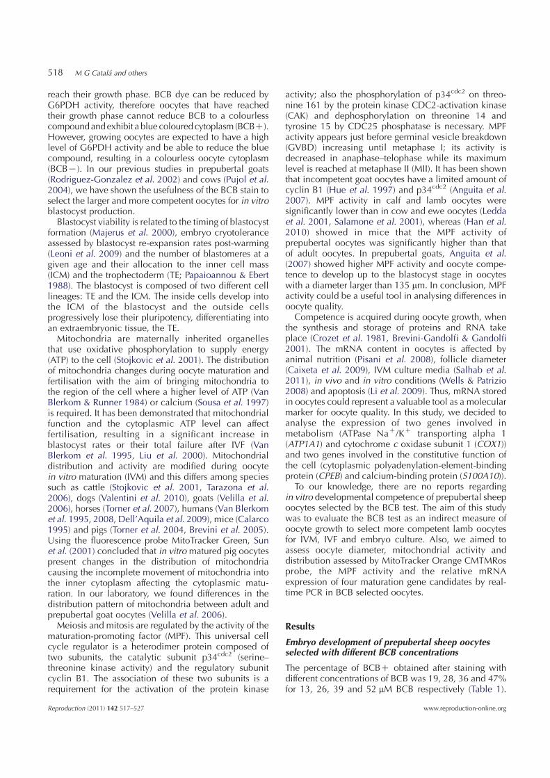
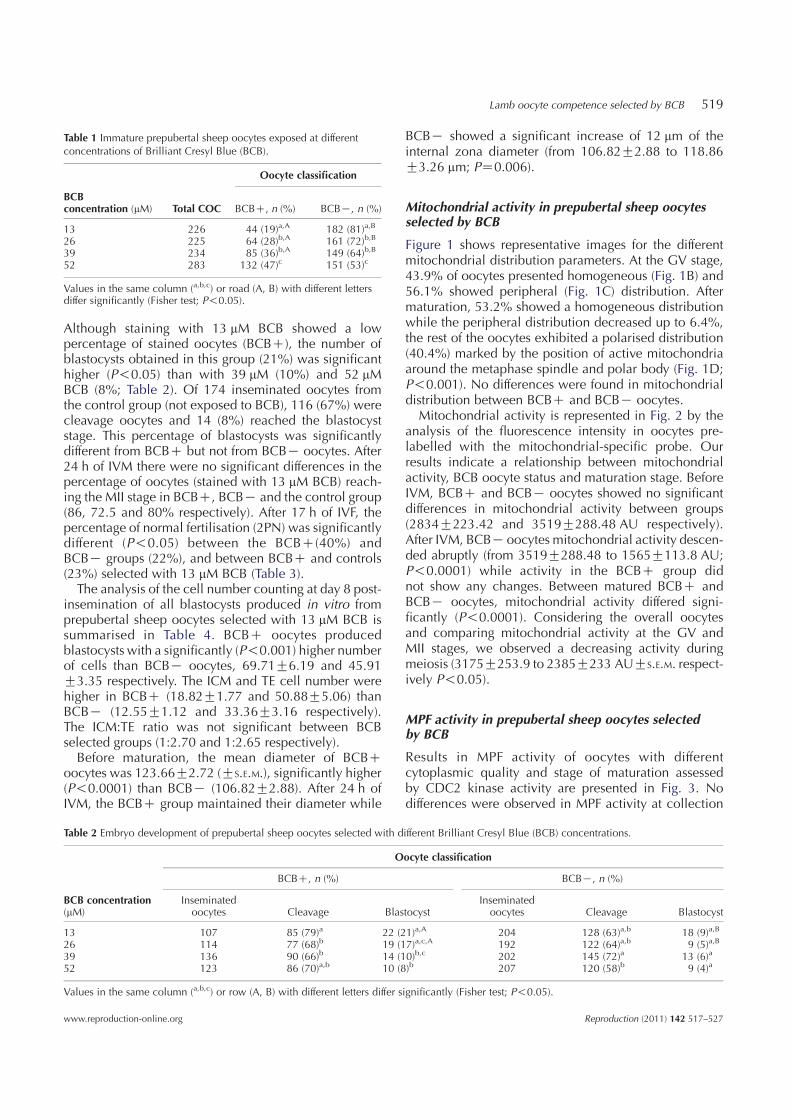
Mitochondrial activity in prepubertal sheep oocytesselected by BCB
Figure 1 shows representative images for the differentmitochondrial distribution parameters. At the GV stage,43.9% of oocytes presented homogeneous (Fig. 1B) and56.1% showed peripheral (Fig. 1C) distribution. Aftermaturation, 53.2% showed a homogeneous distributionwhile the peripheral distribution decreased up to 6.4%,the rest of the oocytes exhibited a polarised distribution(40.4%) marked by the position of active mitochondriaaround the metaphase spindle and polar body (Fig. 1D;P!0.001). No differences were found in mitochondrialdistribution between BCBC and BCBK oocytes.
Mitochondrial activity is represented in Fig. 2 by theanalysis of the fluorescence intensity in oocytes pre-labelled with the mitochondrial-specific probe. Ourresults indicate a relationship between mitochondrialactivity, BCB oocyte status and maturation stage. BeforeIVM, BCBC and BCBK oocytes showed no significantdifferences in mitochondrial activity between groups(2834G223.42 and 3519G288.48 AU respectively).After IVM, BCBK oocytes mitochondrial activity descen-ded abruptly (from 3519G288.48 to 1565G113.8 AU;P!0.0001) while activity in the BCBC group didnot show any changes. Between matured BCBC andBCBK oocytes, mitochondrial activity differed signi-ficantly (P!0.0001). Considering the overall oocytesand comparing mitochondrial activity at the GV andMII stages, we observed a decreasing activity duringmeiosis (3175G253.9 to 2385G233 AUGS.E.M. respect-ively P!0.05).
MPF activity in prepubertal sheep oocytes selectedby BCB
Results in MPF activity of oocytes with differentcytoplasmic quality and stage of maturation assessedby CDC2 kinase activity are presented in Fig. 3. Nodifferences were observed in MPF activity at collection
Table 1 Immature prepubertal sheep oocytes exposed at differentconcentrations of Brilliant Cresyl Blue (BCB).
Oocyte classification
BCBconcentration (mM) Total COC BCBC, n (%) BCBK, n (%)
13 226 44 (19)a,A 182 (81)a,B
26 225 64 (28)b,A 161 (72)b,B
39 234 85 (36)b,A 149 (64)b,B
52 283 132 (47)c 151 (53)c
Values in the same column (a,b,c) or road (A, B) with different lettersdiffer significantly (Fisher test; P!0.05).
Table 2 Embryo development of prepubertal sheep oocytes selected with different Brilliant Cresyl Blue (BCB) concentrations.
Oocyte classification
BCBC, n (%) BCBK, n (%)
BCB concentration(mM)
Inseminatedoocytes Cleavage Blastocyst
Inseminatedoocytes Cleavage Blastocyst
13 107 85 (79)a 22 (21)a,A 204 128 (63)a,b 18 (9)a,B
26 114 77 (68)b 19 (17)a,c,A 192 122 (64)a,b 9 (5)a,B
39 136 90 (66)b 14 (10)b,c 202 145 (72)a 13 (6)a
52 123 86 (70)a,b 10 (8)b 207 120 (58)b 9 (4)a
Values in the same column (a,b,c) or row (A, B) with different letters differ significantly (Fisher test; P!0.05).
Lamb oocyte competence selected by BCB 519
www.reproduction-online.org Reproduction (2011) 142 517–527
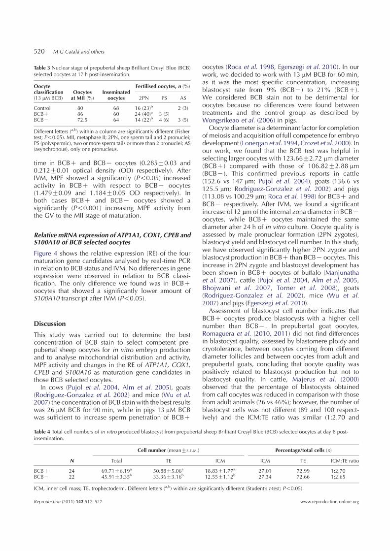
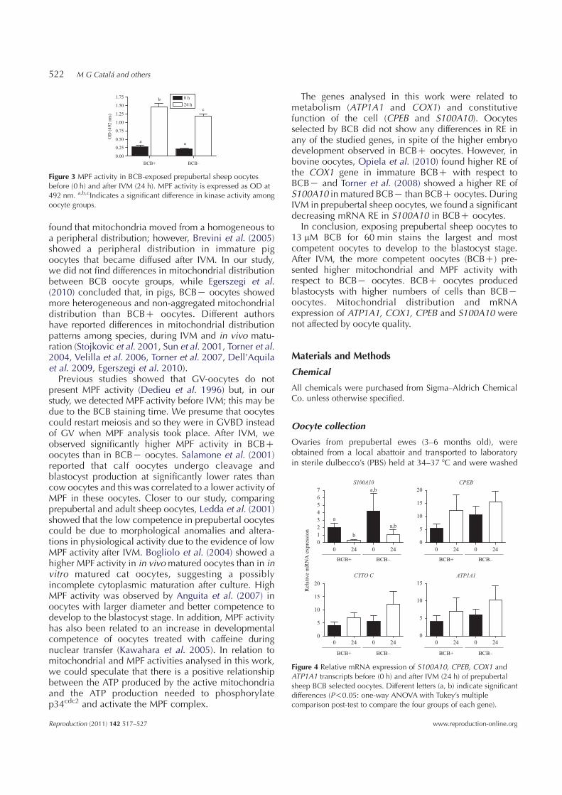
time in BCBC and BCBK oocytes (0.285G0.03 and0.212G0.01 optical density (OD) respectively). AfterIVM, MPF showed a significantly (P!0.05) increasedactivity in BCBC with respect to BCBK oocytes(1.479G0.09 and 1.184G0.05 OD respectively). Inboth cases BCBC and BCBK oocytes showed asignificantly (P!0.001) increasing MPF activity fromthe GV to the MII stage of maturation.
Relative mRNA expression of ATP1A1, COX1, CPEB andS100A10 of BCB selected oocytes
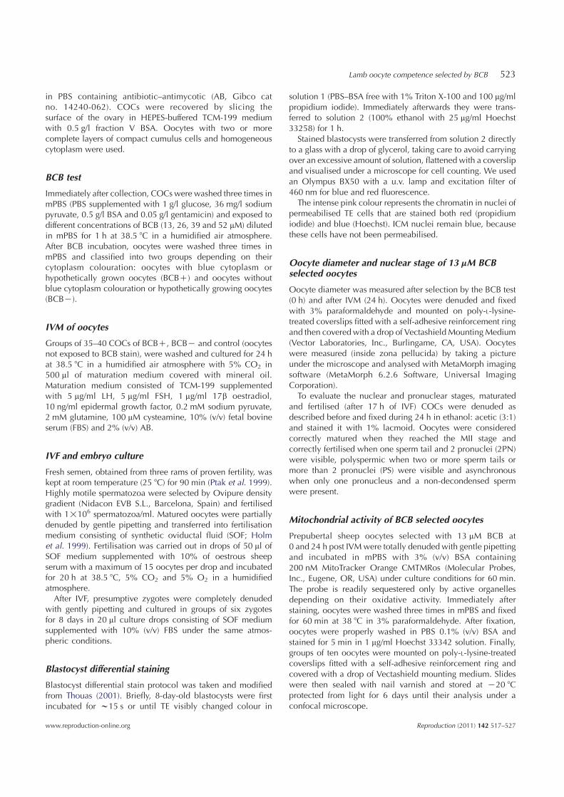
Figure 4 shows the relative expression (RE) of the fourmaturation gene candidates analysed by real-time PCRin relation to BCB status and IVM. No differences in geneexpression were observed in relation to BCB classi-fication. The only difference we found was in BCBCoocytes that showed a significantly lower amount ofS100A10 transcript after IVM (P!0.05).
Discussion
This study was carried out to determine the bestconcentration of BCB stain to select competent pre-pubertal sheep oocytes for in vitro embryo productionand to analyse mitochondrial distribution and activity,MPF activity and changes in the RE of ATP1A1, COX1,CPEB and S100A10 as maturation gene candidates inthose BCB selected oocytes.In cows (Pujol et al. 2004, Alm et al. 2005), goats
(Rodriguez-Gonzalez et al. 2002) and mice (Wu et al.2007) the concentration of BCB stain with the best resultswas 26 mM BCB for 90 min, while in pigs 13 mM BCBwas sufficient to increase sperm penetration of BCBC
oocytes (Roca et al. 1998, Egerszegi et al. 2010). In ourwork, we decided to work with 13 mM BCB for 60 min,as it was the most specific concentration, increasingblastocyst rate from 9% (BCBK) to 21% (BCBC).We considered BCB stain not to be detrimental foroocytes because no differences were found betweentreatments and the control group as described byWongsrikeao et al. (2006) in pigs.
Oocyte diameter is a determinant factor for completionof meiosis and acquisition of full competence for embryodevelopment (Lonergan et al. 1994, Crozet et al. 2000). Inour work, we found that the BCB test was helpful inselecting larger oocytes with 123.66G2.72 mm diameter(BCBC) compared with those of 106.82G2.88 mm(BCBK). This confirmed previous reports in cattle(152.6 vs 147 mm; Pujol et al. 2004), goats (136.6 vs125.5 mm; Rodriguez-Gonzalez et al. 2002) and pigs(113.08 vs 100.29 mm; Roca et al. 1998) for BCBC andBCBK respectively. After IVM, we found a significantincrease of 12 mmof the internal zona diameter in BCBKoocytes, while BCBC oocytes maintained the samediameter after 24 h of in vitro culture. Oocyte quality isassessed by male pronuclear formation (2PN zygotes),blastocyst yield and blastocyst cell number. In this study,we have observed significantly higher 2PN zygote andblastocyst production in BCBC than BCBKoocytes. Thisincrease in 2PN zygote and blastocyst development hasbeen shown in BCBC oocytes of buffalo (Manjunathaet al. 2007), cattle (Pujol et al. 2004, Alm et al. 2005,Bhojwani et al. 2007, Torner et al. 2008), goats(Rodriguez-Gonzalez et al. 2002), mice (Wu et al.2007) and pigs (Egerszegi et al. 2010).
Assessment of blastocyst cell number indicates thatBCBC oocytes produce blastocysts with a higher cellnumber than BCBK. In prepubertal goat oocytes,Romaguera et al. (2010, 2011) did not find differencesin blastocyst quality, assessed by blastomere ploidy andcryotolerance, between oocytes coming from differentdiameter follicles and between oocytes from adult andprepubertal goats, concluding that oocyte quality waspositively related to blastocyst production but not toblastocyst quality. In cattle, Majerus et al. (2000)observed that the percentage of blastocysts obtainedfrom calf oocytes was reduced in comparison with thosefrom adult animals (26 vs 46%); however, the number ofblastocyst cells was not different (89 and 100 respect-ively) and the ICM:TE ratio was similar (1:2.70 and
Table 3 Nuclear stage of prepubertal sheep Brilliant Cresyl Blue (BCB)selected oocytes at 17 h post-insemination.
Oocyteclassification(13 mM BCB)
Oocytesat MII (%)
Inseminatedoocytes
Fertilised oocytes, n (%)
2PN PS AS
Control 80 68 16 (23)b 2 (3)BCBC 86 60 24 (40)a 3 (5)BCBK 72.5 64 14 (22)b 4 (6) 3 (5)
Different letters (a,b) within a column are significantly different (Fishertest; P!0.05). MII, metaphase II; 2PN, one sperm tail and 2 pronuclei;PS (polyspermic), two or more sperm tails or more than 2 pronuclei; AS(asynchronous), only one pronucleus.
Table 4 Total cell numbers of in vitro produced blastocyst from prepubertal sheep Brilliant Cresyl Blue (BCB) selected oocytes at day 8 post-insemination.
Cell number (meanGS.E.M.) Percentage/total cells (n)
N Total TE ICM ICM TE ICM:TE ratio
BCBC 24 69.71G6.19a 50.88G5.06a 18.83G1.77a 27.01 72.99 1:2.70BCBK 22 45.91G3.35b 33.36G3.16b 12.55G1.12b 27.34 72.66 1:2.65
ICM, inner cell mass; TE, trophectoderm. Different letters (a,b) within are significantly different (Student’s t-test; P!0.05).
520 M G Catala and others
Reproduction (2011) 142 517–527 www.reproduction-online.org
1:2.85 respectively). Selecting lamb oocytes according tothenumberof cumulus layers,Kellyet al. (2007) concludedthat the percentage of day 8 blastocysts was affectedby COC grade but the number of blastocyst cells was notsignificantly different (range 49.2–54.6 cells per blasto-cyst). To our knowledge, no studies on oocytes selectedby BCB and embryo quality have been done. In this study,we have shown a positive relationship between BCBCoocytes and the number of blastomeres per blastocyst.Mitochondrial distribution and activity inside the
oocyte could be a good marker of oocyte competenceto develop to the blastocyst stage. The primary functionof mitochondria is to generate ATP. Van Blerkom et al.(1995) described in human oocytes the relationshipbetween ATP content and embryo developmentalcapacity where a transient decrease in ATP content canlead to embryo arrest. Therefore, these data suggest thatmitochondrial activity is a determinant factor of qualityand changes in mitochondrial activity can alter oocytequality in a remarkable way. In cattle (Tarazona et al.2006), horses (Torner et al. 2007), humans (Van Blerkom2004) and pigs (Torner et al. 2004) an increase inmitochondrial activity after IVM was described. In cattle,Torner et al. (2008) observed higher mitochondrialactivity in BCBK oocytes than in BCBC. These authorsspeculated that the reason for the increasing respiratoryactivity in low-quality oocytes was to provide ATP forstill unfinished processes for cytoplasmic maturation. Inpig oocytes, Egerszegi et al. (2010) found a highermitochondrial activity in BCBC compared with BCBKoocytes before IVM, but after IVM, no differences werefound between either kind of oocyte. They attributethis to BCBC oocytes increasing their respiratory activityto provide ATP for the energy-demanding processesof GVBD and the subsequent condensation of chromatinup to MII, while BCBK oocytes showed no changesin mitochondrial activity during meiosis and only afew of them reached MII stage. In our study withprepubertal sheep oocytes, we found a decrease inmitochondrial activity from the GV to the MII stage(3175G253.9–2385G233 AUGS.E.M.). Analysing BCBC
and BCBK oocytes separately, we found that at theGV stage there were no differences in mitochondrialactivity between groups. However, after IVM, BCBKoocytes showed a significant reduction in mitochondrialactivity while BCBC mitochondrial activity remainedconstant. This would indicate a positive relationshipbetween mitochondrial activity at MII stage and embryodevelopment.
Stojkovic et al. (2001) showed that mitochondrialreorganisation was different between morphologicallygood and poor quality oocytes. In our study, mito-chondria migrated throughout the IVM process. Oocytesat the GV stage presented a homogeneous (43.9%)or peripheral (56.1%) mitochondrial distribution. After24 h of IVM, MII oocytes presented a homogeneous(53.2%) distribution or mitochondria polarised aroundthe metaphase spindle and inside the polar body (PB;40.4%). We have previously shown (Velilla et al. 2006),in prepubertal goat IVM oocytes, that total mitochondriamigrate from a cortical and perinuclear distributionin GV oocytes to a polarised distribution oppositethe metaphase spindle and inside the PB (86%) afterIVM, whereas ovulated adult goat oocytes presented amitochondrial distribution inside the PB and aggregatedto the metaphase spindle (Velilla et al. 2006) as we havefound here in lamb oocytes. In pigs, Torner et al. (2004)
A
High
Low
CB D
Figure 1 Representative images of active mitochondrial distribution of prepubertal sheep oocytes taken by a confocal microscope. Images werespectrally (A) coded to represent staining intensity (red is the highest intensity). Representative images of (B) homogeneous, (C) peripheral and (D)polarised mitochondrial activity distribution in lamb oocytes.
0.00
500.00
1000.00
1500.00
2000.00
2500.00
3000.00
3500.00
4000.00
4500.00
5000.00
BCB + BCB –
Oocyte classification
Mea
n f
luore
scen
ce a
ctiv
ity (
arbit
rary
unit
s)
0 h
24 h
Total
**
#
Figure 2 Mitochondrial activity of BCB selected oocytes assessed byfluorescence intensity before (0 h) and after IVM (24 h). Differentsymbols (#, *) indicate significant differences (*P!0.0001; #P!0.05).
Lamb oocyte competence selected by BCB 521
www.reproduction-online.org Reproduction (2011) 142 517–527
found that mitochondria moved from a homogeneous toa peripheral distribution; however, Brevini et al. (2005)showed a peripheral distribution in immature pigoocytes that became diffused after IVM. In our study,we did not find differences in mitochondrial distributionbetween BCB oocyte groups, while Egerszegi et al.(2010) concluded that, in pigs, BCBK oocytes showedmore heterogeneous and non-aggregated mitochondrialdistribution than BCBC oocytes. Different authorshave reported differences in mitochondrial distributionpatterns among species, during IVM and in vivo matu-ration (Stojkovic et al. 2001, Sun et al. 2001, Torner et al.2004, Velilla et al. 2006, Torner et al. 2007, Dell’Aquilaet al. 2009, Egerszegi et al. 2010).Previous studies showed that GV-oocytes do not
present MPF activity (Dedieu et al. 1996) but, in ourstudy, we detected MPF activity before IVM; this may bedue to the BCB staining time. We presume that oocytescould restart meiosis and so they were in GVBD insteadof GV when MPF analysis took place. After IVM, weobserved significantly higher MPF activity in BCBCoocytes than in BCBK oocytes. Salamone et al. (2001)reported that calf oocytes undergo cleavage andblastocyst production at significantly lower rates thancow oocytes and this was correlated to a lower activity ofMPF in these oocytes. Closer to our study, comparingprepubertal and adult sheep oocytes, Ledda et al. (2001)showed that the low competence in prepubertal oocytescould be due to morphological anomalies and altera-tions in physiological activity due to the evidence of lowMPF activity after IVM. Bogliolo et al. (2004) showed ahigher MPF activity in in vivomatured oocytes than in invitro matured cat oocytes, suggesting a possiblyincomplete cytoplasmic maturation after culture. HighMPF activity was observed by Anguita et al. (2007) inoocytes with larger diameter and better competence todevelop to the blastocyst stage. In addition, MPF activityhas also been related to an increase in developmentalcompetence of oocytes treated with caffeine duringnuclear transfer (Kawahara et al. 2005). In relation tomitochondrial and MPF activities analysed in this work,we could speculate that there is a positive relationshipbetween the ATP produced by the active mitochondriaand the ATP production needed to phosphorylatep34cdc2 and activate the MPF complex.
The genes analysed in this work were related tometabolism (ATP1A1 and COX1) and constitutivefunction of the cell (CPEB and S100A10). Oocytesselected by BCB did not show any differences in RE inany of the studied genes, in spite of the higher embryodevelopment observed in BCBC oocytes. However, inbovine oocytes, Opiela et al. (2010) found higher RE ofthe COX1 gene in immature BCBC with respect toBCBK and Torner et al. (2008) showed a higher RE ofS100A10 in matured BCBK than BCBC oocytes. DuringIVM in prepubertal sheep oocytes, we found a significantdecreasing mRNA RE in S100A10 in BCBC oocytes.
In conclusion, exposing prepubertal sheep oocytes to13 mM BCB for 60 min stains the largest and mostcompetent oocytes to develop to the blastocyst stage.After IVM, the more competent oocytes (BCBC) pre-sented higher mitochondrial and MPF activity withrespect to BCBK oocytes. BCBC oocytes producedblastocysts with higher numbers of cells than BCBKoocytes. Mitochondrial distribution and mRNAexpression of ATP1A1, COX1, CPEB and S100A10 werenot affected by oocyte quality.
Materials and Methods
Chemical
All chemicals were purchased from Sigma–Aldrich ChemicalCo. unless otherwise specified.
Oocyte collection
Ovaries from prepubertal ewes (3–6 months old), wereobtained from a local abattoir and transported to laboratoryin sterile dulbecco’s (PBS) held at 34–37 8C and were washed
BCB+ BCB–
0.00
0.25
0.50
0.75
1.00
1.25
1.50
1.75 0 h
24 h
a
c
a
b
OD
(492 n
m)
Figure 3 MPF activity in BCB-exposed prepubertal sheep oocytesbefore (0 h) and after IVM (24 h). MPF activity is expressed as OD at492 nm. a,b,cIndicates a significant difference in kinase activity amongoocyte groups.
S100A10
0 24 0 24
0
1
2
3
4
5
6
7
a
b
a,b
a,b
CPEB
0
5
10
15
20
CYTO C
0
5
10
15
20
ATP1A1
0
5
10
15
Rel
ativ
e m
RN
A e
xpre
ssio
n
BCB+ BCB–
0 24 0 24
BCB+ BCB–
0 24 0 24
BCB+ BCB–
0 24 0 24
BCB+ BCB–
Figure 4 Relative mRNA expression of S100A10, CPEB, COX1 andATP1A1 transcripts before (0 h) and after IVM (24 h) of prepubertalsheep BCB selected oocytes. Different letters (a, b) indicate significantdifferences (P!0.05: one-way ANOVA with Tukey’s multiplecomparison post-test to compare the four groups of each gene).
522 M G Catala and others
Reproduction (2011) 142 517–527 www.reproduction-online.org
in PBS containing antibiotic–antimycotic (AB, Gibco catno. 14240-062). COCs were recovered by slicing thesurface of the ovary in HEPES-buffered TCM-199 mediumwith 0.5 g/l fraction V BSA. Oocytes with two or morecomplete layers of compact cumulus cells and homogeneouscytoplasm were used.
BCB test
Immediately after collection, COCs were washed three times inmPBS (PBS supplemented with 1 g/l glucose, 36 mg/l sodiumpyruvate, 0.5 g/l BSA and 0.05 g/l gentamicin) and exposed todifferent concentrations of BCB (13, 26, 39 and 52 mM) dilutedin mPBS for 1 h at 38.5 8C in a humidified air atmosphere.After BCB incubation, oocytes were washed three times inmPBS and classified into two groups depending on theircytoplasm colouration: oocytes with blue cytoplasm orhypothetically grown oocytes (BCBC) and oocytes withoutblue cytoplasm colouration or hypothetically growing oocytes(BCBK).
IVM of oocytes
Groups of 35–40 COCs of BCBC, BCBK and control (oocytesnot exposed to BCB stain), were washed and cultured for 24 hat 38.5 8C in a humidified air atmosphere with 5% CO2 in500 ml of maturation medium covered with mineral oil.Maturation medium consisted of TCM-199 supplementedwith 5 mg/ml LH, 5 mg/ml FSH, 1 mg/ml 17b oestradiol,10 ng/ml epidermal growth factor, 0.2 mM sodium pyruvate,2 mM glutamine, 100 mM cysteamine, 10% (v/v) fetal bovineserum (FBS) and 2% (v/v) AB.
IVF and embryo culture
Fresh semen, obtained from three rams of proven fertility, waskept at room temperature (25 8C) for 90 min (Ptak et al. 1999).Highly motile spermatozoa were selected by Ovipure densitygradient (Nidacon EVB S.L., Barcelona, Spain) and fertilisedwith 1!106 spermatozoa/ml. Matured oocytes were partiallydenuded by gentle pipetting and transferred into fertilisationmedium consisting of synthetic oviductal fluid (SOF; Holmet al. 1999). Fertilisation was carried out in drops of 50 ml ofSOF medium supplemented with 10% of oestrous sheepserum with a maximum of 15 oocytes per drop and incubatedfor 20 h at 38.5 8C, 5% CO2 and 5% O2 in a humidifiedatmosphere.After IVF, presumptive zygotes were completely denuded
with gently pipetting and cultured in groups of six zygotesfor 8 days in 20 ml culture drops consisting of SOF mediumsupplemented with 10% (v/v) FBS under the same atmos-pheric conditions.
Blastocyst differential staining
Blastocyst differential stain protocol was taken and modifiedfrom Thouas (2001). Briefly, 8-day-old blastocysts were firstincubated for w15 s or until TE visibly changed colour in
solution 1 (PBS–BSA free with 1% Triton X-100 and 100 mg/mlpropidium iodide). Immediately afterwards they were trans-ferred to solution 2 (100% ethanol with 25 mg/ml Hoechst33258) for 1 h.Stained blastocysts were transferred from solution 2 directly
to a glass with a drop of glycerol, taking care to avoid carryingover an excessive amount of solution, flattened with a coverslipand visualised under a microscope for cell counting. We usedan Olympus BX50 with a u.v. lamp and excitation filter of460 nm for blue and red fluorescence.The intense pink colour represents the chromatin in nuclei of
permeabilised TE cells that are stained both red (propidiumiodide) and blue (Hoechst). ICM nuclei remain blue, becausethese cells have not been permeabilised.
Oocyte diameter and nuclear stage of 13 mM BCBselected oocytes
Oocyte diameter was measured after selection by the BCB test(0 h) and after IVM (24 h). Oocytes were denuded and fixedwith 3% paraformaldehyde and mounted on poly-L-lysine-treated coverslips fitted with a self-adhesive reinforcement ringand then covered with a drop of VectashieldMounting Medium(Vector Laboratories, Inc., Burlingame, CA, USA). Oocyteswere measured (inside zona pellucida) by taking a pictureunder the microscope and analysed with MetaMorph imagingsoftware (MetaMorph 6.2.6 Software, Universal ImagingCorporation).To evaluate the nuclear and pronuclear stages, maturated
and fertilised (after 17 h of IVF) COCs were denuded asdescribed before and fixed during 24 h in ethanol: acetic (3:1)and stained it with 1% lacmoid. Oocytes were consideredcorrectly matured when they reached the MII stage andcorrectly fertilised when one sperm tail and 2 pronuclei (2PN)were visible, polyspermic when two or more sperm tails ormore than 2 pronuclei (PS) were visible and asynchronouswhen only one pronucleus and a non-decondensed spermwere present.
Mitochondrial activity of BCB selected oocytes
Prepubertal sheep oocytes selected with 13 mM BCB at0 and 24 h post IVMwere totally denuded with gentle pipettingand incubated in mPBS with 3% (v/v) BSA containing200 nM MitoTracker Orange CMTMRos (Molecular Probes,Inc., Eugene, OR, USA) under culture conditions for 60 min.The probe is readily sequestered only by active organellesdepending on their oxidative activity. Immediately afterstaining, oocytes were washed three times in mPBS and fixedfor 60 min at 38 8C in 3% paraformaldehyde. After fixation,oocytes were properly washed in PBS 0.1% (v/v) BSA andstained for 5 min in 1 mg/ml Hoechst 33342 solution. Finally,groups of ten oocytes were mounted on poly-L-lysine-treatedcoverslips fitted with a self-adhesive reinforcement ring andcovered with a drop of Vectashield mounting medium. Slideswere then sealed with nail varnish and stored at K20 8Cprotected from light for 6 days until their analysis under aconfocal microscope.
Lamb oocyte competence selected by BCB 523
www.reproduction-online.org Reproduction (2011) 142 517–527

Confocal analysis
A laser scanning confocal microscope (Espectral Leica TCS-SP5, Mannheim, Germany) was used to examine activemitochondria (MitoTracker Orange CMTMRos, excitation554 nm) and chromatin (Hoechst; excitation 405 nm). Themitochondrial distribution pattern (representative images inFig. 1) was characterised with 60! magnification undermineral oil and classified as homogeneous (fluorescencethroughout the cytoplasm, Fig. 1B), peripheral (fluorescencein the cortex, Fig. 1C) or polarised (fluorescence near theMII spindle, Fig. 1D) according to mitochondrial distribution.For mitochondrial intensity the microscope objective,
pinhole, filters, offset, gain, pixels and laser potency werekept constant throughout the experiment. Twenty serial cuts of1 mm each in the region of major intensity were performed. The20 images of each oocyte were added in a unique picture andanalysed with MetaMorph imaging software. To expressthe fluorescence intensity, we used arbitrary unit of the meanfluorescence intensity, which is defined as the sum offluorescence intensity in greyscale of every pixel in thecytoplasm of the oocyte divided by the sum of all pixels ofthe same region.
MPF activity of BCB selected oocytes
Before and after IVM, groups of 20 oocytes BCBC, BCBK(three replicates) were washed three times in PBS, and placed intubes containing 5 ml of lysis buffer (50 mM Tris–HCl, pH 7.5,0.5 M NaCl, 5 mM EDTA, 0.01% Brij35, 1 mM PMSF,0.05 mg/ml leupeptin, 50 mM 2-mercaptoethanol, 25 mMb-glycerophosphate and 1 mM Na-orthovanadate). Thesamples were frozen in liquid nitrogen and sonicatedthree times at 1 8C for 25 s. Cell extracts were stored atK80 8C until use.CDC2 assay was performed by the MESACUP CDC2 kinase
assay kit (MBL, Madrid, Spain) following the manufacturer’sprotocol. Oocyte extracts (5 ml) were mixed with 10! CDC2Reaction Buffer (25 mM Hepes buffer pH 7.5 and 10 mMMgCl2) and 10% biotinylated MV Peptide (SLYSSPGGAYC).The phosphorylation reaction was started adding 0.1 mM ATP(Sigma–Aldrich), in a final volume of 50 ml. The mixture wasincubated at 30 8C for 30 min. The reaction was finished by
adding 200 ml of phosphorylation Stop Reagent (PBScontaining 50 mM EGTA). The phosphorylated MV peptidewas detected by ELISA at 492 nm and expressed as OD.
Real-time PCR quantification of ATP1A1, COX1, CPEBand S100A10 of BCB selected oocytes
Groups of 15 denuded prepubertal sheep oocytes (fourreplicates) stained with 13 mM BCB were taken at 0 and 24 hpost IVM and stored at K80 8C in 100 ml Trizol (Invitrogen)until use. For RNA extraction, the addition of a known amountof luciferase (0.2 pg/ml, Promega) was added as an exogenousstandard. To avoid contamination with genomic DNA, totalRNA preparations were incubated during 10 min with RQ1DNase (Promega) as described in the manufacturer’s protocol.Reverse transcription was performed by extended cDNA usingOligo (dT) 15 primers during 5 min at 70 8C and 1 h at 65 8Cusing superscript III (200 U/mM; Invitrogen). Relative quali-tative PCR analysis was performed in MyiQ apparatus (Bio-RadLaboratories). Samples were distributed in the plate by arobotic distributor (Eppendorf) and reactions were performed induplicate using SYBR Green Fluorophore kit (Bio-Rad).Reactions were performed in 20 ml final volume (in duplicate)and PCR cycling conditions were 95 8C for 3 min followed by40 cycles of denaturing (30 s, 95 8C), annealing (30 s, 60 8C)and elongation (20 s, 72 8C). The specificity of each PCRproduct was determined by a melting curve analysis and theamplicon size determination in agarose gels. For each gene, astandard curve was included, consisting of correspondingplasmid DNA fragments from 1 pg to 0.1 fg, purified withQIAquick PCR Purification Kit (Qiagen). Correlation coeffi-cients and PCR efficiencies were considered between 85 and100%. Primer sequences are listed in Table 5. The results formRNAwere normalised according to the relative concentrationof the internal standard, luciferase and 18S.
Statistical analysis
Analysis among treatments (BCB stain, IVM and IVF) wereperformed by Fisher’s exact test. Blastocyst cell numbers wereanalysed by Student’s t-test. Gene and MPF analysis wasperformed by one-way ANOVA in GraphPad Prism v 3(GraphPad Software, San Diego, CA, USA).
Table 5 Oligonucleotide primer sequences used in this study.
Gene Primer Sequence 5 0–3 0 GenBank accession no. Product size (bp)
S100A10 Sense CCGCCAAGGTTTCAACAGACTTC EE822394 271Antisense ATGGTGAGCCCAGCGATTAGC
CPEB Sense CCTCCCAGATGCAAATGACT DY514003 235Antisense CTTAATGGAGGGTGCTGGAA
COX1 Sense TGGAGGACAATATCAAGGGAGGAG CO000988 471Antisense GGACCGAAACCTGAACACAACC
ATP1A1 Sense GAACGGCTTCCTCCCTAATC NM_001009360 207Antisense ACGGAATTCCTCCTGGTCTT
LUCI Sense TCATTCTTCGCCAAAAGCACTC 140Antisense AGCCCATATCCTTGTCGTATCC
18S Sense AGAAACGGCTACCACATCCAA DQ222453.1 90Antisense CCTGTATTGTTATTTTTCGT
524 M G Catala and others
Reproduction (2011) 142 517–527 www.reproduction-online.org

The individual mitochondrial intensity (arbitrary unit) datawere analysed as log10 to normalise them. Statistical analysiswas conducted by PROCMIXED (with number of treatments asrandom parameter) of SAS (version 9.2 Inst.), Inc., Cary, NC,USA) according to Tukey’s multiple comparison test. Thestatistical model contained the fixed effects of BCB (C versusK) and maturation state (0 vs 24 h) and first-order interactionbetween BCB and maturation state and residual error.Differences were declared significant at P!0.05.
Declaration of interest
The authors declare that there is no conflict of interest thatcould be perceived as prejudicing the impartiality of theresearch reported.
Funding
This study was supported by grants from the Spanish Ministry ofEducation and Science (project number: AGL2007-60227/GAN) and from the Universitat Autonoma de Barcelona(2007-FPI00193).
Acknowledgements
The authors thank Ph.D. Maria Rodriguez Prado for herstatistical assistance.
References
Alm H, Torner H, Lochrke B, Viergutz T, Ghoneim IM & Kanitz W 2005Bovine blastocyst development rate in vitro is influenced by selection ofoocytes by brillant cresyl blue staining before IVM as indicator forglucose-6-phosphate dehydrogenase activity. Theriogenology 632194–2205. (doi:10.1016/j.theriogenology.2004.09.050)
Anguita B, Jimenez-Macedo AR, Izquierdo D, Mogas T & ParamioMT 2007Effect of oocyte diameter on meiotic competence, embryo development,p34 (cdc2) expression and MPF activity in prepubertal goat oocytes.Theriogenology67526–536. (doi:10.1016/j.theriogenology.2006.09.003)
Armstrong DT 2001 Effects of maternal age on oocyte developmentalcompetence. Theriogenology 55 1303–1322. (doi:10.1016/S0093-691X(01)00484-8)
Barnes FL & Sirard MA 2000 Oocyte maturation. Seminars in ReproductiveMedicine 18 123–131. (doi:10.1055/s-2000-12551)
Bhojwani S, Alm H, Torner H, Kanitz W & Poehland R 2007 Selection ofdevelopmentally competent oocytes through Brilliant Cresyl Blue stainenhances blastocyst development rate after bovine nuclear transfer.Theriogenology67341–345. (doi:10.1016/j.theriogenology.2006.08.006)
Bogliolo L, Leoni G, Ledda S, Zedda MT, Bonelli P, Madau L, Santucciu C,Naitana S & Pau S 2004 M-phase promoting factor (MPF) and mitogenactivated protein kinases (MAPK) activities of domestic cat oocytesmatured in vitro and in vivo. Cloning and Stem Cells 6 15–23. (doi:10.1089/15362300460743790)
Brevini TA, Vassena R, Francisci C & Gandolfi F 2005 Role of adenosinetriphosphate, active mitochondria, and microtubules in the acquisitionof developmental competence of parthenogenetically activated pigoocytes. Biology of Reproduction 72 1218–1223. (doi:10.1095/biolre-prod.104.038141)
Brevini-Gandolfi TAL & Gandolfi F 2001 The maternal legacy to theembryo: cytoplasmic components and their effects on early develop-ment. Theriogenology 55 1255–1276. (doi:10.1016/S0093-691X(01)00481-2)
Caixeta ES, Ripamonte P, Franco MM, Buratini J & Dode MAN 2009 Effectof follicle size on mRNA expression in cumulus cells and oocytes of
Bos indicus: an approach to identify marker genes for developmentalcompetence. Reproduction, Fertility, and Development 21 655–664.(doi:10.1071/RD08201)
Calarco PG 1995 Polarization of mitochondria in the unfertilizedmouse oocyte. Developmental Genetics 16 36–43. (doi:10.1002/dvg.1020160108)
Cognie Y, Baril G, Poulin N & Mermillod P 2003 Current status ofembryo technologies in sheep and goat. Theriogenology 59 171–188.(doi:10.1016/S0093-691X(02)01270-0)
Crozet N, Motlij J & Szollosi D 1981 Nucleolar fine-structure and RNA-synthesis in porcine oocytes during the early stages of antrum formation.Biology of the Cell 41 35–41.
Crozet N, Dahirel M & Gall L 2000 Meiotic competence of in vitrogrown goat oocytes. Journal of Reproduction and Fertility 118 367–373.(doi:10.1530/reprod/118.2.367)
Dedieu T, Gall L, Crozet N, Sevellec C & Ruffini S 1996 Mitogen-activatedprotein kinase activity during goat oocyte maturation and theacquisition of meiotic competence. Molecular Reproduction andDevelopment 45 351–358. (doi:10.1002/(SICI)1098-2795(199611)45:3!351::AID-MRD12O3.0.CO;2-1)
Dell’Aquila ME, Ambruosi B, De Santis T & Cho YS 2009 Mitochondrialdistribution and activity in human mature oocytes: gonadotropin-releasing hormone agonist versus antagonist for pituitary down-regulation. Fertility and Sterility 91 249–255. (doi:10.1016/j.fertnstert.2007.10.042)
Egerszegi I, Alm H, Ratky J, Heleil B, Bruessow K-P & Torner H 2010Meiotic progression, mitochondrial features and fertilisation character-istics of porcine oocytes with different G6PDH activities. Reproduction,Fertility, and Development 22 830–838. (doi:10.1071/RD09140)
Gilchrist RB, Nayudu PL, Nowshari MA & Hodges JK 1995 Meioticcompetence of marmoset monkey oocytes is related to follicle size andoocyte-somatic cell associations. Biology of Reproduction 521234–1243. (doi:10.1095/biolreprod52.6.1234)
Han D, Cao X, Wang H, Li J, Wang Y & Tan J 2010 Effects of pubertyand gonadotropins on the molecular events controlling meioticresumption of mouse oocytes. Reproduction 139 959–969. (doi:10.1530/REP-09-0485)
Holm P, Booth PJ, Schmidt MH, Greve T & Callesen H 1999 High bovineblastocyst development in a static in vitro production system using sofamedium supplemented with sodium citrate and myo-inositol with orwithout serum-proteins. Theriogenology 52 683–700. (doi:10.1016/S0093-691X(99)00162-4)
Hue I, Thierry D, Huneau D, Ruffini S, Gall L & Crozet N 1997 Cyclin B-1expression in meiotically competent and incompetent goat oocytes.Molecular Reproduction and Development 47 222. (doi:10.1002/(SICI)1098-2795(199706)47:2!222::AID-MRD14O3.0.CO;2-1)
Jimenez-Macedo AR, Paramio MT, Anguita B, Morato R, Romaguera R,Mogas T & Izquierdo D 2007 Effect of ICSI and embryo biopsy onembryo development and apoptosis according to oocyte diameter inprepubertal goats. Theriogenology 67 1399–1408. (doi:10.1016/j.theriogenology.2007.03.003)
Kawahara M, Wakai T, Yamanaka K, Kobayashi J, Sugimura S, Shimizu T,Matsumoto H, Kim J, Sasada H & Sato E 2005 Caffeine promotespremature chromosome condensation formation and in vitro develop-ment in porcine reconstructed embryos via a high level of maturationpromoting factor activity during nuclear transfer. Reproduction 130351–357. (doi:10.1530/rep.1.00644)
Kelly JM, Kleemann DO, Rudiger SR & Walker SK 2007 Effects of grade ofoocyte–cumulus complex and the interactions between grades on theproduction of blastocysts in the cow, ewe and lamb. Reproduction inDomestic Animals 42 577–582. (doi:10.1111/j.1439-0531.2006.00823.x)
Ledda S, Bogliolo L, Leoni G & Naitana S 2001 Cell coupling andmaturation-promoting factor activity in in vitro-matured prepubertal andadult sheep oocytes. Biology of Reproduction 65 247–252. (doi:10.1095/biolreprod65.1.247)
Leoni G, Succu S, Satta V, Paolo M, Bogliolo L, Bebbere D, Spezzigo A,Madeddu M, Berlinguer F, Ledda S et al. 2009 In vitro production andcryotolerance of prepubertal and adult goat blastocysts obtained fromoocytes collected by laparoscopic oocyte-pick-up (LOPU) after FSHtreatment. Reproduction, Fertility, and Development 21 901–908.(doi:10.1071/RD09015)
Lamb oocyte competence selected by BCB 525
www.reproduction-online.org Reproduction (2011) 142 517–527

Li J, Liu DJ, Cang M, Wang LM, Jin MZ, Ma YZ & Shorgan B 2009 Earlyapoptosis is associated with improved developmental potential in bovineoocytes. Animal Reproduction Science 114 89–98. (doi:10.1016/j.anireprosci.2008.09.018)
Liu L, Trimarchi JR & Keefe DL 2000 Involvement of mitochondria inoxidative stress-induced cell death in mouse zygotes. Biology ofReproduction 62 1745–1753. (doi:10.1095/biolreprod62.6.1745)
Lonergan P, Monaghan P, Rizos D, Boland MP & Gordon I 1994 Effectof follicle size on bovine oocyte quality and developmental competencefollowing maturation, fertilization, and culture in-vitro. MolecularReproduction and Development 37 48–53. (doi:10.1002/mrd.1080370107)
Majerus V, Lequarre A, Ferguson EM, Kaidi S, Massip A, Dessy F &Donnay I 2000 Characterization of embryos derived from calfoocytes: kinetics of cleavage, cell allocation to inner cell mass, andtrophectoderm and lipid metabolism. Molecular Reproduction andDevelopment 57 346–352. (doi:10.1002/1098-2795(200012)57:4!346::AID-MRD6O3.0.CO;2-M)
Manjunatha BM, Gupta PSP, Devaraj M, Ravindra JP & Nandi S 2007Selection of developmentally competent buffalo oocytes by BrilliantCresyl Blue staining before IVM. Theriogenology 68 1299–1304. (doi:10.1016/j.theriogenology.2007.08.031)
Martino A, Palomo MJ, Mogas T & Paramio MT 1994 Influence of thecollection technique of prepubertal goat oocytes on in vitro maturationand fertilization. Theriogenology 42 859–873. (doi:10.1016/0093-691X(94)90454-Q)
O’Brien JK, Dwarte D, Ryan JP, Maxwell WM & Evans G 1996Developmental capacity, energy metabolism and ultrastructure ofmature oocytes from prepubertal and adult sheep. Reproduction,Fertility, and Development 7 1029–1037. (doi:10.1071/RD9961029)
Opiela J, Lipinski D, Slomski R & Katska-Ksiazkiewicz L 2010 Transcriptexpression of mitochondria related genes is correlated with bovineoocyte selection by BCB test. Animal Reproduction Science 118188–193. (doi:10.1016/j.anireprosci.2009.07.007)
Papaioannou VE & Ebert KM 1988 The preimplantation pig embryo: cellnumber and allocation to trophectoderm and inner cell mass of theblastocyst in vivo and in vitro. Development 102 793–803.
Peters JK, Milliken G & Davis DL 2001 Development of porcine embryosin vitro: effects of culture medium and donor age. Journal of AnimalScience 79 1578–1583.
Pisani LF, Antonini S, Pocar P, Ferrari S, Brevini TA, Rhind SM & Gandolfi F2008 Effects of pre-mating nutrition on mRNA levels of developmentallyrelevant genes in sheep oocytes and granulosa cells. Reproduction 136303–312. (doi:10.1530/REP-07-0394)
Ptak G, Loi P, Dattena M, Tischner M & Cappai P 1999 Offspring from one-month-old lambs: studies on the developmental capability of prepubertaloocytes. Biology of Reproduction 61 1568–1574. (doi:10.1095/biolre-prod61.6.1568)
Pujol M, Lopez-Bejar M & Paramio MT 2004 Developmental competenceof heifer oocytes selected using the Brilliant Cresyl Blue (BCB) test.Theriogenology 61 735–744. (doi:10.1016/S0093-691X(03)00250-4)
Revel F, Mermillod P, Peynot N, Renard JP & Heyman Y 1995 Lowdevelopmental capacity of in vitro matured and fertilized oocytes fromcalves compared with that of cows. Journal of Reproduction and Fertility103 115–120. (doi:10.1530/jrf.0.1030115)
Rizos D, Ward F, Duffy P, Boland MP & Lonergan P 2002 Consequences ofbovine oocyte maturation, fertilization or early embryo developmentin vitro versus in vivo: implications for blastocyst yield and blastocystquality.Molecular Reproduction and Development 61 234–248. (doi:10.1002/mrd.1153)
Roca J, Martinez E, Vazquez JM & Lucas X 1998 Selection of immature pigoocytes for homologous in vitro penetration assays with the BrilliantCresyl Blue test. Reproduction, Fertility, and Development 6 479–485.(doi:10.1071/RD98060)
Rodriguez-Gonzalez E, Lopez-Bejar M, Velilla E & Paramio MT 2002Selection of prepubertal goat oocytes using the Brilliant Cresyl Bluetest. Theriogenology 57 1397–1409. (doi:10.1016/S0093-691X(02)00645-3)
Romaguera R, Casanovas A, Morato R, Izquierdo D, Catala M, Jimenez-Macedo AR, Mogas T & Paramio MT 2010 Effect of follicle diameter on
oocyte apoptosis, embryo development and chromosomal ploidy inprepubertal goats. Theriogenology 74 364–373. (doi:10.1016/j.therio-genology.2010.02.019)
Romaguera R, Moll X, Morato R, Roura M, Palomo MJ, Catala MG,Jimenez-Macedo AR, Hammami S, Izquierdo D, Mogas T et al. 2011Prepubertal goat oocytes from large follicles result in similar blastocystproduction and embryo ploidy than those from adult goats.Theriogenology 76 1–11. (doi:10.1016/j.theriogenology.2010.12.014)
Salamone DF, Damiani P, Fissore RA, Roble JM & Duby RT 2001Biochemical and developmental evidence that ooplasmic maturationof prepubertal bovine oocytes is compromised. Biology of Reproduction64 1761–1768. (doi:10.1095/biolreprod64.6.1761)
Salhab M, Tosca L, Cabau C, Papillier P, Perreau C, Dupont J, Mermillod P &Uzbekova S 2011 Kinetics of gene expression and signaling in bovinecumulus cells throughout IVM in different mediums in relation tooocyte developmental competence, cumulus apoptosis and progesteronesecretion. Theriogenology 1 90–104. (doi:10.1016/j.theriogenology.2010.07.014)
Sousa M, Barros A, Silva J & Tesarik J 1997 Developmental changes incalcium content of ultrastructurally distinct subcellular compartments ofpreimplantation human embryos. Molecular Human Reproduction 383–90. (doi:10.1093/molehr/3.2.83)
Stojkovic M, Machado SA, Stojkovic P, Zakhartchenko V, Hutzler P,Goncalves PB & Wolf E 2001 Mitochondrial distribution and adenosinetriphosphate content of bovine oocytes before and after in vitromaturation: correlation with morphological criteria and developmentalcapacity after in vitro fertilization and culture. Biology of Reproduction64 904–909. (doi:10.1095/biolreprod64.3.904)
Sun QY, Wu GM, Lai L, Park KW, Cabot R, Cheong HT, Day BN, Prather RS& Schatten H 2001 Translocation of active mitochondria during pigoocyte maturation, fertilization and early embryo development in vitro.Reproduction 122 155–163. (doi:10.1530/rep.0.1220155)
Tarazona AM, Rodriguez JI, Restrepo LF & Olivera-Angel M 2006Mitochondrial activity, distribution and segregation in bovine oocytesand in embryos produced in vitro. Reproduction in Domestic Animals 415–11. (doi:10.1111/j.1439-0531.2006.00615.x)
Thouas G 2001 Simplified technique for differential staining of inner cellmass and trophectoderm cells of mouse and bovine blastocyst.Reproductive Biomedicine Online 3 25–29. (doi:10.1016/S1472-6483(10)61960-8)
Torner H, Bruessow K-P, Alm H, Ratky J, Poehland R, Tuchscherer A &Kanitz W 2004 Mitochondrial aggregation patterns and activity inporcine oocytes and apoptosis in surrounding cumulus cells depends onthe stage of pre-ovulatory maturation. Theriogenology 61 1675–1689.(doi:10.1016/j.theriogenology.2003.09.013)
Torner H, Alm H, Kanitz W, Goellnitz K, Becker F, Poehland R,Bruessow K-P & Tuchscherer A 2007 Effect of initial cumulusmorphology on meiotic dynamic and status of mitochondria in horseoocytes during IVM. Reproduction in Domestic Animals 42 176–183.(doi:10.1111/j.1439-0531.2006.00749.x)
Torner H, Ghanem N, Ambros C, Holker M, Tomek W, Phatsara C, Alm H,Sirard MA, Kanitz W, Schellander K et al. 2008 Molecular andsubcellular characterisation of oocytes screened for their developmentalcompetence based on glucose-6-phosphate dehydrogenase activity.Reproduction 135 197–212. (doi:10.1530/REP-07-0348)
Valentini L, Iorga AI, De santis T, Ambruosi B, Reynaud K, Chastant-Maillard S, Guaricci AC, Caira M & Dell’Aquila ME 2010 Mitochondrialdistribution patterns in canine oocytes as related to the reproductivecycle stage. Animal Reproduction Science 117 166–177. (doi:10.1016/j.anireprosci.2009.03.008)
Van Blerkom J 2004Mitochondria in human oogenesis and preimplantationembryogenesis: engines of metabolism, ionic regulation and develop-mental competence. Reproduction 128 269–280. (doi:10.1530/rep.1.00240)
Van Blerkom J & Runner MN 1984 Mitochondrial reorganization duringresumption of arrested meiosis in the mouse oocyte. American Journal ofAnatomy 171 335–355. (doi:10.1002/aja.1001710309)
Van Blerkom J, Davis PW & Lee J 1995 ATP content of human oocytes anddevelopmental potential and outcome after in-vitro fertilization andembryo-transfer. Human Reproduction 10 415–424.
526 M G Catala and others
Reproduction (2011) 142 517–527 www.reproduction-online.org
Van Blerkom J, Davis P & Thalhammer V 2008 Regulation of mitochondrialpolarity in mouse and human oocytes: the influence of cumulus derivednitric oxide.Molecular Human Reproduction 14 431–444. (doi:10.1093/molehr/gan037)
Velilla E, Rodriguez-Gonzalez E, Vidal F, Izquierdo D & Paramio MT2006 Mitochondrial organization in prepubertal goat oocytes duringin vitro maturation and fertilization. Molecular Reproduction andDevelopment 73 617–626. (doi:10.1002/mrd.20426)
Wells D & Patrizio P 2008 Gene expression profiling of human oocytes atdifferent maturational stages and after in vitro maturation. AmericanJournal of Obstetrics and Gynecology 198 455.e1–455.e11. (doi:10.1016/j.ajog.2007.12.030)
Wongsrikeao P, Otoi T, Yamasaki H, Agung B, Taniguchi M, Naoi H,Shimizu R & Nagai T 2006 Effects of single and double exposure to
Brilliant Cresyl Blue on the selection of porcine oocytes for in vitroproduction of embryos. Theriogenology 66 366–372. (doi:10.1016/j.theriogenology.2005.12.001)
Wu YG, Liu Y, Zhou P, Lan GC, Han D, Miao DQ & Tan JH 2007 Selectionof oocytes for in vitro maturation by Brilliant Cresyl Blue staining: astudy using the mouse model.Cell Research 17 722–731. (doi:10.1038/cr.2007.66)
Received 20 December 2010First decision 3 February 2011Revised manuscript received 7 June 2011Accepted 7 July 2011
Lamb oocyte competence selected by BCB 527
www.reproduction-online.org Reproduction (2011) 142 517–527

49
50
Chapter 5
Effect of insulin transferrin and selenium and Ascorbic Acid in
maturation media on embryo development, MPF activity and ATP
content of prepubertal sheep oocytes selected by brilliant cresyl blue test.

51
52
Effect of insulin transferrin selenium (ITS) and Ascorbic Acid in maturation 1
media on embryo development, MPF activity and ATP content of prepubertal 2
sheep oocytes selected by brilliant cresyl blue (BCB) test. 3
Catalá María Gracia1)
, Roura Montserrat1)
, Izquierdo Dolors1)
, Morato Roser2)
, 4
Hammami Sondes1)
, Paramio María-Teresa1)
. 5
1) Departament de Ciència Animal i dels Aliments, Facultat de Veterinària, Universitat 6
Autònoma de Barcelona, Bellaterra, Barcelona, Spain. 2)
Departament de Medicina i 7
Cirurgia Animal, Facultat de Veterinària, Universitat Autònoma de Barcelona, 8
Bellaterra, Barcelona, Spain 9
Keywords: IVF, blastocyst, 10
Running Head: Growth medium on BCB prepubertal sheep oocytes 11
12
53
Abstract 13
Oocytes from prepubertal females are small and with reduced embryo development. In 14
pig, Wu et al. (Biol. Reprod. 2006; 75(4): 547-554) increased blastocyst production of 15
small oocytes using a growth medium (GM) during the first 24 hours of IVM. GM was 16
composed by low hormone concentration and the addition of Insulin Transferrin 17
Selenium (ITS) and ascorbic acid (AA). The aim of this study was to test in prepubertal 18
sheep oocytes the GM and moreover to test a modified medium (MM) composed by the 19
addition of ITS and AA to a conventional IVM. Immature oocytes were exposed to 20
Brilliant Cresyl Blue (BCB) and classified according to their cytoplasm coloration as 21
grown BCB+ (blue cytoplasm) and growing BCB- (colorless cytoplasm). The BCB+ 22
and control oocytes (oocytes not exposed to BCB) were matured in a conventional 23
medium (CM) for 24 hours. BCB- oocytes were placed in different IVM groups: 1) GM 24
for 12 h and then 12 h in CM. 2) GM for 12 h and 12 h in MM, 3) GM for 24 h and 4) 25
MM for 24 h. There were no differences in blastocyst production among BCB- groups 26
and the control group (3.7, 4.8, 1.3, 3.1% and 4.3%, respectively) but, BCB+ oocytes 27
development was higher (9.1%). MPF activity and ATP content were assessed before 28
and after IVM. MPF and ATP increased in all groups (P<0.001) after in vitro 29
maturation. In conclusion, the lack of response in BCB- oocytes could be due to their 30
early stage of growth. 31
32
54
Introduction 33
Ovaries from prepubertal animals have high percentage of antral follicles with a 34
diameter smaller than 3 mm (Martino et al. 1994). There is a direct and positive 35
relationship between follicle size, oocyte diameter and oocyte competence to develop up 36
to blastocyst stage (Gilchrist et al. 1995; Barnes and Sirard. 2000). In prepubertal goats, 37
oocytes with a diameter bigger than 125 µm produced higher percentage of blastocyst 38
than the smallest ones after IVF (Anguita et al. 2007) and ICSI (Jimenez-Macedo et al. 39
2007). Using a non invasive technique like Brilliant Cresyl Blue (BCB) test can be a 40
suitable method for selection of larger and more competent oocytes (Roca et al. 1998; 41
Rodriguez-Gonzalez et al. 2002; Alm et al. 2005). The BCB test determines the 42
intracellular activity of glucose-6-phosphate dehydrogenize (G6PDH), a pentose 43
phosphate pathway enzyme. It is particularly active in developing oocytes with a clear 44
decrease in activity when the oocytes have finished their growth phase. Fully grown 45
oocytes show a decreased G6PDH activity, remaining with blue colored cytoplasm 46
following the uptake of BCB (BCB+), while growing oocytes show a colorless 47
cytoplasm (BCB-). We have previously showed the effectiveness of BCB stain in 48
prepubertal sheep oocytes by selecting larger oocytes (123.66 µm) which produced 49
higher blastocyst rates (24%) in BCB+ than in BCB- group (106.82 µm and 9%, 50
respectively) (Catalá et al. 2011). The inconvenient is that only 10 to 20 % of 51
prepubertal sheep (Catalá et al. 2011) and goat (Rodriguez-Gonzalez et al. 2002) 52
oocytes are BCB+, while in adult females the percentage of BCB+ oocytes is higher 53
[goat: 85%, (Kątska-Książkiewicz et al. 2007) buffalo: 60% (Manjunatha et al. 2007); 54
cattle: 60% (Alm et al. 2005)]. In pig, Wu et.al ( 2006) demonstrated that placing the 55
small follicle-derived COCs in a more growth-supporting and less maturation-56
promoting environment in the first phase (24 h) of in vitro maturation culture allows 57
55
higher blastocyst development compared with the conventional maturation-stimulating 58
system. Wu’s hypothesis was that oocytes that were still at their last stage of growth and 59
have been liberated from the follicle could not complete meiosis since their intracellular 60
environment is not completely ready for the process. So, adding Ascorbic Acid (AA) 61
and Insulin Transferrin Selenium (ITS) as supplements and reducing FSH and LH 62
(1/250 of concentration used in the IVM medium) during the first 24 h (called Growth 63
Medium, GM) and subsequently transfer them into conventional IVM for further 20 h 64
improved significantly the embryo development of the smallest oocytes. 65
Oocyte maturation involves the activation of various signal transduction pathways that 66
converge to activate maturation promoting factor (MPF). In prepubertal goat oocytes we 67
have shown a positive relationship between oocyte diameter, blastocyst development 68
and MPF activity (Anguita et al. 2007). In prepubertal sheep oocytes, MPF and 69
mitochondria activity were higher in BCB+ oocytes than BCB- oocytes assessed after 70
24 h of IVM (Catalá et al. 2011). Related to mitochondria activity significant increase 71
of ATP content in oocytes during maturation has been reported in pig (Brevini et al. 72
2005) and cattle (Stojkovic et al. 2001), and this ATP increase is correlated with the 73
success rates in embryo development in cattle (Stojkovic et al. 2001). 74
The aim of this study is to improve embryo development of small and growing (BCB-) 75
oocytes of prepubertal sheep. Thus, in Experiment 1, according to the results of Wu et 76
al. ( 2006), we will test the supplementation with ITS and Ascorbic Acid (AA) in a 77
medium with low concentration of hormones (called Growth Medium, GM) during the 78
first 12 h of the IVM. In another study we will test ITS and AA supplementation in the 79
conventional IVM during the 24 h of culture. In Experiment 2, we will analyze the MPF 80

56
activity and the ATP content in control, BCB+ and BCB- before (0 h) and after 24 h of 81
IVM. 82
83
Materials and methods 84
Chemical 85
All chemicals were purchased from Sigma-Aldrich Chemical Co (St. Louis, MO, USA) 86
unless otherwise is specified. 87
Oocyte collection 88
Ovaries from prepubertal ewes (3-6 month old), were obtained from a local abattoir and 89
transported to laboratory in sterile Dulbecco’s phosphate-buffered saline (PBS) held at 90
34-37 C and were washed in PBS containing antibiotic-antimicotic (AB, Gibco cat Nro 91
14240-062). Cumulus oocytes complexes (COCs) were recovered by slicing the surface 92
of the ovary in HEPES-buffered TCM199 medium with 0.5 g/l Fraction V Bovine 93
Serum Albumine (BSA). Oocytes with two or more complete layers of compact 94
cumulus cells and homogeny cytoplasm were used. 95
Brilliant Cresyl Blue test 96
Immediately after collection, oocytes were washed three times in mPBS (PBS 97
supplemented with 1 g/l glucose, 36 mg/l sodium piruvate, 0.5 g/l BSA, 0.05 g/l 98
Gentamicine,) and exposed to 13 µM of BCB diluted in mPBS during 1 h at 38.5 C in a 99
humidified air atmosphere. After BCB incubation, oocytes were washed three times in 100
mPBS and classified into two groups depending on their cytoplasm coloration: oocytes 101
with a blue cytoplasm or hypothetically grown oocytes (BCB+) and oocytes without a 102
blue cytoplasm or hypothetically growing oocytes (BCB103
In vitro maturation of oocytes104
Groups of 35-40 COCs of105
mPBS without BCB colorant106
under mineral oil according to experimental treatments and culture 107
humidified air atmosphere with 5% CO108
analyzed. 109
CM: Conventional maturation medium consisting in TCM199 supplemented with 5 110
µl/ml FSH, 5 µg/ml LH, 1 µ111
GM: Growth maturation medium112
LH, 0.004 µl/ml estradiol, 113
100 µg/ml AA (adapted to sheep protocol, 114
MM Modified Maturation medium, consisting in 115
µl/ml ITS and 100 µg/ml AA. 116
All media were also supplemented with 10 ng/ml EGF, 0.2 mM sodium pyruvate, 2 mM 117
-glutamine, 100 µM Cysteamine, 10% (v/v) FBS (Fetal Bovine Serum), 2% (v/v) 118
antibiotic-antimitotic. 119
In vitro Cdc2 kinase activity assay120
Before and after IVM, groups of 20 oocytes BCB+, BCB121
during 24 h in CM and 20 oocytes BCB122
57
with a blue cytoplasm or hypothetically grown oocytes (BCB+) and oocytes without a
blue cytoplasm or hypothetically growing oocytes (BCB-).
In vitro maturation of oocytes
40 COCs of BCB+, BCB- and control (oocytes exposed during 1 h to
colorant), were washed and culture in 500 µl of the IVM medium
under mineral oil according to experimental treatments and culture
humidified air atmosphere with 5% CO2. Three different maturation mediums were
: Conventional maturation medium consisting in TCM199 supplemented with 5
µl/ml 17β estradiol.
medium, consisting in TCM 199, 0.04 µl/ml
estradiol, 5 µl/ml of ITS solution (Invitrogen, Cat No:
to sheep protocol, from Wu et al ( 2006).
Modified Maturation medium, consisting in CM medium supplemented with 5
AA.
were also supplemented with 10 ng/ml EGF, 0.2 mM sodium pyruvate, 2 mM
M Cysteamine, 10% (v/v) FBS (Fetal Bovine Serum), 2% (v/v)
activity assay
Before and after IVM, groups of 20 oocytes BCB+, BCB- and control group matured
during 24 h in CM and 20 oocytes BCB- oocytes matured during 24 h in GM (three
with a blue cytoplasm or hypothetically grown oocytes (BCB+) and oocytes without a
exposed during 1 h to
of the IVM medium
under mineral oil according to experimental treatments and culture at 38.5 C in a
Three different maturation mediums were
: Conventional maturation medium consisting in TCM199 supplemented with 5
FSH, 0.04 µl/ml
of ITS solution (Invitrogen, Cat No: 41400-045) and
supplemented with 5
were also supplemented with 10 ng/ml EGF, 0.2 mM sodium pyruvate, 2 mM
M Cysteamine, 10% (v/v) FBS (Fetal Bovine Serum), 2% (v/v)
and control group matured
oocytes matured during 24 h in GM (three

58
replicates) were washed three times in PBS, and placed in tubes containing 5 µl of lysis 123
buffer (50 mM Tris–HCl pH 7.5, 0.5 M NaCl, 5 mM EDTA, 0.01% Brij35, 1 mM 124
PMSF, 0.05 mg/ml leupeptin, 50 mM 2-mercaptoethanol, 25 mM β-glycerophosphate, 1 125
mM Na-orthovanadate). The samples were frozen in liquid nitrogen and sonicated three 126
times at 1 C for 25 sec. Cell extracts were stored at −80 C until use. 127
Cdc2 assay was performed using the MESACUP cdc2 kinase assay kit (MBL, Japan) 128
following the manufacturer's protocol. Five micro-liters of oocyte extract were mixed 129
with 10X cdc2 Reaction Buffer (25 mM Hepes buffer pH 7.5, 10 mM MgCl2) and 10% 130
biotynilated MV Peptide (SLYSSPGGAYC). The phosphorylation reaction was started 131
adding 0.1 mM ATP (Sigma, USA), in a final volume of 50 µl. The mixture was 132
incubated at 30 C for 30 min. The reaction was finished by adding 200 µl of 133
phosphorylation Stop Reagent (PBS containing 50 mM EGTA). The phosphorylated 134
MV peptide was detected by ELISA at 492 nm. 135
Adenosine 5’-thriphosphate (ATP) analysis. 136
The ATP analysis was performed using the Adenosine 5’-thriphosphate bioluminescent 137
somatic cell assay kit (FLASC), as manufacture’s protocol. Briefly, groups of 6 BCB+ 138
BCB- and control oocytes before and after IVM were completely denuded by gently 139
pipetting and placed into an eppendorf with 200 µl ultrapure water and stored at -80 C 140
until use. 141
For reaction analysis, 0.1 ml of ATP assay mix working solution was added to a 142
reaction vial during 3 min to hydrolyze any endogenous ATP. Then 0.05 ml of ultrapure 143
water and 0.05 ml of sample were added to a separate vial containing 0.1 ml of 1x 144
59
somatic cell releasing reagent. Finally 0.1 ml of this solution was transferred to the 145
reaction vial and immediately measured the light emitted with a luminometer. 146
In vitro fertilization and embryo culture 147
Fresh semen, obtained of 3 rams of proven fertility, was keep at room temperature (25 148
C) during 90 min. Highly motile spermatozoa were selected by using Ovipure density 149
gradient (Nidacon EVB S.L.) and fertilized with 1x10⁶
spermatozoa/ml. Matured 150
oocytes were partially denuded and transferred into fertilization medium consisting of 151
Synthetic Oviductal Fluid (SOF) supplemented with 10% of estrous sheep serum. 152
Fertilization was carried out in drops of 50 µl of SOF medium with a maximum of 15 153
oocytes per drop and incubated during 20 h at 38.5 C, 5% CO2 and 5% O2 in humidified 154
atmosphere. 155
After IVF, groups of 6 presumptive zygotes were completely denuded by gently 156
pippeting and cultured for 8 days in 20 µL culture drops consisting of SOF medium 157
supplemented with 10% (v:v) FBS at same atmosphere conditions. 158
Experiment 1 159
In experiment 1 we analyzed the effect of using a 2 step IVM protocol in BCB- oocytes 160
in 7 replicates. GM media was used in BCB- oocytes during the first 12 h of IVM. After 161
that, oocytes were placed for another 12 h in CM and MM to complete the 2 step 162
protocol as Wu et al ( 2006) described it. This experiment was performed with 5 163
treatments: 1) Control (not BCB treated oocytes) matured for 24 h in CM 2) BCB+ 164
oocytes matured for 24 h in CM and 3) BCB- oocytes matured for 24 h in CM. 4) BCB- 165
matured during 12 h in GM and 12 h in CM and 5) BCB- oocytes matured during 12 h 166

60
in GM and 12 h in MM. After IVM, all of experimental groups were fertilized and 167
cultured for 8 days after insemination. 168
In this experiment moreover we analyzed the effect of ITS and AA during the whole 169
process of in vitro maturation (1 step maturation protocol). This experiment was 170
performed with 3 treatments: 1) Control oocytes (not BCB treated) matured for 24 h in 171
CM 2) BCB+ oocytes matured for 24 h in CM and 3) BCB- oocytes matured for 24 h in 172
CM 4) BCB- oocytes matured during 24 h with GM and 5) BCB- oocytes matured 173
during 24 h with MM. 174
Experiment 2 175
We studied the effect of the conventional 1 step maturation protocol using CM as 176
maturation medium in Control, BCB+ and BCB- group by analyzing the activity of the 177
MPF (3 replicates of 20 oocytes each) and ATP (3 replicates of 6 oocyte each) content. 178
MPF and ATP were also analyzed in oocytes matured in GM and MM. 179
Statistical analysis 180
Analysis among treatments (IVC) were performed by Fisher’s exact test and for MPF 181
activity and ATP analysis One-way ANOVA in GraphPad Prism v 3 (GraphPad 182
Software, San Diego California USA) was used. 183
184

61
Results 185
Experiment 1. 186
The results of embryo development using different IVM media are shown in Table 1. 187
Using the conventional medium (CM), the percentage of blastocysts obtained was 188
significantly higher in BCB+ than BCB- and control oocytes (P<0.01). Comparing 189
BCB- oocytes matured in different media in 1 and 2- steps protocol, cleavage rate was 190
improved in GM and MM matured oocytes compared to conventional matured BCB- 191
oocytes however, we did not find differences in blastocyst production among groups. 192
Regardless of IVM treatment, BCB- oocyte groups developed to blastocyst stage a 193
lower percentage than BCB+ oocytes. 194
Table 1. In vitro embryo development of prepubertal sheep oocytes selected by Brilliant 195
Cresyl Blue (BCB) and matured in different IVM media (7replicates). 196
48 post insemination Embryo Development at 8 days post
insemination
Treatments Inseminated oocytes Cleavage,n(%) Blastocyst
/total,n(%)
Blastocyst
/Cleavage(%)
CM. Control 282 192 (68.1)a 12 (4.3)a 6.3a
CM. BCB+ 243 166 (68.3)a 22 (9.1)b 13.3b
CM. BCB- 216 125 (57.9)b 5 (2.3)a 4.0a
2 Steps IVM of
BCB- oocytes
12 h GM+12 h CM 268 167 (62.3)a 10 (3.7)a 5.9a
12 h GM+ 12 h MM 208 138 (66.4)a 10 (4.8)a,b 7.3a,b
1 Step IVM of BCB-
oocytes
GM 238 103 (43.3)c 3 (1.3)a 2.9a
MM 227 180 (79.3)d 7 (3.1)a 3.9a
CM: conventional media. GM: growth media. MM: modified media. Values in the same column with 197
different letters (a, b)
differ significantly (Fisher Test, P<0,05). 198
199
200
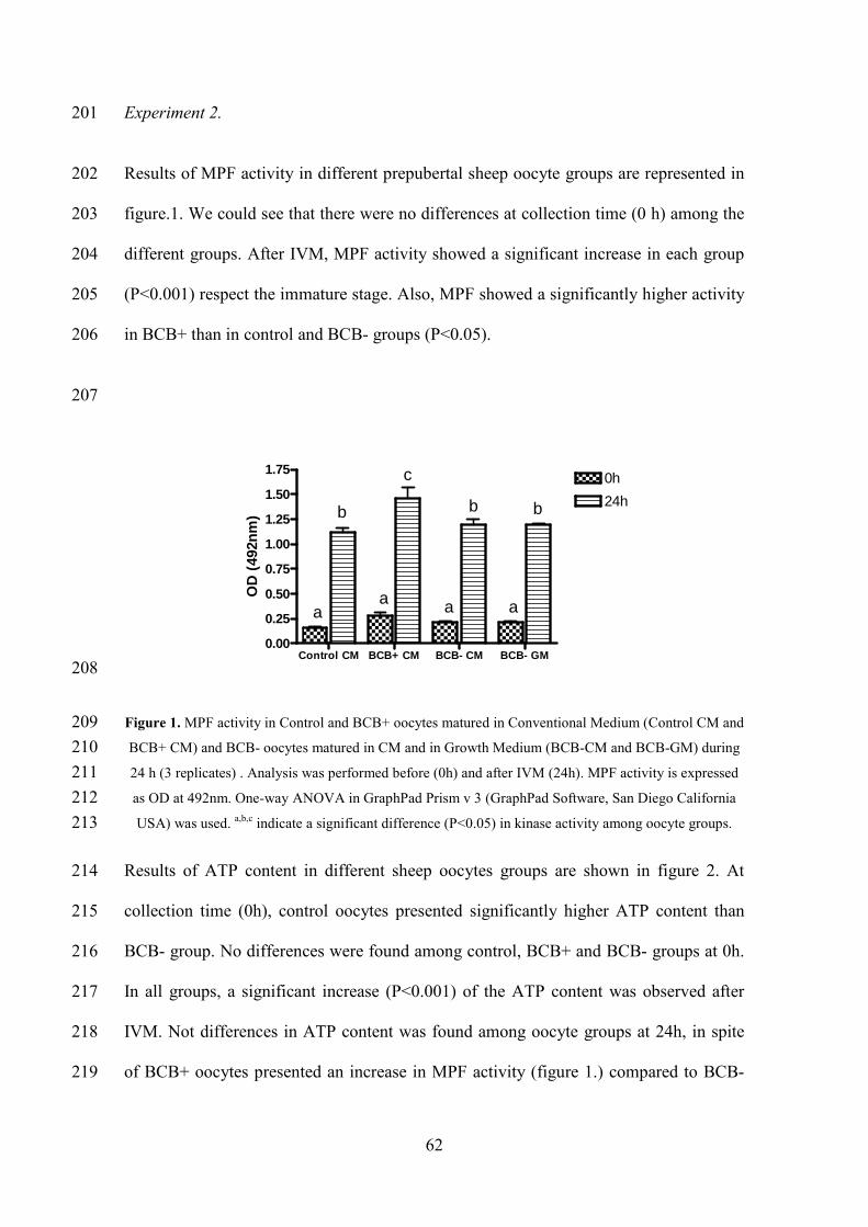
62
Experiment 2. 201
Results of MPF activity in different prepubertal sheep oocyte groups are represented in 202
figure.1. We could see that there were no differences at collection time (0 h) among the 203
different groups. After IVM, MPF activity showed a significant increase in each group 204
(P<0.001) respect the immature stage. Also, MPF showed a significantly higher activity 205
in BCB+ than in control and BCB- groups (P<0.05). 206
207
Control CM BCB+ CM BCB- CM BCB- GM0.00
0.25
0.50
0.75
1.00
1.25
1.50
1.750h
24h
aa a a
b
c
b b
OD
(49
2nm
)
208
Figure 1. MPF activity in Control and BCB+ oocytes matured in Conventional Medium (Control CM and 209
BCB+ CM) and BCB- oocytes matured in CM and in Growth Medium (BCB-CM and BCB-GM) during 210
24 h (3 replicates) . Analysis was performed before (0h) and after IVM (24h). MPF activity is expressed 211
as OD at 492nm. One-way ANOVA in GraphPad Prism v 3 (GraphPad Software, San Diego California 212
USA) was used. a,b,c
indicate a significant difference (P<0.05) in kinase activity among oocyte groups. 213
Results of ATP content in different sheep oocytes groups are shown in figure 2. At 214
collection time (0h), control oocytes presented significantly higher ATP content than 215
BCB- group. No differences were found among control, BCB+ and BCB- groups at 0h. 216
In all groups, a significant increase (P<0.001) of the ATP content was observed after 217
IVM. Not differences in ATP content was found among oocyte groups at 24h, in spite 218
of BCB+ oocytes presented an increase in MPF activity (figure 1.) compared to BCB- 219
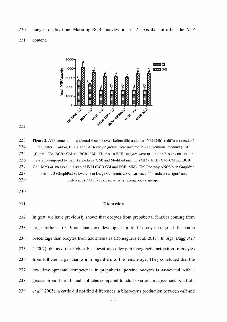
63
oocytes at this time. Maturing BCB- oocytes in 1 or 2-steps did not affect the ATP 220
content. 221
Contro
l CM
BCB+ CM
BCB- CM
BCB- GM+C
M
BCB- GM+M
M
BCB- GM
BCB- MM
0
1000
2000
3000
4000
5000
0h24h
aa,b
b
c
cc c c
c c
b b bb
fmol A
TP
/oocy
te
222
Figure 2: ATP content in prepubertal sheep oocytes before (0h) and after IVM (24h) in different media (3 223
replicates). Control, BCB+ and BCB- oocyte groups were matured in a conventional medium (CM) 224
(Control CM, BCB+ CM and BCB- CM). The rest of BCB- oocytes were matured in 2- steps maturation 225
system composed by Growth medium (GM) and Modified medium (MM) (BCB- GM+CM and BCB- 226
GM+MM) or matured in 1 step of IVM (BCB-GM and BCB- MM). GM One-way ANOVA in GraphPad 227
Prism v 3 (GraphPad Software, San Diego California USA) was used. a,b,c
indicate a significant 228
difference (P<0.05) in kinase activity among oocyte groups. 229
230
Discussion 231
In goat, we have previously shown that oocytes from prepubertal females coming from 232
large follicles (> 3mm diameter) developed up to blastocyst stage at the same 233
percentage than oocytes from adult females (Romaguera et al. 2011). In pigs, Bagg et al 234
( 2007) obtained the highest blastocyst rate after parthenogenetic activation in oocytes 235
from follicles larger than 5 mm regardless of the female age. They concluded that the 236
low developmental competence in prepubertal porcine oocytes is associated with a 237
greater proportion of small follicles compared to adult ovaries. In agreement, Kauffold 238
et al ( 2005) in cattle did not find differences in blastocysts production between calf and 239

64
cow if oocytes coming from follicles larger than 8 mm. In conclusion, we suggest that 240
the low embryo development of prepubertal female oocytes is due to high percentage of 241
unfinished growth follicle and oocyte of these ovaries. In order to increase the number 242
of in vitro blastocysts obtained from prepubertal ovaries we have tested in lamb oocytes 243
the oocyte Growth Maturation protocol described by Wu et al ( 2006) and different 244
variations of it. Previously, we had shown the utility of the BCB test as a technique to 245
discriminate larger and more competent oocytes (BCB+) from smaller and growing 246
lamb oocytes (Catalá et al. 2011). In the present study we confirmed the significantly 247
higher blastocyst yield of BCB+ oocytes (9.1%) compared to BCB- oocytes (2.3%) and 248
control group (4.3%). Different authors in cattle (Pujol et al. 2004) ), pig (Roca et al. 249
1998) and goat (Rodriguez-Gonzalez et al. 2003) have confirmed this usefulness of 250
BCB test to select the more competent oocytes. 251
In the present study we have tested in lamb BCB- oocytes the 2 steps Growth 252
Maturation protocol described by Wu et al ( 2006). We have not found significant 253
differences in blastocyst production between BCB- oocytes matured in the conventional 254
maturation medium (2.3%) and the 2 steps oocyte growth maturation medium (3.7%). 255
In calf oocytes, Cordova et al ( 2010) adding for 12 h Ascorbic Acid (AA) and ITS to 256
the IVM (without changing hormone concentration) found a significantly higher 257
blastocyst production and cytoplasmic maturation assessed by Cicline B protein, 258
cortical granule distribution and chromosome organization than control group. This lack 259
of effect in lamb oocytes in our study could be due to the highly immature BCB- 260
oocytes or to the low hormone concentrations used in GM. Thus, to enhance the 261
beneficial effect of the AA plus ITS supplementation on BCB- lamb oocytes we 262
increased the hormone concentrations to a conventional level. This new medium was 263
called Modified Medium (MM). Here again, we did not find statistically significant 264

65
differences in blastocyst rate (4.8%) but we have observed an increase compared to 265
conventional medium (2.3%). ITS and AA supplementation for 24 hours, with 266
conventional or low hormone concentrations, did not improved blastocyst rate (3.1 and 267
1.3%, respectively) and this was significantly lower than BCB+ oocytes (9.1%). Several 268
authors have shown the positive effect of ITS and AA on embryo development. In pig, 269
Jeong et al.( 2008) found a significantly increase in blastocyst production by ITS 270
addition during in vitro maturation in a chemically defined and porcine Folliclular-Fluid 271
(pFF) media. Although the best results of blastocyst production were found in pFF 272
medium. Also in buffalo, Raghu et al. ( 2002) concluded that the addition of ITS in a 273
maturation medium supplemented with gonadotrophins, EGF and serum improved 274
blastocyst production. Various physiological roles of ITS in oocyte and embryo 275
development have been postulated. Insulin stimulates the synthesis of DNA, RNA, 276
protein and lipid, and the utilization of glucose, and thereby regulates cellular functions 277
by its action on the plasma membrane, cytoskeleton, intracellular enzymes and the 278
nucleus (Harvey and Kaye. 1988; Rao et al. 1990). Transferrin, a serum globulin, acts as 279
a detoxifying protein by removing toxic metals from the medium (Barnes and Sato. 280
1980). Selenium may help to combat the oxidative stress by regulating the activity of 281
glutathione peroxidase (Stadtman. 1974; Lee et al. 2001). In our study, BCB- oocytes 282
matured in a complex medium with gonadotrophins, estradiol, EGF, serum, ITS and AA 283
not increased its embryo development competence. In these BCB- oocytes, we did not 284
find any effect of hormones concentrations on embryo development. This lack of effect 285
of maturation media on BCB- oocytes is also represented by MPF activity and ATP 286
content. In our study, MPF activity after 24 h of IVM is significantly higher in BCB+ 287
oocytes than in control and BCB- oocytes. No differences were found in BCB- oocytes 288
matured in conventional or ITS and AA supplemented media. In our previous study 289

66
(Catalá et al. 2011) we have observed higher MPF and mitochondrial activity in BCB+ 290
than in BCB- oocytes after 24 h of IVM, speculating that there could be a positive 291
relation between the ATP produced by the active mitochondria and the ATP content 292
needed to phosphorylate p34cdc2 and activate the MPF complex. In the present study 293
we observed a significantly increase in ATP content after 24 h of in vitro maturation 294
compared to oocyte at collection time (0 hours) in all of oocytes regardless of 295
maturation media and oocyte quality. However, we did not find differences among the 296
different oocyte groups. 297
Higher MPF activity has been observed in oocytes from adult females compared to 298
oocytes from prepubertal females in sheep (Ledda et al. 2001) and cattle (Salamone et 299
al. 2001). Moreover, assessing MPF activity according to oocyte diameter, Anguita et 300
al. ( 2007) showed a positive correlation among MPF activity, oocyte diameter and 301
embryo development competence in prepubertal goat oocytes. 302
Ptak et al. ( 2006) comparing oocytes from prepubertal sheep classified as small and 303
adult-size oocytes concluded that small oocytes lack both cytoplasmic and nuclear 304
competence with a fibrilogranular nucleolus in small oocytes and compact nucleolus in 305
large oocytes. Moreover blastocyst development after ICSI was 3% in small and 15% in 306
large prepubertal sheep oocytes. In adult sheep, Mohammadi-Sangcheshmeh et al ( 307
2011) observed that after IVM, BCB+ oocytes reached MII stage at a higher ratio than 308
BCB- oocytes (79 and 52%, respectively) and blastocyst development (34 and 4%, 309
respectively). In our study with lamb oocytes, the reason for the failure of BCB- oocytes 310
to increase blastocyst production after a growth-maturation system and to the ITS and 311
AA supplementation could be due to their reduced meiotic competence and abnormal 312
cytoplasmic maturation. 313
67
In conclusion, the BCB staining has allowed us to confirm the higher percentage of 314
blastocyst obtained of BCB+ oocytes and their higher MPF activity compared to BCB- 315
oocytes. However, despite changes in the maturation media, the BCB-oocytes have not 316
improved MPF activity, ATP content and embryo development up to blastocyst. These 317
results may be due to the early stage of growth of these oocytes that unable them to 318
respond to maturation supplementation with ITS and AA. 319
320
Conflicts of interest 321
The author does not have any conflict of interest to declare. 322
Author contributions 323
All authors were involved in all phases of the research and paper. 324
Acknowledgments 325
This study was funded by a grant of Ministry of Science and Innovation: AGL2011-326
23784 and Maria G. Catalá was supported by a scholarship from Universitat Autònoma 327
de Barcelona UAB-2007FI00193 328
Corresponding author. Maria-Teresa Paramio, Departament de Ciència Animal i dels 329
Aliments, Facultat de Veterinària, Universitat Autònoma de Barcelona, Bellaterra, 330
Barcelona, Spain. Phone: +34-93-5811456, Fax: +34-93-5811494. Email: 331
333

68
References 334
Alm H, Torner H, Löhrke B, Viergutz T, Ghoneim IM, Kanitz W, 2005: Bovine 335
blastocyst development rate in vitro is influenced by selection of oocytes by brillant 336
cresyl blue staining before IVM as indicator for glucose-6-phosphate dehydrogenase 337
activity. Theriogenology 63 2194-2205. 338
Anguita B, Jimenez-Macedo AR, Izquierdo D, Mogas T, Paramio MT, 2007: Effect of 339
oocyte diameter on meiotic competence, embryo development, p34 (cdc2) expression 340
and MPF activity in prepubertal goat oocytes. Theriogenology 67 526-536. 341
Bagg M, Nottle M, Armstrong D, Grupen C, 2007: Relationship between follicle size 342
and oocyte developmental competence in prepubertal and adult pigs Reproduction 343
Fertility and Development 19 797. 344
Barnes D, Sato G, 1980: Methods for growth of cultured-cells in serum-free medium. 345
Anal.Biochem. 102 255-270. 346
Barnes FL, Sirard MA, 2000: Oocyte maturation. Semin.Reprod.Med. 18 123-131. 347
Brevini TA, Vassena R, Francisci C, Gandolfi F, 2005: Role of adenosine triphosphate, 348
active mitochondria, and microtubules in the acquisition of developmental competence 349
of parthenogenetically activated pig oocytes. Biol.Reprod. 72 1218-1223. 350
Catalá M, Izquierdo D, Uzbekova S, Morato R, Roura M, Romaguera R, Papillier P, 351
Paramio T, 2011: Brilliant Cresyl Blue (BCB) stain selects largest oocytes with highest 352
mitochondria activity, MPF activity and embryo development competence in 353
prepubertal sheep. Reproduction 142 517-527. 354
Córdova B, Morató R, Izquierdo D, Paramio T, Mogas T, 2010: Effect of the addition 355
of insulin-transferrin-selenium and/or L-ascorbic acid to the in vitro maturation of 356
prepubertal bovine oocytes on cytoplasmic maturation and embryo development. 357
Theriogenology 74 1341-1348. 358
69
Gilchrist RB, Nayudu PL, Nowshari MA, Hodges JK, 1995: Meiotic competence of 359
marmoset monkey oocytes is related to follicle size and oocyte-somatic cell 360
associations. Biol.Reprod. 52 1234-1243. 361
Harvey M, Kaye P, 1988: Insulin stimulates protein synthesis in compacted mouse 362
embryos. Endocrinology 122 1182-1184. 363
Jeong Y, Hossein M, Bhandari D, Kim Y, Kim J, Park S, Lee E, Park S, Jeong Y, Lee J, 364
Kim S, Hwang W, 2008: Effects of insulin–transferrin–selenium in defined and porcine 365
follicular fluid supplemented IVM media on porcine IVF and SCNT embryo 366
production. Anim.Reprod.Sci. 106 13-24. 367
Jimenez-Macedo AR, Paramio MT, Anguita B, Morato R, Romaguera R, Mogas T, 368
Izquierdo D, 2007: Effect of ICSI and embryo biopsy on embryo development and 369
apoptosis according to oocyte diameter in prepubertal goats. Theriogenology 67 1399-370
1408. 371
Kątska-Książkiewicz L, Opiela J, Ryńska B, 2007: Effects of oocyte quality, semen 372
donor and embryo co-culture system on the efficiency of blastocyst production in goats. 373
Theriogenology 68 736-744. 374
Kauffold J, Amer H, Bergfeld U, Weber W, Sobiraj A, 2005: The in vitro 375
developmental competence of oocytes from juvenile calves is related to follicular 376
diameter. Journal of Reproduction and Development 51 325-332. 377
Ledda S, Bogliolo L, Leoni G, Naitana S, 2001: Cell coupling and maturation-378
promoting factor activity in in vitro-matured prepubertal and adult sheep oocytes. 379
Biol.Reprod. 65 247-252. 380
Lee J, Park J, Choi K, Im K, Jin D, 2001: Improvement of in vitro development of 381
bovine embryos in a medium containing selenium. Asian-Australasian Journal of 382
Animal Sciences 14 170-173. 383
Manjunatha BM, Gupta PSP, Devaraj M, Ravindra JP, Nandi S, 2007: Selection of 384
developmentally competent buffalo oocytes by brilliant cresyl blue staining before 385
IVM. Theriogenology 68 1299-1304. 386
70
Martino A, Palomo MJ, Mogas T, Paramio MT, 1994: Influence of the collection 387
technique of prepubertal goat oocytes on in vitro maturation and fertilization. 388
Theriogenology 42 859-873. 389
Mohammadi-Sangcheshmeh A, Soleimani M, Deldar H, Salehi M, Soudi S, Hashemi 390
SM, Schellander K, Hoelker M, 2011: Prediction of oocyte developmental competence 391
in ovine using glucose-6-phosphate dehydrogenase (G6PDH) activity determined at 392
retrieval time. J. Assist. Reprod. Genet . 393
Ptak G, Matsukawa K, Palmieri C, Salda LD, Scapolo PA, Loi P, 2006: Developmental 394
and functional evidence of nuclear immaturity in prepubertal oocytes. Hum.Reprod. 21 395
2228-2237. 396
Pujol M, Lopez-Bejar M, Paramio MT, 2004: Developmental competence of heifer 397
oocytes selected using the brilliant cresyl blue (BCB) test. Theriogenology 61 735-744. 398
Raghu H, Nandi S, Reddy S, 2002: Effect of insulin, transferrin and selenium and 399
epidermal growth factor on development of buffalo oocytes to the blastocyst stage in 400
vitro in serum-free, semidefined media. 151 260. 401
Rao L, Wikarczuk M, Heyner S, 1990: Functional roles of insulin and insulin-like 402
growth factors in preimplantation mouse embryo development. In Vitro Cellular & 403
Developmental Biology 26 1043-1048. 404
Roca J, Martinez E, Vazquez JM, Lucas X, 1998: Selection of immature pig oocytes for 405
homologous in vitro penetration assays with the brilliant cresyl blue test. Reproduction 406
Fertility and Development 6 479-485. 407
Rodriguez-Gonzalez E, Lopez-Bejar M, Izquierdo D, Paramio MT, 2003: 408
Developmental competence of prepubertal goat oocytes selected with brilliant cresyl 409
blue and matured with cysteamine supplementation. Reproduction Nutrition 410
Development 43 179-187. 411
Rodriguez-Gonzalez E, Lopez-Bejar M, Velilla E, Paramio MT, 2002: Selection of 412
prepubertal goat oocytes using the brilliant cresyl blue test. Theriogenology 57 1397-413
1409. 414

71
Romaguera R, Moll X, Morató R, Roura M, Palomo MJ, Catalá MG, Jiménez-Macedo 415
AR, Hammami S, Izquierdo D, Mogas T, Paramio MT, 2011: Prepubertal goat oocytes 416
from large follicles result in similar blastocyst production and embryo ploidy than those 417
from adult goats. Theriogenology 76 1-11. 418
Salamone DF, Damiani P, Fissore RA, Roble JM, Duby RT, 2001: Biochemical and 419
developmental evidence that ooplasmic maturation of prepubertal bovine oocytes is 420
compromised. Biol.Reprod. 64 1761-1768. 421
Stadtman T, 1974: Selenium biochemistry. Science 183 915-922. 422
Stojkovic M, Machado SA, Stojkovic P, Zakhartchenko V, Hutzler P, Gonïzalves PB, 423
Wolf E, 2001: Mitochondrial distribution and adenosine triphosphate content of bovine 424
oocytes before and after in vitro maturation: correlation with morphological criteria and 425
developmental capacity after in vitro fertilization and culture. Biol.Reprod. 64 904-909. 426
Wu D, Cheung QCK, Wen L, Li J, 2006: A growth-maturation system that enhances the 427
meiotic and developmental competence of porcine oocytes isolated from small follicles. 428
Biol.Reprod. 75 547-554. 429
430
72
Chapter 6
Effect of oocyte quality on blastocyst development after in vitro
fertilization (IVF) and intracytoplasmic sperm injection (ICSI) in a sheep
model.

73
Effect of oocyte quality on blastocystdevelopment after in vitrofertilization (IVF) andintracytoplasmic sperm injection(ICSI) in a sheep modelMaria G. Catal!a, M.Sc., Dolors Izquierdo, Ph.D., Maria Rodríguez-Prado, Ph.D.,Sondes Hammami, M.Sc., and Maria-Teresa Paramio, Ph.D.
Department of Animal and Food Science, Universitat Aut"onoma de Barcelona, Barcelona, Spain
Objective: To compare blastocyst production, after IVF and ICSI, from sheep oocytes of various quality. Sham-injected oocytes and parthenogeneticactivated oocyte groups were considered as control.Design: Prospective experimental study.Setting: University.Animal(s): Three- to 6-month-old sheep.Intervention(s): Oocyte quality was assessed with the use of brilliant cresyl blue (BCB) stain. Adenosine triphosphate content was measured. Intracy-toplasmic sperm injection and IVF were performed and blastocyst development and cell numbers were analyzed.Main Outcome Measure(s): Adenosine triphosphate content, embryo development and blastomere numbers.Result(s): After IVF, BCB-stained (BCBþ) oocytes developed up to the blastocyst stage at higher percentages and with more cells per embryo (24.1% vs4.0% and 69.7 vs 43.9, respectively) than unstained (BCB") oocytes. Using intracytoplasmic sperm injection, no differences were found in blastocystproduction (14.3% vs 11.8%) and number of cells per embryo (71.1 vs 54.3) between BCBþ and BCB" oocytes. Adenosine triphosphate content washigher before in vitro maturation than after in both types of oocytes. Brilliant cresyl blue–stained oocytes had more adenosine triphosphate contentthan BCB" oocytes.Conclusion(s): Brilliant cresyl blue–stained oocytes show more adenosine triphosphate content than BCB" oocytes. Results from IVF were affected bythe oocyte quality while ICSI did not produce differences in embryo development or blastomere numbers. (Fertil Steril! 2012;-:-–-."2012 by Amer-ican Society for Reproductive Medicine.)Key Words: Oocyte quality, ICSI, IVF, embryos
Intracytoplasmic sperm injection
(ICSI) is the conventional assisted
reproductive technology (ART)
used in cases of severe male factor in-
fertility, rather than in vitro fertiliza-
tion (IVF). In cases of normospermia,
total fertilization failure and low fertil-
ization (defined as <25% fertilization)
occur in 5%–15% and 20%, respec-
tively, of the couples undergoing ART
(1). Recurrent failure in ART could be
explained by low oocyte quality ex-
pressed by the lack of sperm penetra-
tion, oocyte activation failure, and
blockage of embryo development. In
a clinical context, selecting good-
quality oocytes and embryos is the
key to improving ART. Whereas the nu-
clear maturation of the oocyte is easily
identified by the appearance of the first
polar body, cytoplasmic maturation,
which is also essential for successful
fertilization and early embryo develop-
ment, remains difficult to measure. We
have previously shown using sheep
oocytes that brilliant cresyl blue (BCB)
staining promotes the selection of the
largest and most competent oocytes or
development to the blastocyst stage
with highest mitochondria and MPF
activity (2). The stain is based on the
ability of the BCB dye to be reduced
by glucose-6-phosphate dehydroge-
nase (G6PDH) activity; thus, oocytes
that have reached their growth phase
and have low G6PDH activity cannot
reduce BCB to a colorless compound
exhibiting a blue-colored cytoplasm
(BCBþ) and the growing oocytes with
high G6PDH activity are able to reduce
the blue compound, which results in
a colorless oocyte cytoplasm (BCB").
Received October 25, 2011; revised December 15, 2011; accepted December 21, 2011.M.G.C. has nothing to disclose. D.I. has nothing to disclose. M.R.-P. has nothing to disclose. S.H. has
nothing to disclose. M.-T.P. has nothing to disclose.Supported by a grant (AGL2011-23784) from the Ministry of Science and Innovation, and in part by
a scholarship (UAB-2007FI00193) from Universitat Aut"onoma de Barcelona (M.G.C.).Reprint requests: Maria-Teresa Paramio, Ph.D., Departament de Ciencia Animal i dels aliments, Facul-
tat de Veterinaria, Universitat Aut"onoma de Barcelona, 08193 Bellaterra, Barcelona, Spain(E-mail: [email protected]).
Fertility and Sterility® Vol.-, No.-,- 2012 0015-0282/$36.00Copyright ©2012 American Society for Reproductive Medicine, Published by Elsevier Inc.doi:10.1016/j.fertnstert.2011.12.043
VOL.- NO.- /- 2012 1
ORIGINAL ARTICLE: REPRODUCTIVE BIOLOGY
In conclusion, BCB staining is a good system for selection of
oocytes according to their competence.
Mitochondria are key players in cytoplasmic activity
providing, among other biochemical factors, adenosine tri-
phosphate (ATP) through oxidative phosphorylation and the
citric acid cycle. Better-quality oocytes contain significantly
higher levels of ATP and produce significantly higher blasto-
cyst rates after fertilization (3–5).
The difficulty in acquiring human oocytes for research
has hindered the development of reproductive technology.
A valuable source of oocytes for research purposes are
oocytes from animal donors. The aim of this study, therefore,
is to use sheep as a model of good (BCBþ) and low (BCB")
quality oocytes and to see how they react to ICSI and IVF pro-
cedures. In the current study, the use of sham-injected oocytes
and parthenogenetically activated (PA) oocytes eliminates the
influence of the sperm, and these procedures were also
considered as a control for mechanical and chemical
activation in selected BCB oocytes. Moreover, we analyze
oocyte ATP production, pronuclear stage, blastocyst develop-
ment, and inner cell mass and trophoectoderm cell number.
MATERIALS AND METHODSChemical
All chemicals were purchased from Sigma-Aldrich Chemical
Co (St. Louis, MO, USA) unless otherwise specified.
Oocyte Collection
Ovaries from prepubertal ewes (3–6 months old) were ob-
tained from a local abattoir and transported to the laboratory
in sterile Dulbecco’s phosphate-buffered saline (PBS) held at
34–37#C and were washed in PBS containing antibiotic-
antimycotic (AB, GIBCO cat 14240-062). Because of the small
follicle size of these ovaries, cumulus oocyte complexes were
recovered by slicing the surface of the ovary in N-2-
hydroxyethylpiperazine-N0-2-ethanesulfonic acid–buffered
TCM-199 medium with 0.5 g/L fraction V bovine serum
albumin (BSA). Oocytes with two or more complete layers
of compact cumulus cells and homogenous cytoplasm were
accepted for use.
Brilliant Cresyl Blue Test
Immediately after collection, morphologically selected cumu-
lus oocyte complexes were washed three times in modified
PBS (mPBS [PBS supplemented with 1 g/L glucose, 36 mg/L
sodium pyruvate, 0.5 g/L BSA, and 0.05 g/L gentamicin])
and exposed to 26 mM BCB diluted in mPBS for 1 hour at
38.5#C in a humidified air atmosphere. After BCB incubation,
oocytes were washed three times in mPBS and classified into
two groups depending on their cytoplasm coloration: oocytes
with blue cytoplasm or hypothetically grown oocytes (BCBþ)
and oocytes without blue cytoplasm coloration or hypotheti-
cally growing oocytes (BCB").
In Vitro Maturation of Oocytes
Groups of 35–40 cumulus oocyte complexes of BCBþ and
BCB" were washed and cultured for 24 hours at 38.5#C in
a humidified air atmosphere with 5% CO2 in 500 mL of
maturation medium covered with mineral oil. Maturation
medium consisted of TCM-199 supplemented with 5 mg/mL
LH, 5 mg/mL FSH, 1 mg/mL 17b-E2, 10 ng/mL epidermal
growth factor, 0.2 mM sodium pyruvate, 2 mM L-glutamine,
100 mM cystamine, 10% (v/v) fetal bovine serum, and 2%
(v/v) AB. After in vitro maturation, oocytes were fertilized
in vitro, injected intracytoplasmically with sperm or not
(without sperm), or activated parthenogenetically.
Adenosine-50-triphosphate Analysis
Adenosine-50-triphosphate analysis was performed using the
ATP bioluminescent somatic cell assay kit (FLASC), per the
manufacturer’s protocol. Briefly, groups of 6 BCBþ and
BCB" oocytes before (0 hours) and after (24 hours) in vitro
maturation were completely denuded by gently pipetting
and placed into an Eppendorf tube with 200 mL ultrapure
water and stored at "80#C until use.
For reaction analysis, 0.1 mL of ATP assay mix working
solution was added to a reaction vial for 3 minutes to hydro-
lyze any endogenous ATP. Then 0.05 mL of ultrapure water
and 0.05 mL of sample were added to a separate vial contain-
ing 0.1 mL of 1x somatic cell–releasing reagent. Finally
0.1 mL of this solution was transferred to the reaction vial
and the light emitted was immediately measured with
a luminometer.
Intracytoplasmic Sperm and Sham Injection
Intracytoplasmic sperm injection was performed using mor-
phologically selected thawed sperm in microdroplets of 10
mL of SOF media. Brilliant cresyl blue–stained and unstained
metaphase II (MII) oocytes were injected at the 3 o’clock posi-
tion with one spermatozoon with a minimum volume of me-
dium (<5 pL PVP). Sham injections were performed as in ICSI
excluding the spermatozoa and expelling a similar volume of
PVP into the ooplasm. Injected oocytes were immediately
activated in mPBS containing 5 mmol/L ionomycin (I24222;
Invitrogen) for 4 min. Oocytes were washed and cultured
in vitro.
In Vitro Fertilization
Highly motile spermatozoa from thawed semen were selected
by using commercial Ovipure density gradient (Nidacon EVB
S.L.) and fertilized with 1 % 106 spermatozoa/mL. Matured
oocytes were partially denuded by gentle pipetting and trans-
ferred into fertilization medium consisting of synthetic ovi-
ductal fluid (SOF) (6). Fertilization was performed in 50-mL
droplets of SOF medium supplemented with 20% of estrous
sheep serum with a maximum of 15 oocytes per drop and
incubated for 20 hours at 38.5#C in a humidified atmosphere
containing 5% CO2 and 5% O2.
Parthenogenetic Activation
Matured oocytes were denuded by gently pipetting and acti-
vated in mPBS containing 5 mmol/L ionomycin for 4 minutes.
After this initial activation, oocytes were clearly washed and
2 VOL.- NO.- /- 2012
ORIGINAL ARTICLE: REPRODUCTIVE BIOLOGY

placed in TCM-199 containing 1.9 mmol/L DMAP for 3 hours.
After PA, presumptive zygotes were cultured in vitro.
In Vitro Culture
Intracytoplasmic sperm injection, IVF, PA, and sham pre-
sumptive zygotes were completely denuded by gently
pipetting and cultured in groups of 6 zygotes for 7 days in
20 mL culture drops of SOF medium supplemented with 10%
(v/v) fetal bovine serum at the same atmosphere conditions.
Cleavage was evaluated after 24 hours of fertilization and
blastocyst development at day 7 of in vitro culture.
Assessment of Pronuclear Formation
At 17 h after initial ionomycin exposure or coincubation with
semen, presumptive zygotes were fixed in acetic acid-ethanol
(3:1) and stained with 1% lacmoid to evaluate the nuclear
stage. Zygotes were categorized as normally fertilized if one
female and one male pronuclei were formed (2 PN). Zygotes
with 3 PN were considered as polyspermic, zygotes with 1
PN or 1 PN and visible sperm head were considered activated.
Injected sham oocytes were considered activated when one or
more pronuclei were observed.
Blastocyst Differential Staining
Blastocyst differential stain protocol was adopted from
Thouas (7) and modified. Seven-day-old BCBþ and BCB"
blastocyst were first incubated for approximately 15 seconds
or until trophectoderm visibly changed color in solution with
1% Triton X-100 and 100 mg/mL propidium iodide. They were
immediately transferred to a solution with ethanol and
Hoechst 33258 for 1 hour. Stained blastocysts were trans-
ferred to a glass with a drop of glycerol, flattened with a cover
slip, and then the cells were counted under a microscope. An
Olympus BX50 was used with an ultraviolet lamp and excita-
tion filter of 460 nm for blue and red fluorescence. The intense
pink color represented the chromatin in nuclei of permeabi-
lized trophectoderm cells that are stained both red (propidium
iodide) and blue (Hoechst). Inner cell mass nuclei remained
blue, because these cells had not been permeabilized.
Statistical Analysis
For the statistical analysis all the repetitions that were done
were included in the analysis. Data was analyzed by using
the mixed procedure of SAS (version 9.1; SAS Institute Inc.).
For ATP, the model included the effects of treatment, number
of hours, and their interaction. Although for PN, the model
included only the treatment effect, and a square root–arcsine
transformation was applied, but means are presented as
back-transformed. Results are reported as least square means
and residual errors of the means. For embryo development
analysis, one-way ANOVA with Tukey’s multiple comparison
posttestwas performed and for cell counting Fisher’s exact test
was performed (GraphPad Software, San Diego, CA, USA).
RESULTS
The results of the ATP content (fmoles) analysis is shown in
Figure 1. Our findings indicate that selected BCBþ (3286.9
fmoles) oocytes produced more ATP than BCB" (2701.4
fmoles) oocytes. In addition, the ATP content in the oocytes
FIGURE 1
Adenosine triphosphate content in BCBþ and BCB" sheep oocytesbefore (0 hours) and after (24 hours) in vitro maturation. Differentletters differ significantly (mixed procedure of SAS).
Catal!a. Blastocyst production, after IVF and ICSI. Fertil Steril 2012.
TABLE 1
Nuclear stage of BCBD and BCBL oocytes after 17 hours of ICSI, IVF, PA, and sham procedures in sheep.
ICSI IVF PA Sham
P value SEMBCBD BCBL BCBD BCBL BCBD BCBL BCBD BCBL
VG 0 0 1.45 3.33 3.52 5.00 0 0 .179 1.65MII 10.14 9.22 14.95 12.90 20.18 31.51 30.99 18.33 .340 7.92PN1 0a 0a 1.45a 0a 30.93b 29.28b 52.10c 34.00b .0001 5.09PN2 43.20a 37.58a 57.61a 42.73a 31.30a 23.07a,b 11.33b 29.67a,b .002 8.76PN3 0a 1.82a 24.54b 41.04d 14.07b,c 11.14b,c 5.58a,c 18.00b,c .0002 4.94MII-H 2.22 1.82 0 0 0 0 0 0 .758 1.34PN1-H 44.44a 47.57a 0b 0b 0b 0b 0b 0b .0001 5.34PN3-H 0 2.00 0 0 0 0 0 0 .654 0.93Oocytes analyzed 57 51 57 55 56 52 57 58
Note: Values in the same row (a-c) with different letters differ significantly (mixed procedure of SAS). GV¼ germinal vesicle; MII¼Metaphase II; PN1, PN2, and PN3¼ one, two or three pronuclei;MII-H ¼ Metaphase II oocytes with non-decondense sperm head; PN1-H, PN3-H ¼ one or three pronuclei with a non-decondense sperm head; SEM ¼ standard error of the mean.
Catal!a. Blastocyst production, after IVF and ICSI. Fertil Steril 2012.
VOL.- NO.- /- 2012 3
Fertility and Sterility®
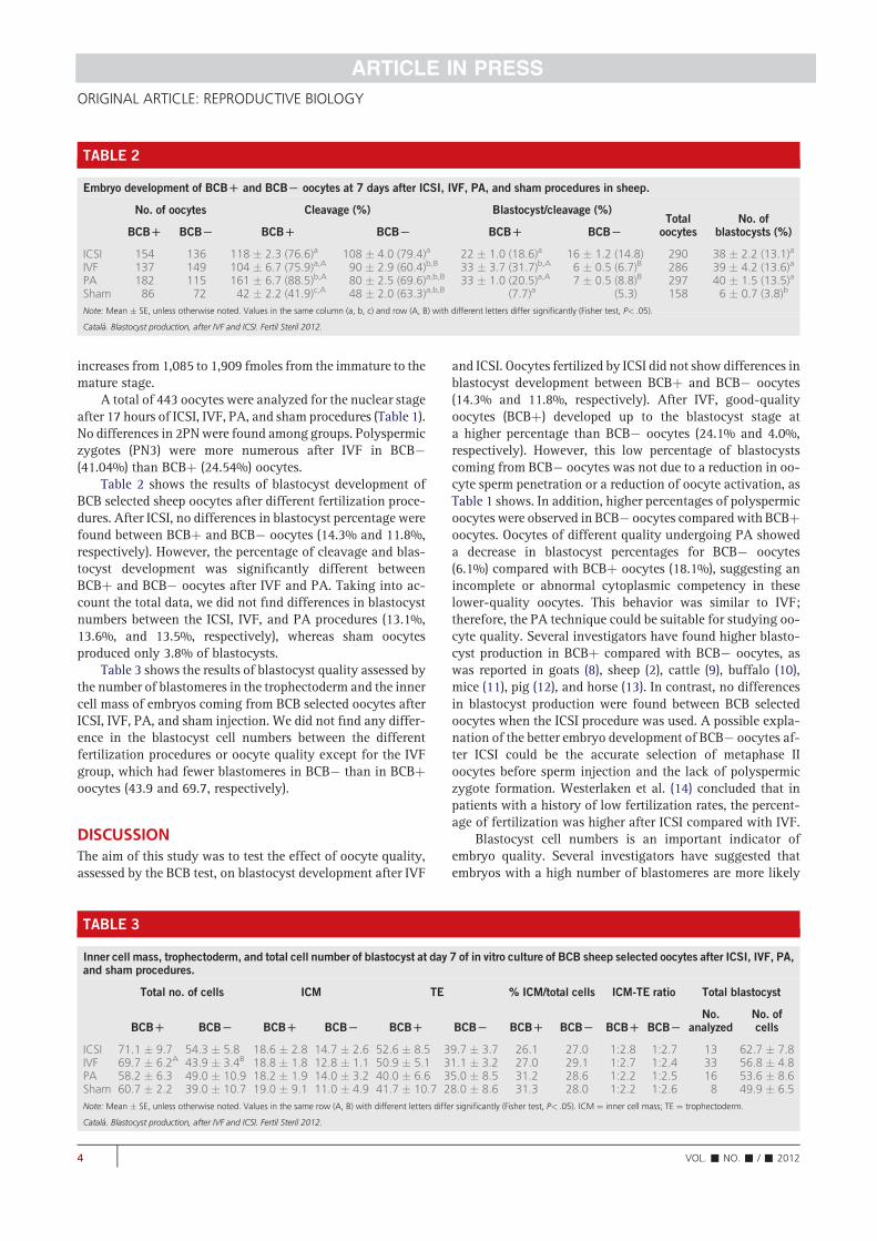
increases from 1,085 to 1,909 fmoles from the immature to the
mature stage.
A total of 443 oocytes were analyzed for the nuclear stage
after 17 hours of ICSI, IVF, PA, and sham procedures (Table 1).
No differences in 2PNwere found among groups. Polyspermic
zygotes (PN3) were more numerous after IVF in BCB"
(41.04%) than BCBþ (24.54%) oocytes.
Table 2 shows the results of blastocyst development of
BCB selected sheep oocytes after different fertilization proce-
dures. After ICSI, no differences in blastocyst percentage were
found between BCBþ and BCB" oocytes (14.3% and 11.8%,
respectively). However, the percentage of cleavage and blas-
tocyst development was significantly different between
BCBþ and BCB" oocytes after IVF and PA. Taking into ac-
count the total data, we did not find differences in blastocyst
numbers between the ICSI, IVF, and PA procedures (13.1%,
13.6%, and 13.5%, respectively), whereas sham oocytes
produced only 3.8% of blastocysts.
Table 3 shows the results of blastocyst quality assessed by
the number of blastomeres in the trophectoderm and the inner
cell mass of embryos coming from BCB selected oocytes after
ICSI, IVF, PA, and sham injection. We did not find any differ-
ence in the blastocyst cell numbers between the different
fertilization procedures or oocyte quality except for the IVF
group, which had fewer blastomeres in BCB" than in BCBþ
oocytes (43.9 and 69.7, respectively).
DISCUSSION
The aim of this study was to test the effect of oocyte quality,
assessed by the BCB test, on blastocyst development after IVF
and ICSI. Oocytes fertilized by ICSI did not show differences in
blastocyst development between BCBþ and BCB" oocytes
(14.3% and 11.8%, respectively). After IVF, good-quality
oocytes (BCBþ) developed up to the blastocyst stage at
a higher percentage than BCB" oocytes (24.1% and 4.0%,
respectively). However, this low percentage of blastocysts
coming from BCB" oocytes was not due to a reduction in oo-
cyte sperm penetration or a reduction of oocyte activation, as
Table 1 shows. In addition, higher percentages of polyspermic
oocytes were observed in BCB" oocytes compared with BCBþ
oocytes. Oocytes of different quality undergoing PA showed
a decrease in blastocyst percentages for BCB" oocytes
(6.1%) compared with BCBþ oocytes (18.1%), suggesting an
incomplete or abnormal cytoplasmic competency in these
lower-quality oocytes. This behavior was similar to IVF;
therefore, the PA technique could be suitable for studying oo-
cyte quality. Several investigators have found higher blasto-
cyst production in BCBþ compared with BCB" oocytes, as
was reported in goats (8), sheep (2), cattle (9), buffalo (10),
mice (11), pig (12), and horse (13). In contrast, no differences
in blastocyst production were found between BCB selected
oocytes when the ICSI procedure was used. A possible expla-
nation of the better embryo development of BCB" oocytes af-
ter ICSI could be the accurate selection of metaphase II
oocytes before sperm injection and the lack of polyspermic
zygote formation. Westerlaken et al. (14) concluded that in
patients with a history of low fertilization rates, the percent-
age of fertilization was higher after ICSI compared with IVF.
Blastocyst cell numbers is an important indicator of
embryo quality. Several investigators have suggested that
embryos with a high number of blastomeres are more likely
TABLE 2
Embryo development of BCBD and BCBL oocytes at 7 days after ICSI, IVF, PA, and sham procedures in sheep.
No. of oocytes Cleavage (%) Blastocyst/cleavage (%)Total
oocytesNo. of
blastocysts (%)BCBD BCBL BCBD BCBL BCBD BCBL
ICSI 154 136 118 ' 2.3 (76.6)a 108 ' 4.0 (79.4)a 22 ' 1.0 (18.6)a 16 ' 1.2 (14.8) 290 38 ' 2.2 (13.1)a
IVF 137 149 104 ' 6.7 (75.9)a,A 90 ' 2.9 (60.4)b,B 33 ' 3.7 (31.7)b,A 6 ' 0.5 (6.7)B 286 39 ' 4.2 (13.6)a
PA 182 115 161 ' 6.7 (88.5)b,A 80 ' 2.5 (69.6)a,b,B 33 ' 1.0 (20.5)a,A 7 ' 0.5 (8.8)B 297 40 ' 1.5 (13.5)a
Sham 86 72 42 ' 2.2 (41.9)c,A 48 ' 2.0 (63.3)a,b,B (7.7)a (5.3) 158 6 ' 0.7 (3.8)b
Note: Mean ' SE, unless otherwise noted. Values in the same column (a, b, c) and row (A, B) with different letters differ significantly (Fisher test, P< .05).
Catal!a. Blastocyst production, after IVF and ICSI. Fertil Steril 2012.
TABLE 3
Inner cell mass, trophectoderm, and total cell number of blastocyst at day 7 of in vitro culture of BCB sheep selected oocytes after ICSI, IVF, PA,and sham procedures.
Total no. of cells ICM TE % ICM/total cells ICM-TE ratio Total blastocyst
BCBD BCBL BCBD BCBL BCBD BCBL BCBD BCBL BCBD BCBLNo.
analyzedNo. ofcells
ICSI 71.1 ' 9.7 54.3 ' 5.8 18.6 ' 2.8 14.7 ' 2.6 52.6 ' 8.5 39.7 ' 3.7 26.1 27.0 1:2.8 1:2.7 13 62.7 ' 7.8IVF 69.7 ' 6.2A 43.9 ' 3.4B 18.8 ' 1.8 12.8 ' 1.1 50.9 ' 5.1 31.1 ' 3.2 27.0 29.1 1:2.7 1:2.4 33 56.8 ' 4.8PA 58.2 ' 6.3 49.0 ' 10.9 18.2 ' 1.9 14.0 ' 3.2 40.0 ' 6.6 35.0 ' 8.5 31.2 28.6 1:2.2 1:2.5 16 53.6 ' 8.6Sham 60.7 ' 2.2 39.0 ' 10.7 19.0 ' 9.1 11.0 ' 4.9 41.7 ' 10.7 28.0 ' 8.6 31.3 28.0 1:2.2 1:2.6 8 49.9 ' 6.5
Note: Mean ' SE, unless otherwise noted. Values in the same row (A, B) with different letters differ significantly (Fisher test, P< .05). ICM ¼ inner cell mass; TE ¼ trophectoderm.
Catal!a. Blastocyst production, after IVF and ICSI. Fertil Steril 2012.
4 VOL.- NO.- /- 2012
ORIGINAL ARTICLE: REPRODUCTIVE BIOLOGY
to implant and give rise to live offspring (15–17). In our study,
we did not find differences in cell numbers among the ICSI,
IVF, PA, and sham procedures, suggesting there are no
differences in embryo quality according to the technique
used. Westerlaken et al. (14) found no significant
differences in pregnancy rate between transfers of ICSI
embryos and transfers of a combination of one ICSI and
one IVF embryo, concluding that there is no difference in
embryo quality between the two procedures after embryo
transfer. Our results showed that IVF BCBþ oocytes
produced blastocysts with higher total cell numbers than
those produced from BCB" oocytes. Similar results were
found in cows after IVF (18) and nuclear transfer (9).
In our study, we found higher levels of ATP in BCBþ than
in BCB" oocytes. Our findings also indicate that the ATP
content increases during in vitro maturation as was previ-
ously reported in cows (5). The developmental potential of
the embryo and the outcome of IVF have been shown to be
related to both ATP and mitochondrial content in human
oocytes (3). Santos (19) showed an increase in mitochondrial
DNA (mtDNA) copy number in fertilized oocytes (250,454
copies) with respect to unfertilized oocytes (163,698 copies).
We have previously found similar results (2) in which BCBþ
oocytes contain more active mitochondria than BCB"
oocytes. These results lead us to suggest that BCB staining
is a good indicator of oocyte quality as observed in the ATP
content and IVF embryos.
In conclusion, oocyte quality, assessed by BCB staining,
significantly affects the percentage and quality of the blasto-
cysts obtained after IVF. However, this effect was not ob-
served in blastocysts from sheep oocytes after ICSI.
REFERENCES
1. McLachlan R, O’Bryan M. State of the art for genetic testing of infertile men.
J Clin Endocrinol Metab 2010;95:1013–24.
2. Catal!aM, Izquierdo D, Uzbekova S, Morato R, Roura M, Romaguera R, et al.
Brilliant cresyl blue (BCB) stain selects largest oocytes with highest mitochon-
dria activity, MPF activity and embryo development competence in prepu-
bertal sheep. Reproduction 2011;142:517–27.
3. Van Blerkom J, Davis PW, Lee J. ATP content of human oocytes and
developmental potential and outcome after in-vitro fertilization and em-
bryo-transfer. Hum Reprod 1995;10:415–24.
4. Van Blerkom J. Mitochondria in human oogenesis and preimplantation em-
bryogenesis: engines of metabolism, ionic regulation and developmental
competence. J Reprod Fertil 2004;128:269–80.
5. Stojkovic M, Machado SA, Stojkovic P, Zakhartchenko V, Hutzler P,
Gonïzalves PB, et al. Mitochondrial distribution and adenosine triphosphate
content of bovine oocytes before and after in vitro maturation: correlation
with morphological criteria and developmental capacity after in vitro fertil-
ization and culture. Biol Reprod 2001;64:904–9.
6. Holm P, Booth PJ, Schmidt MH, Greve T, Callesen H. High bovine blastocyst
development in a static in vitro production system using sofaa medium
supplemented with sodium citrate and myo-inositol with or without
serum-proteins. Theriogenology 1999;52:683–700.
7. Thouas G. Simplified technique for differential staining of inner cell mass
and trophoectoderm cells of mouse and bovine blastocyst. Reprod Biomed
Online 2001;3:25–9.
8. Katska-Ksia _zkiewicz L, Opiela J, Ry!nska B. Effects of oocyte quality, semen
donor and embryo co-culture system on the efficiency of blastocyst produc-
tion in goats. Theriogenology 2007;68:736–44.
9. Bhojwani S, Alm H, Torner H, Kanitz W, Poehland R. Selection of develop-
mentally competent oocytes through brilliant cresyl blue stain enhances
blastocyst development rate after bovine nuclear transfer. Theriogenology
2007;67:341–5.
10. Manjunatha BM, Gupta PSP, Devaraj M, Ravindra JP, Nandi S. Selection of
developmentally competent buffalo oocytes by brilliant cresyl blue staining
before IVM. Theriogenology 2007;68:1299–304.
11. Wu YG, Liu Y, Zhou P, Lan GC, Han D, Miao DQ, et al. Selection of oocytes
for in vitro maturation by brilliant cresyl blue staining: a study using the
mouse model. Cell Res 2007;17:722–31.
12. El Shourbagy SH, Spilkings EC, Freitas M, St John JC. Mitochondria directly
influence fertilisation outcome in the pig. J Reprod Fertil 2006;131:233–45.
13. Mohammadi-Sangcheshmeh A, Held E, Ghanem N, Rings F, Salilew-
Wondim D, Tesfaye D, et al. G6PDH-activity in equine oocytes correlates
with morphology, expression of candidate genes for viability, and preim-
plantative in vitro development. Theriogenology 2011;76:1215–26.
14. Westerlaken L, Helmerhorst F, Dieben S, Naaktgeboren N. Intracytoplasmic
sperm injection as a treatment for unexplained total fertilization failure or
low fertilization after conventional in vitro fertilization. Fertil Steril 2005;
83:612–7.
15. Dokras A, Sargent IL, Barlow DH. Fertilization and early embryology: human
blastocyst grading: an indicator of developmental potential? Hum Reprod
1993;8:2119–27.
16. Fouladi-Nashta A, Alberio R, Kafi M, Nicholas B, Campbell K, Webb R.
Differential staining combined with TUNEL labelling to detect apoptosis
in preimplantation bovine embryos. Reprod Biomed Online 2005;10:
497–502.
17. Holm P, Booth PJ, Callesen H. Kinetics of early in vitro development of bovine
in vivo- and in vitro-derived zygotes produced and/or cultured in chemically
defined or serum-containing media. Reproduction 2002;123:553–65.
18. Alm H, Torner H, L€ohrke B, Viergutz T, Ghoneim IM, Kanitz W. Bovine
blastocyst development rate in vitro is influenced by selection of oocytes
by brillant cresyl blue staining before IVM as indicator for glucose-6-
phosphate dehydrogenase activity. Theriogenology 2005;63:2194–205.
19. Santos T, El Shourbagy S, St John J. Mitochondrial content reflects oocyte
variability and fertilization outcome. Fertil Steril 2006;85:584–91.
VOL.- NO.- /- 2012 5
Fertility and Sterility®

79

80
Chapter 7
General discussion

81

General discussion
82
Oocytes recovered from ovaries of prepubertal and adult animals in sheep (O'Brien et al. 1996;
O'Brien et al. 1997) and cattle (Revel et al. 1995) showed that although meiotic maturation,
fertilization and cleavage rates did not differ between oocyte donor ages, the blastocyst
formation rates were significantly lower in prepubertal oocytes than in adult oocytes. Armstrong
( 2001) concluded that oocytes coming from prepubertal donors have a lower quality versus
oocytes from adult donors. In our first study using prepubertal sheep oocytes (Catalá et al. 2011)
we produced an average of 13% of blastocyst from the total oocytes fertilized, similar to
O’Brien’s et al. ( 1997) 12.6% of blastocyst with prepubertal donor. Using BCB test, oocytes
stained with 13 µM or 26 µM BCB showed an increase of blastocyst to 21% and 17%,
respectively for the BCB+ group compared to 9% and 5% of blastocyst for the BCB- group. In
this first approach using the BCB stain, the results obtained were positive and good prospects
for continuing using this technique.
Previous studies in our laboratory using prepubertal goat oocytes showed that not only the age
of the oocyte donor is crucial for embryo production, but also the size of oocyte plays an
important role in embryo production. Goat oocytes with a diameter bigger than 125 µm showed
significant higher MPF activity and blastocyst cell number after IVF (Anguita et al. 2007) and
ICSI (Jimenez-Macedo et al. 2007).
The BCB test is a non invasive methodology that allows the selection of oocytes with bigger
diameter among a heterogeneous pool. The BCB compound is used to determine the
intracellular activity of G6PDH. The activity of this enzyme gradually decreases as oocytes
reach their growth phase (Mangia and Epstein. 1975). BCB dye can be reduced by the G6PDH
enzyme activity, thus oocytes that have reached their growth phase cannot reduce BCB to a
colorless compound exhibiting a blue colored cytoplasm (BCB+). In our work with prepubertal
sheep oocytes, BCB differentiate two populations of oocytes: the BCB+ oocytes with a diameter
media of 123.66 µm producing significantly more blastocysts than oocytes BCB- with a
diameter of 106.62 µm (Catalá et al. 2011). To our knowledge this was the first time that the
BCB test was used in sheep. The BCB has been previously used in other species at different
concentrations, 13 µM BCB was the best choice in selecting pig oocytes (Egerszegi et al. 2010)
and 26 µM for cow (Pujol et al. 2004; Bhojwani et al. 2007) and goat (Rodriguez-Gonzalez et
al. 2002). In our laboratory, with our pool of oocytes and under our conditions, both
concentrations showed good results in differentiating the two population of oocytes, that is why
we used 13 µM in the first and second work and in the third work we have changed to 26 µM
BCB. The differences in using these 2 concentrations is that 13 µM seemed to be slightly more
selective but only 19% were BCB+ oocytes, while 26 µM BCB appears to be slightly less
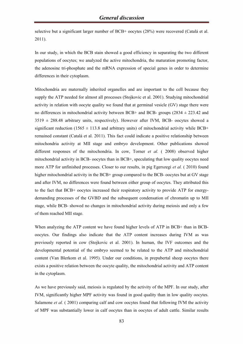
General discussion
83
selective but a significant larger number of BCB+ oocytes (28%) were recovered (Catalá et al.
2011).
In our study, in which the BCB stain showed a good efficiency in separating the two different
populations of oocytes; we analyzed the active mitochondria, the maturation promoting factor,
the adenosine tri-phosphate and the mRNA expression of special genes in order to determine
differences in their cytoplasm.
Mitochondria are maternally inherited organelles and are important to the cell because they
supply the ATP needed for almost all processes (Stojkovic et al. 2001). Studying mitochondrial
activity in relation with oocyte quality we found that at germinal vesicle (GV) stage there were
no differences in mitochondrial activity between BCB+ and BCB- groups (2834 ± 223.42 and
3519 ± 288.48 arbitrary units, respectively). However after IVM, BCB- oocytes showed a
significant reduction (1565 ± 113.8 and arbitrary units) of mitochondrial activity while BCB+
remained constant (Catalá et al. 2011). This fact could indicate a positive relationship between
mitochondria activity at MII stage and embryo development. Other publications showed
different responses of the mitochondria. In cow, Torner et al. ( 2008) observed higher
mitochondrial activity in BCB- oocytes than in BCB+, speculating that low quality oocytes need
more ATP for unfinished processes. Closer to our results, in pig Egerszegi et al. ( 2010) found
higher mitochondrial activity in the BCB+ group compared to the BCB- oocytes but at GV stage
and after IVM, no differences were found between either group of oocytes. They attributed this
to the fact that BCB+ oocytes increased their respiratory activity to provide ATP for energy-
demanding processes of the GVBD and the subsequent condensation of chromatin up to MII
stage, while BCB- showed no changes in mitochondrial activity during meiosis and only a few
of them reached MII stage.
When analyzing the ATP content we have found higher levels of ATP in BCB+ than in BCB-
oocytes. Our findings also indicate that the ATP content increases during IVM as was
previously reported in cow (Stojkovic et al. 2001). In human, the IVF outcomes and the
developmental potential of the embryo seemed to be related to the ATP and mitochondrial
content (Van Blerkom et al. 1995). Under our conditions, in prepubertal sheep oocytes there
exists a positive relation between the oocyte quality, the mitochondrial activity and ATP content
in the cytoplasm.
As we have previously said, meiosis is regulated by the activity of the MPF. In our study, after
IVM, significantly higher MPF activity was found in good quality than in low quality oocytes.
Salamone et al. ( 2001) comparing calf and cow oocytes found that following IVM the activity
of MPF was substantially lower in calf oocytes than in oocytes of adult cattle. Similar results
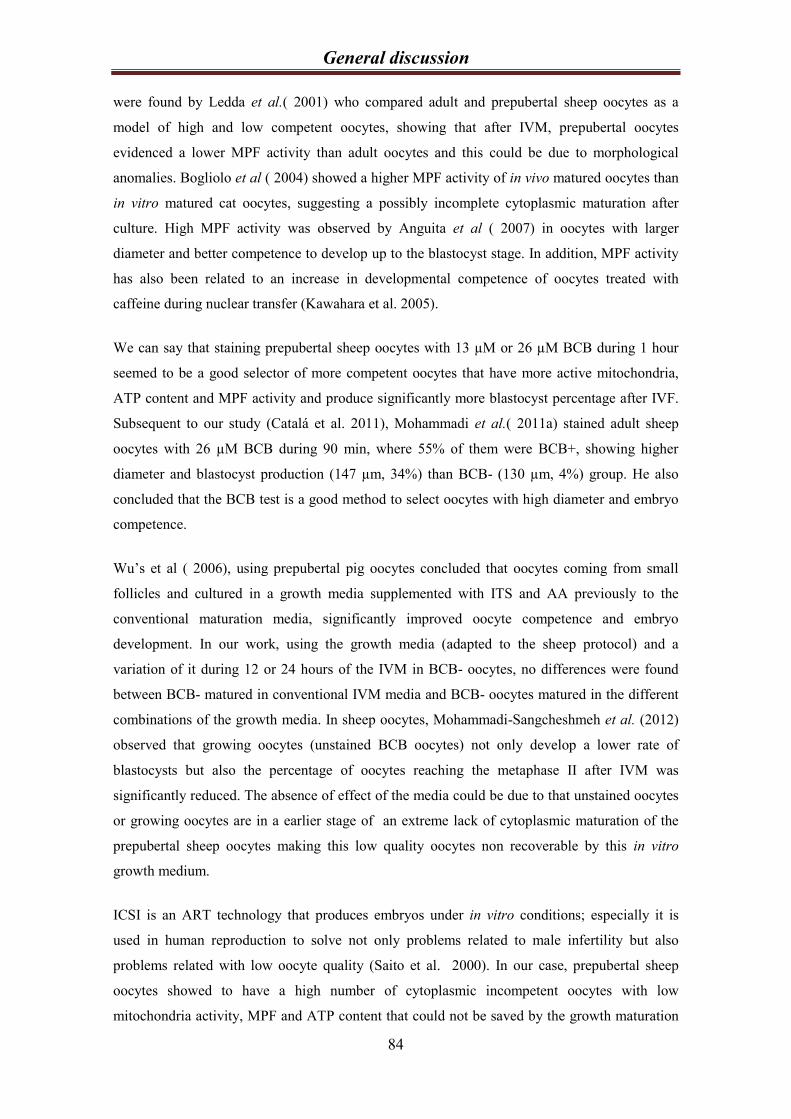
General discussion
84
were found by Ledda et al.( 2001) who compared adult and prepubertal sheep oocytes as a
model of high and low competent oocytes, showing that after IVM, prepubertal oocytes
evidenced a lower MPF activity than adult oocytes and this could be due to morphological
anomalies. Bogliolo et al ( 2004) showed a higher MPF activity of in vivo matured oocytes than
in vitro matured cat oocytes, suggesting a possibly incomplete cytoplasmic maturation after
culture. High MPF activity was observed by Anguita et al ( 2007) in oocytes with larger
diameter and better competence to develop up to the blastocyst stage. In addition, MPF activity
has also been related to an increase in developmental competence of oocytes treated with
caffeine during nuclear transfer (Kawahara et al. 2005).
We can say that staining prepubertal sheep oocytes with 13 µM or 26 µM BCB during 1 hour
seemed to be a good selector of more competent oocytes that have more active mitochondria,
ATP content and MPF activity and produce significantly more blastocyst percentage after IVF.
Subsequent to our study (Catalá et al. 2011), Mohammadi et al.( 2011a) stained adult sheep
oocytes with 26 µM BCB during 90 min, where 55% of them were BCB+, showing higher
diameter and blastocyst production (147 µm, 34%) than BCB- (130 µm, 4%) group. He also
concluded that the BCB test is a good method to select oocytes with high diameter and embryo
competence.
Wu’s et al ( 2006), using prepubertal pig oocytes concluded that oocytes coming from small
follicles and cultured in a growth media supplemented with ITS and AA previously to the
conventional maturation media, significantly improved oocyte competence and embryo
development. In our work, using the growth media (adapted to the sheep protocol) and a
variation of it during 12 or 24 hours of the IVM in BCB- oocytes, no differences were found
between BCB- matured in conventional IVM media and BCB- oocytes matured in the different
combinations of the growth media. In sheep oocytes, Mohammadi-Sangcheshmeh et al. (2012)
observed that growing oocytes (unstained BCB oocytes) not only develop a lower rate of
blastocysts but also the percentage of oocytes reaching the metaphase II after IVM was
significantly reduced. The absence of effect of the media could be due to that unstained oocytes
or growing oocytes are in a earlier stage of an extreme lack of cytoplasmic maturation of the
prepubertal sheep oocytes making this low quality oocytes non recoverable by this in vitro
growth medium.
ICSI is an ART technology that produces embryos under in vitro conditions; especially it is
used in human reproduction to solve not only problems related to male infertility but also
problems related with low oocyte quality (Saito et al. 2000). In our case, prepubertal sheep
oocytes showed to have a high number of cytoplasmic incompetent oocytes with low
mitochondria activity, MPF and ATP content that could not be saved by the growth maturation
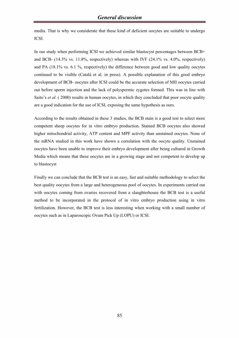
General discussion
85
media. That is why we considerate that these kind of deficient oocytes are suitable to undergo
ICSI.
In our study when performing ICSI we achieved similar blastocyst percentages between BCB+
and BCB- (14.3% vs. 11.8%, respectively) whereas with IVF (24.1% vs. 4.0%, respectively)
and PA (18.1% vs. 6.1 %, respectively) the difference between good and low quality oocytes
continued to be visible (Catalá et al. in press). A possible explanation of this good embryo
development of BCB- oocytes after ICSI could be the accurate selection of MII oocytes carried
out before sperm injection and the lack of polyspermic zygotes formed. This was in line with
Saito’s et al. ( 2000) results in human oocytes, in which they concluded that poor oocyte quality
are a good indication for the use of ICSI, exposing the same hypothesis as ours.
According to the results obtained in these 3 studies, the BCB stain is a good test to select more
competent sheep oocytes for in vitro embryo production. Stained BCB oocytes also showed
higher mitochondrial activity, ATP content and MPF activity than unstained oocytes. None of
the mRNA studied in this work have shown a correlation with the oocyte quality. Unstained
oocytes have been unable to improve their embryo development after being cultured in Growth
Media which means that these oocytes are in a growing stage and not competent to develop up
to blastocyst
Finally we can conclude that the BCB test is an easy, fast and suitable methodology to select the
best quality oocytes from a large and heterogeneous pool of oocytes. In experiments carried out
with oocytes coming from ovaries recovered from a slaughterhouse the BCB test is a useful
method to be incorporated in the protocol of in vitro embryo production using in vitro
fertilization. However, the BCB test is less interesting when working with a small number of
oocytes such as in Laparoscopic Ovum Pick Up (LOPU) or ICSI.

86
Chapter 8
Conclusions

87
Conclusions
88
1- The BCB stain is a useful methodology to select from a large and heterogeneous pool of
oocytes, the largest and most competent sheep oocytes for blastocyst production after
IVF.
2- Oocyte stained by BCB show higher mitochondrial activity, higher MPF activity and
more ATP content than unstained oocytes.
3- Unstained BCB oocytes did not improve their blastocyst production after being exposed
to a Growth Media.
4- After IVF, selected BCB oocytes develop up to blastocyst stage in significantly higher
percentage than unstained BCB oocytes. However, ICSI improve blastocyst production
in unstained oocytes without differences to BCB selected oocytes.

89

90
Chapter 9
Bibliography

91
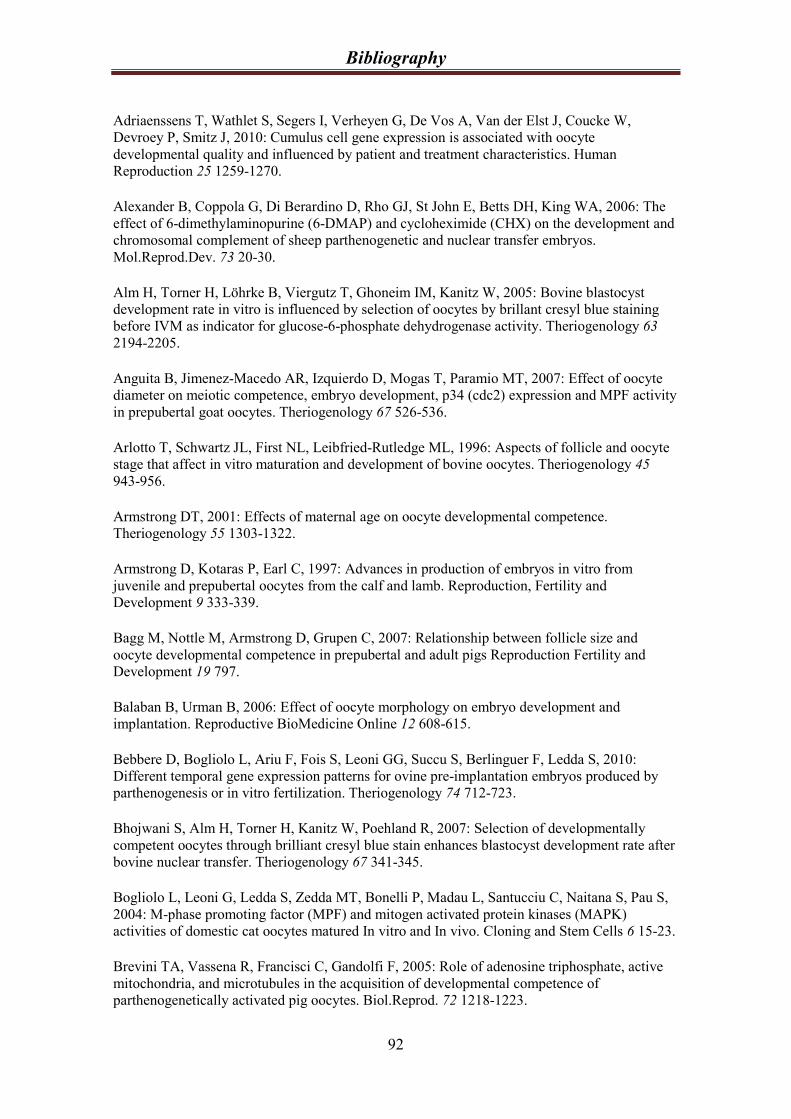
Bibliography
92
Adriaenssens T, Wathlet S, Segers I, Verheyen G, De Vos A, Van der Elst J, Coucke W,
Devroey P, Smitz J, 2010: Cumulus cell gene expression is associated with oocyte
developmental quality and influenced by patient and treatment characteristics. Human
Reproduction 25 1259-1270.
Alexander B, Coppola G, Di Berardino D, Rho GJ, St John E, Betts DH, King WA, 2006: The
effect of 6-dimethylaminopurine (6-DMAP) and cycloheximide (CHX) on the development and
chromosomal complement of sheep parthenogenetic and nuclear transfer embryos.
Mol.Reprod.Dev. 73 20-30.
Alm H, Torner H, Löhrke B, Viergutz T, Ghoneim IM, Kanitz W, 2005: Bovine blastocyst
development rate in vitro is influenced by selection of oocytes by brillant cresyl blue staining
before IVM as indicator for glucose-6-phosphate dehydrogenase activity. Theriogenology 63
2194-2205.
Anguita B, Jimenez-Macedo AR, Izquierdo D, Mogas T, Paramio MT, 2007: Effect of oocyte
diameter on meiotic competence, embryo development, p34 (cdc2) expression and MPF activity
in prepubertal goat oocytes. Theriogenology 67 526-536.
Arlotto T, Schwartz JL, First NL, Leibfried-Rutledge ML, 1996: Aspects of follicle and oocyte
stage that affect in vitro maturation and development of bovine oocytes. Theriogenology 45
943-956.
Armstrong DT, 2001: Effects of maternal age on oocyte developmental competence.
Theriogenology 55 1303-1322.
Armstrong D, Kotaras P, Earl C, 1997: Advances in production of embryos in vitro from
juvenile and prepubertal oocytes from the calf and lamb. Reproduction, Fertility and
Development 9 333-339.
Bagg M, Nottle M, Armstrong D, Grupen C, 2007: Relationship between follicle size and
oocyte developmental competence in prepubertal and adult pigs Reproduction Fertility and
Development 19 797.
Balaban B, Urman B, 2006: Effect of oocyte morphology on embryo development and
implantation. Reproductive BioMedicine Online 12 608-615.
Bebbere D, Bogliolo L, Ariu F, Fois S, Leoni GG, Succu S, Berlinguer F, Ledda S, 2010:
Different temporal gene expression patterns for ovine pre-implantation embryos produced by
parthenogenesis or in vitro fertilization. Theriogenology 74 712-723.
Bhojwani S, Alm H, Torner H, Kanitz W, Poehland R, 2007: Selection of developmentally
competent oocytes through brilliant cresyl blue stain enhances blastocyst development rate after
bovine nuclear transfer. Theriogenology 67 341-345.
Bogliolo L, Leoni G, Ledda S, Zedda MT, Bonelli P, Madau L, Santucciu C, Naitana S, Pau S,
2004: M-phase promoting factor (MPF) and mitogen activated protein kinases (MAPK)
activities of domestic cat oocytes matured In vitro and In vivo. Cloning and Stem Cells 6 15-23.
Brevini TA, Vassena R, Francisci C, Gandolfi F, 2005: Role of adenosine triphosphate, active
mitochondria, and microtubules in the acquisition of developmental competence of
parthenogenetically activated pig oocytes. Biol.Reprod. 72 1218-1223.

Bibliography
93
Brevini-Gandolfi TAL, Gandolfi F, 2001: The maternal legacy to the embryo: cytoplasmic
components and their effects on early development. Theriogenology 55 1255-1276.
Cai C, Tamai K, Molyneaux K, 2010: KHDC1B is a novel CPEB binding partner specifically
expressed in mouse oocytes and early embryos. Molecular Biology of the Cell 21 3137-3148.
Caixeta ES, Ripamonte P, Franco MM, Buratini J, Dode MAN, 2009: Effect of follicle size on
mRNA expression in cumulus cells and oocytes of Bos indicus: an approach to identify marker
genes for developmental competence. Reproduction Fertility and Development 21 655-664.
Campbell BK, Scaramuzzi RJ, Webb R, 1995: Control of antral follicle development and
selection in sheep and cattle. J.Reprod.Fertil. 335-350.
Carolan C, Lonergan P, Van Langendonckt A, Mermillod P, 1995: Factors affecting bovine
embryo development in synthetic oviduct fluid following oocyte maturation and fertilization in
vitro. Theriogenology 43 1115-1128.
Catalá MG, Izquierdo D, Rodríguez-Prado M, Hammami S, Paramio MT, in press: Effect of
oocyte quality on blastocyst development after in vitro fertilization (IVF) and intracytoplasmic
sperm injection (ICSI) in a sheep model. Fertil.Steril. .
Catalá M, Izquierdo D, Uzbekova S, Morato R, Roura M, Romaguera R, Papillier P, Paramio T,
2011: Brilliant Cresyl Blue (BCB) stain selects largest oocytes with highest mitochondria
activity, MPF activity and embryo development competence in prepubertal sheep. Reproduction
142 517-527.
Catt SL, Catt JW, Gomez MC, Maxwell WM, Evans G, 1996: Birth of a male lamb derived
from an in vitro matured oocyte fertilised by intracytoplasmic injection of a single presumptive
male sperm. Veterinary Reserch 139 494-495.
Channing C, Tsalv V, Sachs D, 1976: Role of insulin, thyroxine and cortisol in luteinization of
porcine granullosa-cells grown in chemically defined media. Biol.Reprod. 15 235-247.
Cognie Y, Benoit F, Poulin N, Khatir H, Driancourt M, 1998: Effect of follicle size and of the
Fec(B) Booroola gene on oocyte function in sheep. J.Reprod.Fertil. 112 379-386.
Córdova B, Morató R, Izquierdo D, Paramio T, Mogas T, 2010: Effect of the addition of
insulin-transferrin-selenium and/or L-ascorbic acid to the in vitro maturation of prepubertal
bovine oocytes on cytoplasmic maturation and embryo development. Theriogenology 74 1341-
1348.
Crichton RR, Charloteaux-Wauters M, 1987: Iron transport and storage. European Journal of
Biochemistry 164 485-506.
Crozet N, Dahirel M, Gall L, 2000: Meiotic competence of in vitro grown goat oocytes. Journal
Reproduction and Fertility 118 367-373.
Crozet N, Motlij J, Szollosi D, 1981: Nucleolar fine-structure and RNA-synthesis in porcine
oocytes during the early stages of antrum formation. Biology of the Cell 41 35-41.
Dawson A, Griesinger G, Diedrich K, 2006: Screening oocytes by polar body biopsy.
Reproductive BioMedicine Online 13 104-109.

Bibliography
94
de Matos DG, Gasparrini B, Pasqualini SR, Thompson JG, 2002: Effect of glutathione synthesis
stimulation during in vitro maturation of ovine oocytes on embryo development and
intracellular peroxide content. Theriogenology 57 1443-1451.
Dell'Aquila ME, Ambruosi B, De santis T, Cho YS, 2009: Mitochondrial distribution and
activity in human mature oocytes: gonadotropin-releasing hormone agonist versus antagonist
for pituitary down-regulation. Fertility and Sterility 91 249-255.
Duby RT, Damiani P, Looney CR, Fissore RA, Robl JM, 1996: Prepuberal calves as oocyte
donors: promises and problems. 45 121.
Duleba A, Pehlivan T, Carbone R, Spaczynski R, 2001: Activin stimulates proliferation of rat
ovarian thecal-interstitial cells. Biol.Reprod. 65 704-709.
Egerszegi I, Alm H, Ratky J, Heleil B, Bruessow K, Torner H, 2010: Meiotic progression,
mitochondrial features and fertilisation characteristics of porcine oocytes with different G6PDH
activities. Reproduction Fertility and Development 22 830-838.
El Shourbagy SH, Spilkings EC, Freitas M, St John JC, 2006: Mitochondria directly influence
fertilisation outcome in the pig. J.Reprod.Fertil. 131 233-245.
Eppig J, Hosoe M, O'Brien M, Pendola F, Requena A, Watanabe S, 2000: Conditions that affect
acquisition of developmental competence by mouse oocytes in vitro: FSH, insulin, glucose and
ascorbic acid. Molecular and Cellular Endocrinology 163 109-116.
Eppig JJ, Schultz RM, O'Brien M, Chesnel F, 1994: Relationship between the Developmental
Programs Controlling Nuclear and Cytoplasmic Maturation of Mouse Oocytes. Dev.Biol. 164 1-
9.
Ericsson SA, Boice ML, Funahashi H, Day BN, 1993: Assessment of porcine oocytes using
brilliant cresyl blue. Theriogenology 39 214.
Fair T, Hyttel P, Greve T, 1995: Bovine oocyte diameter in relation to maturational competence
and transcriptional activity. Mol.Reprod.Dev. 42 437-442.
Fouladi-Nashta AA, Gutierrez CG, Gong JG, Garnsworthy PC, Webb R, 2007: Impact of
Dietary Fatty Acids on Oocyte Quality and Development in Lactating Dairy Cows. Biology of
Reproduction 77 9-17.
Gandolfi F, Milanesi E, Pocar P, Luciano AM, Brevini TAL, Acocella F, Lauria A, Armstrong
DT, 1998: Comparative analysis of calf and cow oocytes during in vitro maturation.
Mol.Reprod.Dev. 49 168-175.
Ghasemzadeh Nava H, Tajik P, 2000: In vitro maturation of ovine follicular oocytes in different
concentrations of fetal calf serum and estrous sheep serum. Theriogenology 53 453.
Gomez MC, Catt JW, Evans G, Maxwell WM, 1998: Cleavage, development and competence
of sheep embryos fertilized by intracytoplasmic sperm injection and in vitro. Theriogenology 49
1143-1154.
Grazul-Bilska AT, Vonnahme KA, Bilski JJ, Borowczyk E, Soni D, Mikkelson B, Johnson ML,
Reynolds LP, Redmer DA, Caton JS, 2011: Expression of gap junctional connexin proteins in
ovine fetal ovaries: Effects of maternal diet. Domest.Anim.Endocrinol. 41 185-194.
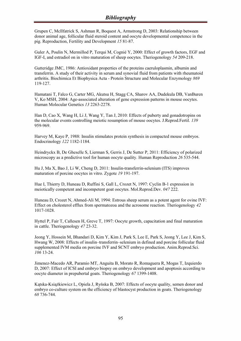
Bibliography
95
Grupen C, McIlfatrick S, Ashman R, Boquest A, Armstrong D, 2003: Relationship between
donor animal age, follicular fluid steroid content and oocyte developmental competence in the
pig. Reproduction, Fertility and Development 15 81-87.
Guler A, Poulin N, Mermillod P, Terqui M, Cognié Y, 2000: Effect of growth factors, EGF and
IGF-I, and estradiol on in vitro maturation of sheep oocytes. Theriogenology 54 209-218.
Gutteridge JMC, 1986: Antioxidant properties of the proteins caeruloplasmin, albumin and
transferrin. A study of their activity in serum and synovial fluid from patients with rheumatoid
arthritis. Biochimica Et Biophysica Acta - Protein Structure and Molecular Enzymology 869
119-127.
Hamatani T, Falco G, Carter MG, Akutsu H, Stagg CA, Sharov AA, Dudekula DB, VanBuren
V, Ko MSH, 2004: Age-associated alteration of gene expression patterns in mouse oocytes.
Human Molecular Genetics 13 2263-2278.
Han D, Cao X, Wang H, Li J, Wang Y, Tan J, 2010: Effects of puberty and gonadotropins on
the molecular events controlling meiotic resumption of mouse oocytes. J.Reprod.Fertil. 139
959-969.
Harvey M, Kaye P, 1988: Insulin stimulates protein synthesis in compacted mouse embryos.
Endocrinology 122 1182-1184.
Heindryckx B, De Gheselle S, Lierman S, Gerris J, De Sutter P, 2011: Efficiency of polarized
microscopy as a predictive tool for human oocyte quality. Human Reproduction 26 535-544.
Hu J, Ma X, Bao J, Li W, Cheng D, 2011: Insulin-transferrin-selenium (ITS) improves
maturation of porcine oocytes in vitro. Zygote 19 191-197.
Hue I, Thierry D, Huneau D, Ruffini S, Gall L, Crozet N, 1997: Cyclin B-1 expression in
meiotically competent and incompetent goat oocytes. Mol.Reprod.Dev. 047 222.
Huneau D, Crozet N, Ahmed-Ali M, 1994: Estrous sheep serum as a potent agent for ovine IVF:
Effect on cholesterol efflux from spermatozoa and the acrosome reaction. Theriogenology 42
1017-1028.
Hyttel P, Fair T, Callesen H, Greve T, 1997: Oocyte growth, capacitation and final maturation
in cattle. Theriogenology 47 23-32.
Jeong Y, Hossein M, Bhandari D, Kim Y, Kim J, Park S, Lee E, Park S, Jeong Y, Lee J, Kim S,
Hwang W, 2008: Effects of insulin–transferrin–selenium in defined and porcine follicular fluid
supplemented IVM media on porcine IVF and SCNT embryo production. Anim.Reprod.Sci.
106 13-24.
Jimenez-Macedo AR, Paramio MT, Anguita B, Morato R, Romaguera R, Mogas T, Izquierdo
D, 2007: Effect of ICSI and embryo biopsy on embryo development and apoptosis according to
oocyte diameter in prepubertal goats. Theriogenology 67 1399-1408.
Kątska-Książkiewicz L, Opiela J, Ryńska B, 2007: Effects of oocyte quality, semen donor and
embryo co-culture system on the efficiency of blastocyst production in goats. Theriogenology
68 736-744.
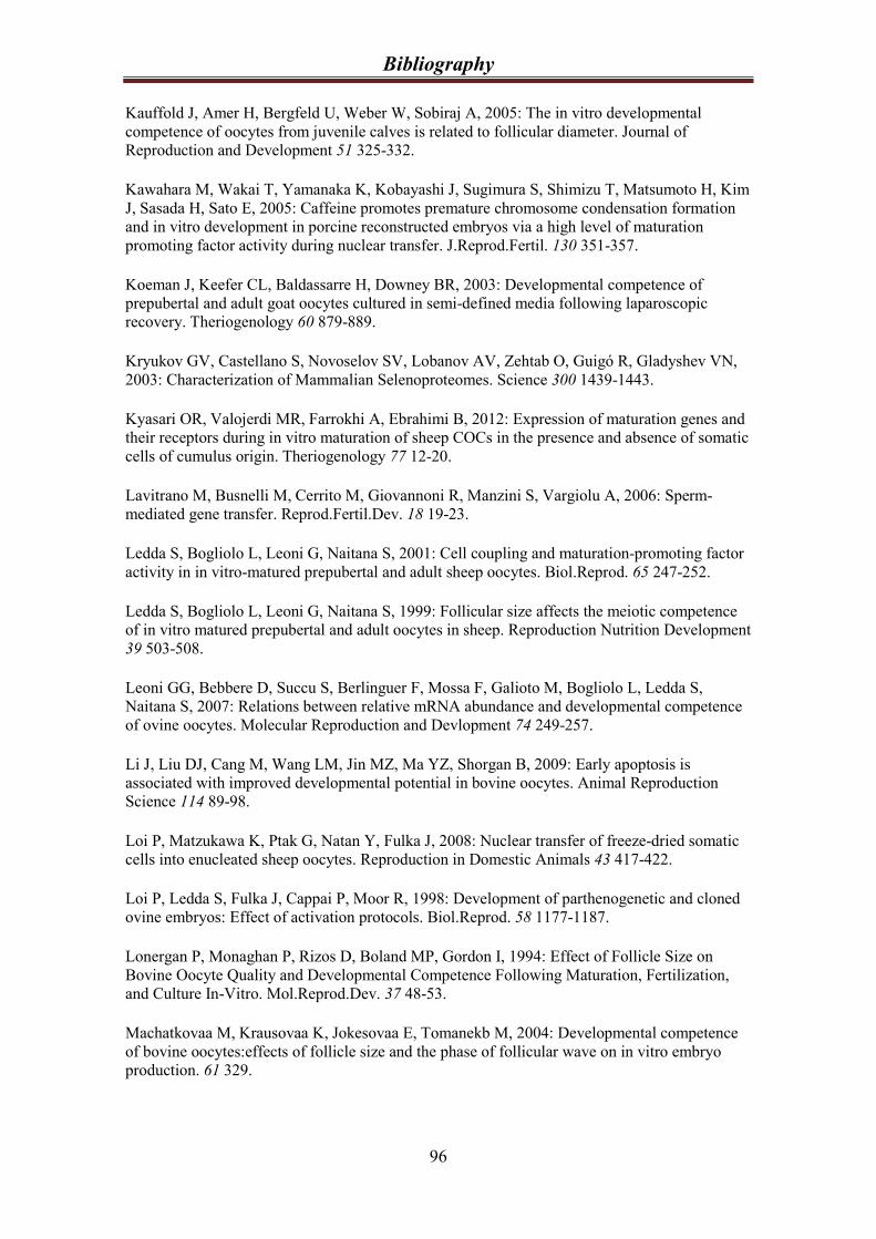
Bibliography
96
Kauffold J, Amer H, Bergfeld U, Weber W, Sobiraj A, 2005: The in vitro developmental
competence of oocytes from juvenile calves is related to follicular diameter. Journal of
Reproduction and Development 51 325-332.
Kawahara M, Wakai T, Yamanaka K, Kobayashi J, Sugimura S, Shimizu T, Matsumoto H, Kim
J, Sasada H, Sato E, 2005: Caffeine promotes premature chromosome condensation formation
and in vitro development in porcine reconstructed embryos via a high level of maturation
promoting factor activity during nuclear transfer. J.Reprod.Fertil. 130 351-357.
Koeman J, Keefer CL, Baldassarre H, Downey BR, 2003: Developmental competence of
prepubertal and adult goat oocytes cultured in semi-defined media following laparoscopic
recovery. Theriogenology 60 879-889.
Kryukov GV, Castellano S, Novoselov SV, Lobanov AV, Zehtab O, Guigó R, Gladyshev VN,
2003: Characterization of Mammalian Selenoproteomes. Science 300 1439-1443.
Kyasari OR, Valojerdi MR, Farrokhi A, Ebrahimi B, 2012: Expression of maturation genes and
their receptors during in vitro maturation of sheep COCs in the presence and absence of somatic
cells of cumulus origin. Theriogenology 77 12-20.
Lavitrano M, Busnelli M, Cerrito M, Giovannoni R, Manzini S, Vargiolu A, 2006: Sperm-
mediated gene transfer. Reprod.Fertil.Dev. 18 19-23.
Ledda S, Bogliolo L, Leoni G, Naitana S, 2001: Cell coupling and maturation-promoting factor
activity in in vitro-matured prepubertal and adult sheep oocytes. Biol.Reprod. 65 247-252.
Ledda S, Bogliolo L, Leoni G, Naitana S, 1999: Follicular size affects the meiotic competence
of in vitro matured prepubertal and adult oocytes in sheep. Reproduction Nutrition Development
39 503-508.
Leoni GG, Bebbere D, Succu S, Berlinguer F, Mossa F, Galioto M, Bogliolo L, Ledda S,
Naitana S, 2007: Relations between relative mRNA abundance and developmental competence
of ovine oocytes. Molecular Reproduction and Devlopment 74 249-257.
Li J, Liu DJ, Cang M, Wang LM, Jin MZ, Ma YZ, Shorgan B, 2009: Early apoptosis is
associated with improved developmental potential in bovine oocytes. Animal Reproduction
Science 114 89-98.
Loi P, Matzukawa K, Ptak G, Natan Y, Fulka J, 2008: Nuclear transfer of freeze-dried somatic
cells into enucleated sheep oocytes. Reproduction in Domestic Animals 43 417-422.
Loi P, Ledda S, Fulka J, Cappai P, Moor R, 1998: Development of parthenogenetic and cloned
ovine embryos: Effect of activation protocols. Biol.Reprod. 58 1177-1187.
Lonergan P, Monaghan P, Rizos D, Boland MP, Gordon I, 1994: Effect of Follicle Size on
Bovine Oocyte Quality and Developmental Competence Following Maturation, Fertilization,
and Culture In-Vitro. Mol.Reprod.Dev. 37 48-53.
Machatkovaa M, Krausovaa K, Jokesovaa E, Tomanekb M, 2004: Developmental competence
of bovine oocytes:effects of follicle size and the phase of follicular wave on in vitro embryo
production. 61 329.
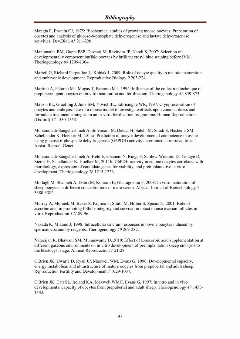
Bibliography
97
Mangia F, Epstein CJ, 1975: Biochemical studies of growing mouse oocytes: Preparation of
oocytes and analysis of glucose-6-phosphate dehydrogenase and lactate dehydrogenase
activities. Dev.Biol. 45 211-220.
Manjunatha BM, Gupta PSP, Devaraj M, Ravindra JP, Nandi S, 2007: Selection of
developmentally competent buffalo oocytes by brilliant cresyl blue staining before IVM.
Theriogenology 68 1299-1304.
Marteil G, Richard Parpaillon L, Kubiak J, 2009: Role of oocyte quality in meiotic maturation
and embryonic development. Reproductive Biology 9 203-224.
Martino A, Palomo MJ, Mogas T, Paramio MT, 1994: Influence of the collection technique of
prepubertal goat oocytes on in vitro maturation and fertilization. Theriogenology 42 859-873.
Matson PL, Graefling J, Junk SM, Yovich JL, Edirisinghe WR, 1997: Cryopreservation of
oocytes and embryos: Use of a mouse model to investigate effects upon zona hardness and
formulate treatment strategies in an in-vitro fertilization programme. Human Reproduction
(Oxford) 12 1550-1553.
Mohammadi-Sangcheshmeh A, Soleimani M, Deldar H, Salehi M, Soudi S, Hashemi SM,
Schellander K, Hoelker M, 2011a: Prediction of oocyte developmental competence in ovine
using glucose-6-phosphate dehydrogenase (G6PDH) activity determined at retrieval time. J.
Assist. Reprod. Genet .
Mohammadi-Sangcheshmeh A, Held E, Ghanem N, Rings F, Salilew-Wondim D, Tesfaye D,
Sieme H, Schellander K, Hoelker M, 2011b: G6PDH-activity in equine oocytes correlates with
morphology, expression of candidate genes for viability, and preimplantative in vitro
development. Theriogenology 76 1215-1226.
Motlagh M, Shahneh A, Daliri M, Kohram H, Gharagozlou F, 2008: In vitro maturation of
sheep oocytes in different concentrations of mare serum. African Journal of Biotechnology 7
3380-3382.
Murray A, Molinek M, Baker S, Kojima F, Smith M, Hillier S, Spears N, 2001: Role of
ascorbic acid in promoting follicle integrity and survival in intact mouse ovarian follicles in
vitro. Reproduction 121 89-96.
Nakada K, Mizuno J, 1998: Intracellular calcium responses in bovine oocytes induced by
spermatozoa and by reagents. Theriogenology 50 269-282.
Natarajan R, Bhawani SM, Munuswamy D, 2010: Effect of L-ascorbic acid supplementation at
different gaseous environments on in vitro development of preimplantation sheep embryos to
the blastocyst stage. Animal Reproduction 7 21-28.
O'Brien JK, Dwarte D, Ryan JP, Maxwell WM, Evans G, 1996: Developmental capacity,
energy metabolism and ultrastructure of mature oocytes from prepubertal and adult sheep.
Reproduction Fertility and Development 7 1029-1037.
O'Brien JK, Catt SL, Ireland KA, Maxwell WMC, Evans G, 1997: In vitro and in vivo
developmental capacity of oocytes from prepubertal and adult sheep. Theriogenology 47 1433-
1443.
Bibliography
98
Opiela J, Lipinski D, Slomski R, Katska-Ksiazkiewicz L, 2010: Transcript expression of
mitochondria related genes is correlated with bovine oocyte selection by BCB test. Animal
Reproduction Science 118 188-193.
Ortavant R, Thibault CH, Wintenberger S, 1949: Experimental superovulation in the sheep. Ann
Endocrinol 10 170-173.
Otoi T, Yamamoto K, Koyama N, Tachikawa S, Suzuki T, 1997: Bovine oocyte diameter in
relation to developmental competence. Theriogenology 48 769-774.
Patel OV, Bettegowda A, Ireland JJ, Coussens PM, Lonergan P, Smith GW, 2007: Functional
genomics studies of oocyte competence: evidence that reduced transcript abundance for
follistatin is associated with poor developmental competence of bovine oocytes. Reproduction
133 95-106.
Patrizio P, Fragouli E, Bianchi V, Borini A, Wells D, 2007: Molecular methods for selection of
the ideal oocyte. Reproductive BioMedicine Online 15 346-353.
Pereyra-Bonnet F, ibbons A, ueto M, ipowicz P, ernandez-Martin R, Salamone D, 2011:
Efficiency of Sperm-Mediated Gene Transfer in the Ovine by Laparoscopic Insemination, In
Vitro Fertilization and ICSI. Journal of Reproduction and Development .
Pisani LF, Antonini S, Pocar P, Ferrari S, Brevini TA, Rhind SM, Gandolfi F, 2008: Effects of
pre-mating nutrition on mRNA levels of developmentally relevant genes in sheep oocytes and
granulosa cells. J.Reprod.Fertil. 136 303-312.
Pope CE, Gomez MC, Kagawa N, Kuwayama M, Leibo SP, Dresser BL, 2012: In vivo survival
of domestic cat oocytes after vitrification, intracytoplasmic sperm injection and embryo transfer.
Theriogenology 77 531-538.
Pujol M, Lopez-Bejar M, Paramio MT, 2004: Developmental competence of heifer oocytes
selected using the brilliant cresyl blue (BCB) test. Theriogenology 61 735-744.
Quirke JF, Hanrahan JP, 1977: Comparison of the survival in the uteri of adult ewes of cleaved
ova from adult ewes and ewe lambs. Journal of Reproduction and Fertility 51 487-489.
Raghu HM, Nandi S, Reddy SM, 2002a: Follicle size and oocyte diameter in relation to
developmental competence of buffalo oocytes in vitro. Reproduction Fertility and Development
14 55-61.
Raghu H, Nandi S, Reddy S, 2002b: Effect of insulin, transferrin and selenium and epidermal
growth factor on development of buffalo oocytes to the blastocyst stage in vitro in serum-free,
semidefined media. 151 260.
Rao BS, Naidu KS, Amarnath D, Vagdevi R, Rao AS, Brahmaiah KV, Rao VH, 2002: In vitro
maturation of sheep oocytes in different media during breeding and non-breeding seasons.
Small Ruminant Research 43 31-36.
Rao L, Wikarczuk M, Heyner S, 1990: Functional roles of insulin and insulin-like growth
factors in preimplantation mouse embryo development. In Vitro Cellular & Developmental
Biology 26 1043-1048.

Bibliography
99
Revel F, Mermillod P, Peynot N, Renard JP, Heyman Y, 1995: Low developmental capacity of
in vitro matured and fertilized oocytes from calves compared with that of cows. Journal
Reproduction and Fertility 103 115-120.
Rizos D, Burke L, Duffy P, Wade M, Mee JF, O’Farrell KJ, MacSiurtain M, Boland MP,
Lonergan P, 2005: Comparisons between nulliparous heifers and cows as oocyte donors for
embryo production in vitro. Theriogenology 63 939-949.
Roca J, Martinez E, Vazquez JM, Lucas X, 1998: Selection of immature pig oocytes for
homologous in vitro penetration assays with the brilliant cresyl blue test. Reproduction Fertility
and Development 6 479-485.
Rodriguez-Gonzalez E, Lopez-Bejar M, Izquierdo D, Paramio MT, 2003: Developmental
competence of prepubertal goat oocytes selected with brilliant cresyl blue and matured with
cysteamine supplementation. Reproduction Nutrition Development 43 179-187.
Rodriguez-Gonzalez E, Lopez-Bejar M, Velilla E, Paramio MT, 2002: Selection of prepubertal
goat oocytes using the brilliant cresyl blue test. Theriogenology 57 1397-1409.
Romaguera R, Moll X, Morató R, Roura M, Palomo MJ, Catalá MG, Jiménez-Macedo AR,
Hammami S, Izquierdo D, Mogas T, Paramio MT, 2011: Prepubertal goat oocytes from large
follicles result in similar blastocyst production and embryo ploidy than those from adult goats.
Theriogenology 76 1-11.
Romaguera R, Casanovas A, Morató R, Izquierdo D, Jimenez-Macedo AR, Mogas T, Paramio
MT, 2010: Effect of follicle diameter on oocyte apoptosis, embryo development and
chromosomal ploidy in prepubertal goats. Theriogenology 74 364-373.
Saadeldin IM, Kim B, Lee BC, Jang G, 2011: Effect of different culture media on the temporal
gene expression in the bovine developing embryos. Theriogenology 75 995-1004.
Saito H, Saito T, Kaneko T, Sasagawa I, Kuramoto T, Hiroi M, 2000: Relatively poor oocyte
quality is an indication for intracytoplasmic sperm injection. Fertil.Steril. 73 465-469.
Salamone DF, Damiani P, Fissore RA, Roble JM, Duby RT, 2001: Biochemical and
developmental evidence that ooplasmic maturation of prepubertal bovine oocytes is
compromised. Biol.Reprod. 64 1761-1768.
Salhab M, Tosca L, Cabau C, Papillier P, Perreau C, Dupont J, Mermillod P, Uzbekova S, 2011:
Kinetics of gene expression and signaling in bovine cumulus cells throughout IVM in different
mediums in relation to oocyte developmental competence, cumulus apoptosis and progesterone
secretion. Theriogenology 1 90-104.
Sartori R, Sartor-Bergfelt R, Mertens SA, Guenther JN, Parrish JJ, Wiltbank MC, 2002:
Fertilization and Early Embryonic Development in Heifers and Lactating Cows in Summer and
Lactating and Dry Cows in Winter. 85 2803.
Shirazi A, Ostad-Hosseini S, Ahmadi E, Heidari B, Shams-Esfandabadi N, 2009: In vitro
developmental competence of ICSI-derived activated ovine embryos. Theriogenology 71 342-
348.
Sirard M, Blondin P, 1996: Oocyte maturation and IVF in cattle. Anim.Reprod.Sci. 42 417-426.

Bibliography
100
Sirard MA, 2011: Follicle environment and quality of in vitro matured oocytes.
J.Assist.Reprod.Genet. 28 483-488.
Sirard MA, Richard F, Blondin P, Robert C, 2006: Contribution of the oocyte to embryo
quality. Theriogenology 65 126-136.
Slavik T, Fulka J, Goll I, 1992: Pregnancy rate after the transfer of sheep embryos originated
from randomly chosen oocytes matured and fertilized in vitro. Theriogenology 38 749-756.
Snijders SEM, Dillon P, O'Callaghan D, Boland MP, 2000: Effect of genetic merit, milk yield,
body condition and lactation number on in vitro oocyte development in dairy cows.
Theriogenology 53 981-989.
Sousa M, Barros A, Silva J, Tesarik J, 1997: Developmental changes in calcium content of
ultrastructurally distinct subcellular compartments of preimplantation human embryos.
Mol.Hum.Reprod. 3 83-90.
Spicer LJ, Echternkamp SE, 1995: The ovarian insulin and insulin-like growth factor system
with an emphasis on domestic animals. Domest.Anim.Endocrinol. 12 223-245.
Stojkovic M, Machado SA, Stojkovic P, Zakhartchenko V, Hutzler P, Gonïzalves PB, Wolf E,
2001: Mitochondrial distribution and adenosine triphosphate content of bovine oocytes before
and after in vitro maturation: correlation with morphological criteria and developmental
capacity after in vitro fertilization and culture. Biol.Reprod. 64 904-909.
Surai PF, 2002: Natural Antioxidants in Avian Nutrition and Reproduction. .
Susko-Parrish JL, Leibfried-Rutledge ML, Northey DL, Schutzkus V, First NL, 1994: Inhibition
of Protein Kinases after an Induced Calcium Transient Causes Transition of Bovine Oocytes to
Embryonic Cycles without Meiotic Completion. Dev.Biol. 166 729-739.
Tagawa M, Matoba M, Okada K, Metoki K, Imai K, 2006: Developmental competence of
oocytes selected by the brilliant cresyl blue staining in prepubertal and adult cattle. Reprod
Fertil Dev 19 273-274.
Tao Y, Zhou B, Xia G, Wang F, Wu Z, Fu M, 2004: Exposure to L-Ascorbic Acid or ?-
Tocopherol Facilitates the Development of Porcine Denuded Oocytes from Metaphase I to
Metaphase II and Prevents Cumulus Cells from Fragmentation. Reproduction in Domestic
Animals 39 52-57.
Tarazona AM, Rodriguez JI, Restrepo LF, Olivera-Angel M, 2006: Mitochondrial activity,
distribution and segregation in bovine oocytes and in embryos produced in vitro. Reproduction
in Domestic Animal 41 5-11.
Tervit HR, Rowson LEA, 1974: Birth of lambs after culture of sheep ova in vitro for up to 6
days. Journal of Reproduction and Fertility 38 177-179.
Tervit HR, Whittingham DG, Rowson LEA, 1972: Successful culture in vitro of sheep and
cattle ova. Journal of Reproduction and Fertility 30 493-497.
Thatcher WW, Staples CR, Danet-Desnoyers G, Oldick B, Schmitt EP, 1994: Embryo Health
and Mortality in Sheep and Cattle. Journal of Animal Science 72 16-30.

Bibliography
101
Thompson JG, Allen NW, McGowan LT, Bell ACS, Lambert MG, Tervit HR, 1998: Effect of
delayed supplementation of fetal calf serum to culture medium on bovine embryo development
in vitro and following transfer. Theriogenology 49 1239-1249.
Tingaud-Sequeira A, Chauvigne F, Lozano J, Agulleiro MJ, Asensio E, Cerda J, 2009: New
insights into molecular pathways associated with flatfish ovarian development and atresia
revealed by transcriptional analysis. BMC Genomics 10 434.
Torner H, Ghanem N, Ambros C, Holker M, Tomek W, Phatsara C, Alm H, Sirard MA, Kanitz
W, Schellander K, Tesfaye D, 2008: Molecular and subcellular characterisation of oocytes
screened for their developmental competence based on glucose-6-phosphate dehydrogenase
activity. J.Reprod.Fertil. 135 197-212.
Torner H, Alm H, Kanitz W, Goellnitz K, Becker F, Poehland R, Bruessow K, Tuchscherer A,
2007: Effect of initial cumulus morphology on meiotic dynamic and status of mitochondria in
horse oocytes during IVM. Reproduction in Domestic Animal 42 176-183.
Torner H, Bruessow K, Alm H, Ratky J, Poehland R, Tuchscherer A, Kanitz W, 2004:
Mitochondrial aggregation patterns and activity in porcine oocytes and apoptosis in surrounding
cumulus cells depends on the stage of pre-ovulatory maturation. Theriogenology 61 1675-1689.
Tsafriri A, Channing CP, 1975: Influence of follicular maturation and culture conditions on the
meiosis of pig oocytes in vitro. Journal of Reproduction and Fertility 43 149-152.
Uehara T, Yanagimachi R, 1976: Microsurgical Injection of Spermatozoa into Hamster Eggs
with Subsequent Transformation of Sperm Nuclei into Male Pronuclei. Biology of
Reproduction 15 467-470.
Van Blerkom J, Davis P, Thalhammer V, 2008: Regulation of mitochondrial polarity in mouse
and human oocytes: the influence of cumulus derived nitric oxide. Mol.Hum.Reprod. 14 431-
444.
Van Blerkom J, 2004: Mitochondria in human oogenesis and preimplantation embryogenesis:
engines of metabolism, ionic regulation and developmental competence. J.Reprod.Fertil. 128
269-280.
Van Blerkom J, Davis PW, Lee J, 1995: ATP content of human oocytes and developmental
potential and outcome after in-vitro fertilization and embryo-transfer. Hum.Reprod. 10 415-424.
Van Blerkom J, Runner MN, 1984: Mitochondrial reorganization during resumption of arrested
meiosis in the mouse oocyte. Animal Journal Anatomy 171 335-355.
Vishwanath R, 2003: Artificial insemination: the state of the art. Theriogenology 59 571-584.
Walker SK, Hill JL, Bee CA, Warnes DM, 1994: Improving the rate of production of sheep
embryos using in vitro maturation and fertilization. Theriogenology 41 330.
Walker S, Hill J, Kleemann D, Nancarrow C, 1996: Development of ovine embryos in synthetic
oviductal fluid containing amino acids at oviductal fluid concentrations. Biol.Reprod. 55 703-
708.
Wani NA, Wani GM, Khan MZ, Salahudin S, 2000: Effect of oocyte harvesting techniques on
in vitro maturation and in vitro fertilization in sheep. Small Ruminant Research 36 63-67.
Bibliography
102
Warzych E, Peippo J, Szydlowski M, Lechniak D, 2007:
Supplements to in vitro maturation media affect the production of bovine blastocysts and their
apoptotic index but not the proportions of matured and apoptotic oocytes. 97 334.
Watson AJ, Westhusin ME, De Sousa PA, Betts DH, Barcroft LC, 1999: Gene expression
regulating blastocyst formation. Theriogenology 51 117-133.
Wells D, Patrizio P, 2008: Gene expression profiling of human oocytes at different maturational
stages and after in vitro maturation. Obstet.Gynecol. 198 455.e1-455.e11.
Williams TJ, 1986: A technique for sexing mouse embryos by a visual colorimetric assay of the
X-linked enzyme, glucose 6-phosphate dehydrogenase. Theriogenology 25 733-739.
Wilson R, Fricke P, Leibfried-Rutiedge M, Rutledge J, Penfield C, Weigel K, 2006: In vitro
production of bovine embryos using sex-sorted sperm. Theriogenology 65 1007-1015.
Wu D, Cheung QCK, Wen L, Li J, 2006: A growth-maturation system that enhances the meiotic
and developmental competence of porcine oocytes isolated from small follicles. Biol.Reprod. 75
547-554.
Wu SH, OLDFIELD J, WHANGER P, WESWIG P, Oldfield JE, Whanger PD, Weswig PH,
1973: Effect of selenium, vitamin E, and antioxidants on testicular function in rats. Biol.Reprod.
8 625-629.
Wu YG, Liu Y, Zhou P, Lan GC, Han D, Miao DQ, Tan JH, 2007: Selection of oocytes for in
vitro maturation by brilliant cresyl blue staining: a study using the mouse model. Cell Res. 17
722-731.

103

104
Thanks to,

105
106
Parece increíble estar escribiendo los agradecimientos de mi tesis. Cuando empecé con el doctorado hace
4 años y medio esta parte se veía muy pero muy lejana y a medida que fui avanzando con los
experimentos llego a verse como algo totalmente imposible!
Antes que nada quiero agradecer a Maite por haberme dado la oportunidad de hacer la tesis en su
laboratorio. Por los consejos, las risas, por haber confiado en mí y ante todo por haber sido muy
comprensible en los momentos difíciles. Gracias Maite!!!
A Dolors, Roser Morató y Maria Rodriguez que más allá de pasar buenos momentos siempre pude
contar con ustedes para los experimentos, correcciones, consejos, estadística y opiniones de la tesis.
A Furius, Cavrini y Ghizlane…las tres fueron y seguirán siendo, eso espero, unas buenas AMIGAS!
A mis amigas y amigos argentinos, que siempre estuvieron conmigo desde la distancia, con un llamadito,
un mail, una visita o los que se actualizaron con la tecnología del whatsapp: euge, vale, coty, anto, flopy,
Amanda, ro, fer, Horacio no saben lo importante que son todos ustedes para mí. También a mis
abuelos, Tili, Heidi, Karina y Luciana, Gracias por estar!
A mis primos Gerald, Nelson y Alex que nos reencontramos hace poco pero como si el tiempo no
hubiese pasado.
A la gente UAB, Arturo, María R, Diego, Sergio I, Adri, Rosa, Sondes, Roser R, Ceci, Marta, Román,
Janet, Marthali, Carmen, Blas, Montse, a las secretarias, a la gente de la granja: Los Ramones, Pepe,
Cristobal… . Gracias a todos ustedes por hacerme reír y darme ánimos en los momentos depre de la
tesis, por ayudarme en el laboratorio, en la granja, con las comandas y papales para la tesis!.
A l'équipe de l'INRA de Tours qui m'ont aidé dans une partie de mes expériences. Merci à Sveta pour
ses conseils et bon humeur! A Pascal Papillier, pour m'avoir donné un grand coup de main dans le
laboratoires et pour m'avoir expliqué toutes les techniques avec beaucoup de patience. Pour Hanna, Prya
et Belen qui furent pour moi plus que des simples collègues, le fait d'avoir fait leur connaissance a fait de
mon séjour en France une très belle expérience.
A mi familia Uruguaya: Moni, Rafa, Magui, Marto, Nico, Pao, el Negro.... A todos ustedes que siempre
supieron estar conmigo en todo momento. Y a mi nueva familia tunecina, que nos adoptamos
mutuamente!!.
107
Muy en especial a mi mama que seguro que estaría muy contenta en este momento y a mi papa que
tanto en las buenas como en las malas siempre me apoyo en todas las decisiones que tomé.
A mi hermanita del alma Meli, a Álvaro y a Amine no tengo más que palabras de agradecimientos para
ustedes. Saben cuánto los quiero a los tres. Meli por ser alguien incondicional, por estar siempre, en las
buenas y en las malas. Álvaro, mi hermanito… que puedo decir… sos lo mas pendex, te adoro!!!!.
Amine, el destino nos cruzó para que me hagas muy muy feliz, te amo!
3/09/09: Después de horas hablando del BCB con María, Dolors y Roser, Montse concluye, “Pero….
El BCB que es?”-