BriefCommunications PKM ... · BriefCommunications PKM RestrictsDendriticArborGrowthbyFilopodial...
7
Brief Communications PKM Restricts Dendritic Arbor Growth by Filopodial and Branch Stabilization within the Intact and Awake Developing Brain Xue Feng Liu, Parisa Karimi Tari, and Kurt Haas Graduate Program in Neuroscience, Brain Research Centre, Department of Cellular and Physiological Sciences, University of British Columbia, Vancouver, British Columbia V6T 2B5, Canada The molecular mechanisms underlying activity-dependent neural circuit growth and plasticity during early brain development remain poorly understood. Protein kinase M (PKMz), an endogenous constitutively active kinase associated with late-phase long-term synaptic potentiation and memory in the mature brain, is expressed in the embryonic Xenopus retinotectal system with heightened levels during peak periods of dendrite growth and synaptogenesis. In vivo rapid time-lapse imaging of actively growing tectal neurons and compre- hensive three-dimensional tracking of dynamic dendritic growth behavior finds that altered PKMz activity affects morphologic stabili- zation. Exogenous expression of PKMz within single neurons stabilizes dendritic filopodia by increasing dendritic filopodial lifetimes and decreasing filopodial additions, eliminations, and motility, whereas long-term in vivo imaging demonstrates restricted expansion of the den- dritic arbor. Alternatively, blocking endogenous PKMz activity in individual growing tectal neurons with an inhibitory peptide (-inhibitory peptide) destabilizes dendritic filopodia and over long periods promotes excessive arbor expansion. Furthermore, inhibiting endogenous PKMz throughout the tectum decreases colocalization of immunostained presynaptic and postsynaptic markers, SNAP-25 and PSD-95, respectively, suggesting impaired synapse maintenance. Together, these results implicate PKMz activity in restricting dendritic arborization during embry- onic brain circuit development through synaptotropic stabilization of dynamic processes. Introduction The creation of functional neural networks during early brain development requires appropriate axonal and dendritic struc- tural growth and formation of precise synaptic connections. Although neuronal transmission and correlated interneuronal activity are associated with synaptic plasticity underlying learning and memory in the mature brain, these factors likely influence neural circuit morphological growth during embryo- genesis by directing synapse formation and maturation. The “synaptotropic model” of dendrite growth suggests that activity- dependent synaptogenesis directs growth by conferring morpho- logical stabilization to otherwise labile processes, optimizing dendritic arborization and network connections to process the specific afferent activity innervating the maturing circuit. Den- dritic arbor structural growth is critical to circuit function, be- cause dendritic surface area and length limits the number and type of synaptic contacts that can be formed, and arbor morphol- ogy influences the biophysical integration of synaptic potentials. Evidence for activity-dependent synaptotropic regulation of den- dritogenesis comes from in vivo rapid time-lapse imaging of neu- ronal growth within the developing brain, which reveals that glutamatergic transmission and synapse formation influences stability of highly dynamic filopodia and branches (Wu et al., 1999; Sin et al., 2002; Niell et al., 2004; Haas et al., 2006). The molecular mechanisms underlying synaptotropic den- dritic arborization within developing brain circuits are poorly understood; however, mounting evidence implicates shared path- ways with those underlying synaptic plasticity in the mature brain. Glutamatergic transmission and activation of CaMKII, critical components of both initiation and the early phase of long-term potentiation (LTP) at mature synapses, have been shown to reg- ulate dendritic arborization in developing central neurons in vivo (Rajan and Cline, 1998; Wu and Cline, 1998; Rajan et al., 1999; Haas et al., 2006). Here, we investigate whether mechanisms un- derlying late-phase LTP in the mature brain are also involved in developmental morphologic plasticity. Protein kinase M (PKMz), a recently discovered constitutively active isoform of protein kinase C (PKC), has been implicated in late-phase hip- pocampal LTP, as well as learning and memory in Drosophila and rodents (Jiang et al., 1994; Drier et al., 2002; Ling et al., 2002; Pastalkova et al., 2006). PKMz has high homology to the catalytic domain of PKC (PKCz), yet lacks the PKC autoinhibitory do- main and thereby exhibits persistent kinase activity (Sacktor et al., 1993). Intracellular introduction of PKMz to hippocampal pyramidal neurons in mature brain slices potentiates synaptic transmission and occludes LTP induction. Inhibiting endoge- nous PKMz activity with the specific -inhibitory peptide (ZIP) peptide inhibitor abolishes late-phase LTP maintenance but not LTP induction or short-term potentiation (Hrabetova and Sack- Received June 16, 2009; revised Aug. 12, 2009; accepted Aug. 28, 2009. This work was supported by operating funds from the Canadian Institutes of Health Research, the Michael Smith Foundation, the Tula Foundation, and the EJLB Foundation. Correspondence should be addressed to Dr. Kurt Haas, Brain Research Centre, University of British Columbia, 2211 Wesbrook Mall, Rm F154, Vancouver, BC V6T 2B5, Canada. E-mail: [email protected]. DOI:10.1523/JNEUROSCI.2842-09.2009 Copyright © 2009 Society for Neuroscience 0270-6474/09/2912229-07$15.00/0 The Journal of Neuroscience, September 30, 2009 • 29(39):12229 –12235 • 12229
Transcript of BriefCommunications PKM ... · BriefCommunications PKM RestrictsDendriticArborGrowthbyFilopodial...
Brief Communications
PKM� Restricts Dendritic Arbor Growth by Filopodialand Branch Stabilization within the Intact and AwakeDeveloping Brain
Xue Feng Liu, Parisa Karimi Tari, and Kurt HaasGraduate Program in Neuroscience, Brain Research Centre, Department of Cellular and Physiological Sciences, University of British Columbia, Vancouver,British Columbia V6T 2B5, Canada
The molecular mechanisms underlying activity-dependent neural circuit growth and plasticity during early brain development remainpoorly understood. Protein kinase M� (PKMz), an endogenous constitutively active kinase associated with late-phase long-term synapticpotentiation and memory in the mature brain, is expressed in the embryonic Xenopus retinotectal system with heightened levels duringpeak periods of dendrite growth and synaptogenesis. In vivo rapid time-lapse imaging of actively growing tectal neurons and compre-hensive three-dimensional tracking of dynamic dendritic growth behavior finds that altered PKMz activity affects morphologic stabili-zation. Exogenous expression of PKMz within single neurons stabilizes dendritic filopodia by increasing dendritic filopodial lifetimes anddecreasing filopodial additions, eliminations, and motility, whereas long-term in vivo imaging demonstrates restricted expansion of the den-dritic arbor. Alternatively, blocking endogenous PKMz activity in individual growing tectal neurons with an inhibitory peptide (�-inhibitorypeptide) destabilizes dendritic filopodia and over long periods promotes excessive arbor expansion. Furthermore, inhibiting endogenous PKMzthroughout the tectum decreases colocalization of immunostained presynaptic and postsynaptic markers, SNAP-25 and PSD-95, respectively,suggesting impaired synapse maintenance. Together, these results implicate PKMz activity in restricting dendritic arborization during embry-onic brain circuit development through synaptotropic stabilization of dynamic processes.
IntroductionThe creation of functional neural networks during early braindevelopment requires appropriate axonal and dendritic struc-tural growth and formation of precise synaptic connections.Although neuronal transmission and correlated interneuronalactivity are associated with synaptic plasticity underlyinglearning and memory in the mature brain, these factors likelyinfluence neural circuit morphological growth during embryo-genesis by directing synapse formation and maturation. The“synaptotropic model” of dendrite growth suggests that activity-dependent synaptogenesis directs growth by conferring morpho-logical stabilization to otherwise labile processes, optimizingdendritic arborization and network connections to process thespecific afferent activity innervating the maturing circuit. Den-dritic arbor structural growth is critical to circuit function, be-cause dendritic surface area and length limits the number andtype of synaptic contacts that can be formed, and arbor morphol-ogy influences the biophysical integration of synaptic potentials.Evidence for activity-dependent synaptotropic regulation of den-dritogenesis comes from in vivo rapid time-lapse imaging of neu-ronal growth within the developing brain, which reveals that
glutamatergic transmission and synapse formation influencesstability of highly dynamic filopodia and branches (Wu et al.,1999; Sin et al., 2002; Niell et al., 2004; Haas et al., 2006).
The molecular mechanisms underlying synaptotropic den-dritic arborization within developing brain circuits are poorlyunderstood; however, mounting evidence implicates shared path-ways with those underlying synaptic plasticity in the mature brain.Glutamatergic transmission and activation of CaMKII, criticalcomponents of both initiation and the early phase of long-termpotentiation (LTP) at mature synapses, have been shown to reg-ulate dendritic arborization in developing central neurons in vivo(Rajan and Cline, 1998; Wu and Cline, 1998; Rajan et al., 1999;Haas et al., 2006). Here, we investigate whether mechanisms un-derlying late-phase LTP in the mature brain are also involved indevelopmental morphologic plasticity. Protein kinase M�(PKMz), a recently discovered constitutively active isoform ofprotein kinase C (PKC), has been implicated in late-phase hip-pocampal LTP, as well as learning and memory in Drosophila androdents (Jiang et al., 1994; Drier et al., 2002; Ling et al., 2002;Pastalkova et al., 2006). PKMz has high homology to the catalyticdomain of PKC� (PKCz), yet lacks the PKC autoinhibitory do-main and thereby exhibits persistent kinase activity (Sacktor etal., 1993). Intracellular introduction of PKMz to hippocampalpyramidal neurons in mature brain slices potentiates synaptictransmission and occludes LTP induction. Inhibiting endoge-nous PKMz activity with the specific �-inhibitory peptide (ZIP)peptide inhibitor abolishes late-phase LTP maintenance but notLTP induction or short-term potentiation (Hrabetova and Sack-
Received June 16, 2009; revised Aug. 12, 2009; accepted Aug. 28, 2009.This work was supported by operating funds from the Canadian Institutes of Health Research, the Michael Smith
Foundation, the Tula Foundation, and the EJLB Foundation.Correspondence should be addressed to Dr. Kurt Haas, Brain Research Centre, University of British Columbia, 2211
Wesbrook Mall, Rm F154, Vancouver, BC V6T 2B5, Canada. E-mail: [email protected]:10.1523/JNEUROSCI.2842-09.2009
Copyright © 2009 Society for Neuroscience 0270-6474/09/2912229-07$15.00/0
The Journal of Neuroscience, September 30, 2009 • 29(39):12229 –12235 • 12229
tor, 1996; Osten et al., 1996; Ling et al., 2002). Here, we testwhether PKMz mediates morphological plasticity in early braindevelopment by controlling expression and activity of PKMz insingle neurons within the retinotectal system of the intact albinoXenopus laevis tadpole brain. We use rapid and long-interval invivo two-photon time-lapse imaging combined with comprehen-sive three-dimensional (3D) quantification involving tracking ofall dendritic filopodia and branches. Results demonstrate bidi-rectional regulation of dendritic stabilization by PKMz activityresulting in altered arbor expansion during periods of dynamicdendritogenesis.
Materials and MethodsAnimals. Freely swimming stage 47 albino Xenopus laevis tadpoles (Nieu-wkoop and Faber, 1994) were reared in 10% Steinberg’s solution [1�Steinberg’s: 10 mM HEPES, 60 mM NaCl, 0.67 mM KCl, 0.34 mM
Ca(NO3)2, 0.83 mM MgSO4, pH 7.4] and housed at 22°C on a 12 hlight/dark cycle. Experiments were conducted at room temperature(22°C). All experimental procedures were conducted according to theguidelines of the Canadian Council on Animal Care and approved by theAnimal Care Committee of the University of British Columbia’s Facultyof Medicine.
DNA constructs. Xenopus PKMz was cloned from brain cDNA usingreverse transcriptase-PCR and amplified using primers (upstream:5�-GGCAAGGCTACAAGTGTATCAACTGC-3�, downstream: 5�-GC-TATAGAGAGATCAGCTTATACGGTCTCC-3�) based on the se-quences flanking the coding region of the Xenopus laevis PKMz cDNA(GI: 1220553), and the PCR product was inserted into a pCDNA expres-sion vector. Green fluorescent protein (GFP) fusions of Xenopus andmouse forms of PKMz were constructed and used for overexpressionstudies (mouse PKMz was a gift from Dr. Jerry Yin, University ofWisconsin, Madison, WI). Mouse and Xenopus laevis sequences share87% homology and have conserved ATP and substrate binding mo-tifs. The kinase activity of fusion proteins was verified by using aPKCz-specific kinase assay. No significant differences were foundbetween the effects of expression of the Xenopus or mouse PKMz ondendrite arborization, so data from these groups were combined.
Single-cell electroporation for transfection of individual neurons in vivo.Individual immature neurons within the optic tectum of the intact tad-pole brain were fluorescently labeled using single-cell electroporation(SCE) (Haas et al., 2001; Hewapathirane et al., 2008). Tadpoles werebriefly anesthetized with 0.02% 3-aminobenzoic acid ethyl ester (MS222).Under visual guidance using an upright stereomicroscope, a sharp glasspipette (tip diameter, �0.6 �m) loaded with a solution of plasmid DNA(1–2 �g/�l) was inserted into the optic tectum with the tip opposed tonewly differentiated neurons in the tectal proliferative zone. An Axopo-rator 800A (Molecular Devices) was used to deliver electric pulses. Stim-ulus parameters were as follows: pulse intensity, 1.5 �A; pulse duration,1 ms; pulse frequency, 300 Hz; train duration, 300 ms.
PKMz inhibitor peptide application. Tadpoles were anesthetized with0.02% MS222, and cell-permeable myristoylated-PKMz inhibitor pep-tide (mZIP) (QCB) dissolved in PBS at 0.5 nmol/�l and a total volume of200 nL was slowly perfused into tadpole tectum using a fine glass pipetteattached to a Picospritzer (General Valve). PBS was used as vehicle con-trol. Alternatively, targeted inhibition of PKMz solely in individual neu-rons being imaged was achieved by SCE of ZIP peptide along with AlexaFluor 488, 3000MW (Invitrogen) to visualize neuron morphology. Anintrapipette concentration of ZIP peptide of 5 nmol/�l and 3 mM forAlexa Fluor 488 was used. Delivery stimulus parameters for SCE of pep-tide and dye were as follows: pulse intensity, 2 �A; pulse duration, 700 �s;pulse frequency, 700 Hz; train duration, 20 ms. In some experiments,TAMRA dye-labeled ZIP peptide (Pepmetric) was used to determineduration of peptide retention within neurons over days. Loading ofTAMRA-labeled peptide was confirmed by fluorescence and confocalmicroscopy, and coelectroporation with Alexa Fluor 488 yielded 100%coloading. No significant differences were found between the effects oflabeled or non-labeled ZIP on dendrite arborization, so data from thesegroups were combined.
Immunoblotting. Tadpoles were anesthetized with 0.02% MS222,killed, and brains were dissected and homogenized in lysis buffer (50 mM
Tris-HCl, 1� Roche protease inhibitor mixture, 1% Triton X-100, pH7.4). After centrifuge separation at 15000 g for 10 min at 4°C, the super-natant was denatured, separated on a 4 –12% Bis-Tris acrylamide gel(Invitrogen), and transferred to a nitrocellulose membrane. The blots
Figure 1. Characterization of Xenopus PKMz. A, Alignment of predicted PKMz amino acid sequences from Xenopus laevis and other species. Regions corresponding to postulated major motifs arehighlighted. B, Western blot using an antibody raised against the catalytic domain of rat PKMz identifies a 50 kDa protein from Xenopus brain extracts, demonstrates a progressive developmentalincrease in expression. C, Schematic drawing of tadpole brain (left) and horizontal section stained with PKMz antibody demonstrating enriched immunoreactivity in the neuropil region (NP) and themembrane portion of cell body (CB) region of the tectum.
12230 • J. Neurosci., September 30, 2009 • 29(39):12229 –12235 Liu et al. • PKMz Stabilizes Growing Dendrites

were then probed with a PKMz antibody (a gift from Dr. T. C. Sacktor,SUNY Downstate Medical Center, Brooklyn, NY).
Immunohistochemistry. For immunostaining of brain sections, tad-poles were fixed in 4% paraformaldehyde in PBS for 2 h at room temper-ature followed by cryoprotection in a solution of 30% sucrose at 4°Covernight and sliced at 20 �m (horizontal cryosections). The sectionswere blocked in 5% normal goat serum for 1 h and incubated in primaryantibodies at 4°C overnight. After washing with PBS buffer, sections wereincubated in Alexa Fluor-conjugated secondary antibodies (Invitrogen)for 1 h at room temperature, mounted, and imaged with an OlympusFV-1000 confocal microscope. The primary antibodies and dilutionsused were: PKMz antibody (rabbit polyclonal, 1:100), SNAP-25 (rabbitpolyclonal, 1:400; Stressgen), and PSD-95 (mouse IgG, 1:200; MilliporeBioscience Research Reagents). For secondary antibodies, we used Alexa568-conjugated goat anti-rabbit antibody and Alexa 633-conjugated goatanti-mouse antibody (1:200). For measurements of immunostainedPSD-95 and SNAP-25 puncta density, all puncta larger than 0.2 �m 2
were included. The cutoff size was selected based on the size distribution
of “false-positive” puncta on images from brain sections incubated withonly secondary antibodies. The density of total PSD-95 and SNAP-25puncta, as well as colocalized puncta, was determined using NationalInstitutes of Health ImageJ software. Colocalization probability was cal-culated by multiplying the percentage of colocalized puncta in each chan-nel. Results from mZIP sections were normalized to control sections onthe same staining slide and tested for significance with unpaired Stu-dent’s t tests. Colocalization was dramatically decreased by rotating ei-ther PSD-95 or SNAP-25 images by 90° to each other (43.2 � 2.6% oforiginal value).
In vivo time-lapse two-photon imaging of dendritogenesis. Images ofneurons were captured using a custom-built two-photon microscopeconsisting of a modified Olympus Fluoview 300V confocal coupled to aChameleon Ti:Sapphire laser (Coherent). An Olympus LUMPlanFl/IR60�, water-immersion objective (1.1 NA) was used, with a z-axis stepsize of 1.5 �m. To capture rapid dendritic filopodial dynamics, unanes-thetized tadpoles were immobilized for imaging using the reversibleparalytic panacurarium dibromide (3 mM; Tocris) and embedded
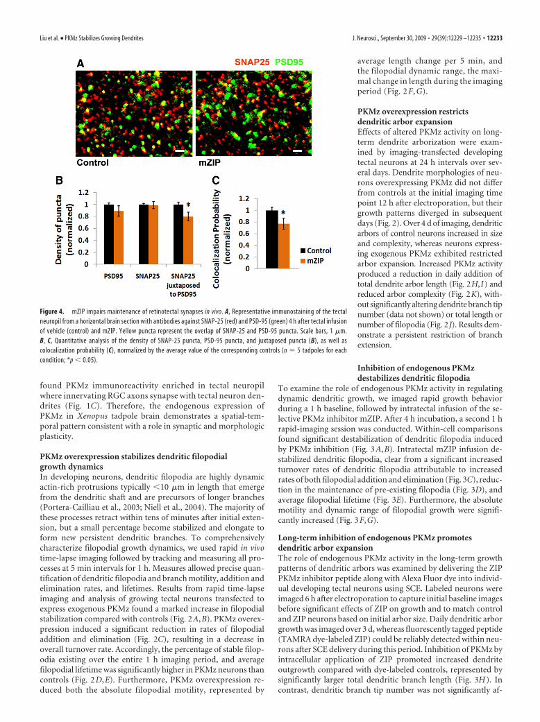
Figure 2. PKMz overexpression stabilizes dendritic filopodia and restricts arbor extension. A, B, Superimposed images of six successive time points (each a different color, with overlap � white)at 10 min imaging intervals of a neuron expressing exogenous PKMz (A) and a control neuron (B). Area within the boxed region is enlarged on the right. Scale bars, 20 �m. C–G, Morphometricanalysis of dendritic filopodial dynamics of PKMz neurons (N � 6 cells; n � 269 filopodia) and control neurons (N � 7 cells; n � 255 filopodia). PKMz overexpression significantly decreases the ratesof filopodial addition and elimination (C); increases the fraction of filopodia that persist for 1 h (D) and mean filopodial lifetime (E); decreases filopodial motility (F ) and dynamic range (G). H–K, 3Dmorphometric analysis of tectal neurons expressing GFP alone (control, n � 12) and GFP with PKMz (n � 10) 1– 4 d after electroporation. PKMz overexpression restricts daily addition of dendriticarbor size (H ), attributable to decreased extension of branches (I) but not filopodia (J ). K, 3D Sholl analysis of dendritic arbor complexity 1– 4 d after transfection. *p � 0.05; **p � 0.01.
Liu et al. • PKMz Stabilizes Growing Dendrites J. Neurosci., September 30, 2009 • 29(39):12229 –12235 • 12231

under a thin layer of agarose (0.8%) in a chamber continuously per-fused with oxygenated 10% Steinberg’s solution. 3D stacks of imagesthrough entire dendritic arbors were captured every 5 min over 1 h.Long-term dendrite growth was imaged with 24 h intervals. Singlestacks of images through the entire dendritic arbor were captured bybriefly anesthetizing tadpoles with 0.02% MS222. After imaging, tad-poles were returned to rearing solution where they rapidly recoveredfrom anesthesia.
Morphometric analysis of dendritic growth. Three-dimensional skele-tonized reconstructions of entire dendritic arbors were constructed usingNeurolucida software (MicroBrightfield) and used to analyze total den-dritic branch length, branch tip number, and 3D Sholl analysis. All mor-phometric data analysis, except for Sholl analysis, was tested forsignificance with unpaired Student’s t tests. Results from Sholl analysiswere tested with one-way ANOVA tests, followed by Turkey post hocanalysis. Rapid dendritic filopodial and branch growth dynamics wereanalyzed using custom-written software using the IGOR platform (basedon a program created by Dr. Jamie Boyd, University of British Columbia,Vancouver, BC, Canada). All filopodia and branches were identified andtracked across multiple time point image stacks.
ResultsEndogenous expression of PKMz in Xenopus tadpole brainA PKMz mRNA previously identified in Xenopus laevis (GI:1220553) was used as the basis for PCR primers to clone PMKzand confirm expression in tadpole brain. Putative translationof this mRNA generates a protein containing 401 aa with 87%homology to rat PKMz (Hernandez et al., 2003) and conservedATP and substrate-binding motifs (Fig. 1 A). Using an anti-body raised against the catalytic domain of rat PKCz and im-munoblots, we detected PKMz in tadpole brain lysates as a50 kDa protein, consistent with its putative translation prod-uct. Western blot analysis found developmental regulation ofPKMz expression in tadpole brain, with levels increasing sig-nificantly from stage 35 to stage 50 (Fig. 1 B). This period spansinitial innervation and subsequent activity-dependent refine-ment of afferent axonal input from retinal ganglion cells(RGCs) and tectal neuron dendritogenesis (Grant et al., 1980).Immunohistochemistry staining of tadpole brain cryosections
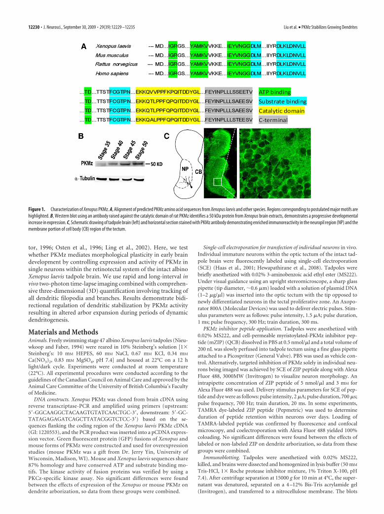
Figure 3. ZIP destabilizes dendritic filopodia and promotes arbor extension. A, B, Overlay of six successive images at 10 min of a GFP neuron before (A) and 4 h after mZIP treatment (B). Whiteportions of the arbor are stable, and colored portions are dynamic over 1 h imaging period. The area within the boxed region is enlarged on the right. Scale bar, 20 �m. C–G, Morphometric analysisof dendritic filopodial dynamics of neurons before and after mZIP delivery to tectum (N � 5 cells; n � 213 filopodia for PRE and 215 filopodia for POST) and vehicle (N � 5 cells; n � 221 filopodiafor PRE and 210 filopodia for POST). mZIP treatment significantly increases the rates of filopodial addition and elimination (C), decreases the proportion of stabilized pre-existing filopodia (D) andmean filopodial lifetime (E); increases the filopodial motility (F ) and dynamic range (G). H–K, 3D morphometric analysis of tectal neurons labeled with Alexa Fluor 488 dye alone (control, n � 12)and with ZIP (n �13) 1–3 d after electroporation. ZIP increases total dendritic arbor size (H ) and branch length (I ) but not total filopodial length (J ). K, 3D Sholl analysis of dendritic arbor complexity1–3 d after electroporation. *p � 0.05; **p � 0.01.
12232 • J. Neurosci., September 30, 2009 • 29(39):12229 –12235 Liu et al. • PKMz Stabilizes Growing Dendrites
found PKMz immunoreactivity enriched in tectal neuropilwhere innervating RGC axons synapse with tectal neuron den-drites (Fig. 1C). Therefore, the endogenous expression ofPKMz in Xenopus tadpole brain demonstrates a spatial-tem-poral pattern consistent with a role in synaptic and morphologicplasticity.
PKMz overexpression stabilizes dendritic filopodialgrowth dynamicsIn developing neurons, dendritic filopodia are highly dynamicactin-rich protrusions typically �10 �m in length that emergefrom the dendritic shaft and are precursors of longer branches(Portera-Cailliau et al., 2003; Niell et al., 2004). The majority ofthese processes retract within tens of minutes after initial exten-sion, but a small percentage become stabilized and elongate toform new persistent dendritic branches. To comprehensivelycharacterize filopodial growth dynamics, we used rapid in vivotime-lapse imaging followed by tracking and measuring all pro-cesses at 5 min intervals for 1 h. Measures allowed precise quan-tification of dendritic filopodia and branch motility, addition andelimination rates, and lifetimes. Results from rapid time-lapseimaging and analysis of growing tectal neurons transfected toexpress exogenous PKMz found a marked increase in filopodialstabilization compared with controls (Fig. 2A,B). PKMz overex-pression induced a significant reduction in rates of filopodialaddition and elimination (Fig. 2C), resulting in a decrease inoverall turnover rate. Accordingly, the percentage of stable filop-odia existing over the entire 1 h imaging period, and averagefilopodial lifetime was significantly higher in PKMz neurons thancontrols (Fig. 2D,E). Furthermore, PKMz overexpression re-duced both the absolute filopodial motility, represented by
average length change per 5 min, andthe filopodial dynamic range, the maxi-mal change in length during the imagingperiod (Fig. 2 F, G).
PKMz overexpression restrictsdendritic arbor expansionEffects of altered PKMz activity on long-term dendrite arborization were exam-ined by imaging-transfected developingtectal neurons at 24 h intervals over sev-eral days. Dendrite morphologies of neu-rons overexpressing PKMz did not differfrom controls at the initial imaging timepoint 12 h after electroporation, but theirgrowth patterns diverged in subsequentdays (Fig. 2). Over 4 d of imaging, dendriticarbors of control neurons increased in sizeand complexity, whereas neurons express-ing exogenous PKMz exhibited restrictedarbor expansion. Increased PKMz activityproduced a reduction in daily addition oftotal dendrite arbor length (Fig. 2H,I) andreduced arbor complexity (Fig. 2K), with-out significantly altering dendrite branch tipnumber (data not shown) or total length ornumber of filopodia (Fig. 2J). Results dem-onstrate a persistent restriction of branchextension.
Inhibition of endogenous PKMzdestabilizes dendritic filopodia
To examine the role of endogenous PKMz activity in regulatingdynamic dendritic growth, we imaged rapid growth behaviorduring a 1 h baseline, followed by intratectal infusion of the se-lective PKMz inhibitor mZIP. After 4 h incubation, a second 1 hrapid-imaging session was conducted. Within-cell comparisonsfound significant destabilization of dendritic filopodia inducedby PKMz inhibition (Fig. 3A,B). Intratectal mZIP infusion de-stabilized dendritic filopodia, clear from a significant increasedturnover rates of dendritic filopodia attributable to increasedrates of both filopodial addition and elimination (Fig. 3C), reduc-tion in the maintenance of pre-existing filopodia (Fig. 3D), andaverage filopodial lifetime (Fig. 3E). Furthermore, the absolutemotility and dynamic range of filopodial growth were signifi-cantly increased (Fig. 3F,G).
Long-term inhibition of endogenous PKMz promotesdendritic arbor expansionThe role of endogenous PKMz activity in the long-term growthpatterns of dendritic arbors was examined by delivering the ZIPPKMz inhibitor peptide along with Alexa Fluor dye into individ-ual developing tectal neurons using SCE. Labeled neurons wereimaged 6 h after electroporation to capture initial baseline imagesbefore significant effects of ZIP on growth and to match controland ZIP neurons based on initial arbor size. Daily dendritic arborgrowth was imaged over 3 d, whereas fluorescently tagged peptide(TAMRA dye-labeled ZIP) could be reliably detected within neu-rons after SCE delivery during this period. Inhibition of PKMz byintracellular application of ZIP promoted increased dendriteoutgrowth compared with dye-labeled controls, represented bysignificantly larger total dendritic branch length (Fig. 3H). Incontrast, dendritic branch tip number was not significantly af-
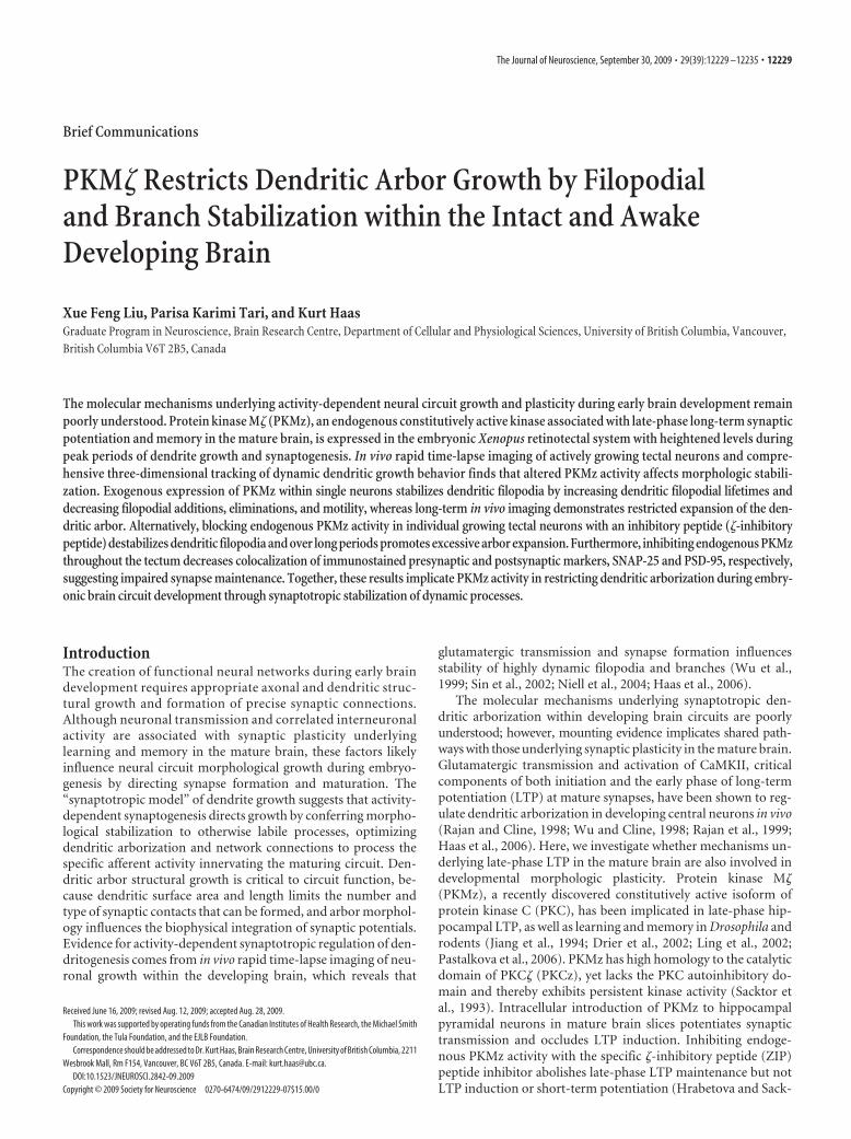
Figure 4. mZIP impairs maintenance of retinotectal synapses in vivo. A, Representative immunostaining of the tectalneuropil from a horizontal brain section with antibodies against SNAP-25 (red) and PSD-95 (green) 4 h after tectal infusionof vehicle (control) and mZIP. Yellow puncta represent the overlap of SNAP-25 and PSD-95 puncta. Scale bars, 1 �m.B, C, Quantitative analysis of the density of SNAP-25 puncta, PSD-95 puncta, and juxtaposed puncta (B), as well ascolocalization probability (C), normalized by the average value of the corresponding controls (n � 5 tadpoles for eachcondition; *p � 0.05).
Liu et al. • PKMz Stabilizes Growing Dendrites J. Neurosci., September 30, 2009 • 29(39):12229 –12235 • 12233

fected by ZIP application (data not shown). 3D Sholl analysisfound that PKMz inhibition increases arbor expansion as well ascomplexity (Fig. 3K). The primary effect of inhibition of endog-enous PKMz by ZIP was an enhanced extension of branches, withno effects on the total filopodial length or number (Fig. 3 I, J).
PKMz inhibition impairs synapse maintenance in vivoGiven the role of PKMz in synapse plasticity in the adult brainand the importance of synaptogenesis in filopodial stabilizationduring dendritogenesis, we examined the effects of PKMz onsynapse density in tadpole tectum. We used immunostaining oftadpole brain cryosections for the presynaptic and postsynapticmarkers SNAP-25 and PSD-95. PKMz inhibition by intratectalinfusion of mZIP significantly reduced tectal synapse density af-ter 4 h, evident from decreased density of PSD-95 puncta juxta-posed to SNAP-25 puncta and colocalization probability. Nosignificant changes were found in the densities of either puncta(Fig. 4). These results demonstrate that inhibition of PKMz dur-ing active periods of developmental synaptogenesis impairsmaintenance of excitatory synapses.
DiscussionThe synaptotropic model of dendritogenesis postulates that syn-apses stabilize growing dendritic processes, thereby preventingretraction, yet allowing further growth and branching from syn-aptic sites (Vaughn, 1989; Niell et al., 2004; Cline and Haas,2008). However, this model does not explain the progressive de-cline in dendritic growth and structural plasticity as neurons ma-ture, characterized by reduced growth, motility, and additions ofdendritic filopodia and branches (Wu et al., 1999). A possibleexplanation is that synaptotropic dendritogenesis is influencedby the maturational state of constituent glutamatergic synapses,thereby linking growth with the functional maturation of neu-rons (Wu et al., 1996). In this framework, nascent immaturesynapses forming on new filopodia act to prevent retraction whilepermitting further growth. Mature synapses, in contrast, wouldrestrict continued growth by conferring further structural stabi-lization. Here, we identify PKMz, a naturally expressed constitu-tively active kinase mediating late-phase synaptic LTP in themature brain (Ling et al., 2002), as a potential regulator of syn-apse maturation state-dependent synaptotropic dendrite growth.
Increasing or decreasing levels of PKMz activity in individualactively growing brain neurons produces a similar shift in den-dritic growth plasticity. Increasing PKMz activity in immatureneurons dramatically reduces dendritic morphological plasticity,resulting in stunted arbors demonstrating precocious stabiliza-tion similar to mature neurons. PKMz’s stabilizing effects ongrowing dendrites are likely mediated through synapse matura-tion rather than a direct effect on the cytoskeleton, since we foundno effects on the motility of new filopodia within the first 30 minof extension, a period before detectable synapse formation (Niellet al., 2004) (data not shown). Inhibiting endogenous PKMz ac-tivity with ZIP, shown to prevent late-phase synaptic potentia-tion in the mature brain (Ling et al., 2002), and here to reducesynapse density in the Xenopus tectum, destabilizes growing den-drites with increased dendritic motility and turnover.
The identification of a role for PKMz in dendritogensis addsto a growing list of molecular pathways shared between develop-mental neuronal morphogenesis and synaptic plasticity in themature brain. Glutamateric transmission, critical to initiation ofmature synaptic plasticity, promotes dendrite growth (Sin et al.,2002), whereas inhibition of NMDA or AMPA receptor trans-mission destabilizes filopodia and inhibits growth (Rajan and
Cline, 1998; Sin et al., 2002; Haas et al., 2006). At mature syn-apses, calcium entry through NMDA receptors activates CaMKII,a critical component of early phase LTP. Exogenous expression ofa constitutively active form of CaMKII in immature Xenopustectal neurons both promotes maturation of retinotectal gluta-matergic synapses (Wu et al., 1996) and restricts dendritic arborplasticity similar to increasing PKMz activity (Wu and Cline,1998). Inhibition of endogenous CaMKII activity destabilizesgrowing dendrites. Because PKMz expression is regulated by ac-tivity of CaMKII during induction of LTP in the mature hip-pocampus (Kelly et al., 2007), these kinases may act in concertduring development to confer early and late-phase synaptic mat-uration and associated morphologic stabilization. These findingssuggests that synapse maturation during brain circuit develop-ment which underlies morphological stabilization involves mo-lecular mechanisms common to synaptic plasticity associatedwith learning and memory in the mature brain.
Manipulations predicted to shift synaptic plasticity in growingneurons in vivo, however, do not produce arbor morphologiespredicted from a simple interpretation of the synaptotropicmodel, where enhancing synapse formation and maturationwould be expected to promote growth, whereas the inhibition ofsynaptic strengthening would limit growth. Rather, enhancedPKMz or CaMKII activity, expected to lower a neuron’s thresh-old for synapse maturation, results in stunted, nonmotile arbors,whereas inhibiting these plasticity-associated kinases producesunrestricted arbor elongation. Thus, synapse maturation duringdendritogenesis appears to function not only to stabilize nascentprocesses to prevent retraction but also to restrict excessive ar-borization and may underlie the loss of dendrite morphologicalplasticity with neuronal maturation.
ReferencesCline H, Haas K (2008) The regulation of dendritic arbor development and
plasticity by glutamatergic synaptic input: a review of the synaptotrophichypothesis. J Physiol 586:1509 –1517.
Drier EA, Tello MK, Cowan M, Wu P, Blace N, Sacktor TC, Yin JC (2002)Memory enhancement and formation by atypical PKM activity in Dro-sophila melanogaster. Nat Neurosci 5:316 –324.
Grant P, Rubin E, Cima C (1980) Ontogeny of the retina and optic nerve inXenopus laevis. I. Stages in the early development of the retina. J CompNeurol 189:593– 613.
Haas K, Sin WC, Javaherian A, Li Z, Cline HT (2001) Single-cell electropo-ration for gene transfer in vivo. Neuron 29:583–591.
Haas K, Li J, Cline HT (2006) AMPA receptors regulate experience-dependent dendritic arbor growth in vivo. Proc Natl Acad Sci U S A103:12127–12131.
Hernandez AI, Blace N, Crary JF, Serrano PA, Leitges M, Libien JM,Weinstein G, Tcherapanov A, Sacktor TC (2003) Protein kinase Mzeta synthesis from a brain mRNA encoding an independent proteinkinase C zeta catalytic domain. Implications for the molecular mech-anism of memory. J Biol Chem 278:40305– 40316.
Hewapathirane DS, Dunfield D, Yen W, Chen S, Haas K (2008) In vivoimaging of seizure activity in a novel developmental seizure model. ExpNeurol 211:480 – 488.
Hrabetova S, Sacktor TC (1996) Bidirectional regulation of protein kinaseM� in the maintenance of long-term potentiation and long-term depres-sion. J Neurosci 16:5324 –5333.
Jiang X, Naik MU, Hrabe J, Sacktor TC (1994) Developmental expression ofthe protein kinase C family in rat hippocampus. Brain Res Dev Brain Res78:291–295.
Kelly MT, Crary JF, Sacktor TC (2007) Regulation of protein kinase M�synthesis by multiple kinases in long-term potentiation. J Neurosci27:3439 –3444.
Ling DS, Benardo LS, Serrano PA, Blace N, Kelly MT, Crary JF, Sacktor TC(2002) Protein kinase Mzeta is necessary and sufficient for LTP mainte-nance. Nat Neurosci 5:295–296.
12234 • J. Neurosci., September 30, 2009 • 29(39):12229 –12235 Liu et al. • PKMz Stabilizes Growing Dendrites

Niell CM, Meyer MP, Smith SJ (2004) In vivo imaging of synapse formationon a growing dendritic arbor. Nat Neurosci 7:254 –260.
Nieuwkoop PD, Faber J (1994) Normal table of Xenopus laevis. Amsterdam:Elsevier.
Osten P, Valsamis L, Harris A, Sacktor TC (1996) Protein synthesis-dependent formation of protein kinase M� in long-term potentiation.J Neurosci 16:2444 –2451.
Pastalkova E, Serrano P, Pinkhasova D, Wallace E, Fenton AA, Sacktor TC(2006) Storage of spatial information by the maintenance mechanism ofLTP. Science 313:1141–1144.
Portera-Cailliau C, Pan DT, Yuste R (2003) Activity-regulated dynamic be-havior of early dendritic protrusions: evidence for different types of den-dritic filopodia. J Neurosci 23:7129 –7142.
Rajan I, Cline HT (1998) Glutamate receptor activity is required for nor-mal development of tectal cell dendrites in vivo. J Neurosci 18:7836 –7846.
Rajan I, Witte S, Cline HT (1999) NMDA receptor activity stabilizes presyn-
aptic retinotectal axons and postsynaptic optic tectal cell dendrites invivo. J Neurobiol 38:357–368.
Sacktor TC, Osten P, Valsamis H, Jiang X, Naik MU, Sublette E (1993)Persistent activation of the zeta isoform of protein kinase C in themaintenance of long-term potentiation. Proc Natl Acad Sci U S A90:8342– 8346.
Sin WC, Haas K, Ruthazer ES, Cline HT (2002) Dendrite growth increasedby visual activity requires NMDA receptor and Rho GTPases. Nature419:475– 480.
Vaughn JE (1989) Fine structure of synaptogenesis in the vertebrate centralnervous system. Synapse 3:255–285.
Wu G, Malinow R, Cline HT (1996) Maturation of a central glutamatergicsynapse. Science 274:972–976.
Wu GY, Cline HT (1998) Stabilization of dendritic arbor structure in vivoby CaMKII. Science 279:222–226.
Wu GY, Zou DJ, Rajan I, Cline H (1999) Dendritic dynamics in vivo changeduring neuronal maturation. J Neurosci 19:4472– 4483.
Liu et al. • PKMz Stabilizes Growing Dendrites J. Neurosci., September 30, 2009 • 29(39):12229 –12235 • 12235