
BKV as a Cofactor in the Etiology of Prostate Cancer in...
36
1 BKV as a Cofactor in the Etiology of Prostate Cancer in its Early Stages 1 Dweepanita Das 1,3 , Kirk Wojno 2 and Michael J. Imperiale* , 1, 3 2 1 Department of Microbiology and Immunology, 2 Department of Urology and 3 Comprehensive 3 Cancer Center, University of Michigan Medical School, Ann Arbor, Michigan 48109-5942 4 5 *Correspondence: 6 Michael J. Imperiale 7 Department of Microbiology and Immunology 8 University of Michigan Medical School 9 1500 E. Medical Center Dr., 6304 Cancer Center, SPC 5942 10 Ann Arbor, Michigan 48109-5942 11 Ph# 734-763-9162 12 Fax# 734-615-6560 13 E-mail: [email protected] 14 15 Running Title: BKV and early stage prostate cancer 16 17 ACCEPTED Copyright © 2007, American Society for Microbiology and/or the Listed Authors/Institutions. All Rights Reserved. J. Virol. doi:10.1128/JVI.02461-07 JVI Accepts, published online ahead of print on 26 December 2007 on July 26, 2018 by guest http://jvi.asm.org/ Downloaded from
Transcript of BKV as a Cofactor in the Etiology of Prostate Cancer in...
1
BKV as a Cofactor in the Etiology of Prostate Cancer in its Early Stages 1
Dweepanita Das1,3
, Kirk Wojno2 and Michael J. Imperiale*
, 1, 3 2
1Department of Microbiology and Immunology,
2Department of Urology and
3Comprehensive 3
Cancer Center, University of Michigan Medical School, Ann Arbor, Michigan 48109-5942 4
5
*Correspondence: 6
Michael J. Imperiale 7
Department of Microbiology and Immunology 8
University of Michigan Medical School 9
1500 E. Medical Center Dr., 6304 Cancer Center, SPC 5942 10
Ann Arbor, Michigan 48109-5942 11
Ph# 734-763-9162 12
Fax# 734-615-6560 13
E-mail: [email protected] 14
15
Running Title: BKV and early stage prostate cancer 16
17
ACCEPTED
Copyright © 2007, American Society for Microbiology and/or the Listed Authors/Institutions. All Rights Reserved.J. Virol. doi:10.1128/JVI.02461-07 JVI Accepts, published online ahead of print on 26 December 2007
on July 26, 2018 by guesthttp://jvi.asm
.org/D
ownloaded from
2
ABSTRACT 1
Prostate cancer has been projected to cause almost 10% of all male cancer deaths in 2007 in 2
the US. The incidence of mutations in the tumor suppressor genes, Rb1 and p53, especially in the 3
early stages of the disease, is low when compared to other cancers. This has led to the hypothesis 4
that a human virus such as BK virus (BKV), which establishes a subclinical persistent infection in 5
the urinary tract and encodes oncoproteins that interfere with these tumor suppressor pathways, is 6
involved. Previously, we detected BKV DNA in the epithelial cells of benign and proliferative 7
inflammatory atrophy ducts of cancerous prostate specimens. In the present report, we demonstrate 8
that BKV is present at a much lower frequency in non-cancerous prostates. Additionally, in normal 9
prostates, TAg expression is only observed in specimens harboring proliferative inflammatory 10
atrophy and prostatic intraepithelial neoplasia. We further demonstrate that the p53 gene from 11
atrophic cells expressing TAg is wild type, whereas tumor cells expressing detectable nuclear p53 12
contain a mix of wild type and mutant p53 genes, suggesting that TAg may inactivate p53 in the 13
atrophic cells. Our results point towards a role for BKV in early prostate cancer progression. 14
15
16 ACCEPTED
on July 26, 2018 by guesthttp://jvi.asm
.org/D
ownloaded from
3
INTRODUCTION 1
Prostate cancer is the leading cause of male cancer deaths in the United States. The latest 2
statistics for 2007 estimate 218,890 new prostate cancer cases and 27,050 deaths (39). Prostate 3
cancer is a slowly progressing disease; therefore, identification of its precursor lesions has become 4
the primary focus of many studies (reviewed in 14, 20, 30, 55). An increased understanding of the 5
molecular mechanisms associated with prostate cancer initiation and progression would be useful for 6
developing strategies for its early detection and treatment. 7
Proliferative inflammatory atrophy (PIA), proposed as the potential precursor to 8
adenocarcinoma due to its proliferative nature, is prevalent in the peripheral zone of the prostate 9
gland, where prostatic intraepithelial neoplasia (PIN) and carcinoma also occur (reviewed in 20, 59, 10
68, 80, 91). It has been postulated that PIA may transition to carcinoma without any intermediate 11
stage or may lead to carcinoma through PIN. Frequent histological transitions between PIA and PIN 12
have been observed (19, 68). Several crucial prostate tumor suppressor genes such as NKX3.1; 13
CDKN1B, which codes for p27, a cyclin-dependent kinase inhibitor that regulates cell cycle 14
progression; and PTEN (phosphatase and tensin homologue) are all expressed at very low levels in 15
PIA, similar to their expression pattern in PIN and carcinoma (reviewed in 30). 16
The occurrence of mutations in p53 and Rb1 in the early stages of prostate cancer is 17
relatively low (reviewed in 1, 7, 40, 57). This has suggested the possibility that a viral agent like BK 18
virus (BKV), which infects the urinary tract and encodes tumor antigens that inactivate these tumor 19
suppressors, may play a role in the etiology of prostate cancer. This would be very similar to how the 20
E6 and E7 oncogene products of human papillomavirus (HPV) inactivate p53 and pRB, respectively, 21
in cervical carcinomas (13, 22, 27, 37, 50, 74, 75). It has been suggested that exposure to infectious 22
ACCEPTED
on July 26, 2018 by guesthttp://jvi.asm
.org/D
ownloaded from
4
agents can cause injury to the normal prostate epithelium, leading to the development of PIA 1
(reviewed in 20). 2
BKV, a member of the polyomavirus family, was first isolated from the urine of a renal 3
transplant patient (28) and infects almost 90% of the human population by early childhood (reviewed 4
in 34, 42, 78). It resides in a subclinical persistent state in the urinary tract of healthy individuals and 5
reactivates in immunosuppressed transplant patients, in whom it is associated with hemorrhagic 6
cystitis and polyomavirus nephropathy (5, 35, 58, 70). BKV transforms rodent cells in culture (65), 7
causes kidney tumors in transgenic mice (15), and immortalizes primary human cells alone (32, 67, 8
79, 84) or in the presence of other oncogenes such as c-ras (60) and adenovirus E1A (92). A possible 9
role for BKV in human cancers is controversial because such a high percentage of the human 10
population is exposed to the virus at a very early age, precluding the use of epidemiologic methods 11
to test an association (reviewed in 16) 12
The genome of BKV is divided into early, late, and regulatory regions and codes for at least 13
six proteins, two from the early region and four from the late region. The early proteins, large tumor 14
antigen (TAg) and small tumor antigen (tAg), are the first to be expressed during infection. When 15
TAg accumulates to high levels, it initiates viral DNA replication in the cell nucleus by recruiting 16
the DNA polymerase α/primase complex to the viral origin of DNA replication, shuts off early gene 17
transcription, and stimulates expression of the late genes, VP1, VP2, VP3, and agnoprotein 18
(reviewed in 36). In a non-productive infection, which usually occurs as a result of a cellular 19
environment that is not conducive to viral gene expression or replication, BKV induces oncogenesis 20
through the expression of its two tumor antigens (reviewed in 88). TAg promotes cellular 21
transformation by interfering with the tumor suppressor functions of p53 and pRB (reviewed in 3). 22
TAg upregulates p53 levels in the cell by stabilizing the protein, but functionally inactivates it by 23
ACCEPTED
on July 26, 2018 by guesthttp://jvi.asm
.org/D
ownloaded from
5
sequestering it in an inert form (reviewed in 45, 46, 49, 63, 93). Therefore, expression of TAg and 1
subsequent inactivation of p53 mimic the same phenotypic effect as those caused by mutations in the 2
p53 gene. Similarly, TAg functionally inactivates pRB by binding it and causing the release of E2F 3
(reviewed in 4). tAg induces tumorigenesis and promotes anchorage-independent growth of 4
transformed cells by the negative regulation of protein phosphatase 2A (64, 95). 5
In a previous report, utilizing in situ analysis, we demonstrated the presence of BKV DNA 6
sequences in epithelial cells of benign and PIA ducts of cancerous prostate specimens (17). 7
Additionally, BKV TAg expression was observed specifically in the atrophic epithelial cells but not 8
in the normal epithelium. TAg was detected in the cytoplasm and co-localized with p53 in the 9
atrophic cells, suggesting that neither protein could carry out its normal nuclear function. However, 10
BKV was not detected in cells in the more advanced stages of cancer progression. In the present 11
study we have extended our analysis to non-cancerous prostates. In normal prostates, BKV was 12
present at a lower frequency than in cancerous ones, and TAg expression is only observed in 13
specimens containing PIA and PIN lesions. We also detected TAg expression in the same PIA ducts 14
containing BKV DNA, confirming that the TAg is that of BKV. Utilizing laser capture 15
microdissection on cancerous prostates, we further show that the p53 gene from TAg-expressing PIA 16
cells was wild type, whereas tumor cells expressing nuclear p53 contained a mix of wild type and 17
mutant p53 genes. Additionally, we demonstrate that the nuclear localization sequences (NLS) of 18
cytoplasmically localized TAg and p53 were wild type, indicating that the sequestration of TAg and 19
p53 in the cytoplasm was not due to mutations in the NLS of these genes. Together, these results 20
support a causal role for BKV in PIA and the early development of prostate cancer. 21
22
ACCEPTED
on July 26, 2018 by guesthttp://jvi.asm
.org/D
ownloaded from
6
MATERIALS AND METHODS 1
Human Tissue Specimens. 2
Paraffin-embedded adenocarcinoma prostate resection specimens from radical 3
prostatectomies and cystoprostatectomy specimens from bladder cancer patients with the diagnosis 4
of muscle invasive high grade urothelial carcinoma, with no prostate cancer histology, were obtained 5
from the Tissue Procurement Core at the University of Michigan Comprehensive Cancer Center. 6
One section from each of the specimens was stained with hematoxylin and eosin and was evaluated 7
for the presence of benign, atrophic, or tumor cells by the pathologist. Additionally, autopsy 8
specimens were obtained commercially as a normal prostate tissue microarray from US BIOMAX, 9
Inc. 10
DNA Extraction and PCR amplification. 11
DNA extraction was performed using the method previously described by Das et al. (2004) 12
in a BKV-free area and cross-contamination was avoided by frequent changing of gloves between 13
samples. The sequences of the oligonucleotide primers used for these studies are listed in Table 1. 14
The BKV early region oligonucleotide probe for ISH (BKV (4434-4478)), and a scrambled control probe 15
with the same length and G+C content were characterized in our previous study (17). 16
Extracted DNA from entire thin tissue sections was amplified using TitaniumTM
Taq DNA 17
polymerase (Clontech) in a ThermoHybaid thermocycler (Px2). All reactions were performed in a 18
final volume of 100 µL containing 200 nM each primer, 200 µM dNTPs, 2 µL template and 1U 19
polymerase in 0.5X buffer (20 mM Tricine-KOH, pH 8.0, 8 mM KCl, 1.75 mM MgCl2 and 1.87 20
µg/mL BSA). For TAg NLS amplification, two rounds of 45 cycles each were used. The template 21
for second round amplification consisted of 30 µL product from the first round amplification. The 22
program consisted of initial denaturation for 5 mins. at 94oC followed by denaturation at 94
oC for 30 23
ACCEPTED
on July 26, 2018 by guesthttp://jvi.asm
.org/D
ownloaded from
7
secs, annealing at 45oC for 30 secs, and elongation at 72
oC for 1 min., followed by final elongation 1
at 72oC for 7 minutes. For p53 gene amplification, 1X buffer was used, with two rounds of 45 cycle 2
amplification. The program for exons 5 through 8 consisted of initial denaturation for 5 mins. at 3
94oC followed by denaturation at 94
oC for 30 secs, annealing at 60
oC for 30 secs, and elongation at 4
72oC for 1 min., followed by final elongation at 72
oC for 7 mins. The program for exon 9 was the 5
same except that the annealing temperature was 51oC. The negative control tube contained all the 6
PCR components except DNA template. p53 NLS amplification was done using identical PCR 7
conditions of exons 7 through 8 and 9. 8
Sequence Analysis. 9
PCR products were separated by agarose gel electrophoresis, extracted (Qiaquick gel 10
extraction kit, Qiagen), and sequenced by the DNA Sequencing Core at the University of Michigan. 11
Sequences were analyzed using Lasergene software from DNA Star. 12
Renal Proximal Tubular Epithelial (RPTE) Cells. 13
RPTE cells were grown and infected with BKV on two well chamber slides (Fisher) as 14
previously reported (47). Cells were fixed and stained 4 days after infection as previously published 15
(17). 16
In Situ DNA Hybridization (ISH) and Immunohistochemistry (IHC). 17
ISH and IHC were performed using our previously published protocol (17). For IHC with 18
anti-VP1, 1% SDS retrieval was performed for 8 mins. at room temperature. Monoclonal antibody 19
anti-VP1 (P5G6 BKV9VP1), a gift from Denise Galloway, was used at a 1:600 dilution. 20
Immuno-Laser Capture Microdissection. 21
Formalin-fixed, paraffin-embedded 4 µm prostate tissue sections were deparaffinized and 22
rehydrated using our previously published protocol. Immunohistochemistry was performed as 23
ACCEPTED
on July 26, 2018 by guesthttp://jvi.asm
.org/D
ownloaded from
8
previously described except that the antibody dilution for the anti-TAg was 1:300. After IHC and 1
counterstaining with hematoxylin, the sections were dehydrated in graded ethanol solutions (95% 2
ethanol for 2 x 5 mins., 100% ethanol for 3 x 5 mins.) and cleared in xylene for 3 x 5 mins. The 3
slides were air dried for 30 min. in a fume hood and stored in a desiccator until laser capture 4
microdissection. Laser capture was performed using the PIXCELL II Laser Capture Microscope 5
from Arcturus Engineering. The TAg positive PIA cells and nuclear p53 immunolabeled areas were 6
visualized directly, after which the laser pulse was applied to activated thermoplastic film mounted 7
on LCM caps to capture cells of interest. The following parameters were set on the PixCell II LCM 8
system: laser spot size 7.5 µm; power 85 mW; current 250 mV. DNA was extracted from 9
approximately 2500 captured cells by treating with 40 µL of proteinase K buffer (10 mM Tris, pH 10
8.0, 1 mM EDTA, pH 8.0, 1% Tween 20 and 0.1 mg/mL proteinase K) for 48 hrs at 45oC. Proteinase 11
K was first inactivated at 95oC for 10 mins. The LCM DNA extracts were flash spun in a 12
microcentrifuge and stored at -20oC. A 20 µL aliquot of the LCM DNA template was routinely used 13
for first round PCR amplification. 14
15
16 ACCEPTED
on July 26, 2018 by guesthttp://jvi.asm
.org/D
ownloaded from
9
RESULTS 1
BKV is present at a significantly higher frequency in cancerous prostates 2
If BKV plays a role in prostate cancer progression, one would expect to find it at lower 3
frequencies in normal prostates than in cancerous prostates. For these experiments, cancerous 4
prostate is defined as a prostate that has been surgically removed due to a diagnosis of prostate 5
cancer and histologically confirmed to contain malignant glands. Normal prostate is defined as a 6
prostate that has been surgically removed as part of a resection to remove a cancerous bladder 7
(cystoprostatectomy) or during autopsy and histologically confirmed to contain only non-malignant 8
glands. 9
We initially analyzed the normal prostates using the same in situ hybridization (ISH) 10
technique that we previously used with cancerous prostates (17). BKV DNA was not detected in the 11
majority of the normal specimens (Figure 1A). However, in a small number of normal specimens, 12
BKV DNA was observed both in normal and atrophic epithelial cells, as demonstrated by nuclear 13
hybridization to the BKV-specific probe (Figure 2, panels B, E and G) but not to the scrambled 14
probe (Figure 2, panels A and D). We did not detect any BKV signal in the stromal cells. The ISH 15
studies on normal prostate demonstrated the presence of BKV DNA sequences in 4/15 of the 16
specimens, which was significantly lower than the frequency of detection in cancerous prostate 17
(Table 2). 18
Next, we used immunohistochemistry (IHC) with a specific anti-TAg monoclonal antibody 19
to determine whether TAg was being expressed in the normal prostate tissue specimens (Figure 1B-20
C). In the majority of the normal prostate tissue specimens there was no detectable TAg (panel B). 21
However, in a small number of normal specimens we observed expression of TAg in the cytoplasm 22
of atrophic epithelium (panel C). The staining pattern of the atrophic cells was identical to that 23
ACCEPTED
on July 26, 2018 by guesthttp://jvi.asm
.org/D
ownloaded from
10
observed with cancerous prostate. The IHC analysis of normal prostate demonstrated the expression 1
of TAg in 4/29 of the specimens, which was significantly lower than cancerous prostate (Table 2). 2
We also examined the status of the p53 protein in the normal prostate tissue specimens. In our 3
previous report, we had observed the cytoplasmic localization of p53 in atrophic cells in the 4
cancerous prostate specimens that also expressed detectable TAg. Representative IHC of anti-p53 5
staining on a serial section from the same normal prostate that was positive for TAg is shown in 6
Figure 1D. p53 expression was cytoplasmic, localized specifically to the atrophic cells that express 7
TAg, and was detectable only in the TAg-positive specimens (Table 3). The ISH and the IHC 8
analysis from normal prostate tissue specimens suggest that BKV is not highly prevalent in the 9
epithelium of normal prostates. 10
We wanted to confirm whether TAg was expressed in the same ducts that contained BKV 11
DNA. To determine this, we aligned ISH and IHC slides from serial sections that showed the 12
presence of viral DNA and TAg expression (Figure 2). In a BKV-positive specimen, BKV DNA 13
(panels B and G) and TAg expression (panels C and H) were observed in the same PIA duct. In 14
contrast, a normal duct from the same BKV-positive specimen showed robust nuclear staining with 15
the BKV probe (panel E) but lacked TAg expression (panel F). 16
Wild type p53 gene in atrophic cells expressing TAg 17
We next wanted to test the hypothesis that TAg inactivates p53 in the early stages of 18
prostate cancer, specifically PIA. Our hypothesis regarding the status of the p53 gene in BKV-19
related prostate cancer is derived from the situation in another virally induced cancer, cervical 20
carcinoma. Like TAg, the HPV E6 oncoprotein inactivates p53. HPV-induced cancers contain 21
wild type p53 while HPV-negative cancers have mutant p53 (13, 27, 37, 74). We predicted that 22
in BKV TAg-expressing cells, a wild type p53 gene would be observed, and in BKV TAg-23
ACCEPTED
on July 26, 2018 by guesthttp://jvi.asm
.org/D
ownloaded from
11
negative tumor cells with detectable nuclear p53, a mutant p53 gene would be detected. To 1
analyze this, we designed PCR primers to amplify exons 5-9 of the p53 gene. This spans the 2
sequence-specific DNA binding domain of p53; these exons are the sites for the most frequent 3
mutations in the p53 gene in various cancers including prostate (reviewed in 21, 94). Utilizing 4
laser capture microdissection (LCM), BKV TAg-positive atrophic cells from PIA and BKV 5
TAg-negative tumor cells expressing nuclear p53 were isolated from cancerous prostate tissue 6
sections immunostained with either anti-TAg or anti-p53, respectively. Amplification was 7
performed both with DNA extracted from entire thin tissue sections and from the laser-captured 8
cells, and the products were sequenced. The entire thin tissue sections contain a mixture of 9
normal, atrophic, tumor, and stromal cells. In both the TAg-positive and TAg-negative captured 10
specimens there was slight stromal cell contamination (Figure 3; C and F). Chromatograms were 11
carefully examined for the presence or absence of mutations in the p53 gene. A partial 12
comparative chromatogram of exon 5 of the p53 gene from isolated TAg-positive cells and entire 13
thin tissue section of one specimen shows that the p53 sequence is wild type in the laser-captured 14
atrophic cells expressing TAg, whereas it is a mixture of wild type and mutant in the entire 15
section (Figure 3). All the laser captured TAg-positive PIA cells we analyzed contained wild 16
type p53, whereas cells from entire thin sections of some of these specimens contained a mixture 17
of wild type and mutant p53 (Table 4). In contrast, captured tumor cells from a specimen that 18
was BKV TAg negative but expressed nuclear p53 contained a mix of wild type and mutant p53 19
(specimen #C6). This may be due to heterozygosity at the locus. 20
21
ACCEPTED
on July 26, 2018 by guesthttp://jvi.asm
.org/D
ownloaded from
12
Absence of BKV replication marker, VP1 in TAg expressing cancerous prostates 1
The exclusive cytoplasmic localization of BKV TAg suggested that viral replication, 2
which relies on the host DNA synthetic machinery in the nucleus, cannot be occurring. To test 3
this, IHC analysis was performed on a subset of the TAg-positive specimens using antibody to 4
the viral late protein, VP1, which is a BKV replication marker (Figure 4). We did not detect any 5
positive signal in the tissue specimens with antibody to VP1, indicating that viral replication was 6
not occurring. As a positive control, BKV-infected human renal proximal tubular epithelial cells 7
were analyzed in parallel (panels C-D). Finally, due to the cytoplasmic localization of the TAg 8
and p53, we determined the integrity of the NLS of these two proteins. To accomplish this, DNA 9
was extracted from three TAg-positive tissue specimens, and nested PCR was used to amplify 10
and sequence the TAg NLS. A wild type NLS was observed in all three specimens (data not 11
shown). Additionally, exons 8 and 9 of the p53 gene, which contain a bipartite NLS, were 12
sequenced from three specimens. In these specimens, the p53 NLS was also wild type (data not 13
shown). 14
15
16 ACCEPTED
on July 26, 2018 by guesthttp://jvi.asm
.org/D
ownloaded from
13
DISCUSSION 1
The experiments in this study tested the hypothesis that BKV plays a role in the etiology of 2
prostate cancer in its early stages. In our initial in situ analysis with cancerous prostates, we had 3
observed the presence of BKV DNA in normal and PIA epithelium, with TAg expression 4
specifically localized to the atrophic but not the normal epithelium (17). BKV was not detected and 5
was assumed to be lost as the cancer advanced to PIN or invasive carcinoma. We have extended our 6
previous studies to determine if BKV is also present in non-diseased prostates, or whether its 7
detection in cancerous tissue is simply a reflection of its ubiquitous presence in the human 8
population. When we compared the presence of BKV between diseased and non-diseased prostates, 9
BKV was found at a higher frequency in cancerous prostates. Viral DNA was detected in 79% of 10
cancerous prostates but only in 27% of non-diseased prostates, and TAg expression was observed in 11
47% of cancerous prostates but only in 14% of non-diseased prostates. Fisher’s exact test both for 12
ISH (p=0.007) and IHC (p= 0.008) shows a significant difference between the presence and 13
expression of BKV in normal and cancerous prostates. Interestingly, in both types of specimen, TAg 14
expression was only detected in PIA cells. In the non-diseased prostates, however, TAg expression 15
in PIA lesions was only observed in those specimens also containing PIN lesions. Additionally, TAg 16
expression is localized to the cytoplasm in both types of specimen, but the NLS is wild type. The 17
comparative analysis between cancerous and non-diseased prostate supports our hypothesis that 18
BKV is a cofactor in PIA. 19
PIA has been observed in the vicinity of carcinoma lesions and has been reported to 20
sometimes merge with adenocarcinoma (18, reviewed in 20). Epithelial cells in PIA are highly 21
proliferative, exhibit phenotypic characteristics that are intermediate between secretory and basal 22
cells, and have been proposed to be precursors to prostatic neoplastic transformation (91). Merging 23
ACCEPTED
on July 26, 2018 by guesthttp://jvi.asm
.org/D
ownloaded from
14
of focal areas of PIA with PIN has also been reported (18). Interestingly, occurrence of mutations in 1
p53 or Rb1 in PIA is low, which supports a viral cause for the transition of benign epithelium to PIA 2
lesions through the inactivation of these tumor suppressors by viral oncoproteins. 3
There are two additional reports on the presence of BKV DNA in prostate carcinomas (44, 4
96). Zambrano et al. (2002) demonstrated the presence of BKV DNA in 3/12 prostate specimens by 5
utilizing PCR, and Lau et al. (2007) detected BKV DNA in tumor cells in 2/30 prostate specimens 6
using ISH. Our previous study, however, was the first to demonstrate the presence of both viral 7
DNA and oncoprotein expression in PIA lesions of neoplastic prostate (17). In the current report, we 8
specifically examined expression of TAg in the same PIA ducts that contain BKV DNA, further 9
supporting our previous conclusion that the TAg is indeed that of BKV. Additionally, in this study, 10
we show that although the normal ducts may have BKV DNA, there is no apparent expression of 11
TAg in those ducts. This suggests the intriguing possibility that BKV infects the normal epithelium 12
and resides in a latent state, and that activation of TAg expression in PIA may promote the transition 13
from benign to atrophic, ultimately leading to prostate cancer. The fact that TAg is only detected in 14
the PIA of non-diseased prostates that also have signs of PIN also supports a possible link between 15
BKV and cancerous lesions. It is tempting to speculate here that these non-diseased prostates which 16
have PIN are already on their way to cancer, and TAg expression may act as a co-factor that 17
promotes this transition. Interestingly, prostate cancer is a slowly progressing disease, focal areas of 18
atrophy are a common occurrence of the ageing prostate (reviewed in 20, 26, 29, 48, 71), and BKV 19
is a relatively poor transforming agent (6, 33). 20
Detection of p53 by IHC usually requires that the p53 be stabilized in some manner (23, 21
24). Two known means of stabilization are binding by TAg (reviewed in 63) and mutation 22
(reviewed in 61, 82). Our analysis of the p53 gene from laser captured TAg-positive PIA cells 23
ACCEPTED
on July 26, 2018 by guesthttp://jvi.asm
.org/D
ownloaded from
15
and TAg-negative tumor cells with nuclear p53 is therefore relevant to whether the virus is 1
involved in oncogenesis. The p53 sequence is wild type in laser-captured PIA cells expressing 2
TAg. This is similar to the status of p53 genes in cervical carcinomas expressing the HPV E6 3
oncoprotein (reviewed in 86), and suggests that the wild type p53 is being inactivated by 4
sequestration in the cytoplasm. However, when the p53 sequence was analyzed from entire thin 5
sections from cancerous prostates, a mixture of wild type and mutant p53 was sometimes, but not 6
always, observed. Since p53 mutations are not frequent in prostate cancer, it is not surprising that 7
we did not detect more mutations (21). We predicted that the mutant p53 sequence that we 8
observed from TAg-positive thin sections was derived from tumor cells. LCM of p53-positive 9
tumor cells supported this, because we detected a mix of wild type and mutant p53 in those 10
specimens. In spite of slight stromal cell contamination in the LCM samples, the mutant form of 11
p53 was readily detectable in the specimen expressing nuclear p53. An earlier study using laser 12
capture microdissection demonstrated a very low occurrence of p53 gene mutations in PIA 13
lesions (90). Since the p53 gene in TAg-positive cells in cancerous prostates is wild type, we did 14
not perform this analysis in normal specimens. p53 expression in both types of specimen was 15
only detectable in PIA cells and was cytoplasmic despite the protein having a wild type NLS. For 16
p53 to function as a tumor suppressor, its translocation and retention in the nucleus is required 17
(41, 53, 73). Nuclear p53 over-expression was not observed in any of the non-diseased prostates. 18
It is interesting that TAg, which is normally a nuclear protein, is localized in the cytoplasm of 19
the epithelial cells in these PIA lesions. The tumorigenic potential of an SV40 TAg containing a 20
mutation in the NLS, causing it to remain in the cytoplasm, has been previously reported (reviewed 21
in 10, 43, 62). This cytoplasmic SV40 TAg has the ability to induce tumors in transgenic mice at a 22
rate equivalent to nuclear TAg (62). However, in our studies the NLS of BKV TAg was wild type. In 23
ACCEPTED
on July 26, 2018 by guesthttp://jvi.asm
.org/D
ownloaded from
16
addition, BKV replication, as measured by VP1 expression, was not observed in the TAg-positive 1
specimens, consistent with the observation of TAg expression in the cytoplasm. Non-productive 2
infections leading to oncogenesis are known to occur with the gamma herpesvirus, EBV, as well as 3
with oncogenic retroviruses, which generally require a helper virus for replication. 4
Additionally, cytoplasmic p53 expression in transformed cells in the presence of a 5
cytoplasmically localized SV40 TAg has been previously reported (43). Cytoplasmic localization 6
of p53 has been also observed in various human tumors (8, 9, 52, 54, 77, 81). This suggests that 7
in certain cancers, functional p53 inactivation occurs by the sequestration of the protein in the 8
cytoplasm, leading to acceleration of tumor progression by the accumulation of chromosomal 9
mutations during cell proliferation. 10
The fact that the NLS of TAg and p53 were wild type in BKV positive prostates suggests that 11
there is a cellular factor(s) in the cytoplasm that is sequestering both proteins. Wild type p53 has 12
been shown to be retained in the cytoplasm as a result of interactions with proteins like Hdm-2 and 13
heat shock proteins (83). Phosphorylation of serine residues adjacent to the NLS of SV40 TAg by 14
casein kinase II facilitates the nuclear translocation of TAg and this process of nuclear import is 15
inhibited by the p34cdc2
- mediated phosphorylation of a nearby threonine residue (38, 72, 76). 16
Complexes of p34cdc2
and p53 have been observed in TAg-transformed cells (51) and TAg has been 17
shown to stimulate the expression of the cdc2 gene (12, 56). Interestingly, serine 315 of p53 is 18
adjacent to one of its NLS and is also phosphorylated by p34cdc2
(2), and exclusion of p53 from the 19
nucleus due to the phosphorylation of serine-315 was recently demonstrated (69). It is tempting to 20
speculate that in the prostate specimens that express TAg, p34cdc2
phosphorylates TAg and/or p53, 21
which impairs the ability of both proteins to translocate to the nucleus. 22
ACCEPTED
on July 26, 2018 by guesthttp://jvi.asm
.org/D
ownloaded from
17
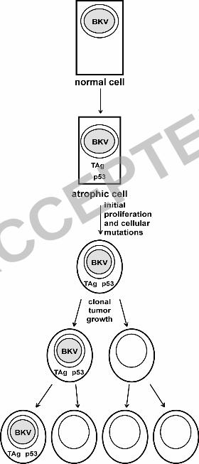
Based on our findings, we present the following model (Figure 5). BKV infects normal 1
epithelial cells and induces a change of the normal cells to PIA through the expression of TAg; 2
alternatively, the transition to PIA induces TAg expression. This results in induction of 3
proliferation and sequestration of p53 in the cytoplasm. As the cells proliferate, they accumulate 4
mutations at a higher-than-normal rate due to the absence of p53 activity. Eventually, a cell 5
accumulates enough mutations to completely lose growth control and clonally expands into a 6
tumor. The loss of BKV in the tumor cells could be due to selection against TAg by the immune 7
system, dilution of viral episomes due to lack of replication, or pro-apoptotic effects mediated by 8
TAg that are not compatible with the other growth control mutations in the tumor cells (85, 87, 9
89), resulting in selection against TAg expression. Loss of TAg expression has been reported in 10
studies of TRAMP (TRansgenic Adenocarcinoma of Mouse Prostate) mice, which develop 11
tumors due to expression of the SV40 early region (31). When tumor cells are removed from 12
these animals and grown in culture, TAg expression is lost (25). A similar loss of oncoprotein 13
expression is seen in cancers of the alimentary canal in cattle caused by bovine papillomavirus 14
type 4 (BPV-4): the virus is required to induce papillomas but its presence is not necessary for 15
progression or maintenance of the transformed state (11). Additional work will be required to 16
determine if a causal connection between BKV and prostate cancer exists. If so, the unique viral 17
properties of BKV can be explored for the possibility of prophylactic or therapeutic vaccination, 18
or treatment by designing drugs that target TAg. 19
20
21
ACCEPTED
on July 26, 2018 by guesthttp://jvi.asm
.org/D
ownloaded from
18
ACKNOWLEDGMENTS 1
We would like to thank the members of our laboratory for useful discussions and comments 2
about this work and Rajal Shah for providing us with prostate tissue specimens. We are very grateful 3
to Kathy Spindler, Jill Macoska and Mark Day for helpful comments on the manuscript. We would 4
also like to thank Tom Giordano for the use of the LCM scope. 5
This work was supported by grant from US Army Medical Research and Materiel Command, 6
Department of Defense (DAMD 17-01-1-0076 and W81xWH-06-1-0132) and NIH CA118970. 7
8
9
ACCEPTED
on July 26, 2018 by guesthttp://jvi.asm
.org/D
ownloaded from
19
REFERENCES 1
2 1. Abate-Shen, C., and M. M. Shen. 2000. Molecular genetics of prostate cancer. Genes Dev. 3
14:2410-2434. 4
2. Addison, C., J. R. Jenkins, and H. W. Stürzbecher. 1990. The p53 nuclear localisation 5
signal is structurally linked to a p34cdc2 kinase motif. Oncogene 5:423-426. 6
3. Ahuja, D., M. T. Saenz-Robles, and J. M. Pipas. 2005. SV40 large T antigen targets 7
multiple cellular pathways to elicit cellular transformation. Oncogene 24:7729-7745. 8
4. Ali, S. H., and J. A. DeCaprio. 2001. Cellular transformation by SV40 large T antigen: 9
interaction with host proteins. Semin Cancer Biol. 11:15-23. 10
5. Barbanti-Brodano, G., F. Martini, M. De Mattei, L. Lazzarin, A. Corallini, and M. 11
Tognon. 1998. BK and JC human polyomaviruses and simian virus 40: natural history of 12
infection in humans, experimental oncogenicity, and association with human tumors. Adv 13
Virus Res 50:69-99. 14
6. Bollag, B., W. F. Chuke, and R. J. Frisque. 1989. Hybrid genomes of the polyomaviruses 15
JC virus, BK virus, and simian virus 40: Identification of sequences important for efficient 16
transformation. J Virol 63:863-872. 17
7. Bookstein, R., D. MacGrogan, S. G. Hilsenbeck, F. Sharkey, and D. C. Allred. 1993. p53 18
Is Mutated in a Subset of Advanced-Stage Prostate Cancers. Cancer Res 53:3369-3373. 19
8. Bosari, S., G. Viale, P. Bossi, M. Maggioni, G. Coggi, J. J. Murray, and A. K. C. Lee. 20
1994. Cytoplasmic Accumulation of p53 Protein: an Independent Prognostic Indicator in 21
Colorectal Adenocarcinomas. J. Natl. Cancer Inst. 86:681-687. 22
9. Bosari, S., G. Viale, M. Roncalli, D. Graziani, G. Borsani, A. K. Lee, and G. Coggi. 23
1995. p53 gene mutations, p53 protein accumulation and compartmentalization in colorectal 24
adenocarcinoma. Am J Pathol. 147:790-798. 25
10. Butel, J. S., and D. L. Jarvis. 1986. The plasma-membrane-associated form of SV40 large 26
tumor antigen: biochemical and biological properties. Biochim Biophys Acta. 865:171-95. 27
11. Campo, M. S., M. H. Moar, M. L. Sartirana, I. M. Kennedy, and W. F. Jarrett. 1985 28
The presence of bovine papillomavirus type 4 DNA is not required for the progression to, or 29
the maintenance of, the malignant state in cancers of the alimentary canal in cattle. EMBO J 30
4:1819-25. 31
12. Chen, H., J. Campisi, and R. Padmanabhan. 1996. SV40 Large T Antigen Transactivates 32
the Human cdc2 Promoter by Inducing a CCAAT Box Binding Factor. J. Biol. Chem. 33
271:13959-13967. 34
13. Choo, K. B., and K. Y. Chong. 1993. Absence of mutation in the p53 and the 35
retinoblastoma susceptibility genes in primary cervical carcinoma. . Virology 193:1042-36
1046. 37
14. Chrisofos, M., A. G. Papatsoris, A. Lazaris, and C. Deliveliotis. 2007. Precursor lesions of 38
prostate cancer. Crit Rev Clin Lab Sci. 44:243-270. 39
15. Dalrymple, S. A., and K. L. Beemon. 1990. BK virus T antigens induce kidney carcinomas 40
and thymoproliferative disorders in transgenic mice. J Virol. 64:1182-1191. 41
16. Das, D., and M. J. Imperiale. 2006. BK virus and human tumors, p. 1-26. In M. Tognon 42
(ed.), Viral Oncogenesis. Research Signpost, Trivandrum. 43
17. Das, D., R. B. Shah, and M. J. Imperiale. 2004. Detection and expression of human BK 44
virus sequences in neoplastic prostate tissues. Oncogene 23:7031-7046. 45
ACCEPTED
on July 26, 2018 by guesthttp://jvi.asm
.org/D
ownloaded from
20
18. De Marzo, A. M., T. L. DeWeese, E. A. Platz, A. K. Meeker, M. Nakayama, J. I. 1
Epstein, W. B. Isaacs, and W. G. Nelson. 2004. Pathological and molecular mechanisms of 2
prostate carcinogenesis: Implications for diagnosis, detection, prevention, and treatment. 3
Journal Of Cellular Biochemistry 91:459-477. 4
19. De Marzo, A. M., V. L. Marchi, J. I. Epstein, and W. G. Nelson. 1999. Proliferative 5
inflammatory atrophy of the prostate - Implications for prostatic carcinogenesis. American 6
Journal Of Pathology 155:1985-1992. 7
20. De Marzo, A. M., E. A. Platz, S. Sutcliffe, J. Xu, H. Gronberg, C. G. Drake, Y. Nakai, 8
W. B. Isaacs, and W. G. Nelson. 2007. Inflammation in prostate carcinogenesis. Nat Rev 9
Cancer 7:256-269. 10
21. Dong, J. T. 2006. Prevalent mutations in prostate cancer. Journal of Cellular Biochemistry 11
97:433-447. 12
22. Dyson, N., P. M. Howley, K. Münger, and E. Harlow. 1989. The human papilloma virus-13
16 E7 oncoprotein is able to bind to the retinoblastoma gene product. Science 243:934-937. 14
23. Esrig, D., D. Elmajian, S. Groshen, J. A. Freeman, J. P. Stein, S.-C. Chen, P. W. 15
Nichols, D. G. Skinner, P. A. Jones, and R. J. Cote. 1994. Accumulation of Nuclear p53 16
and Tumor Progression in Bladder Cancer. N Engl J Med 331:1259-1264. 17
24. Esrig, D., C. H. d. Spruck, P. W. Nichols, B. Chaiwun, K. Steven, S. Groshen, S. C. 18
Chen, D. G. Skinner, P. A. Jones, and R. J. Cote. 1993. p53 nuclear protein accumulation 19
correlates with mutations in the p53 gene, tumor grade, and stage in bladder cancer. Am J 20
Pathol 143:1389-1397. 21
25. Foster, B. A., J. R. Gingrich, E. D. Kwon, C. Madias, and N. M. Greenberg. 1997. 22
Characterization of prostatic epithelial cell lines derived from transgenic adenocarcinoma of 23
the mouse prostate (TRAMP) model. Cancer Res. 57:3325-30. 24
26. Franks, L. M. 1954. Atrophy And Hyperplasia In The Prostate Proper. Journal Of Pathology 25
And Bacteriology 68:617-621. 26
27. Fujita, M., M. Inoue, O. Tanizawa, S. Iwamoto, and T. Enomoto. 1992. Alteration of the 27
p53 Gene in Human Primary Cervical Carcinoma with and without Human Papillomavirus 28
Infection. Cancer Res 52:5323-5328. 29
28. Gardner, S. D., A. M. Field, D. V. Coleman, and B. Hulme. 1971. New human 30
papovavirus (B.K.) isolated from urine after renal transplantation. Lancet 1:1253-7. 31
29. Gardner, W. A., Jr , and D. E. Culberson. 1987. Atrophy and proliferation in the young 32
adult prostate. J. Urol. 137:53-56. 33
30. Gonzalgo, M. L., and W. B. Isaacs. 2003. Molecular pathways to prostate cancer. Journal 34
Of Urology 170:2444-2452. 35
31. Greenberg, N. M., F. DeMayo, M. J. Finegold, D. Medina, W. D. Tilley, J. O. Aspinall, 36
G. R. Cunha, A. A. Donjacour, R. J. Matusik, and J. M. Rosen. 1995. Prostate cancer in a 37
transgenic mouse. Proc Natl Acad Sci U S A. 92:3439-3443. 38
32. Grossi, M. P., A. Caputo, G. Meneguzzi, A. Corallini, L. Carra, M. Portolani, M. 39
Borgatti, G. Milanesi, and G. Barbanti-Brodano. 1982b. Transformation of human 40
embryonic fibroblasts by BK virus, BK virus DNA, and a subgenomic BK virus DNA 41
fragment. J Gen Virol 63:393-403. 42
33. Harris, K. F., J. B. Christensen, and M. J. Imperiale. 1996. BK virus large T antigen: 43
Interactions with the retinoblastoma family of tumor suppressor proteins and effects on 44
cellular growth control. J Virol 70:2378-2386. 45
34. Hirsch, H. H. 2005. BK Virus: Opportunity Makes a Pathogen. Clin Infect Dis 41:354-360. 46
ACCEPTED
on July 26, 2018 by guesthttp://jvi.asm
.org/D
ownloaded from
21
35. Hirsch, H. H. 2002. Polyomavirus BK nephropathy: a (re-)emerging complication in renal 1
transplantation. Am J Transplant. 2:25-30. 2
36. Imperiale, M. J., and E. O. Major. 2007. Polyomaviruses, p. 2263-2298. In D. M. Knipe 3
and P. M. Howley (ed.), Fields Virology, Fifth ed, vol. 2. Lippincott Williams & Wilkins, 4
Philadelphia. 5
37. Iwasaka, T., M. Oh-uchida, N. Matsuo, M. Yokoyama, K. Fukuda, K. Hara, K. 6
Fukuyama, K. Hori, and H. Sugimori. 1993. Correlation between HPV positivity and state 7
of the p53 gene in cervical carcinoma cell lines. Gynecol. Oncol. 48:104-109. 8
38. Jans, D., M. Ackermann, J. Bischoff, D. Beach, and R. Peters. 1991. p34cdc2-mediated 9
phosphorylation at T124 inhibits nuclear import of SV-40 T antigen proteins. J Cell Biol. 10
115:1203-1212. 11
39. Jemal, A., R. Siegel, E. Ward, T. Murray, J. Xu, and M. J. Thun. 2007. Cancer Statistics, 12
2007. CA Cancer J Clin 57:43-66. 13
40. Jin-Tang, D. 2006. Prevalent mutations in prostate cancer. Journal of Cellular Biochemistry 14
97:433-447. 15
41. Knippschild, U., M. Oren, and W. Deppert. 1996. Abrogation of wild-type p53 mediated 16
growth-inhibition by nuclear exclusion. Oncogene 12:1755-1765. 17
42. Knowles, W. A., P. Pipkin, N. Andrews, A. Vyse, P. Minor, D. W. Brown, and E. Miller. 18
2003. Population-based study of antibody to the human polyomaviruses BKV and JCV and 19
the simian polyomavirus SV40. J Med Virol. 71:115-123. 20
43. Lanford, R., C. Wong, and J. Butel. 1985. Differential ability of a T-antigen transport-21
defective mutant of simian virus 40 to transform primary and established rodent cells. Mol 22
Cell Biol. 5:1043-50. 23
44. Lau, S. K., S. F. Lacey, Y.-Y. Chen, W.-G. Chen, and L. M. Weiss. 2007. Low frequency 24
of BK virus in prostatic adenocarcinomas. APMIS 115:743-749. 25
45. Lin, J. Y., and D. T. Simmons. 1990. Transformation by simian virus 40 does not involve 26
the mutational activation of p53 to an oncogenic form. Virology. 176:302-305. 27
46. Liu, X., and R. Marmorstein. 2006. When viral oncoprotein meets tumor suppressor: a 28
structural view. Genes Dev. 20:2332-2337. 29
47. Low, J. A., B. Magnuson, B. Tsai, and M. J. Imperiale. 2006. Identification of 30
Gangliosides GD1b and GT1b as Receptors for BK Virus. J. Virol. 80:1361-1366. 31
48. McNeal, J. E. 1988. Normal histology of the prostate. Am J Surg Pathol. 12:619-633. 32
49. Mietz, J. A., T. Unger, J. M. Huibregtse, and P. M. Howley. 1992. The transcriptional 33
transactivation function of wild-type p53 is inhibited by SV40 large T-antigen and by HPV-34
16 E6 oncoprotein. EMBO J. 11:5013-5020. 35
50. Milde-Langosch, K., K. Albrecht, S. Joram, H. Schlechte, M. Giessing, and T. Löning. 36
1995. Presence and persistence of HPV infection and p53 mutation in cancer of the cervix 37
uteri and the vulva. Int J Cancer 63:639-645. 38
51. Milner, J., A. Cook, and J. Mason. 1990. p53 is associated with p34cdc2 in transformed 39
cells. EMBO J. 9:2885-2889. 40
52. Moll, U. M., M. LaQuaglia, J. Bénard, and G. Riou. 1995. Wild-type p53 protein 41
undergoes cytoplasmic sequestration in undifferentiated neuroblastomas but not in 42
differentiated tumors. Proc Natl Acad Sci U S A. 92:4407-4411. 43
53. Moll, U. M., A. G. Ostermeyer, R. Haladay, B. Winkfield, M. Frazier, and G. Zambetti. 44
1996. Cytoplasmic sequestration of wild-type p53 protein impairs the G1 checkpoint after 45
DNA damage. Mol Cell Biol. 16:1126-1137. 46
ACCEPTED
on July 26, 2018 by guesthttp://jvi.asm
.org/D
ownloaded from
22
54. Moll, U. M., G. Riou, and A. J. Levine. 1992. Two distinct mechanisms alter p53 in breast 1
cancer: mutation and nuclear exclusion. Proc Natl Acad Sci U S A. 89:7262-7266. 2
55. Montironi, R., R. Mazzucchelli, A. Lopez-Beltran, L. Cheng, and M. Scarpelli. 2007. 3
Mechanisms of disease: high-grade prostatic intraepithelial neoplasia and other proposed 4
preneoplastic lesions in the prostate. Nat Clin Pract Urol. 4:321-332. 5
56. Oshima, J., K. Steinmann, J. Campisi, and R. Schlegel. 1993. Modulation of cell growth, 6
p34cdc2 and cyclin A levels by SV-40 large T antigen. Oncogene 8:2987-2993. 7
57. Osman, I., M. Drobnjak, M. Fazzari, J. Ferrara, H. I. Scher, and C. Cordon-Cardo. 8
1999. Inactivation of the p53 Pathway in Prostate Cancer: Impact on Tumor Progression. 9
Clin Cancer Res 5:2082-2088. 10
58. Padgett, B. L., and D. L. Walker. 1976. New human papovaviruses. Prog Med Virol. 22:1-11
35. 12
59. Palapattu, G. S., S. Sutcliffe, P. J. Bastian, E. A. Platz, A. M. De Marzo, W. B. Isaacs, 13
and W. G. Nelson. 2005. Prostate carcinogenesis and inflammation: emerging insights. 14
Carcinogenesis 26:1170-1181. 15
60. Pater, A., and M. M. Pater. 1986. Transformation of primary human embryonic kidney 16
cells to anchorage independence by a combination of BK virus DNA and the Harvey-ras 17
oncogene. J Virol. 58:680-3. 18
61. Petitjean, A., M. I. W. Achatz, A. L. Borresen-Dale, P. Hainaut, and M. Olivier. 2007. 19
TP53 mutations in human cancers: functional selection and impact on cancer prognosis and 20
outcomes. Oncogene 26:2157-2165. 21
62. Pinkert, C. A., R. L. Brinster, R. D. Palmiter, Wong C, and J. S. Butel. 1987. 22
Tumorigenesis in transgenic mice by a nuclear transport-defective SV40 large T-antigen 23
gene. Virology 160:169-75. 24
63. Pipas, J. M., and A. J. Levine. 2001. Role of T antigen interactions with p53 in 25
tumorigenesis. Semin Cancer Biol. 11:23-30. 26
64. Porras, A., J. Bennett, A. Howe, K. Tokos, N. Bouck, B. Henglein, S. Sathyamangalam, 27
B. Thimmapaya, and K. Rundell. 1996. A novel simian virus 40 early-region domain 28
mediates transactivation of the cyclin A promoter by small-t antigen and is required for 29
transformation in small-t antigen-dependent assays. J. Virol. 70:6902-6908. 30
65. Portolani, M., and M. Borgatti. 1978. Stable transformation of mouse, rabbit and monkey 31
cells and abortive transformation of human cells by BK virus, a human papovavirus. J Gen 32
Virol. 38:369-74. 33
66. Preacher, K. J., and N. E. Briggs. 2001. Calculation for Fisher's Exact Test: An interactive 34
calculation tool for Fisher's exact probability test for 2 x 2 tables [Computer software]. 35
Available at www.quantpsy.org. 36
67. Purchio, A. F., and G. C. Fareed. 1979. Transformation of human embryonic kidney cells 37
by human papovarirus BK J Virol. 29:763-9. 38
68. Putzi, M. J., and A. M. De Marzo. 2000. Morphologic transitions between proliferative 39
inflammatory atrophy and high-grade prostatic intraepithelial neoplasia. Urology 56:828-40
832. 41
69. Qu, L., S. Huang, D. Baltzis, A.-M. Rivas-Estilla, O. Pluquet, M. Hatzoglou, C. 42
Koumenis, Y. Taya, A. Yoshimura, and A. E. Koromilas. 2004. Endoplasmic reticulum 43
stress induces p53 cytoplasmic localization and prevents p53-dependent apoptosis by a 44
pathway involving glycogen synthase kinase-3{beta}. Genes Dev. 18:261-277. 45
ACCEPTED
on July 26, 2018 by guesthttp://jvi.asm
.org/D
ownloaded from
23
70. Reese, J. M., M. Reissig, R. W. Daniel, and K. V. Shah. 1975. Occurrence of BK virus and 1
BK virus-specific antibodies in the urine of patients receiving chemotherapy for malignancy 2
Infect Immun. 11:1375-1381. 3
71. Rich, A. R. 1934. On the frequency of occurrence of occult carcinoma of the prostate. J. 4
Urol. 33:215-223. 5
72. Rihs, H., D. Jans, H. Fan, and R. Peters. 1991. The rate of nuclear cytoplasmic protein 6
transport is determined by the casein kinase II site flanking the nuclear localization sequence 7
of the SV40 T-antigen. EMBO J. 10:633-639. 8
73. Ryan, J. J., E. Prochownik, C. A. Gottlieb, I. J. Apel, R. Merino, G. Nunez, and M. F. 9
Clarke. 1994. c-myc and bcl-2 Modulate p53 Function by Altering p53 Subcellular 10
Trafficking During the Cell Cycle. PNAS 91:5878-5882. 11
74. Scheffner, M., K. Munger, J. C. Byrne, and P. M. Howley. 1991. The state of the p53 and 12
retinoblastoma genes in cervical carcinoma cell lines. Proc. Natl. Acad. Sci. USA. 88:5523-13
5527. 14
75. Scheffner, M., B. A. Werness, J. M. Huibregtse, A. J. Levine, and P. M. Howley. 1990. 15
The E6 oncoprotein encoded by human papillomavirus types 16 and 18 promotes the 16
degradation of p53. Cell 63:1129-1136. 17
76. Scheidtmann, K., J. Schickedanz, G. Walter, R. Lanford, and J. Butel. 1984. Differential 18
phosphorylation of cytoplasmic and nuclear variants of simian virus 40 large T antigen 19
encoded by simian virus 40-adenovirus 7 hybrid viruses. J Virol 50:636-640. 20
77. Schlamp, C. L., G. L. Poulsen, T. M. Nork, and R. W. Nickells. 1997. Nuclear exclusion 21
of wild-type p53 in immortalized human retinoblastoma cells. J. Natl. Cancer Inst. 89:1530-22
1536. 23
78. Shah, K. V., R. W. Daniel, and R. M. Warszawski. 1973. High prevalence of antibodies to 24
BK virus, an SV40-related papovavirus, in residents of Maryland. J Infect Dis. 128:784-787. 25
79. Shah, K. V., C. Hudson, J. Valis, and J. D. Strandberg. 1976. Experimental infection of 26
human foreskin cultures with BK virus, a human papovavirus. Proc Soc Exp Biol Med 27
153:180-6. 28
80. Shah, R., N. R. Mucci, A. Amin, J. A. Macoska, and M. A. Rubin. 2001. Postatrophic 29
hyperplasia of the prostate gland: neoplastic precursor or innocent bystander? Am J Pathol. 30
158:1767-1773. 31
81. Soong, R., P. D. Robbins, B. R. Dix, F. Grieu, B. Lim, S. Knowles, K. E. Williams, G. R. 32
Turbett, A. K. House, and B. J. Iacopetta. 1996. Concordance between p53 protein 33
overexpression and gene mutation in a large series of common human carcinomas. Hum 34
Pathol. 27:1050-1055. 35
82. Soussi, T. 2007. p53 alterations in human cancer: more questions than answers. Oncogene 36
26:2145-2156. 37
83. Sun, X. F., H. Zhang, J. Carstensen, A. Jansson, and B. Nordenskjöld. 1997. Heat shock 38
protein 72/73 in relation to cytoplasmic p53 expression and prognosis in colorectal 39
adenocarcinomas. International Journal of Cancer 74:600-604. 40
84. Takemoto, K. K., H. Linke, T. Miyamura, and G. C. Fareed. 1979. Persistent BK 41
papovavirus infection of transformed human fetal brain cells. I. Episomal viral DNA in 42
cloned lines deficient in T-antigen expression. J Virol. 29:1177-85. 43
85. Theile, M., and G. Grabowski. 1990. Mutagenic activity of BKV and JCV in human and 44
other mammalian cells. Arch Virol. 113:221-233. 45
ACCEPTED
on July 26, 2018 by guesthttp://jvi.asm
.org/D
ownloaded from
24
86. Thomas, M., D. Pim, and L. Banks. 1999. The role of the E6-p53 interaction in the 1
molecular pathogenesis of HPV. Oncogene 18:7690-7700. 2
87. Tognon, M., R. Casalone, F. Martini, M. De Mattei, P. Granata, E. Minelli, C. Arcuri, 3
P. Collini, and V. Bocchini. 1996. Large T antigen coding sequences of two DNA tumor 4
viruses, BK and SV40, and non random chromosome changes in two glioblastoma cell lines. 5
Cancer Genet Cytogenet. 90:17-23. 6
88. Tognon, M., A. Corallini, F. Martini, M. Negrini, and G. Barbanti-Brodano. 2003. 7
Oncogene transformation by BK virus and association with human tumors. Oncogene 8
22:5192-5200. 9
89. Trabanelli, C., A. Corallini, R. Gruppioni, A. Sensi, A. Bonfatti, D. Campioni, M. 10
Merlin, N. Calza, L. Possati, and G. Barbanti-Brodano. 1998. Chromosomal aberrations 11
induced by BK virus T antigen in human fibroblasts. Virology. 243:492-6. 12
90. Tsujimoto, Y., H. Takayama, N. Nonomura, A. Okuyama, and K. Aozasa. 2002. 13
Postatrophic hyperplasia of the prostate in Japan: Histologic and immunohistochemical 14
features and p53 gene mutation analysis. The Prostate 52:279-287. 15
91. van Leenders, G., W. R. Gage, J. L. Hicks, B. van Balken, T. W. Aalders, J. A. 16
Schalken, and A. De Marzo. 2003. Intermediate cells in human prostate epithelium are 17
enriched in proliferative inflammatory atrophy. American Journal Of Pathology 162:1529-18
1537. 19
92. Vasavada, R., K. B. Eager, G. Barbanti-Brodano, A. Caputo, and R. P. Ricciardi. 1986. 20
Adenovirus type 12 early region 1A proteins repress class I HLA expression in transformed 21
human cells. Proc Natl Acad Sci U S A 83:5257-61. 22
93. Vousden, K. H., and C. Prives. 2005. P53 and prognosis: New insights and further 23
complexity. Cell 120:7-10. 24
94. Weisz, L., M. Oren, and V. Rotter. 2007. Transcription regulation by mutant p53. 25
Oncogene 26:2202-2211. 26
95. Yu, J., A. Boyapati, and K. Rundell. 2001. Critical role for SV40 small-t antigen in human 27
cell transformation. Virology 290:192-198. 28
96. Zambrano, A., M. Kalantari, A. Simoneau, J. L. Jensen, and L. P. Villarreal. 2002. 29
Detection of human polyomaviruses and papillomaviruses in prostatic tissue reveals the 30
prostate as a habitat for multiple viral infections. The Prostate 53:263-276. 31
32
33
34
ACCEPTED
on July 26, 2018 by guesthttp://jvi.asm
.org/D
ownloaded from
25
Table 1 1
Primers 2
3
4
Target Gene Primer Round Sequence (5’ to 3’)
p53 exons
5-6 (13040-13058) 1 TTCCTCTTCCTACAGTACT
(13493-13475) GGAGGGCCACTGACAACCA
5-6 (13057-13077) 2 CTCCCCTGCCCTCAACAAGAT
(13432-13412) CTCAGGCGGCTCATAGGGCAC
7-8 (13987-14006) 1 TGTTATCTCCTAGGTTGGCT
(14703-14684) GGAGCTGGTGTTGTTGGGCA
7-8 (14003-14022) 2 GGCTCTGACTGTACCACCAT
(14602-14583) TCCTGCTTGCTTACCTCGCT
7 (14217-14198) 2 ATGGGTAGTAGTATGGAAGA
(14003-14022) GGCTCTGACTGTACCACCAT
8 (14591-14572) 2 TACCTCGCTTAGTGCTCCCT
(14438-14457) CCTATCCTGAGTAGTGGTAA
9 (14521-14540) 1 AGAGGAAGAGAATCTCCGCA
(14830-14811) CAGTCAAGAAGAAAACGGCA
(14796-14777) 2 CTGGAAACTTTCCACTTGAT
(14579-14598) ACTAAGCGAGGTAAGCAAGC
BKV TAg
NLS* (4670-4649) 1 ATTGGAGAAACTCCCTTCAGAG
(4315-4334) AGTATACACAGCAAAGCAGG
(4467-4443) 2 GATGAAGAAGCAACAGCAGATTCTC
(4358-4382) GCTTGACTAAGAAACTGGTGTAGAT
5
* NLS, Nuclear Localization Sequence 6
7
ACCEPTED
on July 26, 2018 by guesthttp://jvi.asm
.org/D
ownloaded from
26
Table 2 1
Comparison of Cancerous and Normal Prostate Specimens 2
3
4
Normal Cancer p-value
BKV DNA (pos) 4 11
BKV DNA (neg) 11 3
fraction* 0.27 0.79 0.007
TAg (pos) 4 13
TAg (neg) 25 15
fraction+ 0.14 0.46 0.008
5
p-value was obtained by using Fisher’s exact probability test (66) 6
*fraction of BKV-positive specimens 7
+fraction of TAg-positive specimens 8
9
ACCEPTED
on July 26, 2018 by guesthttp://jvi.asm
.org/D
ownloaded from
27
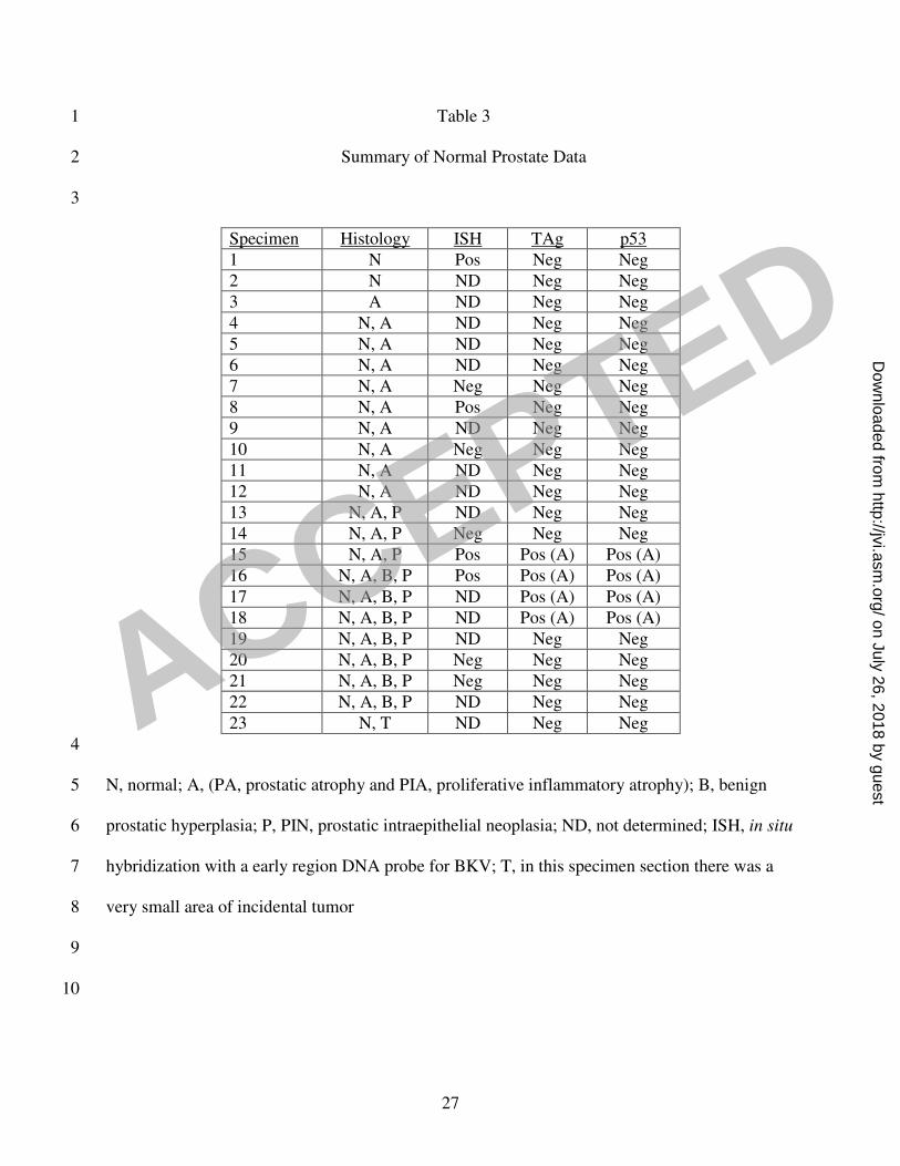
Table 3 1
Summary of Normal Prostate Data 2
3
Specimen Histology ISH TAg p53
1 N Pos Neg Neg
2 N ND Neg Neg
3 A ND Neg Neg
4 N, A ND Neg Neg
5 N, A ND Neg Neg
6 N, A ND Neg Neg
7 N, A Neg Neg Neg
8 N, A Pos Neg Neg
9 N, A ND Neg Neg
10 N, A Neg Neg Neg
11 N, A ND Neg Neg
12 N, A ND Neg Neg
13 N, A, P ND Neg Neg
14 N, A, P Neg Neg Neg
15 N, A, P Pos Pos (A) Pos (A)
16 N, A, B, P Pos Pos (A) Pos (A)
17 N, A, B, P ND Pos (A) Pos (A)
18 N, A, B, P ND Pos (A) Pos (A)
19 N, A, B, P ND Neg Neg
20 N, A, B, P Neg Neg Neg
21 N, A, B, P Neg Neg Neg
22 N, A, B, P ND Neg Neg
23 N, T ND Neg Neg
4
N, normal; A, (PA, prostatic atrophy and PIA, proliferative inflammatory atrophy); B, benign 5
prostatic hyperplasia; P, PIN, prostatic intraepithelial neoplasia; ND, not determined; ISH, in situ 6
hybridization with a early region DNA probe for BKV; T, in this specimen section there was a 7
very small area of incidental tumor 8
9
10
ACCEPTED
on July 26, 2018 by guesthttp://jvi.asm
.org/D
ownloaded from
28
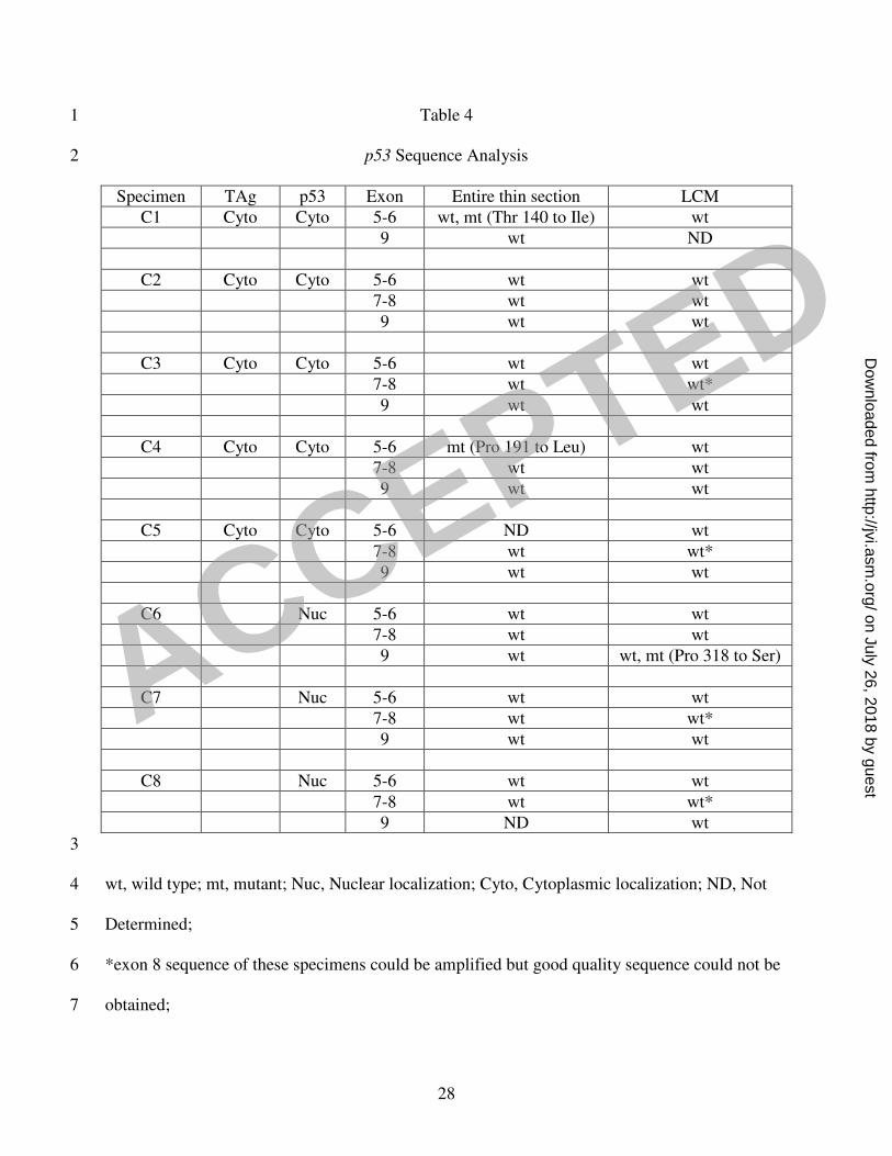
Table 4 1
p53 Sequence Analysis 2
Specimen TAg p53 Exon Entire thin section LCM
C1 Cyto Cyto 5-6 wt, mt (Thr 140 to Ile) wt
9 wt ND
C2 Cyto Cyto 5-6 wt wt
7-8 wt wt
9 wt wt
C3 Cyto Cyto 5-6 wt wt
7-8 wt wt*
9 wt wt
C4 Cyto Cyto 5-6 mt (Pro 191 to Leu) wt
7-8 wt wt
9 wt wt
C5 Cyto Cyto 5-6 ND wt
7-8 wt wt*
9 wt wt
C6 Nuc 5-6 wt wt
7-8 wt wt
9 wt wt, mt (Pro 318 to Ser)
C7 Nuc 5-6 wt wt
7-8 wt wt*
9 wt wt
C8 Nuc 5-6 wt wt
7-8 wt wt*
9 ND wt
3
wt, wild type; mt, mutant; Nuc, Nuclear localization; Cyto, Cytoplasmic localization; ND, Not 4
Determined; 5
*exon 8 sequence of these specimens could be amplified but good quality sequence could not be 6
obtained; 7
ACCEPTED
on July 26, 2018 by guesthttp://jvi.asm
.org/D
ownloaded from

29
Specimen, cancerous prostate; (C1-C5), LCM of TAg+ PIA cells; (C6-C8), LCM of TAg- tumor 1
cells 2
ACCEPTED
on July 26, 2018 by guesthttp://jvi.asm
.org/D
ownloaded from
30
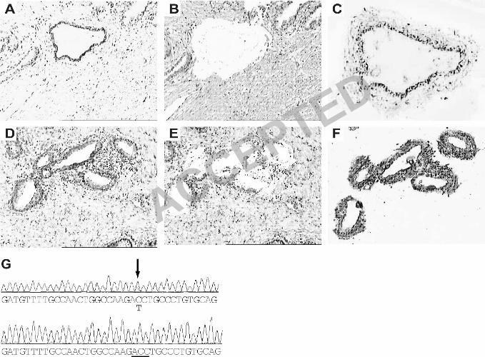
FIGURE LEGENDS 1
2
Figure 1. In situ hybridization and immunohistochemistry of normal prostates. (A), section stained 3
with BKV-specific probe. (B), section immunostained with anti-TAg antibody. (C), PIA duct from a 4
section that was positive for the presence of BKV DNA, showing immunostaining with anti-TAg 5
antibody. (D), PIA duct from the same specimen as (C), immunostained with anti-p53 antibody. 6
Magnification: x 400 (A, C and D); x 100 (B) 7
8
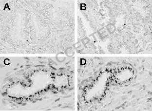
Figure 2. Alignment of ducts for the presence of BKV DNA and TAg expression. (A-C) are serial 9
sections of the same atrophic duct showing hybridization with scrambled probe (A), BKV-specific 10
probe (B) and immunostaining with anti-TAg antibody (C). (D-F) are serial sections of the same 11
normal duct from the same specimen showing hybridization with scrambled probe (D), BKV-12
specific probe (E) and immunostaining with anti-TAg (F). (G) and (H) are identical PIA ducts of the 13
same specimen showing hybridization with BKV-specific probe and immunostaining with anti-TAg, 14
respectively. Magnification: x 100 (A-C); x 200 (D-H). Sections A, B, D, E and F were 15
counterstained with contrast red and C, F and H were counterstained with hematoxylin. The green 16
stain in D, E and F is orientation ink used by the pathologist to maintain the orientation of the 17
exterior surface of the prostate in a paraffin-embedded block. Positive staining for ISH is dark 18
red/black and for IHC is brown. 19
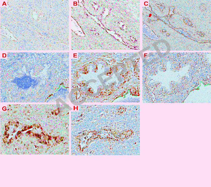
20
Figure 3. Laser capture microdissection of cancerous prostate. (A-C), an atrophic gland 21
immunostained with anti-TAg is shown before laser capture (A, 100X), after the thermoplastic film 22
is removed (B, 100X), and as the captured sample (C, 200X). (D-F), tumor cells immunostained with 23
ACCEPTED
on July 26, 2018 by guesthttp://jvi.asm
.org/D
ownloaded from
31
anti-p53 are shown before laser capture (D, 100X), after removal of thermoplastic film (E, 100X) 1
and in the captured sample (F, 200X). (G), representative chromatogram of specimen 1 from Table 2
3, showing comparison of the DNA sequence of exon 5, nucleotides 13075-13111, of the p53 gene 3
from laser captured TAg-expressing atrophic cells (bottom) and from the entire thin section (top). 4
Arrow shows a mix of wild type (cytosine) and mutant (thymidine) p53sequence, corresponding to 5
amino acid 140. Careful analysis of the chromatogram shows both nucleotides even though the 6
software recognized that position as a cytosine. The underline represents the wild type codon (ACC; 7
Thr) at position140 from laser-captured, TAg-expressing cells. 8
9
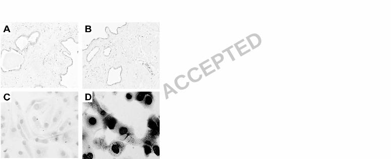
Figure 4. Immunohistochemistry for VP1. A and B are TAg-positive cancerous prostate tissue 10
sections immunostained with IgG2a isotype control or anti-VP1 monoclonal antibody, respectively. 11
(C-D) are mock or BKV-infected kidney epithelial cells in culture, respectively, immunostained with 12
anti-VP1 antibody. Magnification: x 100 (A-B); x 400 (C-D) 13
14
Figure 5. Model of induction of prostate cancer by BKV. See Discussion for details. 15
16 ACCEPTED
on July 26, 2018 by guesthttp://jvi.asm
.org/D
ownloaded from