Biotechnological approaches to the treatment of ... · Biotechnological approaches to the treatment...
11
Biotechnological approaches to the treatment of aspermatogenic men Pedro Manuel Aponte, I,II Stefan Schlatt, III Luiz Renato de Franca I I Federal University of Minas Gerais, Department of Morphology, Minas Gerais, Brazil. II Central University of Venezuela, Department of Anatomy, Maracay, Venezuela. III Center for Reproductive Medicine and Andrology, Mu ¨ nster, Germany. Aspermatogenesis is a severe impairment of spermatogenesis in which germ cells are completely lacking or present in an immature form, which results in sterility in approximately 25% of patients. Because assisted reproduction techniques require mature germ cells, biotechnology is a valuable tool for rescuing fertility while maintaining biological fatherhood. However, this process involves, for instance, the differentiation of preexisting immature germ cells or the production/derivation of sperm from somatic cells. This review critically addresses four potential techniques: sperm derivation in vitro, germ stem cell transplantation, xenologous systems, and haploidization. Sperm derivation in vitro is already feasible in fish and mammals through organ culture or 3D systems, and it is very useful in conditions of germ cell arrest or in type II Sertoli-cell-only syndrome. Patients afflicted by type I Sertoli-cell-only syndrome could also benefit from gamete derivation from induced pluripotent stem cells of somatic origin, and human haploid-like cells have already been obtained by using this novel methodology. In the absence of alternative strategies to generate sperm in vitro, in germ cells transplantation fertility is restored by placing donor cells in the recipient germ-cell-free seminiferous epithelium, which has proven effective in conditions of spermatogonial arrest. Grafting also provides an approach for ex-vivo generation of mature sperm, particularly using prepubertal testis tissue. Although less feasible, haploidization is an option for creating gametes based on biological cloning technology. In conclusion, the aforementioned promising techniques remain largely experimental and still require extensive research, which should address, among other concerns, ethical and biosafety issues, such as gamete epigenetic status, ploidy, and chromatin integrity. KEYWORDS: Spermatogenesis; Azoospermia; Assisted Reproductive Techniques; Transplantation; Spermatozoa; Biotechnology. Aponte PM, Schlatt S, Franca LR. Biotechnological approaches to the treatment of aspermatogenic men. Clinics. 2013;68(S1):157-167. Received for publication on August 21, 2012; Accepted for publication on August 30, 2012 E-mail: [email protected] Tel.: +55 31 3409 2816 & INTRODUCTION Beyond the apparent anatomical simplicity of the male reproductive system, which has a basic design consisting of a pair of gonads with its corresponding excurrent ducts and associated accessory sexual glands, there lies an overwhel- mingly complex system that is responsible for gamete production and transport into the female tract for ultimate sexual reproduction. Although many aspects of the endo- crine regulation of testis function are well understood and therapeutic options for hypogonadal men are available, many aspects of the multiple physiological processes involved in gamete development inside the testis are often deregulated and out of homeostasis, with the subsequent outcome of absent or disturbed spermatogenesis, which leads to infertility. Human infertility is usually defined as the inability of couples to achieve pregnancy after 12 months of unprotected intercourse and is a problem that currently affects 10 to 15% of couples. An outstanding 50% of these cases are associated with male factors (1,2) Specific etiologies of male infertility include systemic diseases (e.g., endocrine, infectious, and cancer), varicoceles, obstructive syndromes, genetic/chromosomal factors, testi- cular failure/hypogonadism, and cryptorchidism. Of all of the causes reported, approximately 12% of the determinants of primary dysfunction in the male reproductive organs are of unknown origin and are usually confounded by the context of multicausal origins (Figure 1). Furthermore, it is clear that many cases of infertility are secondary to general systemic diseases or congenital defects, with reproductive consequences that may be treated when appropriate state-of-the-art procedures are implemen- ted. Data in the literature show that 25 to 75% of cases (depending on the report/study) could theoretically be treated medically and/or surgically (Figure 1). In many cases, very few sperm are present in the testes, and they can Copyright ß 2013 CLINICS – This is an Open Access article distributed under the terms of the Creative Commons Attribution Non-Commercial License (http:// creativecommons.org/licenses/by-nc/3.0/) which permits unrestricted non- commercial use, distribution, and reproduction in any medium, provided the original work is properly cited. No potential conflict of interest was reported. DOI: 10.6061/clinics/2013(Sup01)18 REVIEW 157
Transcript of Biotechnological approaches to the treatment of ... · Biotechnological approaches to the treatment...

Biotechnological approaches to the treatment ofaspermatogenic menPedro Manuel Aponte,I,II Stefan Schlatt,III Luiz Renato de FrancaI
I Federal University of Minas Gerais, Department of Morphology, Minas Gerais, Brazil. II Central University of Venezuela, Department of Anatomy,
Maracay, Venezuela. III Center for Reproductive Medicine and Andrology, Munster, Germany.
Aspermatogenesis is a severe impairment of spermatogenesis in which germ cells are completely lacking orpresent in an immature form, which results in sterility in approximately 25% of patients. Because assistedreproduction techniques require mature germ cells, biotechnology is a valuable tool for rescuing fertility whilemaintaining biological fatherhood. However, this process involves, for instance, the differentiation ofpreexisting immature germ cells or the production/derivation of sperm from somatic cells. This review criticallyaddresses four potential techniques: sperm derivation in vitro, germ stem cell transplantation, xenologoussystems, and haploidization. Sperm derivation in vitro is already feasible in fish and mammals through organculture or 3D systems, and it is very useful in conditions of germ cell arrest or in type II Sertoli-cell-onlysyndrome. Patients afflicted by type I Sertoli-cell-only syndrome could also benefit from gamete derivationfrom induced pluripotent stem cells of somatic origin, and human haploid-like cells have already been obtainedby using this novel methodology. In the absence of alternative strategies to generate sperm in vitro, in germcells transplantation fertility is restored by placing donor cells in the recipient germ-cell-free seminiferousepithelium, which has proven effective in conditions of spermatogonial arrest. Grafting also provides anapproach for ex-vivo generation of mature sperm, particularly using prepubertal testis tissue. Although lessfeasible, haploidization is an option for creating gametes based on biological cloning technology. Inconclusion, the aforementioned promising techniques remain largely experimental and still require extensiveresearch, which should address, among other concerns, ethical and biosafety issues, such as gamete epigeneticstatus, ploidy, and chromatin integrity.
KEYWORDS: Spermatogenesis; Azoospermia; Assisted Reproductive Techniques; Transplantation;Spermatozoa; Biotechnology.
Aponte PM, Schlatt S, Franca LR. Biotechnological approaches to the treatment of aspermatogenic men. Clinics. 2013;68(S1):157-167.
Received for publication on August 21, 2012; Accepted for publication on August 30, 2012
E-mail: [email protected]
Tel.: +55 31 3409 2816
& INTRODUCTION
Beyond the apparent anatomical simplicity of the malereproductive system, which has a basic design consisting of apair of gonads with its corresponding excurrent ducts andassociated accessory sexual glands, there lies an overwhel-mingly complex system that is responsible for gameteproduction and transport into the female tract for ultimatesexual reproduction. Although many aspects of the endo-crine regulation of testis function are well understood andtherapeutic options for hypogonadal men are available,many aspects of the multiple physiological processesinvolved in gamete development inside the testis are oftenderegulated and out of homeostasis, with the subsequent
outcome of absent or disturbed spermatogenesis, which leadsto infertility. Human infertility is usually defined as theinability of couples to achieve pregnancy after 12 months ofunprotected intercourse and is a problem that currentlyaffects 10 to 15% of couples. An outstanding 50% of thesecases are associated with male factors (1,2)
Specific etiologies of male infertility include systemicdiseases (e.g., endocrine, infectious, and cancer), varicoceles,obstructive syndromes, genetic/chromosomal factors, testi-cular failure/hypogonadism, and cryptorchidism. Of all ofthe causes reported, approximately 12% of the determinantsof primary dysfunction in the male reproductive organs areof unknown origin and are usually confounded by thecontext of multicausal origins (Figure 1).
Furthermore, it is clear that many cases of infertility aresecondary to general systemic diseases or congenitaldefects, with reproductive consequences that may be treatedwhen appropriate state-of-the-art procedures are implemen-ted. Data in the literature show that 25 to 75% of cases(depending on the report/study) could theoretically betreated medically and/or surgically (Figure 1). In manycases, very few sperm are present in the testes, and they can
Copyright � 2013 CLINICS – This is an Open Access article distributed underthe terms of the Creative Commons Attribution Non-Commercial License (http://creativecommons.org/licenses/by-nc/3.0/) which permits unrestricted non-commercial use, distribution, and reproduction in any medium, provided theoriginal work is properly cited.
No potential conflict of interest was reported.
DOI: 10.6061/clinics/2013(Sup01)18
REVIEW
157

be retrieved by biopsy and testicular sperm extraction fromthe tissue. Patients with access to assisted reproductivetechnologies (ART) can undergo ICSI (intracytoplasmaticsperm injection), which is routinely used in many fertilitycenters worldwide, to achieve pregnancy. However, thegroup of patients for whom ART procedures are not anoption deserves special attention. These men present withazoospermia and varying degrees of germ cell pathology,from germinal aplasia to germ cell premeoitic/meioticarrest. This group of testicular pathologies has a poorprognosis and holds the status of being untreatable andincurable from a medical point of view. We propose thatmen with incomplete spermatogenesis could be collectivelyclassified as aspermatogenic, i.e., having a severe testicularpathology with a complete absence of spermatids. Such mencomprise from 25 to 50% of andrological cases (3,4) (seeFigure 1), and the only option currently available for thesepatients is adoption or artificial insemination using donorsperm.
Histopathologic findings in biopsies from patients in theaspermatogenic group can be classified as: a) Sertoli cellonly (SCO) syndrome, which strictly consists of testespresenting a complete absence of germ cells; b) arrest atthe level of meiotic germ cells; or c) spermatogonia only(pre-meiotic arrest) (Figure 2).
Sertoli cell only syndrome, i.e., type I SCO, refers to asituation in which testicular biopsy reveals an absence ofgerm cells in the seminiferous tubules (5). The presence offew seminiferous tubules showing active spermatogenesishas also been referred as to focal SCO (6) or type II SCO
(MeSH database PubMed). However, in contrast to patientswith complete SCO, these patients benefit from ARTprocedures, as their sperm can be retrieved by TESE(testicular sperm extraction). Type I SCO can have agenetically determined developmental origin that results inthe disturbance of migration and/or colonization of primor-dial germ cells (PGCs) into the genital ridges. Because thediagnosis of type I or type II SCO depends on histopathologicanalysis, which is of course prone to sampling error (becausebiopsies cover only a very small portion of the testis), thesubtypes of SCO are often misdiagnosed. The situationwould present less of a diagnostic challenge if a reliable, non-invasive method were available to distinguish cases of partialand complete absence of germ cells. The first steps in thisdirection involved the amplification of specific germ cells,seminal vesicles and prostate mRNA from cell-free seminalmRNA by using RT-PCR (7). Combinations of non-invasivediagnostic procedures with other procedures, such ashormonal profiles and histopathology, may unequivocallyhelp to diagnose patients correctly. As type I SCO patientshave no germ cells, the only possible way to generateoffspring would be the use of their somatic cells to generategermline cells in vitro.
Arrest represents a situation in which spermatogenesisstops at a specific stage of germ cell development.McLachlan et al. proposed a nomenclature in which theterm arrest exclusively refers to histological samples inwhich no progression occurs beyond that specific stage inany seminiferous tubule (5). Arrest can represent anintermediate step on the way to more severe germ cell
Figure 1 - Frequency of male infertility etiological groups determined from reproductive clinic records. Percentages for each categoryare presented as ranges (minimum and maximum values in the reported literature) (3,4). Patients from Group A (27.8 to 73.8% of cases)can undergo medical or surgical treatments to address their infertility problems. An unknown percentage of cases within Group B(idiopathic azoospermia) and perhaps even some from Group C can benefit from ART, provided that their sperm can be retrieved. Mensuffering from aspermatogenesis, with some type of premeiotic/meiotic spermatogenic disturbance, are included in Group C.
Artificial sperm generationAponte PM et al.
CLINICS 2013;68(S1):157-167
158

losses. Arrest occurs often at the spermatocytes or sperma-togonial level (12.5% and 1.7% of 534 patients, respectively)(5). Arrest during spermiogenesis is rare, and hyposperma-togenesis (often misdiagnosed as arrest), in which a lowerspermatid count is combined with the presence of allspermatogenic stages, which are reduced to some extent,accounts for up to 63% of cases (7). In any case, becausehypospermatogenesis results in the presence of spermatids,ART procedures are applicable. Given that germ cells arepresent in cases of arrest, an interesting strategy would be todifferentiate these cells in vitro to obtain sperm for ICSI orIVF (in vitro fertilization). Interestingly, in arrest at the levelof meiotic cells, these cells can be injected into oocytes toproduce viable embryos and even offspring (8-10).Nevertheless, intracytoplasmatic spermatocyte injection isonly an experimental option with low efficiency. For instance,only 15% of mouse oocytes injected with secondary sperma-tocytes generated offspring, whereas those injected withprimary spermatocytes generated none (8). Furthermore, inanother set of experiments, from 0 to 9% of eggs ‘‘fertilized’’by primary spermatocytes developed into adult fertile mice(9). In humans, one report showed an offspring-derivingefficiency of approximately 3% (10). For these reasons, theuse of spermatocytes to compensate for a lack of more maturemale germ cells will require further experimentation.
Many patients believe that biological fatherhood has avery high value. In this respect, biotechnology offers, nowmore than ever, a basis on which to develop clinicalapplications for such cases, to restore fertility and conse-quently to obtain offspring. In this context, advances in cell
biology from several animal models are waiting to betranslated into human reproductive medicine, provided thatsome technical issues and biosafety and ethical concerns areovercome. We propose four main groups of technologies that,alone or combined, could bring hope to men with severecases of aspermatogenesis: i) sperm derivation in vitro; ii)germ stem cell transplantation; iii) xenologous systems; andiv) haploidization. These topics are addressed below.
& SPERM DERIVATION IN VITRO
The complex spatial and functional organization of thetestis and its seminiferous epithelium make it difficult toachieve efficient spermatogenesis in artificial systems. Theobservation of at least a few aspects of spermatogenesis inculture dishes has been a scientific aim for decades (17,18).Spermatogenesis in vitro is not only a long-desiredtechnique to gain deeper insight into the complex processof gametogenesis; it is also desired for clinical applicationsto broaden the therapeutic options for men with infertilityproblems. Given the current status of ART and therequirement of only one sperm to fertilize an oocyte throughICSI, the in vivo physiological situation, in which millions ofsperm (many more than needed) are produced on a dailybasis may be surpassed by in vitro strategies, which may beinefficient but which can generate a few fully mature spermfor ART procedures.
Spermatogenesis in vitro was first successfully achievedin vertebrates by using a fish model that consisted of a tissueculture system that supported full spermatogenesis inJapanese eels (19,20), which opened the possibility for
Figure 2 - Schematic summary of the types of seminiferous epithelium impairment that can lead to infertility in men. Group Isummarizes pathologies in which germ cell arrest occurs at spermiogenesis. (a) Presents an apparently normal seminiferous tubule withapparently normal sperm and elongated spermatids as the most advanced cell types present. Several pathologies show amorphologically normal seminiferous epithelium, as depicted, but sperm can still present a pathological condition and therefore beunable to fertilize an egg. Many of these cases can be solved by using ART, i.e., testicular sperm extraction (TESE) combined with ICSI.(b) Arrest at the level of round spermatids, which can be used for ROSI (round spermatid injection). This type of arrest is rare. Group IIrepresents arrest at the levels of (c) spermatocytes (12.5 % of cases) or (d) spermatogonial cells (1.7 % of cases) and (e) total absence ofgerm cells, as in SCO syndrome (estimated to occur in 16.5 % of cases, based on data from Tuttelmann (2010) (3), with detailedetiological entities including chromosomal aberrations, as in Klinefelter (11), XX males (12), translocations (13,14) and microdeletionsat the level of the AZF regions of the Y chromosome (15,16). Prevalence data are from McLachlan et al. (2007) (5), except for the SCOdata (3).
CLINICS 2013;68(S1):157-167 Artificial sperm generationAponte PM et al.
159
similar technological advances in higher vertebrates.Systems for in vitro spermatogenesis in fish seem to bevery simple from a practical point of view. For example, onesuch system based on a spermatogonial cell line withoutSertoli cells and involving medaka fish, allowed postmeioticadvances in the presence of a culture medium containing acrude embryo extract with no specific growth factors added(21), which introduced the idea that germ cells in lowervertebrates are more intrinsically programmed and lessdependent on regulation by extrinsic signals.
Currently, two main experimental approaches dominatethe efforts to generate mammalian sperm in culture systems:organ culture of testes or testicular fragments that maintainthe complex architecture of testicular tissue and itsarrangement inside the epithelium. A recent article bySato et al. convincingly demonstrated, in what has beendescribed as a great scientific breakthrough, the feasibility ofobtaining viable and apparently normal offspring fromsperm generated through testicular tissue explants thatinitially contained immature germ cells (22). In thisapproach, the cytoarchitecture of the gonad remains widelypreserved in the system, many endogenous factors areproduced and released by the mostly intact seminiferousepithelium, and associated somatic cells regulate the germcells in the system similarly to the in vivo condition. Indissected tissue, the supply of oxygen and nutrients isdisturbed, and therefore the spermatogenic process is oftendisrupted and continues, at best, at low efficiency (22).Alternatively, enzymatically digested cell suspensions canbe used for various culture systems. The supply of oxygenand nutrients to isolated cells is achieved by modern cellculture media under appropriate culture conditions.However, the seminiferous tubule microenvironment, withits different, functionally distinct compartments (e.g., inter-stitial, basal, intraepithelial, and adluminal), is destroyed. Ifthese changes in the microenvironment during germ celldifferentiation are significant, then spermatogenesis will bedisrupted. Although many additional strategies of cell andtissue culture have been tested over the last 30 years, successhas been limited (23-29). Recent studies have used mono-layers other than those generated from Sertoli cells assources of differentiating factors (30-33). Because theproduction of sperm in higher vertebrates involves anoverwhelmingly intricate process of germ cell differentia-tion, further research efforts have been directed towardidentifying signaling pathways associated with differentia-tion in the seminiferous epithelium during spermatogenesis.To incorporate this knowledge into the development of invitro spermatogenesis systems, studies that have addressedthe presence of substances such as retinol (34,35), kit ligand(stem cell factor) (36,37), insulin-like growth factor andtransforming growth factor alpha (TGF-alpha) (38), as wellas hormones such as testosterone (39) and FSH (40), amongothers, have been instrumental in recapitulating the entiredifferentiation process. These studies will become particu-larly valuable if the paradigm of spermatogenesis in vitro,as a model of the in vivo situation, is accepted. Therefore,other factors, such as scrotal temperature, the timing ofcellular events and the presence of important culture mediacomponents, e.g., fetal calf serum, oxygen levels, pressure,substrates and the use of feeder layers, should be carefullyevaluated and whenever possible translated from the invivo situation to the artificial system. The use of Sertoli cellsis controversial but, in principle, these cells are important
for the maintenance of germ cells in culture and to providedifferentiating factors (41-44). Among the advantages of cellculture systems is that such systems (cell cultures startedfrom dissociated germ cell suspensions) are much simplerthan tissue cultures, in which cell function is tightlyregulated and more complex, as in the in vivo situation.Cell cultures can be started with purified, selected, isolatedcells, thereby allowing uncomplicated manipulationschemes because signal redundancy, which is observed inintact biological systems, is minimized. Furthermore, as thefirst step of the procedure, germ cells can be stimulated toproliferate in vitro, which creates the conditions suitable toobtain large numbers of spermatogonial stem cells beforeinducing them to differentiate during the process of gameteproduction.
Recently, a novel three-dimensional testicular cell cultureapproach was described. The soft agar culture system(SACS), which was originally described for the culture ofhematopoietic cells in vitro, has created the conditionsnecessary to enable the full development of murine malegerm cells in vitro (45-47). This system provides options formanipulation, e.g., the testing of the effects of hormones,drugs, toxins or other compounds on germ cell develop-ment. We were able to demonstrate the progression ofspermatogonia into meiosis and the cells’ further matura-tion into morphologically normal spermatozoa. However,the efficiency of sperm production with SACS is very low.The success of such strategies may depend on thereconstruction of small functional units of testicular cellsinside the matrix, which occasionally fully resembles themicroenvironment in the seminiferous tubules and thusresults in small islands of complete spermatogenesis.
To recapitulate the situation in the testis, researchers mustsequentially and gradually provide the right substances/conditions at the proper time, thereby emulating the in vivoprocesses that occur in an orderly fashion in differentfunctional compartments (e.g., basal, meiotic, and adlum-inal) of the seminiferous epithelium during normal in vivospermatogenesis. Apparently, the most challenging step ofdifferentiation to achieve in an in vitro spermatogenicsystem is proceeding beyond meiosis (41). Hence, progressmust be experimentally monitored by meiotic/postmeiotic-specific markers, by immunohistochemistry and/or by PCR.Some examples are summarized in Table 1. The need todistinguish somatic cells renders the use of markers for thiscell group useful (Table 2).
These findings have greatly inspired further researchinvolving humans in the hope of reversing cases ofincomplete spermatogenesis. This type of approach wouldrequire biopsy procedures, which usually yield smalltesticular samples with limited numbers of germ cells. Ifthe availability of testicular tissue is not an obstacle, thenfertility patients with immature germ cell arrest couldexpect to overcome infertility by using this procedure.Moreover, cell culture systems for in vitro spermatogenesiswould provide advantages not only to germ cell arrestpatients but also to type II SCO syndrome cases or caseswith other reproductive pathologies involving faulty sper-matogonial stem cell niches.
Current technology allows, at least on the theoreticallevel, SCO syndrome patients, who have no germ cells, toachieve the goal of producing offspring that retain their owngenetic material. To this end, biotechnology must firstprovide gametes to help these men overcome their fertility
Artificial sperm generationAponte PM et al.
CLINICS 2013;68(S1):157-167
160

problems. Pluripotent stem cells could be a reliable andplentiful source of immature germ cells to provide abaseline for a differentiation process that eventually willyield spermatozoa in vitro. Pluripotency is the property of acell to differentiate into somatic cells of ectodermal,endodermal and mesodermal lineages, which constitutethe vast majority of the cells in the body (94). It has beendemonstrated that some pluripotent stem cells can alsogenerate germ cells. Pluripotent stem cells can be basicallyobtained from two sources: a) the epiblast region inblastodermic embryos, and b) adult somatic cells. The lattersource provides an opportunity for success in the greatchallenge of obtaining pluripotent stem cells from adultpatients, despite the conventional wisdom in cell biologythat cells in tissues tend to progress into lineage restrictionsduring development, gradually reaching terminal differen-tiation. The idea of somatic pluripotentiation, or reprogram-ming, has been discussed for quite some time and foundoriginal success in mouse models (95). Efforts continuedshortly thereafter using human cells (96,97). Only recently,independent research groups (98,99) went a step further toachieve the major breakthrough of defining four basicpluripotency-inducing factors, i.e., Oct4 (pouf1), Sox2, c-Myc, and Klf4, which are required to switch somatic cellsinto a pluripotent state, thus opening a window for deeperunderstanding of pluripotency networks, as well as thepossibility of generating germ cells, among other cell types,from somatic cells induced to be pluripotent (iPS cells). Theavailability of such personalized germ cells of somatic originwould be a prerequisite for an in vitro sperm-generatingsystem for SCO patients.
The development of in vitro spermatogenesis has bene-fited from various animal models, including models that usefarm animals. Some culture systems were developed indomestic animal models in which germ cells progressed toadvanced steps of spermatogenesis. For instance, Izadyar etal. observed elongated spermatids in a long-term culture ofbovine germ cells (70), whereas Dong et al. obtainedelongated spermatids from fetal calf gonocyte cultures(100). In these species, the available procedures proved tobe rather inefficient because not many terminally differ-entiated gametes could be derived. In parallel, murinemodels involving ES (embryonic stem) cells resulted inadvances in the field. In principle, germ cell derivation fromES cells does not seem to be practical for infertility patients,as almost no man has access to his own embryonic cells.However, because iPS cells appear to be similar to ES cells(101), and in nature, technically derivable from somaticcells, the resulting scenario becomes very promising to drivethem into the production of gametes in vitro. Efforts togenerate germ cells and gametes from ES cells and iPS cellsin murine models and humans are summarized on Figure 3.
Briefly, three research groups have made advances in thederivation of germ cells from mouse ES cells. One of thesegroups produced gamete-like cells, and using ICSI as afunctional assay, they managed to obtain embryos afterfertilization had reached the blastocyst stage (102). Twolaboratories were able to generate sperm in vitro (31,103),but only one of them obtained offspring by injecting thederived sperm into donor oocytes using ICSI (31). However,in this case, the offspring production had low efficiency (7out of 65 embryos), and the mice that were born died
Table 1 - Testicular germ cell markers.
Cell type Marker
Germ cell general VASA (48) h
Gonocytes ZBTB16 (49) m,h
Undifferentiated type A spermatogonia (single) ID4 (50) m; CSF1R (51) m
Undifferentiated type A spermatogonia (single and paired) EFR3 (52) m
Undifferentiated type A spermatogonia (single, paired and aligned) THY1 (53) m; UTF1 (54) m; PLZF (55) m; LIN28 (TEX17) (56) m; NGN3 (57) m;
NANOS3 (52) m; CD24 (53) m; GFRalpha1 (57) m; CDH1 (58) m; GPR125 (59) m;
SOHLH2 (60) m; RET (61) m; BCL6B (62) m; SOX3 (63) m; SALL4 (64) p
Spermatogonia (general) HSP60 (65) h; MAGEA-4 (66) h; UCHL1 (PGP9.5) (66) h; ITGA6
(alpha6 integrin) (66) h; ZBTB16 (66) h, (49) m,h; Kit (36) m
Pachytene spermatocytes testis specific histone (TH2B)(67) r; phosphoprotein P19 (68) r; SPTRX-3 (69) h
Spermatocytes (general) SCP-3 (70) b, (71) h; GRP78 (65) h; Kit (66) h; MLH1 (71) h
Round spermatids Transition proteins T1, T2 (72,73) h; protamine 2 (PRM-2) (74); CREM (75) h,m;
GRP78 (65) h; Kit (66) h
Elongated spermatids CRES (76) h
Spermatids (general) SPANX (a subset) (77) h; KLF4 (78) h; SPTRX-3 (69) h; SP-10 (79) h; TGFbeta3 (80) h
Sperm outer dense fibers (ODF-2) (70); acrosin (81) b; SPANX (77); HSP60 (65) h; GRP78
(65) h; CRES (76) h; DAZ2 (82) h
m, mouse; r, rat; d, dog; b, bovine; p, non-human primate; h, human.
Table 2 - Testicular somatic cell markers.
Cell type Marker
Sertoli immature AMH (anti-mullerian hormone) (83) p; WT1 (Wilms’ tumor gene)- transcription factor (83) r; aromatase (P450
enzymes) (83) r; NCAM (neural cell adhesion molecule) (83) r; cytokeratin 18 (83,84) h; M2A (83) h
Sertoli mature occludin (85) m,r,d; vimentin (86) r, (43,87) b; P27Kip 1 (cyclin-dependent kinase inhibitor) (83) m,r,h; WT1
(Wilms tumor gene)- transcription factor (83) r; Dmrt1 (88) m; Gata 4 (88) m; Gata 1(83) m; AR (androgen
receptor) (83) r,p,h; transferrin (88) r; ITGA6 (alpha6 integrin) (66) h
Leydig cells ITGalpha6 (alpha6 integrin) (66) h; RLF (89) m; 3 beta-HSD (90) g, (91) r; TGF alpha (92) r
Peritubular myoid cells alpha-smooth muscle actin (93) r, (43,87) b
m, mouse; r, rat; g, guinea pig; d, dog; b, bovine; p, non-human primate; h, human.
CLINICS 2013;68(S1):157-167 Artificial sperm generationAponte PM et al.
161

Figure 3 - Recent advances in the derivation of germ cells / gametes from ES cells and iPS cells in mouse models (A) and in humans (B).ESC, embryonic stem cells; iPS, induced pluripotent stem cells; PGCs, primordial germ cells; SSC, spermatogonial stem cells; HL, haploid-like cells; ICSI, intracytoplasmatic sperm injection. In (A) arrows point into the cell types generated by each specific research group. Greyletter X and arrow in (B) indicate advances not yet accomplished.
Artificial sperm generationAponte PM et al.
CLINICS 2013;68(S1):157-167
162
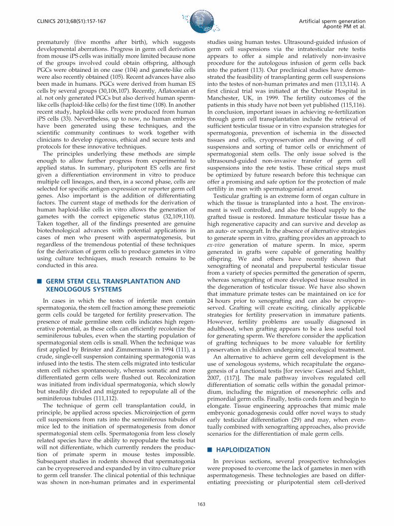
prematurely (five months after birth), which suggestsdevelopmental aberrations. Progress in germ cell derivationfrom mouse iPS cells was initially more limited because noneof the groups involved could obtain offspring, althoughPGCs were obtained in one case (104) and gamete-like cellswere also recently obtained (105). Recent advances have alsobeen made in humans. PGCs were derived from human EScells by several groups (30,106,107). Recently, Aflatoonian etal. not only generated PGCs but also derived human sperm-like cells (haploid-like cells) for the first time (108). In anotherrecent study, haploid-like cells were produced from humaniPS cells (33). Nevertheless, up to now, no human embryoshave been generated using these techniques, and thescientific community continues to work together withclinicians to develop rigorous, ethical and secure tests andprotocols for these innovative techniques.
The principles underlying these methods are simpleenough to allow further progress from experimental toapplied status. In summary, pluripotent ES cells are firstgiven a differentiation environment in vitro to producemultiple cell lineages, and then, in a second phase, cells areselected for specific antigen expression or reporter germ cellgenes. Also important is the addition of differentiatingfactors. The current stage of methods for the derivation ofhuman haploid-like cells in vitro allows the generation ofgametes with the correct epigenetic status (32,109,110).Taken together, all of the findings presented are genuinebiotechnological advances with potential applications incases of men who present with aspermatogenesis, butregardless of the tremendous potential of these techniquesfor the derivation of germ cells to produce gametes in vitrousing culture techniques, much research remains to beconducted in this area.
& GERM STEM CELL TRANSPLANTATION ANDXENOLOGOUS SYSTEMS
In cases in which the testes of infertile men containspermatogonia, the stem cell fraction among these premeioticgerm cells could be targeted for fertility preservation. Thepresence of male germline stem cells indicates high regen-erative potential, as these cells can efficiently recolonize theseminiferous tubules, even when the starting population ofspermatogonial stem cells is small. When the technique wasfirst applied by Brinster and Zimmermann in 1994 (111), acrude, single-cell suspension containing spermatogonia wasinfused into the testis. The stem cells migrated into testicularstem cell niches spontaneously, whereas somatic and moredifferentiated germ cells were flushed out. Recolonizationwas initiated from individual spermatogonia, which slowlybut steadily divided and migrated to repopulate all of theseminiferous tubules (111,112).
The technique of germ cell transplantation could, inprinciple, be applied across species. Microinjection of germcell suspensions from rats into the seminiferous tubules ofmice led to the initiation of spermatogenesis from donorspermatogonial stem cells. Spermatogonia from less closelyrelated species have the ability to repopulate the testis butwill not differentiate, which currently renders the produc-tion of primate sperm in mouse testes impossible.Subsequent studies in rodents showed that spermatogoniacan be cryopreserved and expanded by in vitro culture priorto germ cell transfer. The clinical potential of this techniquewas shown in non-human primates and in experimental
studies using human testes. Ultrasound-guided infusion ofgerm cell suspensions via the intratesticular rete testisappears to offer a simple and relatively non-invasiveprocedure for the autologous infusion of germ cells backinto the patient (113). Our preclinical studies have demon-strated the feasibility of transplanting germ cell suspensionsinto the testes of non-human primates and men (113,114). Afirst clinical trial was initiated at the Christie Hospital inManchester, UK, in 1999. The fertility outcomes of thepatients in this study have not been yet published (115,116).In conclusion, important issues in achieving re-fertilizationthrough germ cell transplantation include the retrieval ofsufficient testicular tissue or in vitro expansion strategies forspermatogonia, prevention of ischemia in the dissectedtissues and cells, cryopreservation and thawing of cellsuspensions and sorting of tumor cells or enrichment ofspermatogonial stem cells. The only issue solved is theultrasound-guided non-invasive transfer of germ cellsuspensions into the rete testis. These critical steps mustbe optimized by future research before this technique canoffer a promising and safe option for the protection of malefertility in men with spermatogonial arrest.
Testicular grafting is an extreme form of organ culture inwhich the tissue is transplanted into a host. The environ-ment is well controlled, and also the blood supply to thegrafted tissue is restored. Immature testicular tissue has ahigh regenerative capacity and can survive and develop asan auto- or xenograft. In the absence of alternative strategiesto generate sperm in vitro, grafting provides an approach toex-vivo generation of mature sperm. In mice, spermgenerated in grafts were capable of generating healthyoffspring. We and others have recently shown thatxenografting of neonatal and prepubertal testicular tissuefrom a variety of species permitted the generation of sperm,whereas xenografting of more developed tissue resulted inthe degeneration of testicular tissue. We have also shownthat immature primate testes can be maintained on ice for24 hours prior to xenografting and can also be cryopre-served. Grafting will create exciting, clinically applicablestrategies for fertility preservation in immature patients.However, fertility problems are usually diagnosed inadulthood, when grafting appears to be a less useful toolfor generating sperm. We therefore consider the applicationof grafting techniques to be more valuable for fertilitypreservation in children undergoing oncological treatment.
An alternative to achieve germ cell development is theuse of xenologous systems, which recapitulate the organo-genesis of a functional testis [for review: Gassei and Schlatt,2007, (117)]. The male pathway involves regulated celldifferentiation of somatic cells within the gonadal primor-dium, including the migration of mesonephric cells andprimordial germ cells. Finally, testis cords form and begin toelongate. Tissue engineering approaches that mimic maleembryonic gonadogenesis could offer novel ways to studyearly testicular differentiation (29) and may, when even-tually combined with xenografting approaches, also providescenarios for the differentiation of male germ cells.
& HAPLOIDIZATION
In previous sections, several prospective technologieswere proposed to overcome the lack of gametes in men withaspermatogenesis. These technologies are based on differ-entiating preexisting or pluripotential stem cell-derived
CLINICS 2013;68(S1):157-167 Artificial sperm generationAponte PM et al.
163

germ cells in vitro (spermatogenesis in vitro) or on in vivosystems (transplantation and xenografting) to producesperm.
Haploidization is an alternative approach to manufacturegametes based on biological cloning technology. In cloning,an entire genome is obtained from a single individual whenhe donates a somatic cell nucleus, which is transferred into anenucleated, developmentally young cell (MII oocyte, zygote,embryo) (118). In contrast, haploidization is a modifiedcloning technique in which there is a genetic haploidcontribution from both parents, which is one reason whythis technique has also been called semi-cloning (119,120).
Although promising, this technology has not beensuccessful for generating live normal offspring in murineexperimental models, and therefore it remains largelyexperimental, with no immediate prospects for clinicalapplication. For instance, in preliminary experiments, only17 to 22% of mouse oocytes ‘‘fertilized’’ with somatic cellsformed blastocysts, and no offspring were derived fromthem (121).
& EXPERT COMMENTS
This review article provides an overview of severalbiotechnological approaches that have the potential to offersolutions for a range of reproductive pathologies that causesterility in a significant number of azoospermic men, whileat the same time providing new tools to contribute to ourcurrent knowledge of male reproductive physiology.Although they are thus far experimental, the proposedemerging technologies could work in combination withother already well-established reproductive technologiesand take advantage of the requirement for just one healthyspermatozoon to ensure successful fertilization and, hope-fully, normal offspring from these individuals.
Thus far, only haploid-like cells, with an apparentlycorrect epigenetic status, have been generated by severalresearch groups. In the near future, we can expect thegeneration of sperm as the most natural, terminallydifferentiated cells to be used in assisted reproduction formen afflicted with aspermatogenesis.
Regardless of the pervading optimism, remaining chal-lenges include the verification of aspects such as normalcytogenetics, epigenetic stability, the tumorogenic potentialof derived germ cells and even ethical aspects, which shouldguarantee the correct implementation of these new repro-ductive technologies. Additionally, the genetic basis formany conditions of aspermatogenesis will necessitate cau-tion, as well as a consideration of the need to develop genetherapies that are specific for these reproductive problems toprevent their transmission to future generations.
& KEY ISSUES
N At least 25% of azoospermic men are sterile becausetheir testes either hold only immature or no germ cells at all,a condition that can be collectively termed as aspermato-genesis.
N Possible solutions to aspermatogenesis require biotech-nological approaches to retain biological fatherhood.
N Azoospermic men with germ cell arrests at early stagesof spermatogenesis would benefit from sperm derivation invitro or ex vivo.
N Azoospermic men with type II SCO would have thealternatives of germ cell derivation from a) somatic cells viainduced pluripotency or b) haploidization.
N The most challenging differentiation step in an in vitrospermatogenic system is completion of meiosis which hasbeen achieved in several animal models by ex vivoapproaches or 3D culture systems.
N In the absence of alternative strategies, testiculargrafting (auto- or xenograft) should be considered to ex-vivo generate mature sperm and would be more valuablefor fertility preservation in children undergoing oncologicaltreatment.
N Haploidization is a modified cloning technique, inwhich each parent genetically contributes with a haploidcomplement, but unfortunately this technique offers noimmediate perspective for clinical application.
N Although extensive research has been and continues tobe performed, biotechnologies described on this reviewarticle still remain largely experimental.
& ACKNOWLEDGMENTS
This study was supported by CNPq and FAPEMIG.
& AUTHOR CONTRIBUTIONS
All of the authors who are listed participated sufficiently in the present
review article and therefore take public responsibility for its content.
& REFERENCES
1. Evers JL. Female subfertility. Lancet. 2002;360(9327):151-9, http://dx.doi.org/10.1016/S0140-6736(02)09417-5.
2. Tuttelmann F, Simoni M, Kliesch S, Ledig S, Dworniczak B, Wieacker P,et al. Copy number variants in patients with severe oligozoospermiaand Sertoli-cell-only syndrome. PloS one. 2011;6(4):e19426, http://dx.doi.org/10.1371/journal.pone.0019426.
3. Tuttelmann F, Werny F, Cooper TG, Kliesch S, Simoni M, Nieschlag E.Clinical experience with azoospermia: aetiology and chances forspermatozoa detection upon biopsy. Int J Androl. 2011;34(4):291-8.
4. Hamada A, Esteves SC, Agarwal A. Unexplained male infertility:potential causes and management. Hum Androl. 2011;1:2-16, http://dx.doi.org/10.1097/01.XHA.0000397686.82729.09.
5. McLachlan RI, Rajpert-De Meyts E, Hoei-Hansen CE, de Kretser DM,Skakkebaek NE. Histological evaluation of the human testis -approaches to optimizing the clinical value of the assessment: MiniReview. Hum Reprod. 2007;22(1):2-16.
6. Anniballo R, Brehm R, Steger K. Recognising the Sertoli-cell-only (SCO)syndrome: a case study. Andrologia. 2011;43(1):78-83, http://dx.doi.org/10.1111/j.1439-0272.2009.01030.x.
7. Li HG, Wu CL, Gu XL, Xiong CL. A novel application of cell-freeseminal mRNA: non-invasive identification of the presence of germcells or complete obstruction in men with azoospermia. Hum Reprod.2012;27(4):991-7, http://dx.doi.org/10.1093/humrep/der481.
8. Kimura Y, Yanagimachi R. Development of Normal Mice from OocytesInjected with Secondary Spermatocyte Nuclei. Biol Reprod. 1995;53(4):855-62, http://dx.doi.org/10.1095/biolreprod53.4.855.
9. Ogura A, Suzuki O, Tanemura K, Mochida K, Kobayashi Y, Matsuda J.Development of normal mice from metaphase I oocytes fertilized withprimary spermatocytes. P Natl Acad Sci USA. 1998;95(10):5611-5,http://dx.doi.org/10.1073/pnas.95.10.5611.
10. Sofikitis N, Mantzavinos T, Loutradis D, Yamamoto Y, Tarlatzis V,Miyagawa I. Ooplasmic injections of secondary spermatocytes for non-obstructive azoospermia. Lancet. 1998;351(9110):1177-8, http://dx.doi.org/10.1016/S0140-6736(05)79121-2.
11. Aksglaede L, Wikstrom AM, Rajpert-De Meyts E, Dunkel L, SkakkebaekNE, Juul A. Natural history of seminiferous tubule degeneration inKlinefelter syndrome. Hum Reprod Update. 2006;12(1):39-48.
12. Yamamoto M, Yokoi K, Katsuno S, Hibi H, Miyake K. A case of sexreversal syndrome with sex-determining region (XX male). Nagoyajournal of medical science. 1995;58(3-4):111-5.
13. Shapiro CE. Unbalanced chromosomal translocation associated withSertoli-cell-only histology. J Urol. 1991;145(3):563-4.
Artificial sperm generationAponte PM et al.
CLINICS 2013;68(S1):157-167
164

14. Cacheiro NL, Russell LB, Swartout MS. Translocations, the predomi-nant cause of total sterility in sons of mice treated with mutagens.Genetics. 1974;76(1):73-91.
15. Kamp C, Huellen K, Fernandes S, Sousa M, Schlegel PN, Mielnik A,et al. High deletion frequency of the complete AZFa sequence in menwith Sertoli-cell-only syndrome. Mol Hum Reprod. 2001;7(10):987-94,http://dx.doi.org/10.1093/molehr/7.10.987.
16. Yang Y, Ma MY, Xiao CY, Li L, Li SW, Zhang SZ. Massive deletion inAZFb/b plus c and azoospermia with Sertoli cell only and/ormaturation arrest. Int J Androl. 2008;31(6):573-8.
17. Steinberger A, Steinberger E. In vitro culture of rat testicular cells. ExpCell Res. 1966;44(2):443-52, http://dx.doi.org/10.1016/0014-4827(66)90451-4.
18. Goldschmidt R. Some experiments on spermatogenesis in vitro. P NatlAcad Sci USA. 1915;1:220-2, http://dx.doi.org/10.1073/pnas.1.4.220.
19. Miura T, Yamauchi K, Takahashi H, Nagahama Y. Human Chorionic-Gonadotropin Induces All Stages of Spermatogenesis Invitro in theMale Japanese Eel (Anguilla-Japonica). Dev Biol. 1991;146(1):258-62,http://dx.doi.org/10.1016/0012-1606(91)90468-I.
20. Miura T, Yamauchi K, Takahashi H, Nagahama Y. Hormonal inductionof all stages of spermatogenesis in vitro in the male Japanese eel(Anguilla japonica). Proc Natl Acad Sci U S A. 1991;88(13):5774-8,http://dx.doi.org/10.1073/pnas.88.13.5774.
21. Hong Y, Liu T, Zhao H, Xu H, Wang W, Liu R, et al. Establishment of anormal medakafish spermatogonial cell line capable of sperm produc-tion in vitro. Proc Natl Acad Sci U S A. 2004;101(21):8011-6, http://dx.doi.org/10.1073/pnas.0308668101.
22. Sato T, Katagiri K, Gohbara A, Inoue K, Ogonuki N, Ogura A, et al. Invitro production of functional sperm in cultured neonatal mouse testes.Nature. 2011;471(7339):504-7, http://dx.doi.org/10.1038/nature09850.
23. Tesarik J, Greco E, Rienzi L, Ubaldi F, Guido M, Cohen-Bacrie P, et al.Differentiation of spermatogenic cells during in-vitro culture oftesticular biopsy samples from patients with obstructive azoospermia:effect of recombinant follicle stimulating hormone. Hum Reprod.1998;13(10):2772-81, http://dx.doi.org/10.1093/humrep/13.10.2772.
24. Tanaka A, Nagayoshi M, Awata S, Mawatari Y, Tanaka I, Kusunoki H.Completion of meiosis in human primary spermatocytes through invitro coculture with Vero cells. Fertil Steril. 2003;79:795-801, http://dx.doi.org/10.1016/S0015-0282(02)04833-1.
25. Parvinen M, Wright WW, Phillips DM, Mather JP, Musto NA, BardinCW. Spermatogenesis Invitro - Completion of Meiosis and EarlySpermiogenesis. Endocrinology. 1983;112(3):1150-2, http://dx.doi.org/10.1210/endo-112-3-1150.
26. Tres LL, Kierszenbaum AL. Viability of Rat Spermatogenic Cells-InvitroIs Facilitated by Their Co-Culture with Sertoli Cells in Serum-FreeHormone-Supplemented Medium. P Natl Acad Sci-Biol. 1983;80(11):3377-81, http://dx.doi.org/10.1073/pnas.80.11.3377.
27. Oatley JM, de Avila DM, Reeves JJ, McLean DJ. Testis tissue explantculture supports survival and proliferation of bovine spermatogonialstem cells. Biol Reprod. 2004;70(3):625-31.
28. Lee JH, Kim HJ, Kim H, Lee SJ, Gye MC. In vitro spermatogenesis by three-dimensional culture of rat testicular cells in collagen gel matrix. Biomaterials.2006;27(14):2845-53, http://dx.doi.org/10.1016/j.biomaterials.2005.12.028.
29. Gassei K, Ehmcke J, Schlatt S. Initiation of testicular tubulogenesis iscontrolled by neurotrophic tyrosine receptor kinases in a three-dimensional Sertoli cell aggregation assay. Reproduction. 2008;136(4):459-69, http://dx.doi.org/10.1530/REP-08-0241.
30. Tilgner K, Atkinson SP, Golebiewska A, Stojkovic M, Lako M,Armstrong L. Isolation of Primordial Germ Cells from DifferentiatingHuman Embryonic Stem Cells. Stem Cells. 2008;26(12):3075-85, http://dx.doi.org/10.1634/stemcells.2008-0289.
31. Nayernia K, Nolte J, Michelmann HW, Lee JH, Rathsack K,Drusenheimer N, et al. In vitro-differentiated embryonic stem cellsgive rise to male gametes that can generate offspring mice. Dev Cell.2006;11(1):125-32, http://dx.doi.org/10.1016/j.devcel.2006.05.010.
32. Medrano JV, Ramathal C, Nguyen HN, Simon C, Pera RAR. DivergentRNA-Binding Proteins, DAZL and VASA, Induce Meiotic Progressionin Human Germ Cells Derived In Vitro. Stem Cells. 2012;30(3):441-51,http://dx.doi.org/10.1002/stem.1012.
33. Eguizabal C, Montserrat N, Vassena R, Barragan M, Garreta E, Garcia-Quevedo L, et al. Complete Meiosis from Human Induced PluripotentStem Cells. Stem Cells. 2011;29(8):1186-95, http://dx.doi.org/10.1002/stem.672.
34. Hogarth CA, Griswold MD. The key role of vitamin A in spermatogen-esis. J Clin Invest. 2010;120(4):956-62, http://dx.doi.org/10.1172/JCI41303.
35. van Pelt AM, Morena AR, van Dissel-Emiliani FM, Boitani C, GaemersIC, de Rooij DG, et al. Isolation of the synchronized A spermatogoniafrom adult vitamin A-deficient rat testes. Biol Reprod. 1996;55(2):439-44,http://dx.doi.org/10.1095/biolreprod55.2.439.
36. Schrans-Stassen BH, van de Kant HJ, de Rooij DG, van Pelt AM.Differential expression of c-kit in mouse undifferentiated and differ-entiating type A spermatogonia. Endocrinology. 1999;140(12):5894-900,http://dx.doi.org/10.1210/en.140.12.5894.
37. Feng LX, Chen Y, Dettin L, Pera RA, Herr JC, Goldberg E, et al. Generationand in vitro differentiation of a spermatogonial cell line. Science.2002;297(5580):392-5, http://dx.doi.org/10.1126/science.1073162.
38. Tajima Y, Watanabe D, Koshimizu U, Matsuzawa T, Nishimune Y.Insulin-like growth factor-I and transforming growth factor-alphastimulate differentiation of type A spermatogonia in organ culture ofadult mouse cryptorchid testes. Int J Androl. 1995;18(1):8-12.
39. Walker WH, Cheng J. FSH and testosterone signaling in Sertoli cells.Reproduction. 2005;130(1):15-28, http://dx.doi.org/10.1530/rep.1.00358.
40. Tesarik J, Mendoza C, Greco E. The effect of FSH on male germ cellsurvival and differentiation in vitro is mimicked by pentoxifylline butnot insulin. Mol Hum Reprod. 2000;6(10):877-81, http://dx.doi.org/10.1093/molehr/6.10.877.
41. Godet M, Sabido O, Gilleron J, Durand P. Meiotic progression of ratspermatocytes requires mitogen-activated protein kinases of Sertolicells and close contacts between the germ cells and the Sertoli cells. DevBiol. 2008;315(1):173-88, http://dx.doi.org/10.1016/j.ydbio.2007.12.019.
42. Aponte PM, Soda T, van de Kant HJ, de Rooij DG. Basic features ofbovine spermatogonial culture and effects of glial cell line-derivedneurotrophic factor. Theriogenology. 2006;65(9):1828-47, http://dx.doi.org/10.1016/j.theriogenology.2005.10.020.
43. Aponte PM, Soda T, Teerds KJ, Mizrak SC, van de Kant HJ, de RooijDG. Propagation of bovine spermatogonial stem cells in vitro.Reproduction. 2008;136(5):543-57, http://dx.doi.org/10.1530/REP-07-0419.
44. Kierszenbaum AL. Mammalian spermatogenesis in vivo and in vitro: apartnership of spermatogenic and somatic cell lineages. Endocr Rev.1994;15(1):116-34.
45. Stukenborg JB, Schlatt S, Simoni M, Yeung CH, Elhija MA, Luetjens CM,et al. New horizons for in vitro spermatogenesis? An update on novelthree-dimensional culture systems as tools for meiotic and post-meioticdifferentiation of testicular germ cells. Mol Hum Reprod. 2009;15(9):521-9, http://dx.doi.org/10.1093/molehr/gap052.
46. Stukenborg JB, Wistuba J, Luetjens CM, Elhija MA, Huleihel M, LunenfeldE, et al. Coculture of spermatogonia with somatic cells in a novel three-dimensional soft-agar-culture-system. J Androl. 2008;29(3):312-29.
47. Abu Elhija M, Lunenfeld E, Schlatt S, Huleihel M. Differentiation ofmurine male germ cells to spermatozoa in a soft agar culture system.Asian J Androl. 2012;14(2):285-93.
48. Castrillon DH, Quade BJ, Wang TY, Quigley C, Crum CP. The humanVASA gene is specifically expressed in the germ cell lineage. Proc NatlAcad Sci U S A. 2000;97(17):9585-90, http://dx.doi.org/10.1073/pnas.160274797.
49. Wu X, Schmidt JA, Avarbock MR, Tobias JW, Carlson CA, Kolon TF,et al. Prepubertal human spermatogonia and mouse gonocytes shareconserved gene expression of germline stem cell regulatory molecules.Proc Natl Acad Sci U S A. 2009;106(51):21672-7, http://dx.doi.org/10.1073/pnas.0912432106.
50. Oatley MJ, Kaucher AV, Racicot KE, Oatley JM. Inhibitor of DNA binding4 is expressed selectively by single spermatogonia in the male germlineand regulates the self-renewal of spermatogonial stem cells in mice.Biol Reprod. 2011;85(2):347-56, http://dx.doi.org/10.1095/biolreprod.111.091330.
51. Oatley JM, Oatley MJ, Avarbock MR, Tobias JW, Brinster RL. Colonystimulating factor 1 is an extrinsic stimulator of mouse spermatogonialstem cell self-renewal. Development. 2009;136(7):1191-9, http://dx.doi.org/10.1242/dev.032243.
52. Tsuda M, Sasaoka Y, Kiso M, Abe K, Haraguchi S, Kobayashi S, et al.Conserved role of nanos proteins in germ cell development. Science.2003;301(5637):1239-41, http://dx.doi.org/10.1126/science.1085222.
53. Kubota H, Avarbock MR, Brinster RL. Spermatogonial stem cells sharesome, but not all, phenotypic and functional characteristics with otherstem cells. Proc Natl Acad Sci U S A. 2003;100(11):6487-92, http://dx.doi.org/10.1073/pnas.0631767100.
54. van Bragt MP, Roepers-Gajadien HL, Korver CM, Bogerd J, Okuda A,Eggen BJ, et al. Expression of the pluripotency marker UTF1 isrestricted to a subpopulation of early A spermatogonia in rat testis.Reproduction. 2008;136(1):33-40, http://dx.doi.org/10.1530/REP-07-0536.
55. Grisanti L, Falciatori I, Grasso M, Dovere L, Fera S, Muciaccia B, et al.Identification of spermatogonial stem cell subsets by morphologicalanalysis and prospective isolation. Stem Cells. 2009;27(12):3043-52.
56. Zheng K, Wu X, Kaestner KH, Wang PJ. The pluripotency factor LIN28marks undifferentiated spermatogonia in mouse. BMC Dev Biol.2009;9:38, http://dx.doi.org/10.1186/1471-213X-9-38.
57. Yoshida S. Stem cells in mammalian spermatogenesis. Dev GrowthDiffer. 2010;52(3):311-7, http://dx.doi.org/10.1111/j.1440-169X.2010.01174.x.
58. Tokuda M, Kadokawa Y, Kurahashi H, Marunouchi T. CDH1 is aspecific marker for undifferentiated spermatogonia in mouse testes.Biol Reprod. 2007;76(1):130-41, http://dx.doi.org/10.1095/biolreprod.106.053181.
CLINICS 2013;68(S1):157-167 Artificial sperm generationAponte PM et al.
165

59. Seandel M, Falciatori I, Shmelkov SV, Kim J, James D, Rafii S. Nicheplayers: spermatogonial progenitors marked by GPR125. Cell Cycle.2008;7(2):135-40, http://dx.doi.org/10.4161/cc.7.2.5248.
60. Ballow DJ, Xin Y, Choi Y, Pangas SA, Rajkovic A. Sohlh2 is a germ cell-specific bHLH transcription factor. Gene Expr Patterns. 2006;6(8):1014-8, http://dx.doi.org/10.1016/j.modgep.2006.04.007.
61. Ebata KT, Zhang X, Nagano MC. Expression patterns of cell-surfacemolecules on male germ line stem cells during postnatal mousedevelopment. Mol Reprod Dev. 2005;72(2):171-81, http://dx.doi.org/10.1002/mrd.20324.
62. Oatley JM, Avarbock MR, Telaranta AI, Fearon DT, Brinster RL.Identifying genes important for spermatogonial stem cell self-renewaland survival. Proc Natl Acad Sci U S A. 2006;103(25):9524-9, http://dx.doi.org/10.1073/pnas.0603332103.
63. Raverot G, Weiss J, Park SY, Hurley L, Jameson JL. Sox3 expression inundifferentiated spermatogonia is required for the progression ofspermatogenesis. . Dev Biol. 2005;283(1):215-25, http://dx.doi.org/10.1016/j.ydbio.2005.04.013.
64. Eildermann K, Aeckerle N, Debowski K, Godmann M, Christiansen H,Heistermann M, et al. Developmental Expression of the PluripotencyFactor Sal-Like Protein 4 in the Monkey, Human and Mouse Testis:Restriction to Premeiotic Germ Cells. Cells Tissues Organs.2012;196(3):206-20, http://dx.doi.org/10.1159/000335031.
65. Lachance C, Fortier M, Thimon V, Sullivan R, Bailey JL, Leclerc P.Localization of Hsp60 and Grp78 in the human testis, epididymis andmature spermatozoa. Int J Androl. 2010;33(1):33-44.
66. He ZP, Kokkinaki M, Jiang JJ, Dobrinski I, Dym M. Isolation,Characterization, and Culture of Human Spermatogonia. Biol Reprod.2010;82(2):363-72, http://dx.doi.org/10.1095/biolreprod.109.078550.
67. Unni E, Mayerhofer A, Zhang Y, Bhatnagar YM, Russell LD, MeistrichML. Increased Accessibility of the N-Terminus of Testis-SpecificHistone Th2b to Antibodies in Elongating Spermatids. Mol ReprodDev. 1995;42(2):210-9, http://dx.doi.org/10.1002/mrd.1080420210.
68. Hue D, Staub C, Perrard-Sapori MH, Weiss M, Nicolle JC, Vigier M,et al. Meiotic differentiation of germinal cells in three-week cultures ofwhole cell population from rat seminiferous tubules. Biol Reprod.1998;59(2):379-87, http://dx.doi.org/10.1095/biolreprod59.2.379.
69. Jimenez A, Zu W, Rawe VY, Pelto-Huikko M, Flickinger CJ, Sutovsky P,et al. Spermatocyte/spermatid-specific thioredoxin-3, a novel Golgiapparatus-associated thioredoxin, is a specific marker of aberrantspermatogenesis. J Biol Chem. 2004;279(33):34971-82, http://dx.doi.org/10.1074/jbc.M404192200.
70. Izadyar F, den Ouden K, Creemers LB, Posthuma G, Parvinen M, deRooij DG. Proliferation and differentiation of bovine type A sperma-togonia during long-term culture. Biol Reprod. 2003;68(1):272-81.
71. Ko E, Martin RH. Immunofluorescence analysis of human spermato-cytes. Methods Mol Biol. 2009;558:401-18, http://dx.doi.org/10.1007/978-1-60761-103-5_23.
72. Alfonso PJ, Kistler WS. Immunohistochemical localization of spermatidnuclear transition protein 2 in the testes of rats and mice. Biol Reprod.1993;48(3):522-9, http://dx.doi.org/10.1095/biolreprod48.3.522.
73. Steger K, Klonisch T, Gavenis K, Drabent B, Doenecke D, Bergmann M.Expression of mRNA and protein of nucleoproteins during humanspermiogenesis. Mol Hum Reprod. 1998;4(10):939-45, http://dx.doi.org/10.1093/molehr/4.10.939.
74. Steger K, Fink L, Klonisch T, Bohle RM, Bergmann M. Protamine-1 and-2 mRNA in round spermatids is associated with RNA-binding proteins.Histochem Cell Biol. 2002;117(3):227-34, http://dx.doi.org/10.1007/s00418-002-0385-3.
75. Steger K, Behr R, Kleiner I, Weinbauer GF, Bergmann M. Expression ofactivator of CREM in the testis (ACT) during normal and impairedspermatogenesis: correlation with CREM expression. Molecular humanreproduction. 2004;10(2):129-35, http://dx.doi.org/10.1093/molehr/gah012.
76. Wassler M, Syntin P, Sutton-Walsh HG, Hsia N, Hardy DM, CornwallGA. Identification and characterization of cystatin-related epididymalspermatogenic protein in human spermatozoa: Localization in theequatorial segment. Biol Reprod. 2002;67(3):795-803, http://dx.doi.org/10.1095/biolreprod.102.003970.
77. Salemi M, Calogero AE, Di Benedetto D, Cosentino A, Barone N,Rappazzo G, et al. Expression of SPANX proteins in human-ejaculatedspermatozoa and sperm precursors. Int J Androl. 2004;27(3):134-9.
78. Behr R, Deller C, Godmann M, Muller T, Bergmann M, Ivell R, et al.Kruppel-like factor 4 expression in normal and pathological humantestes. Mol Hum Reprod. 2007;13(11):815-20, http://dx.doi.org/10.1093/molehr/gam064.
79. Kurth BE, Klotz K, Flickinger CJ, Herr JC. Localization of sperm antigenSP-10 during the six stages of the cycle of the seminiferous epitheliumin man. Biol Reprod. 1991;44(5):814-21, http://dx.doi.org/10.1095/biolreprod44.5.814.
80. Zhang YQ, He XZ, Zhang JS, Wang RA, Zhou J, Xu RJ. Stage-specificlocalization of transforming growth factor beta1 and beta3 and theirreceptors during spermatogenesis in men. Asian J Androl. 2004;6(2):105-9.
81. De los Reyes M, Barros C. Immunolocalization of proacrosin/acrosin inbovine sperm and sperm penetration through the zona pellucida. AnimReprod Sci. 2000;58(3-4):215-28, http://dx.doi.org/10.1016/S0378-4320(99)00077-9.
82. Habermann B, Mi HF, Edelmann A, Bohring C, Backert IT, KiesewetterF, et al. DAZ (Deleted in AZoospermia) genes encode proteins locatedin human late spermatids and in sperm tails. Hum Reprod.1998;13(2):363-9, http://dx.doi.org/10.1093/humrep/13.2.363.
83. Sharpe RM, McKinnell C, Kivlin C, Fisher JS. Proliferation andfunctional maturation of Sertoli cells, and their relevance to disordersof testis function in adulthood. Reproduction. 2003;125(6):769-84,http://dx.doi.org/10.1530/rep.0.1250769.
84. Kruse R, Eigelshoven S, Kaiser A, Ruzicka T, Neumann NJ. Cytokeratin18 expression in immature Sertoli cells: Co-localization with interstitiallymphocytic infiltrates. Folia Histochem Cyto. 2009;47(1):127-30,http://dx.doi.org/10.2478/v10042-009-0009-z.
85. Morrow CMK, Mruk D, Cheng CY, Hess RA. Claudin and occludinexpression and function in the seminiferous epithelium. Philos T R Soc B.2010;365(1546):1679-96.
86. Franke WW, Grund C, Schmid E. Intermediate-Sized Filaments Presentin Sertoli Cells Are of the Vimentin Type. Eur J Cell Biol. 1979;19(3):269-75.
87. Devkota B, Sasaki M, Takahash KI, Matsuzaki S, Matsui M, Haneda S,et al. Postnatal developmental changes in immunohistochemicallocalization of alpha-smooth muscle actin (SMA) and vimentin inbovine testes. J Reprod Develop. 2006;52(1):43-9, http://dx.doi.org/10.1262/jrd.17062.
88. Buzzard JJ, Wreford NG, Morrison JR. Thyroid hormone, retinoic acid,and testosterone suppress proliferation and induce markers ofdifferentiation in cultured rat Sertoli cells. Endocrinology.2003;144(9):3722-31, http://dx.doi.org/10.1210/en.2003-0379.
89. Balvers M, Spiess AN, Domagalski R, Hunt N, Kilic E, MukhopadhyayAK, et al. Relaxin-like factor expression as a marker of differentiation inthe mouse testis and ovary. Endocrinology. 1998;139(6):2960-70, http://dx.doi.org/10.1210/en.139.6.2960.
90. Dupont E, Luuthe V, Labrie F, Pelletier G. Light MicroscopicImmunocytochemical Localization of 3-Beta-Hydroxy-5-Ene-SteroidDehydrogenase Delta-5-Delta-4-Isomerase in the Gonads andAdrenal-Glands of the Guinea-Pig. Endocrinology. 1990;126(6):2906-9,http://dx.doi.org/10.1210/endo-126-6-2906.
91. Dupont E, Zhao HF, Rheaume E, Simard J, Luuthe V, Labrie F, et al.Localization of 3-Beta-Hydroxysteroid Dehydrogenase Delta-5-Delta-4-Isomerase in Rat Gonads and Adrenal-Glands by Immunocytochemistryand Insitu Hybridization. Endocrinology. 1990;127(3):1394-403, http://dx.doi.org/10.1210/endo-127-3-1394.
92. Teerds KJ, de Boer-Brouwer M, Dorrington JH, Balvers M, Ivell R.Identification of markers for precursor and Leydig cell differentiation inthe adult rat testis following ethane dimethyl sulphonate administra-tion. Biol Reprod. 1999;60(6):1437-45, http://dx.doi.org/10.1095/biolreprod60.6.1437.
93. Tung PS, Fritz IB. Characterization of Rat Testicular Peritubular MyoidCells in Culture - Alpha-Smooth Muscle Isoactin Is a SpecificDifferentiation Marker. Biol Reprod. 1990;42(2):351-65, http://dx.doi.org/10.1095/biolreprod42.2.351.
94. Greenow K, Clarke AR. Controlling the Stem Cell Compartment andRegeneration in Vivo: The Role of Pluripotency Pathways. Physiol Rev.2012;92(1):75-99, http://dx.doi.org/10.1152/physrev.00040.2010.
95. Takahashi K, Yamanaka S. Induction of pluripotent stem cells frommouse embryonic and adult fibroblast cultures by defined factors. Cell.2006;126(4):663-76, http://dx.doi.org/10.1016/j.cell.2006.07.024.
96. Takahashi K, Tanabe K, Ohnuki M, Narita M, Ichisaka T, Tomoda K,et al. Induction of pluripotent stem cells from adult human fibroblastsby defined factors. Cell. 2007;131(5):861-72, http://dx.doi.org/10.1016/j.cell.2007.11.019.
97. Yu JY, Vodyanik MA, Smuga-Otto K, Antosiewicz-Bourget J, Frane JL,Tian S, et al. Induced pluripotent stem cell lines derived from humansomatic cells. Science. 2007;318(5858):1917-20, http://dx.doi.org/10.1126/science.1151526.
98. Yamanaka S. Induction of pluripotent stem cells from mouse fibroblastsby four transcription factors. Cell Prolif. 2008;41 Suppl 1:51-6.
99. Park IH, Zhao R, West JA, Yabuuchi A, Huo H, Ince TA, et al.Reprogramming of human somatic cells to pluripotency with definedfactors. Nature. 2008;451(7175):141-6, http://dx.doi.org/10.1038/nature06534.
100. Dong WZ, Hua JL, Shen WZ, Dou ZY. In vitro production of haploidsperm cells from male germ cells of foetal cattle. Anim Reprod Sci.2010;118(2-4):103-9, http://dx.doi.org/10.1016/j.anireprosci.2009.06.018.
101. Yee J. Turning Somatic Cells into Pluripotent Stem Cells. NatureEducation. 2010;3(9):25.
102. Geijsen N, Horoschak M, Kim K, Gribnau J, Eggan K, Daley GQ.Derivation of embryonic germ cells and male gametes from embryonicstem cells. Nature. 2004;427(6970):148-54, http://dx.doi.org/10.1038/nature02247.
Artificial sperm generationAponte PM et al.
CLINICS 2013;68(S1):157-167
166

103. Toyooka Y, Tsunekawa N, Akasu R, Noce T. Embryonic stem cells canform germ cells in vitro. P Natl Acad Sci USA. 2003;100(20):11457-62,http://dx.doi.org/10.1073/pnas.1932826100.
104. Imamura M, Aoi T, Tokumasu A, Mise N, Abe K, Yamanaka S, et al.Induction of Primordial Germ Cells From Mouse Induced PluripotentStem Cells Derived From Adult Hepatocytes. Mol Reprod Dev.2010;77(9):802-11, http://dx.doi.org/10.1002/mrd.21223.
105. Zhu Y, Hu HL, Li P, Yang S, Zhang W, Ding H, et al. Generation of malegerm cells from induced pluripotent stem cells (iPS cells): an in vitroand in vivo study. Asian J Androl. 2012;14(4):574-9.
106. Tilgner K, Atkinson SP, Yung S, Golebiewska A, Stojkovic M, Moreno R,et al. Expression of GFP Under the Control of the RNA Helicase VASAPermits Fluorescence-Activated Cell Sorting Isolation of HumanPrimordial Germ Cells. Stem Cells. 2010;28(1):84-92.
107. Bucay N, Yebra M, Cirulli V, Afrikanova I, Kaido T, Hayek A, et al. ANovel Approach for the Derivation of Putative Primordial Germ Cellsand Sertoli Cells from Human Embryonic Stem Cells. Stem Cells.2009;27(1):68-77, http://dx.doi.org/10.1634/stemcells.2007-1018.
108. Aflatoonian B, Ruban L, Jones M, Aflatoonian R, Fazeli A, Moore HD.In vitro post-meiotic germ cell development from human embryonicstem cells. Human Reproduction. 2009;24(12):3150-9, http://dx.doi.org/10.1093/humrep/dep334.
109. Panula S, Medrano JV, Kee K, Bergstrom R, Ha NN, Byers B, et al.Human germ cell differentiation from fetal- and adult-derived inducedpluripotent stem cells. Hum Mol Genet. 2011;20(4):752-62, http://dx.doi.org/10.1093/hmg/ddq520.
110. Kee K, Angeles VT, Flores M, Nguyen HN, Pera RAR. Human DAZL,DAZ and BOULE genes modulate primordial germ-cell and haploidgamete formation. Nature. 2009;462(7270):222-U95, http://dx.doi.org/10.1038/nature08562.
111. Brinster RL, Zimmermann JW. Spermatogenesis Following Male Germ-Cell Transplantation. P Natl Acad Sci USA. 1994;91(24):11298-302,http://dx.doi.org/10.1073/pnas.91.24.11298.
112. Brinster RL, Avarbock MR. Germline Transmission of Donor HaplotypeFollowing Spermatogonial Transplantation. P Natl Acad Sci USA.1994;91(24):11303-7, http://dx.doi.org/10.1073/pnas.91.24.11303.
113. Schlatt S, Rosiepen G, Weinbauer GF, Rolf C, Brook PF, Nieschlag E.Germ cell transfer into rat, bovine, monkey and human testes. HumReprod. 1999;14(1):144-50, http://dx.doi.org/10.1093/humrep/14.1.144.
114. Schlatt S, Foppiani L, Rolf C, Weinbauer GF, Nieschlag E. Germ celltransplantation into X-irradiated monkey testes. Hum Reprod.2002;17(1):55-62, http://dx.doi.org/10.1093/humrep/17.1.55.
115. Radford J, Shalet S, Lieberman B. Fertility after treatment for cancer.Questions remain over ways of preserving ovarian and testicular tissue.BMJ. 1999;319(7215):935-6, http://dx.doi.org/10.1136/bmj.319.7215.935.
116. Radford J. Restoration of fertility after treatment for cancer. Horm Res.2003;59 Suppl 1:21-3, http://dx.doi.org/10.1159/000067840.
117. Gassei K, Schlatt S. Testicular morphogenesis: comparison of in vivoand in vitro models to study male gonadal development. Ann N Y AcadSci. 2007;1120:152-67, http://dx.doi.org/10.1196/annals.1411.015.
118. Campbell KH, Fisher P, Chen WC, Choi I, Kelly RD, Lee JH, et al.Somatic cell nuclear transfer: Past, present and future perspectives.Theriogenology. 2007;68 Suppl 1:S214-31, http://dx.doi.org/10.1016/j.theriogenology.2007.05.059.
119. Eichenlaub-Ritter U. Reproductive semi-cloning respecting biparen-tal origin. Reconstitution of gametes for assisted reproduction. HumReprod. 2003;18(3):473-5, http://dx.doi.org/10.1093/humrep/deg080.
120. Tesarik J. Reproductive semi-cloning respecting biparental embryoorigin - Embryos from syngamy between a gamete and a haploidizedsomatic cell. Hum Reprod. 2002;17(8):1933-7, http://dx.doi.org/10.1093/humrep/17.8.1933.
121. Lacham-Kaplan O, Daniels R, Trounson A. Fertilization of mouseoocytes using somatic cells as male germ cells. Reprod Biomed Online2001;3(3):205-211, http://dx.doi.org/10.1016/S1472-6483(10)62037-8.
CLINICS 2013;68(S1):157-167 Artificial sperm generationAponte PM et al.
167