Author: Ida Lucy Iacobucci, 2015 License: Unless … · B. Diagnosis (utilizing the cover/uncover...
24
Author: Ida Lucy Iacobucci, 2015 License: Unless otherwise noted, this material is made available under the terms of the Creative Commons Attribution-NonCommercial-Share Alike 4.0 License: http://creativecommons.org/licenses/by-nc-sa/4.0/ The University of Michigan Open.Michigan initiative has reviewed this material in accordance with U.S. Copyright Law and has tried to maximize your ability to use, share, and adapt it. Copyright holders of content included in this material should contact [email protected] with questions, corrections, or clarifications regarding the use of content. For more information about how to attribute these materials, please visit: http://open.umich.edu/education/about/terms-of-use.
Transcript of Author: Ida Lucy Iacobucci, 2015 License: Unless … · B. Diagnosis (utilizing the cover/uncover...

Author: Ida Lucy Iacobucci, 2015
License: Unless otherwise noted, this material is made available under the terms of the Creative Commons Attribution-NonCommercial-Share Alike 4.0 License: http://creativecommons.org/licenses/by-nc-sa/4.0/
The University of Michigan Open.Michigan initiative has reviewed this material in accordance with U.S. Copyright Law and has tried to maximize your ability to use, share, and adapt it. Copyright holders of content included in this material should contact [email protected] with questions, corrections, or clarifications regarding the use of content.
For more information about how to attribute these materials, please visit:
http://open.umich.edu/education/about/terms-of-use.

Chapter 8
VERTICAL DEVIATIONS I. HYPERPHORIA
A. Etiology
1. Abnormal attachments or insertions of vertically-acting muscles.
2. Contracture of the antagonist or yoke of originally paretic muscle which has recovered.
3. Weakness of one or more vertically-acting muscles.
B. Diagnosis (utilizing the cover/uncover test, alternate cover test with prisms)
The following measurements must be performed: 1. Distance and near
a. If greater at distance, may be a rectus muscle involvement. b. If greater at near, may be an oblique muscle involvement.
2. Measure with either eye fixing to check if primary-secondary deviation is present.
3. Check for incomitance. Measure in right and left gazes, straight up and down, up to the right, down to the right, up to the left and down to the left.
C. Comitant Deviation – Hyperphoria (same distance and near)
1. Characteristics
a. Deviation is same for distance and near and in all directions of gaze.
b. Normal versions. c. Vertical deviation may or may not affect horizontal
amplitudes and stereopsis. d. Vertical amplitudes may be larger than normal especially in
direction opposite to that of the deviation especially in longstanding vertical deviation.

D. Incomitant Deviation - Hyperphoria – differs for distance and near and various direction of gazes.
1. Characteristics
a. Greatest deviation found in field (s) of paretic muscle (s) b. Primary and secondary deviations may be present c. Versions may reveal over-and under-action of vertically-
acting muscle (s) d. Compensatory head-tilt or face turn. e. Horizontal amplitudes good to poor.
E. Treatment (only symptomatic patients)
Comitant or incomitant deviation
1. Attempt to increase vertical amplitudes in the opposite
direction of deviation. If a right hyperphoria, increase vertical amplitudes in the opposite direction, i.e. base-up prism amplitudes. This treatment can be performed with a penlight and red filter or physiologic diplopia awareness.
2. Horizontal amplitudes should also be improved if limited. Stable horizontal fusion helps to control the vertical component. Physiologic diplopia awareness can be used as a ‘check mark’.
3. If patient is still symptomatic, give base-up or base-down press-on prisms in the amount to correct the hyper present. If there is a difference for distance and near, use the respective amounts in upper and lower segments of the lens (If no glasses worn, prescribe a pair of plano spectacles).
4. If symptoms are relieved with the press-on prisms, permanent prisms should be given. Slab-off prisms are given at near if there is a difference in distance and near measurements. In cases where the amount of hyper is too great for permanent prisms; surgery is done to correct the deviation.
5. Surgery is also recommended: a. With significant incomitance. b. To eliminate very obvious compensating head or face
posture.

II. MANIFEST VERTICAL DEVIATIONS
Comitant and Incomitant
A. Comitant Hypertropia
1. Etiology a. Originally incomitant vertical strabismus (usually
congenital which has become comitant with time). b. Abnormalities of insertion or attachments of vertical
muscles. c. Asymmetry of orbit or displacement of the globe.
(Hypoglobus)
2. Characteristics a. Frequently vertical deviation is associated with horizontal
strabismus, but vertical component is the primary factor. b. Deviation is the same with either eye fixing and in all
directions of gaze. c. Amblyopia or suppression frequently present. d. ARC may be present due to a constant angle. e. Compensatory head posture frequently present (retained
after spread of comitance).
3. Treatment a. Orthoptics ineffective. b. If fusion present either on the Synoptophore or with Fresnel
prisms (check monthly and gradually reduce prisms). Treat amblyopia if present in children and place Fresnel prism in front of dominant eye, non-dominant eye in adults.
c. If reduction of prism is unsuccessful ground in prisms or surgery.
d. If no fusion – surgery if cosmetically unacceptable.
B. Incomitant Hypertropia
1. Superior Oblique Palsy – Example (Left Superior Oblique Palsy) a. Etiology: Paretic in origin. Superior oblique palsy is the
most commonly seen paretic vertical muscle. b. Causes: Vascular disease, inflammation, viral, trauma,
most frequently congenital. c. Characteristics

i. Head-tilt most characteristic; head tilted to side opposite the paretic muscle.
ii. If head forcibly tilted to side of paralyzed eye, the eye rotates upward.
iii. OS deviated upward and deviation increases down and to the right.
iv. Main limitation of motion is down and to the right. v. Diplopia torsional and vertical. (Separation greatest
down and to the right.)
d. Tests i. Inspection: OS is higher, corneal light displaced
downward, head-tilt to the right shoulder, chin lower. ii. Cover test: Covered, OS deviates upward. Covered,
OD downward (greater amount). Deviation OD is greater under cover with OS fixing (secondary deviation).
iii. Ductions: Limitation down and in. iv. Versions: incomitant increases in lower right field. v. Diplopia test: vertical separation increasing in lower
field – greatest down and to the right. Red glass over OD – distal image is white, therefore LSO is lagging.
vi. Lancaster Red-Green: patient attempts to place his green light over the examiner's red light - the green is higher in primary position. The green light is farther above the red in right 1ower field. If patient holds red light, the red is well below the green in that field. (Demonstrates primary secondary deviations). (Green OS, red OD in Lancaster Red-Green). This is a fovea to fovea test–the light goes where the light is.
vii. Bielschowsky head tilt Test: OS is in a position of extortion since the superior oblique cannot intort. Vertical corneal meridian is adjusted by the patient tilting his head to the right shoulder. If head is forcibly tilted to the left shoulder, intorsion would occur from the action of LSO and LSR. Since LSO is paralyzed, the LSR acts alone and being mainly an elevator, elevates the left eye. This is the diagnostic test for LSO palsy.
viii. 3-Step Test for Single Paretic Muscle. a. Primary Position – Hyper /Hypodeviation b. Lateral Gazes – Deviation is greater in the gaze
the paretic muscle normally functions c. Head Tilt – Superior – Same side
– Inferior – Opposite side
NOTE: One problem with long-standing LSO palsy changes in other muscles occur when patient fixing with the paretic eye. A patient with an LSO palsy will show a lag of RSR on versions. LSO is paralyzed and LIO is not opposed, becomes contracted and requires less innervation. Yoke of LIO is RSR and it receives less innervation (Hering's Law) and lags in versions. This is termed Inhibitional Palsy of the Contralateral Antagonist, or more appropriately Inhibitional Palsy of Yoke of the Antagonist.
e. Treatment
Torsion may be a barrier to fusion of images and should be measured with the Double Maddox Rod Test. If torsion is under 5 ° – no barrier to fusion.
1. If patient fusing with minimal head-tilt, no symptoms
and fair amplitudes, no treatment. 2. If head-tilt minimal, fusing, but has symptoms,
increase and stabilize horizontal amplitudes. 3. If head-tilt marked, fusing with compensatory
position, surgery is indicated. 4. If head-tilt marked, but only to avoid diplopia, no
fusion with compensatory position; surgery may or may not be successful in eliminating the head tilt.
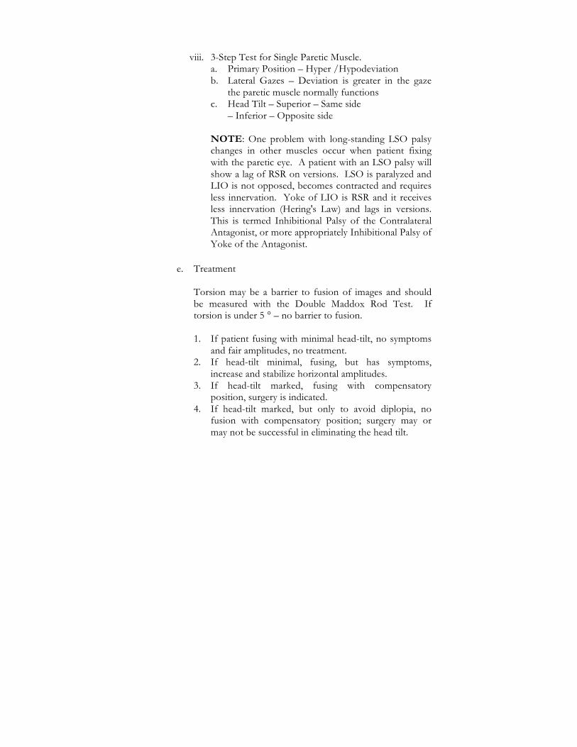
C. Greatest Field Of Action of the Vertical Muscle Using ‘H’ Design–Demonstrating the 3-Step Test

The remaining two muscles in lateral gaze will always be either:
Two Superiors – A rectus of one eye and an oblique of the other eye. Two Inferiors – A rectus of one eye and an oblique of the other eye.

When the choice is:
Two Superiors – The tilt with the greatest vertical is to the same side as the eye with the paretic muscle.
Two Inferiors – The tilt with the greatest vertical is to the
opposite side of the eye with the paretic muscle.
HINT: Head Tilt: Superiors = same side
Inferiors = opposite side
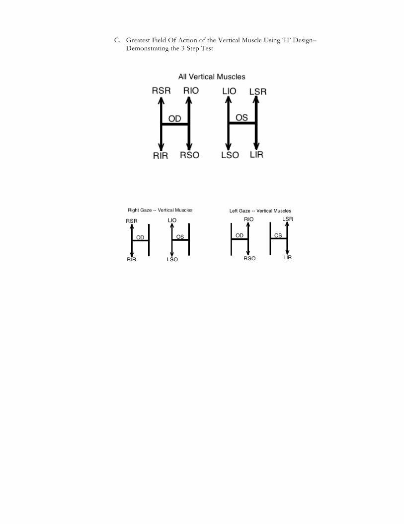
D. Three Step Test for Single Paretic Muscle Using “H” Design

E. DX ‘BISO’ Palsy
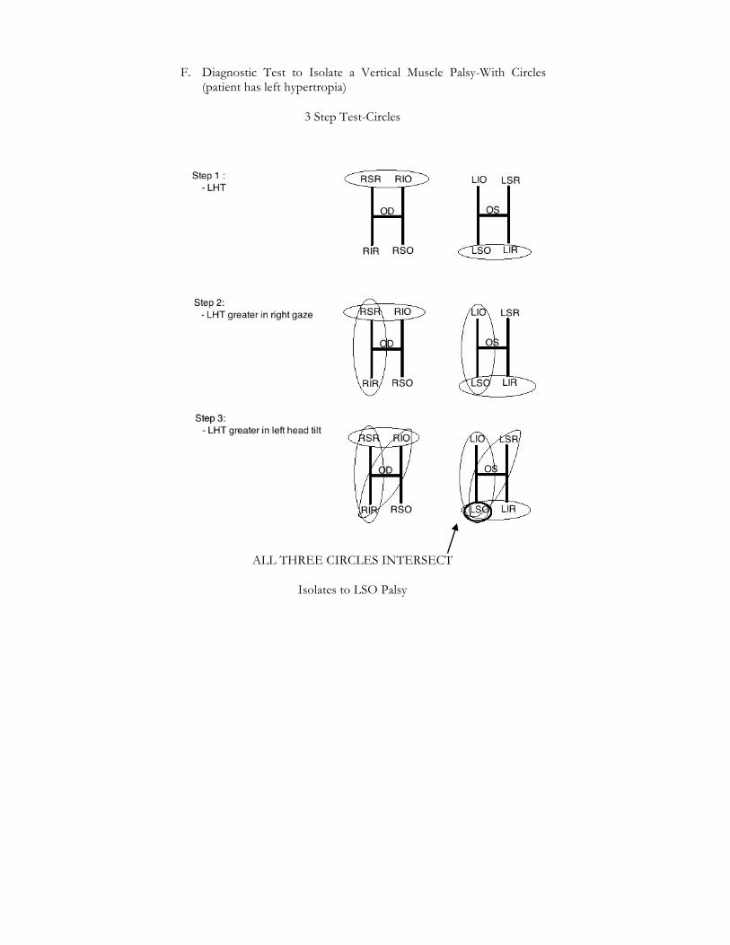
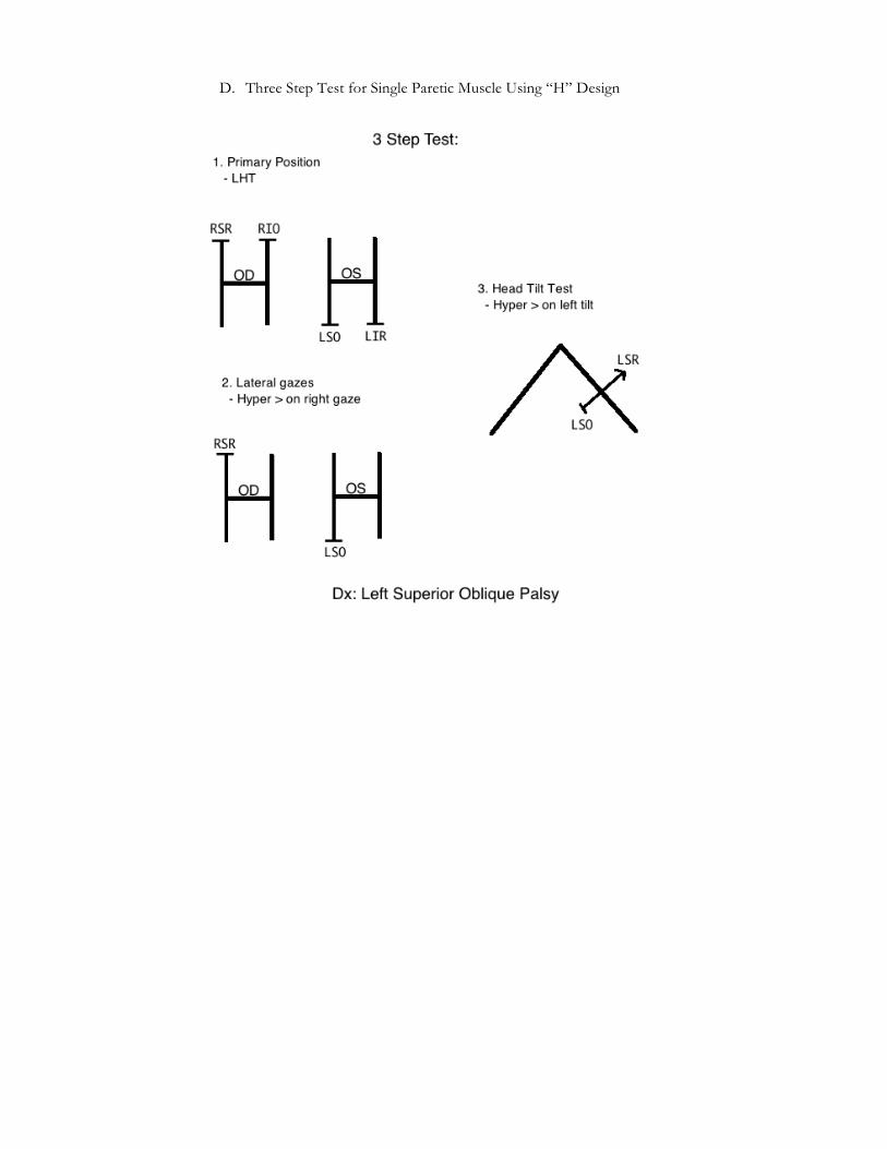
F. Diagnostic Test to Isolate a Vertical Muscle Palsy-With Circles (patient has left hypertropia)
3 Step Test-Circles
ALL THREE CIRCLES INTERSECT
Isolates to LSO Palsy

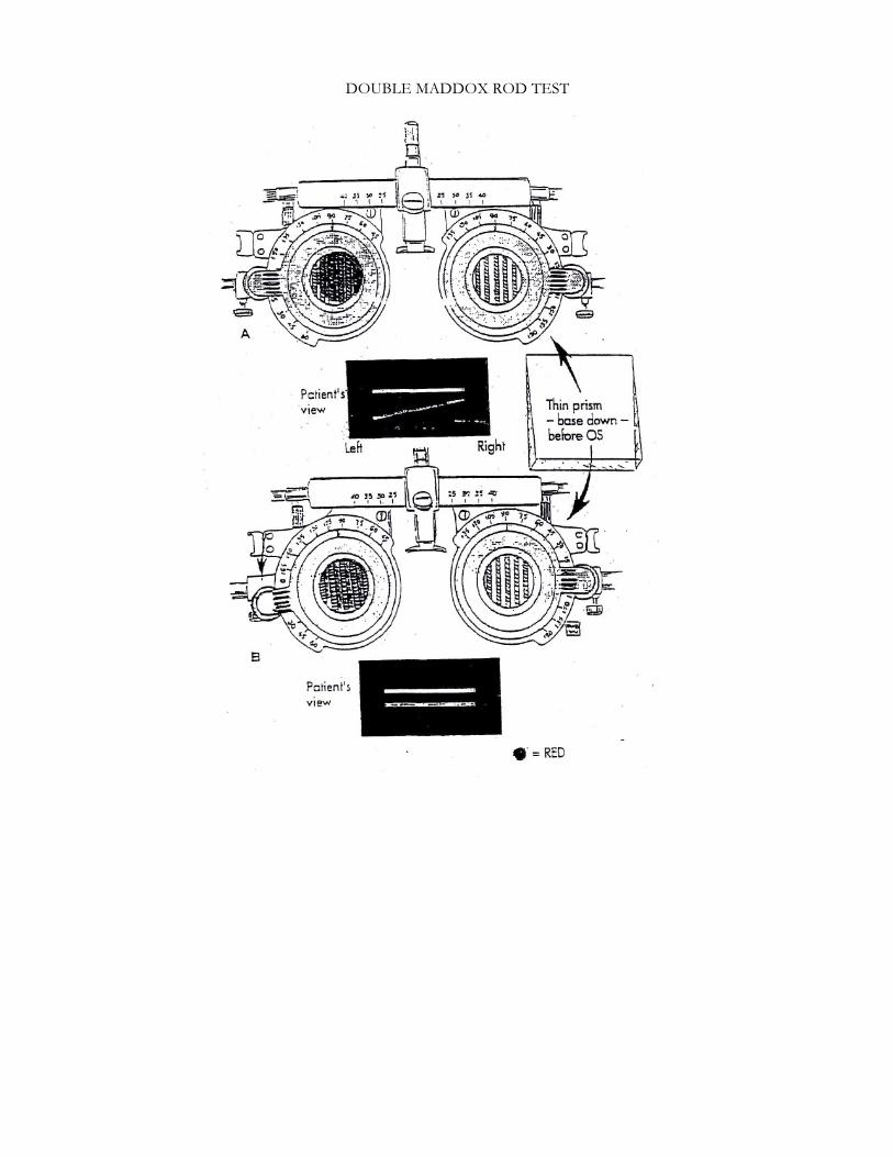
G. Double Maddox Rod Test Evaluate Ex/In Cyclotorsion
For quantitative determination of a cyclodeviation, red and white Maddox rods are placed in a trial frame, the red before the right and the white before the left eye. The direction of the glass rods is aligned with the 90° marks of the trial frame. A small scratch on the metal frame of the Maddox rods facilitates this alignment. Special care must be taken to avoid tilting the trial frames during the test. The patient looks through the Maddox rods and is shown a penlight, the images of which appear as horizontal streaks. A vertical prism may be added to separate the images for easier identification. If one of the lines (say, the red one) appears slanted toward the nose excyclotropia of the right eye is present. The red Maddox rod is then turned by the ophthalmologist (or by the patient) until the red line is seen parallel with the white line. If at the end of this adjustment the scratched mark points for example, toward the 100° mark of the right trial frame, the patient has a right excyclotropia of 10°.
1. Double Maddox Rod Test
LSO Palsy – Extorsion OS Bilateral SO palsy – Bilateral excyclotorsion
IMPORTANT: If 15° torsion bilateral superior oblique palsy is present. (Daryl Ellis, M.D.)
2. Hemifacial Microsomia in SO palsy
Asymmetric Facial Changes Associated With Ocular Torticollis
Patients with multiple types of torticollis including face turns, show similar appearance of facial compression on side of the torticollis, the tilt or turn itself cause the asymmetry. Head tilts are frequently associated with nasal tip deviation to the side of the torticollis in SO palsy.
DOUBLE MADDOX ROD TEST

3. Inferior Oblique Palsy (Example: Right Inferior Oblique Palsy) Much less common than that of SO; less annoying to the patient since it involves the upper fields more than the lower fields.
a. Characteristics
i. Downward deviation of OD. ii. Angle of squint increases looking up and to the left. iii. Deviation OS under cover greater when OD fixing
(secondary deviation). iv. Limitation of rotation up and to the left. v. Vertical diplopia --Image of the right eye is above the
image of the left eye. vi. Head elevated, tilted to the right shoulder.
b. Tests
Inspection -- OD lower, corneal reflex displaced upward OD. Chin up, head-tilt to the right shoulder. i. Cover Test: OD covered, it turns down. Shift cover
to OS, marked upward deviation behind cover (secondary deviation).
ii. Ductions: limitation OD up and in. iii. Versions: incomitance increases in left upper field. iv. Diplopia test with red glass: vertical separation of
image increases in upper fields. This indicates paralysis of an elevator muscle. Separation of images greatest up and to the left, the distal image red over OD), therefore RIO is lagging.
v. Lancaster Red-Green: (the light goes where the eye is) When patient attempts to place green light over red the green light is projected much higher in primary position (secondary deviation). The green light is farther above the red in left upper field. When patient holds the red light, it is below the green in the same field of gaze, but less separated in primary position. (Green OS, red OD in Lancaster Red-Green).

vi. Three Step Test (a) Left hypertropia in primary position. (b) Greater in left gaze. (c) Greater vertical deviation with left head tilt.
vii. Bielschowsky Test: Since RIO normally extorts, in
paralysis, the vertical corneal meridian turns inward. This is adjusted with head tilted to the right. If head forcibly tilted to the left, compensation by extorsion of OD would result from RIO, RIR. Since RIO paralyzed, RIR pulls the eye down due to its main action of depression.
c. Treatment – Surgery
4. Superior Rectus Palsy (Example: Left Superior Rectus Palsy)
May be congenital and associated with ptosis of upper lid.
a. Characteristics
i. OS deviated downward. ii. Vertical deviation increases looking up and to the left. iii. Upward deviation OD under cover greater than left
hypo under cover (secondary deviation). iv. Limitation is greater up and to the left. v. Diplopia – vertical - as image higher. Vertical
separation is greater up and to the left. vi. Seldom any head-tilt.
b. Tests
Inspection - OS lower, corneal reflex displaced upward --chin raised, rare head-tilt to right shoulder.
i. Cover Test: covered OS deviated downward; when
OD is covered, it turns upward with greater vertical deviation (secondary deviation).
ii. Ductions: limitation of movement up and out. iii. Versions: incomitance increases in upper left field. iv. Diplopia Test: vertical separation of images increasing
in upper fields. Indicates paralysis of elevators. Separation of images greatest up and left. Distal image is white (red glass over OD). Therefore LSR is lagging.

v. Lancaster Red-Green: patient attempts to place green over red, the green is lower in the primary position. Green light farther below in left upper field. Patient holds red, the red is farthest above green in upper left field. (secondary deviation). (Green OS, red OD in Lancaster Red-Green).
vi. Three-step test (a) RHT in primary position (b) Greater deviation looking to the left (c) Greater vertical measurements with head tilted to
left shoulder
c. Treatment - Surgery
5. Inferior Rectus Palsy (Example: Right Inferior Rectus Palsy)
More annoying paralysis in fresh case than SR involvement because lower reading field is involved.
a. Characteristics i. Right eye deviated upward. ii. Angle of deviation increases down and right. iii. Deviation of as under cover greater than OD under cover
(secondary deviation). iv. Limitation of movement in OD down and to the right in
the involved eve. v. Diplopia - vertical, image of OD lower, with greatest
separation down and to the right. vi. Slight head-tilt to right.
b. Tests Inspection -- OD appears higher, corneal reflection displaced downward. Head tilted to right shoulder, chin lowered.
i. Cover Test: OD covered, deviated upward; when OS
covered, deviates downward in a greater amount (secondary deviation).
ii. Versions: incomitance increases in lower right field. iii. Diplopia Test: vertical separation of images is greater in
lower fields (indicates paralysis of depressor muscle). Sensation of image greatest down and right. Distal image is red (red OD). Therefore, RIR is lagging.

iv. Lancaster Red-Green: patient attempts to place his green light over the red, green is lower in primary position. Green is farther below in 1ower right field (secondary deviation). Patient holds red, red is above green in lower right field, but less distance away (primary deviation). (Green OS, red OD in Lancaster Red-Green).
v. Three Step Test (a) RHT in primary position (b) Greater deviation in right gaze (c) Greater vertical deviation with left head tilt
c. Treatment - Surgery
III. DOUBLE HYPERTROPIA
A. Etiology
Paresis of similar acting vertical muscles in both eyes. If both muscles are equally paretic, the amount of right and left hyper will be equal; if not, the vertical deviation will be greater when fixing with the eye with the greater involvement. Direction of hyper may change depending which eye is fixing or in the different direction of gaze.
B. Treatment – vertical prisms or surgery
IV. DISSOCIATED VERTICAL DIVERGENCE DVD
A. What is DVD?
Spontaneous slow upward drift associated with abduction and extorsion of the affected eye. The deviated eye extorts during its ascent then intorts as it descended to resume fixation. Becomes manifest when binocular vision input is disrupted mechanically, optically or sensorially.
B. DVD was a term introduced by Bielschowsky in 1943.
Synonyms: 1. Alternating sursumduction – ASD 2. As occlusion hypertropia 3. Anaphoria 4. Dissociated hyperphoria 5. Double hyperphoria
C. Etiology
1. Abnormal excitation of the vertical divergence center (Jampolsky 1986)4
2. Abnormally in the development of normal centers (Harcourt, et al 1980)5
3. A compensated mechanism to dampen cyclovertical latent nystagmus (Guyton 1998)6
4. Recapitulates the primitive Dorsal light reflex which asymmetrical visual input to the 2 eyes evokes a vertical divergence movement of the eyes (Brodsky 2002)7
D. Characteristics (May be present in one or both eyes)
1. Either eye makes a slow and irregular drift upward under cover
which is usually accompanied with some extorsion. When cover is removed, the eye intorts as it drifts down slowly.
2. In changing fixation from one to the other eye, one does not observe an upward movement of one eye by a downward movement of the other as seen in vertical deviations. In DVD, the non-fixing eye consistently drifts upward.
3. DVD occurs in other cardinal positions of gaze as well as in primary position,
4. It may be present in binocular fixation which is called "pure" DVD and only becomes manifest when one eye is occluded or under fatigue and stress. In these individuals, the vertical amplitudes in both directions are higher than in patients found without DVD.
5. Most commonly found superimposed upon a horizontal or vertical deviation or both.
6. VA in both eyes may be normal, but if amblyopia is present, dissociation occurs more readily in the amblyopic eye.
7. Diplopia is usually not present in patients with DVD. Suppression of the deviating eye occurs readily (Bielschowsky emphasized the high incidence of DVD in patients with either congenital or acquired amblyopia.)
8. Nystagmus and DVD frequently co-exist; Billet and Ehrlich stated that latent nystagmus is found in every case of DVD and in fact induces DVD.
9. Vertical amplitudes are variable. 10. First noted two to five years of age. 11. Presence of a manifest head tilt in 35% of patients.

E. Distinguishing Features of DVD and Inferior Oblique (IO) Overaction8
DVD IO Overaction
1. Elevation in adduction 1. Elevation only in adduction and abduction
2. Comitant in fields of gaze 2. Incomitant in action IO
3. Variable hyper 3. Not variable
4. No A or V pattern 4. Normally associated ‘V’ pattern
5. Same hyper up and down 5. More hyper up than down gaze gaze
6. No hypotropia in 6. Corresponding hypotropia corresponding opposite eye in the opposite eye
7. May co-exist with IO 7. IO overaction always Overaction present
8. Frequently associated with 8. Not associated infantile ET and congenital nystagmus
F. Method of Measuring DVD:
Step 1. Concurrent horizontal deviation is neutralized first with
prism alternate cover test. Step 2. Amount of vertical deviation is measured by placing
base down prism in front of the eye with DVD. Step 3. The amount of base down prism is increased until no
downward movement is seen; the induced hypotropia in the opposite eye is ignored.
Step 4. In presence of a co-existing, true hypertropia
(corresponding hypotropia in the opposite eye).
• Amount is measured by increasing base down prism in front of the DVD eye, while observing diminishing hypotropia shift in the fellow eye upon alternate cover test.
• After the full amount of corresponding hypotropia
is neutralized, the additional hypertropia measurement is due to DVD.
G. Associated with Manifest Head Tilt
• A manifested head tilt with DVD may be present in patients
with or without fixation preference, prior vertical muscle surgery.
• In patients with fixation preference, in some case the angle of deviation increased with a forced head tilt to the contralateral side of DVD and decreases with a forced head tilt to the ipsilateral side.
• With alternate fixation the angle of deviation is the same with
right and left head tilt.
• Atypical responses are not influenced by oblique overaction.
• The size and control of DVD is measured in both the natural (if manifested head tilt is present) and forced primary head position and after forced head position to the right and left.
H. Bielschowsky Phenomenon
This phenomenon is an interesting characteristic of DVD. The amplitude of vertical deviation is related to the asymmetry of visual input in the 2 eyes. This effect is clearly shown by the Bielschowsky Phenomenon.
Method 1. Red filters of increment density is placed before the
fixing eye causes the hypertropic eye to gradually descend even to the hypotropic position.
Method 2. Place a dark red wedge occluder before the fixing eye
while the hypertropic eye is occluded. As the field of the fixing eye darkens, the occluded eye moves downward.

I. Treatment for DVD
Orthoptic Management • Eliminate amblyopia • Increase horizontal amplitudes • Base down ground-in prisms in front of DVD eye
J. Surgical Management – moderate to a large DVD or the presence
of manifested head tilt. (OA-SR-IO)
V. DISSOCIATED HORIZONTAL DEVIATION DHD
A. DVD includes movements in both the horizontal and vertical planes. When the horizontal component of DVD is very prominent, the horizontal dissociation movement is labeled DHD
B. DHD is defined as an asymmetric or unilateral slow outward
deviation associated with vertical and torsional dissociated movements that can be manifest or latent. DHD is nonparalytic and does not obey Herrings law of equal innervation
C. Distinguishing Features of DHD
1. Unilaterally or asymmetry of the exodeviation between the 2
eyes. 2. Variability and difficulty in obtaining a clear end point with
prism neutralization. 3. Often times, prism neutralization produces an esodeviation in
the fellow eye. This similar pattern could be produced by a paralytic strabismus in the form of primary and secondary deviation. However, no weakness in ductions, versions and incomitance is present in DHD.
4. Uncorrected refractive error can produce a pseudo DHD. An exodeviation decreases when a patient fixes with a unilateral uncorrected hyperopic eye stimulating accommodation with convergence.
5. Bielschowsky phenomenon is demonstrated in the horizontal plane. Place a darkening wedge in front of fixing eye, and the outwardly drifted eye will return and cross the midline line.

D. Three Criteria Required for True DHD
1. Minimum difference 5 Δ in amount of exodeviation when
alternately fixing. 2. No limitation in ocular motility that may produce primary and
secondary deviation. 3. No uncorrected anisometropia that may produce incomitance
with alternate fixation. VI. DOUBLE ELEVATOR PALSY
This is a lack of elevation of one eye due to a weakness of both the superior rectus and the inferior oblique. The two muscles may be affected equally, but often the paralysis is more pronounced in the inferior oblique than the superior rectus. The forced duction test will differentiate it from a fibrosis of the inferior rectus. Etiology may be a unilateral supranuclear lesion.
A. Characteristics 1. Pseudo-ptosis secondary to hypotropia in opposite eye. A
ptosis may also occur. 2. Marked hypertropia when fixing with the paretic eye. 3. Head-tilt directly back and a head tilt in opposite direction. 4. Phoria is intermittent in down gaze with usually good fusion
and stereopsis. 5. Check if Bells is normal – if not could be due to a mechanical
restriction.
B. Treatment – Surgery
VII. BROWN’S SO TENDON SHEATH SYNDROME
Etiology: Short anterior tendon sheath of SO which normally acts as a check ligament for the IO on the same side.
A. Characteristics 1. Good visual acuity OU. 2. Primary position -- hypotropia in the affected eye or no
vertical deviation present. 3. Absence or gross limitation of elevation in nasal field
(simulates an IO paralysis). In most cases elevation in nasal field is limited to a few degrees below the mid-horizontal plane.
4. Limitation of elevation decreases as involved eye is abducted.

Full elevation with involved eye completely abducted – may demonstrate V pattern.
5. ‘Downshoot’ of depression of affected eye on adduction. 6. Widening of palpebral fissure on adduction of affected eye. 7. Normal excursions of involved eye in temporal field of gaze. 8. No vertical imbalance in lower field. 9. Little or no overaction of SO (no secondary contracture). 10. Backward head-tilt often present. 11. Usually good fusion and stereopsis at near.
B. Treatment – Surgery.